0022-538X/80/01-0920/06$02.00/0
Effect
of
a
tsA
Mutation
on
Simiian
Virus 40 Late Gene
Expression: Variations Between Host Cell Lines
JAMES C.ALWINE ANDGEORGE KHOURY*
Laboratoryof MolecularVirology,National CancerInstitute,National InstitutesofHealth, Bethesda, Maryland 20205
Infection ofAGMKorCV-1 cellsbytheearly simian virus 40 mutant tsA58 at
the permissive temperature (32°C) followed by a shift to the nonpermissive
temperature(410C) causedasubstantial decrease in the levels of late viral RNA
in the cytoplasmof AGMK cells butnotCV-1 cells. At the translationallevel, thisdepression of late viral RNA levelswasreflectedbyadecrease in late viral proteinsynthesis.Thus, in AGMK cells,anearly regiongeneproduct (presumably largeT-antigen) appearedtobecontinuously requiredfor efficientexpression of the late viralgenes. In contrast, late simian virus40 geneexpression, onceit is initiated in CV-1 cells, continued efficiently regardless of the tsA mutation. The difference in expression of the late simian virus 40 genesin these tsA
mutant-infected monkey kidney cell lines mayreflect adifference in host cellproteins whichregulate viralgeneexpression inconjunctionwithearly viral proteins.
The temporal expression of early and late simianvirus40(SV40)genes appearstobe
con-trolledatthelevel oftranscription (2,5, 14,18). Aprimary factor indeterminingthe
transcrip-tional activity of thesegenesisoneof theearly gene products, large T-antigen, which appears tohave arepressor-like activity in modulating
earlytranscription (2,14, 18) and also functions
in theinitiation of viralDNAsynthesis (20, 21). Theseconclusionsarebased, inpart, onstudies
withearlySV40temperature-sensitive mutants
(tsA mutants) whichsynthesize a thermosensi-tivelarge T-antigen (1, 24). At elevated
temper-atureslargeT-antigen becomes defective (1,20,
22-24), causing overproduction of early viral
RNA (2, 14, 18) and curtailment of viral DNA synthesis (20, 21). Theregulation of late SV40 transcription also seems to be dependent, at
least inpart, onthe activity of largeT-antigen orconcomitantDNA replication or both (2, 5,
14). Ifmonkey kidney cultures are infected in
thepresenceof inhibitorsof DNAreplication(7,
14;Alwine,unpublished data) or bytsAmutants atthenonpermissivetemperature (2, 5, 14), the
nornal pattern of abundant late transcription
cannotbe established. If theinhibitors of DNA
synthesis are added after the onset of SV40
DNAreplication,however,theyhave little or no
effect on established late viral transcription. A
more complex effect is seen in tsA
mutant-in-fectedcellswhen they are shifted from the
per-missive tothe nonpermissive temperature after
DNAsynthesis and late transcription have been
initiated. Althoughlatetranscription continues
(2,5, 14),thelevel of this synthesis may depend
onthespecificinfectedmonkey kidneycell line.
Although the level of late viral mRNA remains highaftershifting tsA mutant-infected TC-7or
CV-1 cells (2,5) tononpermissivetemperatures, a decrease of late RNA was noted in shifted
tsA58-infected primary African green monkey
kidney (AGMK) cells (14). Since these earlier
studiesemployeddifferenttechniques andwere,
in somecases, semiquantitative, we have
ana-lyzed late SV40 RNAs from CV-1 and AGMK cells inparallel experimentswithbothwild-type
and tsA mutant viruses. Data are presented which indicate: (i) the continuous requirement
foran earlygene productfornormal late viral
geneexpression,and(ii)adifference in thelevels
of late viral RNA (and late proteins) between CV-1 and AGMKcells which is under the effect ofatemperature-sensitive mutationinthe early
gene.These observationsmayindicatethatthe
early gene product interacts with a host cell
factor(s) for the control of lategene expression. The cells used in this study were primary
AGMK cells and the established AGMK line, CV-1, whichwerecultivatedinminimalessential
mediumplus10% fetal calf serum. Virus strains
werethewild-type VA4554 (21) and a
tempera-ture-sensitive mutantderived from it, tsA 58 (20,
21). Contluent monolayers of cells in 150-cm2 Costarplastic bottles wereinfected at a
multi-plicity of10PFU/cell in 5 ml ofminimal
essen-tial mediumcontaining2%fetalcalf serum. After
rockingat roomtemperature for 1.5 h, 20 ml of
mediumwasadded, andthe cells were incubated at32°C for40 h. At thispoint, cells were
har-vested (32°C samples) or shifted to 41°C for
920
on November 10, 2019 by guest
http://jvi.asm.org/
921
5 h (shifted samples) before harvesting.
Cyto-plasmic RNAfrominfected cellswasprepared as previously described (13). Polyadenylated [poly(A)]RNA was selected on columns of
oli-godeoxythymidylic-cellulose (3). Cytoplasmic
RNA pulse-labeled with [3H]uridine was
pre-pared in a similar manner except that it was
treatedwith 40
jig
ofDNAse I per ml(Worth-ington, RNase free) for 30minat40Cand was notselectedfor poly(A) sequences (6). For
pulse-labeling, cells were infected and incubated as
described above. At the end of the incubation
period at 320C or 4 h after the shift to
410C,
eachflask of cellswaswashed withwarmed (32 or
410C)
serum-free medium and labeledwith 8mlofwarmed serum-freemediumcontaining1.5
mCi of[5,6-3H]uridine (NewEngland Nuclear,
Boston,Mass.). Theperiodofpulse-labelingwas
1.25 h at 320C and 45 min at 410C. For the
preparation of32P-labeled SV40 DNA hybridi-zation probes, SV40 DNA was labeled in vivo and purified as previously described (15).
La-beled form I SV40 DNA
(25,ug)
was digested with the restriction endonucleases HpaII andBamHI (New England Biolabs).Thestrands of thefragmentswereseparatedon a1.4%agarose
gel by the method of Hayward (9). Separated strands were electroeluted from gels, self-an-nealed, and chromatographed on hydroxyapa-tite. The resulting purified single strands were
dialyzed into 10 mM Tris-hydrochloride (pH
7.5)-i mM EDTA. Hybridization analysiswith
single-stranded DNA probes and cytoplasmic poly(A)-containing RNA was performed under conditions of DNA excess. These conditions
were established by titration of different
sam-ples. Between0.1and5ygof poly(A) RNAwas
used in a normal hybridization reaction. For each reaction10ng(10,000 cpm) ofDNAprobe was added. Hybridization samples were dis-solved in30
pi
of 50%formnamide,0.35 MNaCl, 0.04 M PIPES[piperazine-N-N'-bis(2-ethane-sulfonicacid)],pH6.5, heatedto650Cfor3min
and hybridized at 370C for 24 to 48 h. After
hybridization, the samples were treated with
nucleaseS1 and electrophoresed on1.4or1.8% alkaline agarosegels asdescribedby Berk and
Sharp (4).The gelsweredriedonDEAEpaper
and autoradiographed at -70°C with Kodak
XR-1X-rayfilmandaDupontCronex
lighten-ing-plus intensifying screen. For analysis of
pulse-labeled RNA, SV40 DNA was cleaved
with the restriction endonucleases HpaII and
BamHI, and the strands of the two resultant
fragments were separated as described above
and transferred onto nitrocellulose
by
themethod of Southern (19). Each filter
(10
cmwide) contained 30,ugofSV40DNA.
Strips (0.5
cm) ofthe separated strand blot were cut and
used ineach hybridization (approximately0.35 ,ugof eachstrand). Samplescontaining4,000 or
12,000 cpm of virus-specific RNA (previously
determined) were added to the hybridization mixture containing 5X SSC (0.15 M NaCl plus
0.015 M sodium citrate), 0.1% sodium dodecyl
sulfate,and300
Ag
ofyeast tRNA in a volume of1.5ml.Incubations were at
680C
for 24 to 48 h.After hybridizationthe strips weretreated with
RNase A and washed as previously described
(19),and then cut into 1-mm pieces and counted in Econofluor (New England Nuclear). For the
aiialysis of SV40lateproteinsynthesis, confluent monolayers ofAGMK or CV-1 cells grown in 50-mmplates (three plates per set) were infected
with VA4554 or tsA58 at a multiplicity of 10.
The infected cells were incubated for 40 h at
320C.
Atthatpoint thecellswerepulse-labeled for1 hat320C
with[3S]methionineor shifted to41°C for4 hbeforepulse-labelingfor 1 h at410C. The pulse-labeling was accomplished by
washing thecellsthree timeswithappropriately
warmed (32or
410C)
methionine freeminimumessential medium containing 2% dialyzed fetal
bovineserumandthenlabeling eachplatewith
1ml ofprewarmedmediumcontaining200
,uCi
of[3S]methionine (300 Ci/mmol, Amersham). Afterlabeling, theplateswereput onice,washed three times with cold analysis buffer
(Tris-bufferedsaline, pH 8, containing 1 mM dithio-threitol,300
p.g
ofphenylmethylsulfonylfluoride
perml, and 150,ugof
tosylamid-2-phenylethyl-chloromethyl ketone per ml). The cells were
scraped into 1.5 ml of analysis buffer, sedi-mentedat3,000rpmfor 10min,
resuspended
inanalysis buffer containing 0.5% Nonidet
P-40,
and incubated on ice for 30 min,
blending
vig-orously in a Vortex mixer every 5 min. The
lysateswere sedimented at 15,000 rpm for 60
min, and the supernatant fraction was with-drawnand saved. Portions(0.3
ml)
of thesuper-natantswere
immunoprecipitated
withheat-in-activatedStaphylococcusaureusand antiserum directed against the SV40virion
proteins
(pre-dominantly VP1) as
previously
described(12).
Theserum had beentitrated
previously
to de-termineoptimal
condition for thesesamples.
Both the serumandS. aureuswere the
gift
of Gilbert Jay.Immunoprecipitates
(in 15- to20-pl
volumes)wereanalyzed
byelectrophoresis
on 10%sodiumdodecyl
sulfate-polyacrylamide
slab gels (11). Radiolabeledproteins
were detectedbyfluorography (17).
Cytoplasmic
poly(A)
SV40RNAwasanalyzed
bythenucleaseS1
procedure
ofBerk andSharp
(4)followedbyalkaline agarosegel
electropho-resis and
autoradiography.
Toquantitate
theon November 10, 2019 by guest
http://jvi.asm.org/
relativeamountsof latemRNAspeciesfrom the intensity ofautoradiographic bands,
hybridiza-tionswereperformedin DNAexcess, and incu-bation times wereprolonged to ensurethat
re-actionswereessentiallycomplete.The
specific-ityofthe reactionwasfurther ensuredby using a[32P]DNAprobewhich consisted of the
puri-fied late sense (+) strand of the B fragment,
generated by cleavage of SV40 DNA with the restriction endonucleases HpaII and BamHI
(0.72 to 0.14 on the SV40 map). This strand
contains almost all of the known late coding
region.
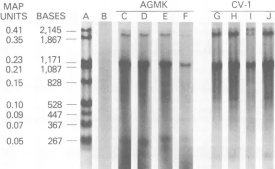
Figure1shows theanalysis of lateRNAfrom wild-type or tsA58-infected cells (CV-1 and
AGMK)whichhad been incubatedat320Cfor
40hafter infection (32°Csamples) orhadbeen incubated at320Cfor40hand thenshifted to
410Cfor 5h(shiftedsamples).Inall tracks(Fig.
1CtoJ), bandsrepresenting the "bodies" of the lateSV4019S RNA (0.38 SV40fractionallength)
and the late 16S RNA (0.21 fractional length)
arepresent.The smallamountof
0.41-fractional-length bandseen atthetop of somegels isthe DNA probe which was either not digested or
whichannealed withtracequantities of
contam-inatingcomplimentaryDNAstrands. Since the
samequantities ofinfected cellRNA were used
MAP
UNITS BASES A B
0.41
2,145
0.35
1.867
0.23 1171 W
0.21 1,087
0.1.5 828 4
0.10 5267 t
0.09 447 X
0.0-7 36 l
306/ * :
-7~~~~~~~~~~~~~~~~~~~~~~~~~~~~~~~~~~~~~
005 261~~~~~~~~~~~~~~~~~~~~~~~~~~~~~~~~~~~~~~~~~~~~~~~~~~~~~~~~~~~~~~~
inmatchingtracks and sincehybridizationswere
performed in DNA excess,the intensity of the
bandsreflects therelative amountsoflateSV40 RNAs. Inwild-type-infected AGMK cells (Fig.
1C andD)andCV-1 cells (Fig. 1GandH), there
is asmallbutdefinite increaseinthequantityof
late viralRNAsin cellsshiftedfrom 32 to410C,
comparedwith thequantityof lateRNAs
pres-ent in cellsmaintained at 320C (1.5- to 2-fold
increase as measured by microdensitometry tracing). This same increase is also seen in
tsA58-infected CV-1 cells (Fig. 1I and J) aftera
shift from 32 to410C.However, tsA58-infected
AGMKcellshavesubstantiallyless late19Sand
16S cytoplasmic RNAs in the shifted sample (Fig. 1F) compared with the 320Csample (Fig. 1E).This decrease is six- toeightfold as
deter-mined by microdensitometry ofthree separate
experiments with different matching sets of
RNA.
To further verify the decrease of late SV40
mRNA in tsA58-infected AGMK cells, we
an-nealedpulse-labeled RNAsamples from tsA 58-infected cells (see above) to the separated
strands of the early (E) and late (L) coding
regions (HpaII-BamHI-cleaved DNA frag-ments) which had been boundtonitrocellulose membranefilters (19).The results of this
exper-AGMK V
D E F -r
~ ~ ~ ~ ..
FIG. 1. NucleaseSI analysisofSV40 cytoplasmic poly(A) -containinglateRNA.Poly(A)-containing cyto-plasmic RNA, isolatedfrom AGMKorCV-1cellsinfectedwith WT-SV40ortsA58(see text), washybridized
to the late (+) strandof thesmallerfragmentof32P-labeled SV40DNAgenerated bydigestion with the
restrictionendonucleases HpaIIandBamHI(0.73-0.14SV40mapunits).Sampleswerehybridized, nuclease Sl treated, electrophoresed, and autoradiographed as described in the text. In these experiments equal
amountsof RNAwereaddedto eachmatchingset ofsamples; thusquantitativecomparisonscan bemade directly withinand between matched lanes. (A)SV40DNAfragmentsizemarkers; (B)hybridization with probeDNA alone; (C) 32°CsamplefromAGMK cells infectedwithwild-typeSV40;(D)shifted sample from AGMKcells infected with wild-type SV40; (E) 32°C samplefromAGMK cells infected withtsA58; (F)shifted
samplefromAGMK cells infectedwith tsA58;(G) 32°C samplefromCV-1cellsinfectedwithwild-typeSV40; (H) shifted samplefrom CV-1cellsinfectedwithwild-typeSV40;(I) 32°CsamplefromCV-1cellsinfectedwith
tsA58; (J) shiftedsamplefromCV-1cellsinfected withtsA58.
on November 10, 2019 by guest
http://jvi.asm.org/
[image:3.514.115.398.374.548.2]NOTES 923
iment indicate the relative synthetic rates of
early and lateSV40 RNA.These ratios are
sum-marized in Table1. In thecytoplasmic fraction
ofboth tsA 58-infected AGMK cells and CV-1
cells,ashift from32to410Cwas accompanied
by anincreaseinpulse-labeledearly RNA (see
the shift/32°C ratios forearly RNA) in agree-mentwith previousresults (2, 14, 18). The
tem-peratureshiftintsA 58-infectedCV-1cellsledto
asmall increase inthe amountofpulse-labeled cytoplasmic late viralRNA (seethe shift/32°C ratio for late RNA). In contrast, pulse-labeled late cytoplasmic viral RNA was significantly decreased in tsA 58-infected AGMK cells after the temperature shift. Wild-type-infected cells do notshow the overproduction ofearlyRNA, andpulse-labeled late RNA doesnotdecrease in AGMK cells after a temperature shift (14, 18; ourdatanotpresented).
Thepulse-labeledRNA from thenuclear
frac-tions oftsA-infected AGMK and CV-1 cellswas
alsoassayed forearly and late SV40 RNA (data
notpresented). In agreement withprevious
re-sults (14, 18), the nuclear early RNA ratios showed anoverproduction similarto thatseen
in thecytoplasmfortsA-infected cultures which
areshiftedto41°C.However,the very low levels ofpulse-labeled nuclear late RNA preventedan
accurate determination of theshift/32°Cratios
for both cell lines. These low levels of
pulse-labeled late nuclear RNA, compared with the late cytoplasmic RNA, are also seen in
wild-type-infected cells and agree with previous
re-sults by a more sensitive (C,t) hybridization
analysis (18).Theseresultssuggestrapid
trans-portofnewly synthesized SV40 late RNA from the nucleus.
From theresults of the nucleaseSiand
pulse-labeling analysis of cytoplasmic late RNA, it
appearsthat in t&A-infected CV-1 cells, ashift
from32 to410C ischaracterizedbyanincrease
(overproduction) inearlyRNA withlittle
alter-ation inthe levels of late RNA. In AGMK
cells,
TABLE 1. Ratiosofviralearlyand late RNA in
tsA58-infectedcellsshiftedto41°Ccompared with
cultured maintainedat32°C(shift/32° ratios)
Shift/32°ratio
Cellline
EarlyRNA Late RNA
AGMK 4.5a 0.1
CV-1 12.8 1.1
nThe numbers represent the ratio of theamounts
ofearlyorlate RNA in tsA58-infected cultures
pulse-labeled aftera5-h shiftto410Ccomparedwith pulse-labeled cultures which had been maintainedat320C (see text). The percentage of total viral RNA in the varioussampleswasbetween5.0and6.0oinAGMK cells and between1.0and 2.0% in CV-1 cells.
however, the early RNA overproduction, after a
shift to 410C, is accompanied by a substantial
decreaseinlate RNA.The fact thatsimilar data
were obtained in experiments designed to
ex-amine notonly steady-state RNA (Fig. 1) but
alsopulse-labeled transcripts (Table 1) suggests
that the alterations in late RNA levels
accom-panying temperature shifts with AGMK cells
may occur at the level of synthesis; however,
otherexplanations arepossible, and amore
ex-tensive examination ofnuclear late RNA is nec-essary to establish the mechanism of the
de-crease.
We next askedwhetherthespecific decrease
in cytoplasmic late RNA in shiftedsamples of
tsA58-infected AGMKcellsaffects the synthesis
oflate viralproteins. Todetermine this, AGMK
and CV-1 cells were infected under the same conditions used previouslyand were then
pulse-labeled with [3S]methionine for 1h atthe end
of theincubation at 320C or atthe endofthe
shift period.Sampleswereimmunoprecipitated
with serum directed against the SV40 virion
proteins andanalyzed byelectrophoresison so-dium dodecyl sulfate-polyacrylamide gels (11,
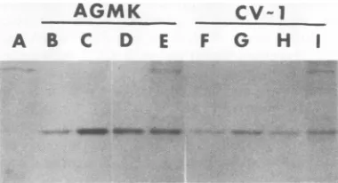
12). Figure 2 shows the VP1 bands resulting from this analysis. Comparing the 320C and
shiftedsamples fromwild-type-infectedAGMK
cells (Fig. 2B and C), wild-type-infected CV-1
cells (Fig. 2F, G), or tsA58-infected CV-1 cells
(Fig. 2H andI),we see adefiniteincreaseinthe
intensity ofthe band in shiftedsamples.
How-AGMK
A
B
C
D
E
Cv-1
F
G
H
I
m . .4bPi. -.
-FIG. 2. Analysis of[35S]methioninepulselabeled VP1. AGMK and CV-1 cellswereinfected with wild typeortsA58 andpulse-labeledwith[3S]methionine
asdescribed in thetext.Immunoprecipitatedsamples
wereanalyzedon 10% sodiumdodecyl sulfate-poly-acrylamide gels, and bandswerevisualizedby fluo-rography. (A)Mock-infectedsample;(B)32°Csample from AGMK cellsinfected with wild-type SV40; (C) shifted sample from AGMK cellsinfected with
wild-typeSV40; (D)32°CsamplefromAGMK cellsinfected
with tsA58; (E) shifted sample from AGMK cells
infectedwithtsA58; (F) 32°Csample fromCV-1cells
infected withwild-type SV40; (G)shifted samplefrom CV-1 cells infected with wild-type SV40; (H) 32°C
sample from CV-1cellsinfectedwithtsA58;(I)shifted
samplefromCV-1 cellsinfectedwithtsA58.
on November 10, 2019 by guest
http://jvi.asm.org/
[image:4.514.274.464.403.506.2]ever,thisincrease in intensity is not seen in the
shifted samplefromtsA58-infected AGMKcells
(Fig. 2D and E). Microdensitometer tracing, in
fact, shows a decrease. By this analysis the
de-creaseinVP1synthesisinAGMKcellsdoesnot
appear to be as great as the decrease in late
mRNA levels. The reasons for this difference
are unclear. Since translation is affected by
many factors besides mRNA concentration, a
directcorrelation ofproteinsynthesisand RNA concentrationmay notalwaysbeexpected.The synthesis of VP1 is reduced only in tsA58-in-fected AGMK cells aftera shift from the
per-missive tothenonpermissive temperature.
Be-causethiscorrelates withaspecific decrease in
late mRNA,weconclude the reduced VP1
syn-thesis isareflection of thedepressionin the late
mRNAlevels. The other late viralproteinsVP2
and VP3 are present in much lower amounts
than VP-1 andmeasurementsoftheir band in-tensities are less accurate. However, with very
long exposure, VP2 appears to have a similar decrease in its synthetic ratein tsA58-infected AGMK cells shiftedtothe nonpermissive
tem-perature (data not shown). This suggests that
the decrease in late RNAs affects all late
pro-teinsintsA 58-infected AGMK cellsat410C. Inconclusion,wehaveshown that theamount oflate SV40 RNAin the cytoplasm of tsA
58-infected cultures, shifted to
410C,
depends largely on the host monkey kidney cell line. Comparing320Csamplestoshiftedsamples,wehavefoundadecrease in the level of late SV40 RNA andlate viral protein synthesisin second-ary AGMK cells. In contrast, normal or even
slightlyincreased levels of late SV40 RNAand
lateproteinsaresynthesized inCV-1cellsunder
similar infection conditions. The decreasein late
RNAlevels and late viral protein productionin
AGMK cells,in response to the tsA mutation,
indicates that a functional early viral protein
(presumably large T-antigen)iscontinuously
re-quired fornormal late viral gene expression in
this cell line. Variations in the effect of early
SV40geneproducts in differentpermissive host
cell lineshavebeendemonstratedin therecent
report that themitogeniceffect of theSV40A
geneproduct differsbetweenAGMKandTC-7
cells (10). In addition, there have been several
recent reports suggesting possible interactions
between large T-antigen and hostproteins (8,
16, 25).Theseobservationssuggest that the
con-trastingresults betweenAGMKand CV-1 cells
may be due to differences in host cell factors
which interactwith early viral proteins for the
regulationoflategeneexpression.
Wewould like to thank S.L. Adams and G. Jay for helpful discussions and critical reading of the manuscript.
LITERATURE CITED
1. Alwine, J. C., S. I. Reed, J. Ferguson, and G. R.
Stark. 1975.Properties of T-antigen induced by wild-type SV40 and tsAmutantsinlyticinfection. Cell 6: 529-533.
2. Alwine, J. C., S. I. Reed, and G. R. Stark. 1977. Characterization of the autoregulation of simian virus 40gene A. J. Virol. 24:22-27.
3. Aviv, H., and P. Leder. 1972. Purification ofbiologically
active globinmessenger RNAby chromatographyon
oligothymidylic acid-cellulose. Proc. Natl. Acad. Sci. U.S.A.69:1408-1412.
4. Berk, A. J., and P. A. Sharp. 1978. Splicedearly mRNAs ofsimian virus 40. Proc. Natl. Acad. Sci. U.S.A. 75: 1274-1278.
5. Cowan, K.,P.Tegtmeyer, and P. D. Anthony. 1973. Relationship of replication and transcription of simian virus40DNA. Proc.Natl. Acad. Sci. U.S.A. 70:1927-1930.
6. Ferdinand, F.-J., M. Brown, and G. Khoury. 1977. Synthesis and characterization of late lytic simian virus 40RNA fromtranscriptional complexes. Virology 78: 150-161.
7.Ferdinand, F.-J., M. Brown, and G. Khoury. 1977. Characterization of early simian virus 40transcriptional complexes: latetranscription in the absence of detect-able DNAreplication. Proc. Natl. Acad. Sci. U.S.A. 74: 5443-5447.
8. Griffin, J. D., G. Spangler, and D.Livingston. 1979. Protein kinase activity associated with simian virus 40 T-antigen. Proc. Natl. Acad. Sci. U.S.A.76:2610-2614. 9. Hayward, G. S. 1972. Gel electrophoretic separation of thecomplementary strands ofbacteriophage DNA. Vi-rology 49:342-344.
10. Hiscott, D., and V. Defendi. 1979.Simian virus 40 gene A regulation of cellular DNAsynthesis. I. In permissive cells. J.Virol. 30:590-599.
11.Jay, G., F. T.Jay, R. M.Friedman,and A. J.Levine. 1977. Simian virus40-specific ribosome-bending pro-teins induced by a nondefective adenovirus 2-simian virus 40hybrid. J. Virol. 23:692-699.
12.Jay,G.,R. P. C.Shiv,F. T.Jay,A. S.Levine,and I. Pastan. 1978. Identification ofa transformation-spe-cificprotein induced by a Rous sarcoma virus.Cell 13: 527-534.
13.Khoury, G., B. J. Carter,F. J. Ferdinand, P. M. Howley, M. Brown, and M. A. Martin. 1976. Genome
localizationof simian virus40 RNAspecies. J. Virol. 17:832-840.
14. Khoury, G.,and E.May.1977.Regulationofearlyand late simian virus40 transcription: overproduction of early viral RNA in the absence of a functional T-anti-gen.J.Virol. 23:167-176.
15.Lai, C.-J.,R.Dhar,and G.Khoury.1978.Mappingthe spliced and unspliced late lytic SV40 RNAs. Cell 14: 971-982.
16. Lane, D., and L.V.Crawford.1979.T-antigen is bound to a host protein in SV40-transformed cells. Nature (London) 278:261-263.
17.Laskey, R. A., and A. D. Mills. 1975. Quantitative film detection of3Hand"4Cinpolyacrylamide gels by fluo-rography.Eur.J.Biochem. 56:335-341.
18. Reed, S. I., G. R. Stark, and J. C. Alwine. 1976. Autoregulation of simian virus 40 gene A by T-antigen. Proc.Natl. Acad. Sci. U.S.A.73:3083-3087.
19.Southern, E. M. 1975. Detection of specificsequences among DNAfragments separated by gel electrophore-sis.J.Mol.Biol. 98:503-517.
20. Tegtmeyer,P.1974.Altered pattems of proteinsynthesis in infection by SV40 mutants. Cold Spring Harbor Symp. Quant. Biol. 39:69-84.
21. Tegtmeyer, P., C. Dohan, and C. Reznikoff. 1970.
on November 10, 2019 by guest
http://jvi.asm.org/
Inactivating and mutagenic effects of nitrosoguanidine onSV40.Proc.Natl. Acad.Sci. U.S.A. 66:745-752. 22. Tegtmeyer, P.,K.Rundell, and J. K. Collins. 1977.
Modification of simian virus 40 protein A. J. Virol. 21: 647-657.
23. Tegtmeyer, P., M. Schwartz, J. K.Collins, and K. Rundell. 1975.Regulation of tumor antigensynthesis by simian virus 40 geneA.J.Virol. 16:168-178.
24. Tenen, D. G., P.Baygel, and D. Livingston. 1975.
Thermolabile T-antigen from celLs transformedbya temperaturesensitive mutant of simianvirus 40. Proc. Natl. Acad. Sci. U.S.A. 72:4351-4355.
25. Tjian, R., and A. Robbins. 1979. Enzymatic activities associated with apurified simian virus 40 T-antigen-related protein. Proc. Natl. Acad. Sci. U.S.A. 76:610-614.
on November 10, 2019 by guest
http://jvi.asm.org/