COMPARATIVE EVALUATION OF THE SHEAR BOND
STRENGTH OF THE BOND BETWEEN CERAMIC AND
ENAMEL PRETREATED WITH DIFFERENT ETCHING
METHODS – AN IN VITRO STUDY
Dissertation Submitted to
THE TAMILNADU DR. M.G.R. MEDICAL UNIVERSITY
In partial fulfillment for the Degree of
MASTER OF DENTAL SURGERY
BRANCH I
PROSTHODONTICS AND CROWN & BRIDGE
CERTIFICATE
This is to certify that the dissertation titled “COMPARATIVE
EVALUATION OF THE SHEAR BOND STRENGTH OF THE BOND
BETWEEN CERAMIC AND ENAMEL PRETREATED WITH
DIFFERENT ETCHING METHODS – AN IN VITRO STUDY” is a bonafide
record work done by Dr. ABDUL RAFE under our guidance and to our
satisfaction during his post graduate study period between 2009 – 2012.
This dissertation is submitted to THE TAMILNADU DR. M.G.R.
MEDICAL UNIVERSITY, in partial fulfillment for the Degree of MASTER OF DENTAL SURGERY – PROSTHODONTICS AND CROWN & BRIDGE,
BRANCH I. It has not been submitted (partial or full) for the award of any other degree or diploma.
Guided by
Dr. N.S. Azhagarasan, M.D.S., Dr. S. Jayakrishnakumar, M.D.S., Professor and Head of the Department, Professor,
Department of Prosthodontics Department of Prosthodontics,
and Crown & Bridge, and Crown & Bridge,
Ragas Dental College & Hospital, Ragas Dental College & Hospital, Chennai. Chennai.
Dr. S. Ramachandran, M.D.S., Principal,
ACKNOWLEDGEMENT
It is said that a man realizes only in the times of adversity how truly blessed he is. This, for me, has been a blessing as it helped me realize the wonderful people I’m surrounded with who’ve helped shape not only this dissertation but also various aspects of my life. This is my humble attempt to express my gratitude towards them.
I would like to thank Almighty God for each time I fumbled He led me on gave me the courage and the strength and wisdom to overcome all hurdles and finish this endeavour of mine successfully. Verily whom He wills to help he shall never fail.
I wish to express my gratitude to Dr. S Ramachandran, M.D.S., Principal, Ragas Dental College and Hospital, Chennai for his encouragement and support throughout my post graduate course. I also thank him for permitting me to make use of the amenities in the institution.
I would like to express my real sense of respect, gratitude and thanks to my
Guide, Dr. S. Jayakrishnakumar, M.D.S., Professor for his guidance, constant support and encouragement extended to me during this study. His patience and perseverance benefited me in every facet of my study and I thoroughly enjoyed every discussion with him. His guidance and supervision helped me to bring the best out of me in this study.
I would also like to thank Dr. K. Chitra Shankar, M.D.S., who has time and again been a pillar of support and a reservoir of knowledge. A large part of my postgraduate programme and also this study is merely a reflection of her massive endurance, proficient guidance and wise counsel.
I am also indebted to Dr. Manoj Rajan, M.D.S. and Dr. Saket Miglani, M.D.S., for their valuable suggestions and unconditional support given throughout my study.
My sincere thanks to Dr. Manikandan, M.D.S., Dr.M.Saravana Kumar, M.D.S., Dr.R. Hariharan, M.D.S., Dr.Vallabh Mahadevan, M.D.S., Dr.Sabarinathan, M.D.S., Dr.Divya Krishnan, M.D.S., for their support during this study.
I would like to thank Dr.Vidya Hari, for helping with the laser procedure selflessly, and also for letting me use her clinical equipment. I am deeply indebted to her for sharing her knowledge and will always admire her voracious appetite for futher enhancing her already vast repertoire of skills.
I would like to convey my sincere thanks to Professor Mr.N.Karthikeyan, and Mr.D.Balamurugan, Department of Mechanical Engineering, Central Institute of Plastic Engineering and Technology, Chennai, for their help and support throughout the study.
I would like to convey my gratitude to Professor Mr.Srinivasan and Mr.Babu, Department of Mechanical Engineering, Anna University, Guindy, Chennai for helping me in Scanning Electron Microscopic analysis.
My thanks to Mr. Raavan, Statistician, Chennai, for his valuable help with the statistical work for this study.
helping me shape this dissertation to its present form. I am also grateful to my fellow colleagues, my seniors, juniors and friends for their constant encouragement and continued support throughout my post graduate course.
CONTENTS
INDEX
PAGE NO.
1.
Introduction
1
2.
Review of Literature
11
3.
Materials and Methods
22
4.
Results
41
5.
Discussion
50
6.
Conclusion
64
7.
Summary
68
LIST OF TABLES
Table
No.
Title
Page
No.
Table I Basic values and mean value of shear bond strength for Group
A (acid etching) test samples
44
Table II Basic values and mean value of shear bond strength for Group
B (laser etching) test samples
44
Table III Basic values and mean value of shear bond strength for Group
C (combination of acid etching followed by laser etching) test samples
45
Table IV Comparison between mean shear bond strength values of
Group A (acid etching), Group B (laser etching) and Group C (combination of acid etching followed by laser etching) test samples using One-way ANOVA
46
Table V Comparison of mean shear bond strength values of Group A
(acid etching) and Group B (laser etching) test samples using Tukey HSD test
47
Table VI Comparison of mean shear bond strength values of Group A
(acid etching) and Group C (combination of acid etching followed by laser etching) test samples using Tukey HSD test
48
Table VII Comparison of mean shear bond strength values of Group B
(laser etching) and Group C (combination of acid etching followed by laser etching) test samples using Tukey HSD test
LIST OF GRAPHS
Graph No. Title
Graph I Basic values of shear bond strength of Group A (acid etching) test
samples
Graph II Basic values of shear bond strength of Group B (laser etching) test
samples
Graph III Basic values of shear bond strength of Group C (combination of acid
etching followed by laser etching) test samples
Graph IV Comparison of mean shear bond strength values of Group A (acid
etching), Group B (laser etching) and Group C (combination of acid
etching followed by laser etching) test samples
Graph V Comparison of mean shear bond strength values of Group A (acid
etching) and B (laser etching) test samples
Graph VI Comparison of mean shear bond strength values of Group A (acid
etching) and C (combination of acid etching followed by laser etching) test samples
Graph VII Comparison of mean shear bond strength values of Group B (laser
etching) and Group C (combination of acid etching followed by laser
ANNEXURE
LIST OF FIGURES
Fig No. Title
Fig. 1 Recently extracted maxillary central incisors
Fig. 2 Separating discs
Fig. 3 Autopolymerizing clear acrylic resin
Fig. 4 Die lubricant
Fig. 5 Inlay wax
Fig. 6 Sprue wax
Fig. 7 Investment ring and crucible former
Fig. 8 Pattern sprue guide
Fig. 9 Phosphate bonded investment material
Fig. 10 Colloidal silica
Fig. 11 Aluminum oxide powder
Fig. 12 Diamond disc
Fig. 13 Silicon carbide impregnated burs coarse
Fig. 14 Silicon carbide impregnated burs fine
Fig. 15a Primer
Fig. 15b Adhesive
Fig. 15d Silane coupling agent
Fig. 16 Dual-cure resin luting cement
Fig. 17 P. K. Thomas wax up instruments
Fig. 18 Aerotor hand piece
Fig. 19 Inverted cone diamond abrasive
Fig. 20 Flat end tapered diamond abrasive
Fig. 21 Light cure unit
Fig. 22 Vacuum mixer
Fig. 23 Burnout furnace
Fig. 24 Sandblaster
Fig. 25 Incubator
Fig. 26 Universal testing machine
Fig. 27 Scanning electron microscope
Fig. 28 Custom-made stainless steel split mounting jig
Fig. 29 Custom-made stainless steel tooth preparation guide
Fig. 30 Custom-made stainless steel split mold
Fig. 31 Lithium disilicate ingots
Fig. 32a Boron nitride
Fig. 32b Plunger
Fig. 34 7% hydrofluoric acid gel
Fig. 35 37% phosphoric acid
Fig. 36 Er;Cr:YSGG laser system
Fig. 37a Selected teeth
Fig. 37b Cleaned tooth
Fig. 37c Sectioned tooth
Fig. 38a Mold filled with acrylic resin and tooth placed
Fig. 38b Finished sample
Fig. 39a Premarked bur
Fig. 39b Tooth preparation guide secured in place over the jig
Fig. 39c Tooth preparation done upto the marking on the bur
Fig. 39d Completed prepartion
Fig. 39e Marked surface outlining completed preparation
Fig. 40a Custom-made split mold placed against flat glass plate
Fig. 40b Mold space filled with inlay wax
Fig. 40c Completed wax blocks
Fig. 41a Wax blocks attached to crucible former
Fig. 41b Angle verification using pattern sprue guide
Fig. 42a Investment being gently applied on with a brush
Fig. 42c Excess investment material being removed
Fig. 42d Set investment mold
Fig. 43 Burnout furnace
Fig. 44a Placement of ingot
Fig. 44b Placement of plunger
Fig. 44c Investment mold placed in press furnace
Fig. 44d Investment mold on completion of pressing
Fig. 44e Sectioned investment mold for divesting
Fig. 45a Divested mold
Fig. 45b Sectioning of sprues
Fig. 45c Finished ceramic blocks
Fig. 46 7% Hydrofluoric acid
Fig. 47a Etching with phosphoric acid
Fig. 47b Laser etching
Fig. 47c Tooth surface after acid etching
Fig. 47d Tooth surface after laser etching
Fig. 47e Tooth surface after combination acid etching followed by laser
etching
Fig. 48a Application of the silane coupling agent to the ceramic block
Fig. 48c Application of primer to the tooth surface
Fig. 48d Application of adhesive to the tooth surface
Fig. 48e Application of bonding agent to the tooth surface
Fig. 48f Dual-cure resin cement mixed and applied to the ceramic block
Fig. 48g Ceramic block pressed against the tooth under light finger
pressure
Fig. 48h Ceramic block bonded to surface treated tooth test sample
Fig. 49a Test samples stored in water kept for aging
Fig. 49b Test samples kept in an incubator
Fig. 50 Test sample undergoing shear bond strength test using universal
testing machine
Fig. 51 Debonded test samples
Fig. 52 SEM analysis being done on the prepared teeth surface before
bonding
Fig. 53 SEM analysis being done on surface of prepared treated teeth
LIST OF SEM PHOTOMICROGRAPHS
Fig. No. Title
Fig.54 SEM of Group A pretreated enamel surface under 10x
Fig.55 SEM of Group A pretreated enamel surface under 500x
Fig.56 SEM of Group A pretreated enamel surface under 1000x
Fig.57 SEM of Group B pretreated enamel surface under 10x
Fig.58 SEM of Group B pretreated enamel surface under 500x
Fig.59 SEM of Group B pretreated enamel surface under 1000x
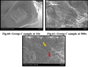
Fig.60 SEM of Group C pretreated enamel surface under 10x
Fig.61 SEM of Group C pretreated enamel surface under 500x
Fig.62 SEM of Group C pretreated enamel surface under 1000x
Fig.63 SEM of Group A debonded test sample under 10x
Fig.64 SEM of Group A debonded test sample under 500x
Fig.65 SEM of Group A debonded test sample under 1000x
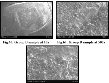
Fig.66 SEM of Group B debonded test sample under 10x
Fig.67 SEM of Group B debonded test sample under 500x
Fig.68 SEM of Group B debonded test sample under 1000x
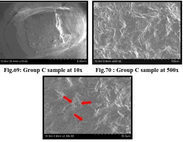
Fig.69 SEM of Group C debonded test sample under 10x
Fig.70 SEM of Group C debonded test sample under 500x
1
INTRODUCTION
The demand for esthetic restorations and for preservation of healthy
tooth structure has lead to the development and improvement of esthetic
restorative materials.10 Dental ceramics are appreciated as highly esthetic
restorative materials with optimal esthetic properties, that simulate the
appearance of natural dentition. Other desirable characteristics include
translucence, fluorescence, chemical stability, biocompatibility, low thermal
and electrical conductivity, compressive strength and a coefficient of thermal
expansion similar to that of tooth structure. In spite of their many advantages,
ceramics are fragile under tensile strain.This makes the ceramic susceptible to
fracture during the luting procedure and under occlusal force.8
Metal-backed ceramics were developed with the objective of
improving the mechanical properties of the overall restoration.46 The
ceramo-metal restoration, which combined the strength of ceramo-metal with the esthetics of
ceramic, improved the success of dental ceramics.8 The presence of the metal
sub-structure limits the optical properties of the ceramic due to reduced light
transmission and the tendency for marginal discolouration that affects the
esthetics.14,46 Developments in dental-ceramic engineering have led to the
introduction of new, commercially available systems that use a ceramic core to
2
The primary weakness of dental ceramics is their brittleness which is
likely to be their most important clinical characteristic. The esthetic and
biological advantages of ceramic restorations have led to many efforts to
improve the mechanical properties of dental ceramics.Several strengthening
techniques and principles were developed and has resulted in improved
mechanical properties and heightened esthetics of dental ceramics.21 These
strengthened ceramics have been named as metal-free ceramics, or
“All-ceramics” and have been indicated for inlays, onlays, crowns and fixed partial
dentures.8
All-ceramic restorations have gained popularity in recent years for the
restoration of anterior teeth due to their excellent esthetic quality,
biocompatibility and fracture resistance. They also have low thermal and
electrical conductance and a coefficient of thermal expansion that is similar to
enamel and dentin, resulting in minimal marginal leakage.29,35 These
restorations offer superior esthetics compared with metal-ceramic restorations.
The inherent brittleness of some ceramic materials, specific treatment
modalities and certain clinical conditions require resin bonding of the
completed ceramic restoration to the supporting tooth structures for long-term
clinical success.6
The success of all-ceramic restorations depends in part on a durable
bond being created between the hard tissues of the tooth and the adhesive
3
also critical throughout the lifetime of a restoration.35 A strong durable resin
bond provides high retention, improves marginal adaptation, prevents
microleakage and increases the fracture resistance of the restored tooth and
restoration.6,38 The bonded all-ceramic restorations provide a successful
esthetic and functional service for patients. Clinical studies show excellent
long-term success of bonded ceramic restorations such as inlays, onlays,
laminate veneers and crowns.27
Contemporary restorative dentistry places a definite emphasis on
adhesion.Accordingly, a long-term survival of adhesive porcelain restorations
depends on the success of a reliable bond between the porcelain, the
composite luting agent and the dental substrates.15,18
The ceramic restorations require considerable support from the
underlying luting agent and enamel/dentin in order to optimize the bond
strength between the restorations and the natural tooth.44,54 The durability and
the clinical performance of bonded porcelain restorations are mainly due to the
cementing agents and adhesive systems. The cementation procedure is one of
the factors for the clinical success of ceramic restoration.35,36 This includes
optimum surface treatment of the ceramic as well as proper choice and
manipulation of the luting agent.27 Therefore, adequate ceramic surface
conditioning is essential in order to have a strong resin bond that relies on the
micromechanical interlocking and chemical bonding to the ceramic surface.
4
diamond rotary instruments, airborne particle abrasion with aluminium oxide,
acid etching and combinations of any of these methods.7,48 Acid etching with
solutions of hydrofluoric acid (HF) or ammonium bifluoride can achieve
proper surface texture and roughness. Hydrofluoric acid solutions between
2.5% and 10% applied for 2 to 3 minutes seem to be most successful.6 Silane
coupling agent application improves the bond strength of porcelain to resin
luting agent.27
The surface treatment of dental substrate prior to adhesive restorative
procedures is an extremely important step of the bonding protocol and
accounts for the clinical success of restorations. In the literature, various
surface treatment methods like air abrasion, acid etching and laser irradiation
have been shown to etch enamel/dentin for the ceramic bonded restorations.4,19
Air abrasion is a technique that involves use of air pressure with aluminium
oxide powders to abrade dental tissues and produce large rough, irregular
surface areas.32 This can be regarded as a form of macroetching. The air
abraded surface (sand blasted) displayed obtuse angularities instead of the
sharp irregularities of etched enamel surfaces which could lead to weak bond
strengths.4
The chemical treatment of enamel was first proposed by Buonocore by
etching the enamel surface with orthophosphoric acid and has been commonly
used to increase the bond strength of bonded ceramic restorations.19 The
5
surface of enamel. This allows an increase in the prepared surface area
available for the retention of the resin cement and an improvement in the
marginal adaptation of all ceramic restorations. The retentive characteristics of
acid conditioned enamel surfaces depend on the type of acid, etching time and
chemical composition of the enamel. Acid etching contributes to
micromechanical retention of the adhesive components between the
restoration and the enamel. The disadvantage of acid etching is that
demineralization of the enamel surface makes it more permeable and prone to
long term acid attack and caries. Currently, the most widelyused protocol for
enamel etching is with 37% phosphoric acid for 15 seconds.19,31,51
Since the development of the ruby laser by Maiman in 1960, lasers
have become widely used in medicine and dentistry. Technological advances
during the last decade have resulted in the increased use of lasers in dentistry.
Many of these advances have been directed at the use of lasers in clinical
applications as an alternative to acid etching of enamel or dentin for bonding
dental materials to the tooth surface.26
The CO2 laser was the first dental laser approved by the US Food and
Drug Administration (FDA) and has been successfully used in soft tissue
surgeries. CO2 lasers have been reported to alter enamel surfaces in such a
way as to strengthen bonding of resin materials and these lased surfaces may
be superior to acid-etched enamel surfaces. The Nd:YAG laser uses a
6
Nd:YAG and ArF:excimer devices have been reported to engender a weaker
bonding surface than can be achieved with acid etching. Other approved
systems include the Er;Cr:YSGG laser and the Er:YAG laser. These systems
can be used for both soft and hard tissue procedures.30,50
The Er:YAG laser, originally developed by Zharikov et al in 1975, was
approved by the FDA in 1997 for removal of caries, cavity preparations and
modification of dentin and enamel surfaces prior to restoring with adhesive
restorations. The Er;Cr:YSGG laser system was investigated in 1995 by
Eversole and Rizolu. This pulsed laser device, when used with an air-water
spray, has cut enamel, dentin, cementum and bone efficiently and cleanly
without creating a significant smear layer. This laser system has been
designated as hydrokinetic system (HKS) and can be used for tooth
preparation without causing deleterious pulpal effects.10,30,50
Laser etching has also become available as an alternative to acid
etching of enamel and dentin. Laser irradiation in particular causes thermally
induced changes in the enamel surfaces. It causes surface roughening and
irregularity similar to those following acid etching. Laser etching is painless
and does not involve either vibration or heat, making it highly attractive for
routine use. Furthermore, laser etching of enamel has been reported to yield an
anfractuous surface (fractured and uneven) and open dentinal tubules, both
7
The surface produced by laser irradiation is also acid resistant. Laser
irradiation of the enamel modifies the calcium-phosphate ratio and leads to the
formation of more stable and less acid-soluble compounds, thus reducing
susceptibility to caries attack. Therefore laser etching of enamel might be
advantageous over phosphoric acid etching.50
The use of both laser and acid together has also been reported to
enhance the strength of bonding to hard tooth surfaces relative to those
exposed to acid alone.19
The type of luting cement has an influence on the long term durability
of bonded ceramic restorations. Since the use of all-ceramic restorations
requires considerable support from underlying composite resin cement and
enamel/dentin for a successful clinical outcome, the luting agent should have
high bond strength, not only to the ceramic surface, but also to tooth structure.
Resin cements have been selected for their advantageous mechanical and
adhesive properties when compared with the conventional luting cements.The
applications of dual-polymerizing resin cements for all-ceramic restorations
have considerably increased due to the ability of these cements to polymerize
completely and their greater resistance to occlusal loading.28,35,37,39,47
The international standards organization document, TR110405 Dental
Materials-Guidance has recommended longer periods of storage in a solution
may be necessary to determine durability of bonds.28 The complex nature of
8
between the interfaces of bonded ceramic restoration especially of the
cementing agent and hard tissue. Water absorption may reduce the mechanical
properties of the resin based luting agents and is detrimental to the
silane-ceramic bond.33,42 Therefore, testing the samples following water storage is
essential to better simulate the oral conditions and achieve predictable results.
The common tests used in literature for measuring the bond strength
are three-point bending, tensile, microtensile and shear bond strength tests.25
Shear strength testing is perhaps more clinically applicable because resistance
to shear stresses are thought to be important in retaining restorations that have
been bonded to enamel surfaces.30 In this study, a conventional shear bond
strength was used to evaluate the bond strength.
Studies that comparatively evaluate the shear bond strength between
ceramic and enamel subjected to acid etching or irradiated with different laser
systems are available.19,30,32,50,51 However, research comparing the effects of
Er;Cr:YSGG irradiated enamel with acid etched enamel on the shear bond
strength with ceramic is sparse.19,50,51 Also there are fewer studies comparing
the combined effects of acid etching followed by laser etching with
Er;Cr:YSGG laser system.
In light of the above, the aim of the present in vitro study was to
comparatively evaluate the shear bond strength of the bond between ceramic
9
The objectives of the present study included the following:
1. To evaluate the shear bond strength of the bond between ceramic and
enamel pretreated with 37% phosphoric acid etching.
2. To evaluate the shear bond strength of the bond between ceramic and
enamel pretreated with Er;Cr:YSGG laser etching.
3. To evaluate the shear bond strength of the bond between ceramic and
enamel pretreated with a combination of 37% phosphoric acid etching
followed by Er;Cr:YSGG laser etching.
4. To compare the shear bond strengths of the bond between ceramic
bonded to acid etched enamel, laser etched enamel and a combination
of acid and laser etched enamel.
5. To qualitatively analyse the surface topography of enamel pretreated
with 37% phosphoric acid etching before ceramic bonding by scanning
electron microscope (SEM) analysis.
6. To qualitatively analyse the surface topography of enamel pretreated
with Er;Cr:YSGG laser etching before ceramic bonding by scanning
electron microscope (SEM) analysis..
7. To qualitatively analyse the surface topography of enamel pretreated
with a combination of 37% phosphoric acid etching followed by
Er;Cr:YSGG laser etching before ceramic bonding by scanning
10
8. To qualitatively evaluate the mode of failure of debonded test sample
of ceramic bonded to enamel pretreated with 37% phosphoric acid
etching by scanning electron microscope (SEM) analysis.
9. To qualitatively evaluate the mode of failure of debonded test sample
of ceramic bonded to enamel pretreated with Er;Cr:YSGG laser
etching by scanning electron microscope (SEM) analysis.
10.To qualitatively evaluate the mode of failure of debonded test sample
of ceramic bonded to enamel pretreated with a combination of 37%
phosphoric acid etching followed by Er;Cr:YSGG laser etching by
11
REVIEW OF LITERATURE
Stangel I et al (1987)47 investigated the shear bond strength of
composite resin to porcelain to optimize variables, of etching and use of type
of composite, for bonding porcelain laminate veneers. Composite was bonded
onto both etched and non etched porcelain using unfilled resin, silane and
silane with dentin adhesive. The conclusion they derived was that porcelain
etching significantly increased bond strength across all variables.
al Edris A et al (1990)1 evaluated the etch patterns produced by 1) a combination of hydrofluoric acid, hydrochloric acid and nitric acid, 2) a
combination of hydrofluoric acid and sulphuric acid and 3) acidulated
phosphate fluoride gel on porcelain surface. They concluded based on the
SEM analysis that hydrofluoric acid and its combinations produced the most
similar etch patterns consistently and produced greater roughness which the
authors concluded to mean greater retention.
Visuri SR et al (1996)53 evaluated the shear bond strength of the
composite bonded to Er:YAG laser prepared dentin. The authors used human
extracted molars and the teeth were prepared to the dentinal surfaces with a
laser or with a dental aerotor handpiece. From these samples a few of them
were further surface treated by etching with acid while others were not before
12
irradiated samples had improved bond strengths when compared with the other
samples.
Brosh T et al (1997)9 evaluated the effect of different combinations of surface treatments and bonding agents on the bond strength of repaired
composites. Three hundred and sixty samples were split into six groups with
one group serving as control and the other five were subjected to the following
surface treatments 1) grinding with a diamond stone 2) sandblasting with
microetcher 3) jet prophylaxis 4) grinding with green carborundum
5) hydrofluoric acid – etching (9%). The authors concluded that different
combinations of surface treatments and bonding agents affect the bond
strength with sandblasting surface treatment recording the highest value and
hydrofluoric acid etching recording the lowest value of shear bond strength.
Chen JH et al (1998)12 investigated the effect of different etching periods on the bond strength of a composite resin to porcelain. They used 5%
hydrofluoric acid and examined different times of 0, 5, 30, 60, 120 and 180
seconds with sixteen samples in each group. The etched patterns created were
observed under a scanning electron microscope and the bond strengths were
tested under a universal testing machine. The authors concluded that etching
porcelain for 120 seconds gives the highest bond strength.
Iwami Y et al (1998)23 investigated the effect of the wetness of enamel and dentin surfaces on the shear bond strength of composites. After testing
13
preparations the authors concluded that some amount of saturation was
necessary for dentin surfaces to obtain high bond strength. However the
relative dryness or the wetness of the enamel surface had no bearing
whatsoever on the bond strength as measured by the authors.
Lin S et al (1999)30 assessed the shear bond strength of composite
bonded to tooth structure treated with an Er;Cr:YSGG laser system and
compared it to a surface treated with carbide burs. The teeth were prepared
along their long axes and were cut into both the enamel and dentin. They were
also divided into two subgroups of etched and non-etched. A SEM analysis of
the laser prepared surface and the carbide bur prepared surface was also done
revealing that laser prepared surface did not cause the formation of a smear
layer and an almost similar topography for the bur prepared surface was
observed. No significant differences were also observed by the authors across
their groups.
Martinez-Insua A et al (2000)31 evaluated the tensile bond strength of
teeth treated with an Er:YAG laser and acid-etched teeth. Eighty healthy
human premolars were used. Brackets were cemented to acid-etched enamel,
laser-etched enamel, acid-etched dentin, or laser-etched dentin (20 teeth per
group). Dentin was previously exposed using a high-speed handpiece.
Acid-etching was with 37% orthophosphoric acid (15 seconds for enamel, 5 seconds
for dentin). Laser etching was with Er:YAG laser (four 200 mJ pulses per
14
bonded with auto – curing resin paste, having first applied a primer (dentin
only) and then light-cured bonding resin. The authors concluded that adhesion
to dental hard tissues after Er:YAG laser etching is inferior to that obtained
after conventional acid etching. Enamel and dentin surfaces prepared by
Er:YAG laser etching show extensive subsurface fissuring that is unfavourable
to adhesion.
Hara AT et al (2001)22 evaluated the influence of different cross head
speeds on shear bond strength test on the tooth surface using one hundred and
twenty extracted bovine incisors, embedded in resin. According to the authors
different cross head speeds influence the shear bond strength of the material
being tested and its fracture pattern. They also advocated cross head speeds of
0.50 and 0.75 mm/min for obtaining accurate results.
Kitasako Y et al (2001)28 studied the shear bond strengths of three resin cements to dentin over a period of three years in vitro. Ten bovine teeth
were used each with three different materials Panvia21, BISTITE and MASA
bond. The bond strengths were evaluated at 1 day, six months, one year and
three years. The samples were stored in plain tap water at 37o C, with the
water being changed on a daily basis. In this study the authors found that
MASA bond an auto polymerizing resin cement recorded the highest bond
strengths throughout the time period but the bond strength of all the cements
15
Shimada Y et al (2002)44 evaluated the shear bond strength of dual-cured resin cement to glass ceramics after sandblasting, acid etching and
silanation. A castable glass class ceramic with a crystalline phase was used as
the substrate material. The glass surfaces, which were sandblasted, polished or
etched with phosphoric acid or hydrofluoric acid and were subsequently
bonded with a dual-cured resin cement both with and without a silane coupling
agent. The authors concluded that a silane coupling agent mixed with an acidic
primer can effectively increase the bonding strength between resin cement and
cast glass ceramics.
Stewart GP et al (2002)48 evaluated in vitro the shear bond strength of resin cements to both ceramic and dentin. The ceramic specimens received six
different surface conditioning treatments: sanding with 600-grit silicon carbide
paper, microetching with aluminium oxide, sanding followed by silane
application, microetching followed by silane application, hydrofluoric
acid-etching and hydrofluoric acid-acid-etching. The authors concluded that bond
strengths were highly dependent on surface conditioning with hydrofluoric
acid etching followed by silane application emerging as the most effective and
reliable method in their study.
Cura C et al (2003)15 evaluated the shear bond strength of a luting
composite to enamel with six different bonding systems. Seventy extracted
human molars and premolars were used for the study onto whom ceramic
16
of ten each, with the last group serving as a control in which no bonding agent
was used. The authors concluded that, though no significant variance was
observed between the systems, the use of a bonding agent greatly increased
the bond strength.
Khoroushi M et al (2003)25 evaluated the effect of thermocycling on
the shear bond strength of composite resin to porcelain. In this experimental
study, forty porcelain blocks were prepared and randomly divided into four
groups (n=10). All porcelain surfaces were etched with 9.6% hydrofluoric
acid, rinsed and air dried. In two groups, silane pre-treatment was done.
Composite-resin was subsequently added on the ceramic surfaces, and
light-cured. A group each of specimens with the silane pre-treatment and without
the silane pretreatment were then subjected to 1000 thermal cycles. The
authors found that the shear bond strengths of sample decreased considerably
after thermocycling.
Spohr AM et al (2003)46 studied the influence of six different surface treatments on the tensile bond strength between a resin cement and ceramic,
with and without the application of a silane coupling agent. The six methods
studied were sandblasting (100 um) with no silanation, sandblasting (100 um)
with silanation, sandblasting (50 um) with no silanation, sandblasting (50 um)
with silanation, hydrofluoric acid etching with no silanation, hydrofluoric acid
17
improved the bond strength within the same groups and hydrofluoric acid
etching recorded the highest bond strength values.
Usumez A et al (2003)51 evaluated in vitro the bond strengths of porcelain laminate veneers to tooth surfaces prepared with acid and
Er;Cr:YSGG laser conditioning. Three surface treatments were used: laser
conditioning with Er;Cr:YSGG, 37% phosphoric acid, 10% maleic acid. The
in vitro bond strengths of porcelain laminate veneers bonded to tooth surfaces
that were laser etched showed results similar to orthophosphoric acid or
maleic acid etched tooth surfaces.
Piwowarczyk A et al (2004)37 investigated the in vitro shear bond
strength of cementing agents to fixed prosthodontic restorative materials.
High-gold-content alloy and high-strength aluminium oxide surfaces were
airborne-particle–abraded, and pressable ceramics were hydrofluoric
acid-etched and silanated prior to cementing. The cementing agents tested were a
zinc-phosphate cement, glass ionomer cements, resin-modified glass ionomer
cements and resin cements. The authors’ findings indicated that resin cements
exhibited strong bond strengths to specific prosthodontic materials.
Ramos RP et al (2004)41 investigated the effect of Er:YAG laser on bonding to dentin and the interaction pattern of different adhesive systems
with the lased substrate. Tensile bond strength of a self-etching and two
total-etch systems to lased and non-lased dentin was evaluated and the adhesive
18
consistent hybrid layers were observed for conventionally treated specimens;
whereas they were either absent or scarce hybridization zones were viewed for
the lased subgroups.
Celik EU et al (2006)10 evaluated the shear bond strength of different
adhesives to Er:YAG laser prepared dentin. Seventy specimens obtained from
35 extracted human molars were embedded in polyester resin and ground with
silicon carbide papers. The authors concluded that Er:YAG laser irradiation
increased the shear bond strength to dentin.
Chimello-Sousa DT et al (2006)13 evaluated the influence of Er:YAG
laser irradiation on the bond strength of a restorative system on enamel,
varying the irradiation distance. The samples were divided into six groups,
with the first five being treated with Er:YAG laser with the irradiation distance
at 11, 12, 14, 16 and 17 mm, while the last group served as the control and
received treatment with phosphoric acid alone. The authors concluded that
with increase in irradiation distance, the bond strength increased.
Souza-Gabriel AE et al (2006)45 investigated the shear bond strength of rein modified glass ionomer cements to ER:YAG laser treated tooth
structure. The authors found that the adhesion for enamel was more efficient
than for dentin. The cavities prepared with a conventional bur (control group)
presented higher bond strength values than those recorded for Er:YAG laser.
Kukiattrakoon B et al (2007)29 studied the effect of different etching
times of acidulated phosphate fluoride gel on the shear bond strength of high
19
was no significant difference in the bond strengths in either of the two
different surface treatments when the times for APF use were between 7 to 10
minutes.
Duarte S et al (2009)18 studied the effectiveness of immediate dentin
sealing (IDS) on the marginal adaptation and tensile bond strength of total –
etch and self etch adhesives. The authors used twenty recently extracted
molars and standard MOD inlay preparations were made on them. The authors
came to the conclusion that immediate dentin sealing greatly improves the
bond strength when compared with conventional composite cementation
technique.
Pekkan G et al (2009)35 examined both the shear and tensile bond strengths between pressable ceramic and resin cements. Three commercially
available dual polymerizing resin cements were used to bond the two different
ceramic systems to a total of one hundred and twenty extracted human molar
teeth. All the specimens were thermocycled before being sent for the tests. The
authors state that cementing agents influence the bond to the hard tissue with
the shear bond strength values being consistently and significantly higher than
tensile bond strength values.
Ritter AV et al (2009)42 evaluated the shear bond strengths of
dual-cure composite luting agents used with dual-dual-cure dental adhesives. The
authors reported that on enamel, the total-etch adhesives performed better than
20
self-etch adhesives performed better than their total-etch counterparts.
Thermocycling for 1800 cycles did not affect the shear bond strength of the
materials tested to dentin and enamel.
Moslemi M et al (2010)32 compared in vitro the shear bond strength of
a fissure sealant to enamel penetrated with Er;Cr:YSGG laser or air abrasion
followed by acid etching. The authors concluded that pretreatment of enamel
surfaces with the Er;Cr:YSGG laser did not increase the effectiveness of
conventional acid etching and subsequently the bond strength as opposed to
pretreatment of the enamel surfaces with air abrasion
Qeblawi DM et al (2010)39 studied the effect of zirconia surface treatment on the flexural strength and shear bond strength to a resin cement.
The mechanical treatments used were: airborne particle abrasion, silicoating
and wet hand grinding. The chemical treatments used were acid etching
followed by silanation, silanation only, and application of zirconia primer. The
authors concluded that a combination of mechanical and chemical
conditioning of the zirconia surface was essential to develop a durable resin
bond to zirconia.
Turkmen et al (2010)50 evaluated the shear bond strength of composite bonded with three different adhesive systems to Er;Cr:YSGG laser
prepared enamel. The bond strengths obtained were not significant between
the non-etched and laser etched groups, however for the etched groups laser
21
Er;Cr:YSGG laser-powered hydrokinetic system etched the enamel more
effectively than 37% phosphoric acid.
Yuasa T et al (2010)55 evaluated the effects of two years of storage on the shear bond strength of two self-etching adhesive systems studied. The
authors concluded that both the self etching primer adhesive systems,
produced adequate shear bond strength even after 2 years of storage and
thermocycling between 5o C and 55°C for 6000 cycles.
Dundar et al (2011)19 evaluated the strength of the bond between porcelain laminate veneers and tooth surfaces etched with acid and laser,
separately and together. The teeth studied comprised 60 incisors extracted for
periodontal reasons. These were divided into four groups according to etching
method: group 1, acid etching alone; group 2, acid etching followed by laser
etching; group 3, laser etching followed by acid etching; group 4, laser etching
alone. The teeth were etched with 37% phosphoric acid and an Er;Cr:YSGG
laser system. After the shear tests, scanning electron microscopy images of the
tooth surfaces were obtained at a magnification of ×3,800. Etching with acid
alone yielded the highest mean value of bond shear strength (15.4±3.8 MPa),
while laser etching followed by acid etching gave the lowest mean value
(11.5±4.6 MPa). The mean values of the bond shear strength for acid etching
followed by laser etching and laser etching alone were 13.8±3.9 MPa and
12.8±4.6 MPa, respectively. Statistical analysis revealed no significant
22
MATERIALS AND METHODS
The present in vitro study was done to comparatively evaluate the
shear bond strength of the bond between ceramic and enamel pretreated with
different etching methods.
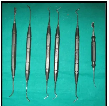
MATERIALS EMPLOYED:
1. 33 recently extracted maxillary central incisors (Fig.1)
2. Separating discs 0.7 mm thickness (Dentorium, New York, USA) (Fig.2)
3. Autopolymerizing clear acrylic resin (Cold cure, DPI-RR, India) (Fig.3)
4. Die lubricant (Yeti Dental, Germany) (Fig.4)
5. Inlay wax (GC Corporation, Tokyo, Japan) (Fig.5)
6. Sprue wax (Bego, Germany) (Fig.6)
7. Investment ring and crucible former (IPS Silicone Ring, Ivoclar
Vivadent, Liechtenstein) (Fig.7)
8. Pattern sprue guide (IPS e.max Press Sprue Guide, Ivoclar Vivadent,
Liechtenstein) (Fig.8)
9. Phosphate bonded investment material (Pressvest, Ivoclar Vivadent,
Liechtenstein) (Fig.9)
10.Colloidal silica (Pressvest Liquid, Ivoclar Vivadent, Liechtenstein)
(Fig.10)
11.Aluminum oxide powder 110 microns (Aluminox 110, Delta , India)
(Fig.11)
23
13.Silicon carbide impregnated burs coarse (Dura Green, Shofu Dental,
Japan) (Fig.13)
14.Silicon carbide impregnated burs fine (Dura White, Shofu Dental, Japan)
(Fig.14)
15.Primer (Syntac Primer, Ivoclar Vivadent, Liechtenstein) (Fig.15a)
16.Adhesive (Syntac Adhesive, Ivoclar Vivadent, Liechtenstein) (Fig.15b)
17.Bonding agent (Heliobond, Ivoclar Vivadent, Liechtenstein) (Fig.15c)
18.Silane coupling agent (Monobond S, Ivoclar Vivadent, Liechtenstein)
(Fig.15d)
19.Dual-cure resin luting cement (Variolink N, Ivoclar Vivadent,
Liechtenstein) (Fig.16)
INSTRUMENTS AND EQUIPMENTS EMPLOYED:
1. P.K. Thomas wax up instruments (Dispodent, India) (Fig.17)
2. Aerotor hand piece (Pana air, NSK, Japan) (Fig.18)
3. Inverted cone diamond abrasive (Dia Burs, Mani, Germany) (Fig.19)
4. Flat end tapered diamond abrasive (Dia Burs, Mani, India) (Fig.20)
5. Light cure unit (Confident, India) (Fig.21)
6. Vacuum mixer (Whipmix, U.S.A) (Fig.22)
7. Burnout furnace (Technico, Technico Laboratory Products Pvt. Ltd.,
Chennai, India) (Fig.23)
8. Sandblaster (Delta, India) (Fig.24)
9. Incubator (Narang Industries Ltd., India) (Fig.25)
24
11.Scanning electron microscope (SA400N, Canada) (Fig.27)
12.Custom-made stainless steel split mounting jig (for mounting teeth in
acrylic) (Fig.28)
13.Custom-made stainless steel tooth preparation guide (Fig.29)
14.Custom-made stainless steel split mold (for fabricating wax blocks)
(Fig.30)
Ceramic system employed:
1. Lithium disilicate ingots MO shade (IPS e.max Press, Ivoclar
Vivadent, Liechtenstein) (Fig.31)
2. Boron nitride (IPS e.max Alox Plunger Separator, Ivoclar Vivadent,
Liechtenstein) (Fig.32a)
3. Plunger (IPS e.max Alox Plunger, Ivoclar Vivadent, Liechtenstein)
(Fig.32b)
4. Ceramic press furnace (Programat EP-3000, Ivoclar Vivadent,
Liechtenstein) (Fig.33)
5. 7% Hydrofluoric acid gel (IPS Ceramic Etching Gel, Ivoclar Vivadent,
Liechtenstein) (Fig.34)
Etching systems employed:
1. 37% Phosphoric acid etching gel (N-Etch, Ivoclar Vivadent,
Liechtenstein) (Fig.35)
2. Er;Cr:YSGG Laser (Waterlase MD Turbo, Biolase Technology, United
25
Description of custom-made stainless steel split mounting jig: (Fig.28)
In the present study, a custom-made stainless steel split jig (Fig.28)
was fabricated to mount the extracted natural teeth. The dimensions of the jig
were 75mm x 25mm x 25mm (l x b x h) with an inner open space of 25 mm x
15mm x 25mm (l x b x h), for embedding the tooth in acrylic resin blocks. The
jig was sectioned into two exact halves along the length and the two parts were
retained by screws. This was done to facilitate easy removal of the acrylic
block with the embedded tooth and also for subsequent reseating during tooth
preparation. The jig had four screw holes on the top to help in positioning and
seating the custom-made tooth preparation guide onto its surface.
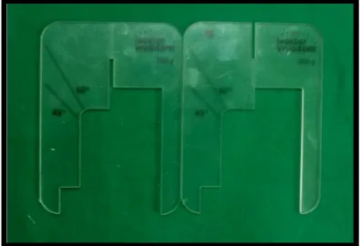
Description of custom-made stainless steel tooth preparation guide:
(Fig.29)
In the present study, a custom-made stainless steel tooth preparation
guide (Fig.29) measuring 65 mm x 50 mm x 5mm (l x b x h) with a central
preparation box measuring 5mm x 5mm was fabricated. The custom-made
tooth preparation guide had four screw holes through which it was secured
onto the top of the mounting jig during tooth preparation. This metallic guide
was used to prepare the natural tooth embedded in the acrylic block which was
secured in the stainless steel split mounting jig.
Description of custom-made stainless steel split mold: (Fig.30)
In the present study, a custom-made stainless steel split mold (Fig.30)
was fabricated to obtain the wax blocks. The dimension of the custom-made
26
Its dimensions are 65 mm x 45 mm x 5 mm (l x b x h). The mold was also
split along the center to help in easy retrieval of the wax blocks. The mold was
first milled to size using a commercial lathe for its exterior dimensions. The
interior dimensions of the mold space (5 x 5 mm) were obtained thereafter.
Description of Er;Cr:YSGG laser system: (Fig.36)
In the present in vitro study the laser system used for surface treatment
was from Biolase Technology, USA and the model was Waterlase MD Turbo
(Fig.36) which is an Er;Cr:YSGG (Erbium; Chromium: Yttrium, Scandium,
Gallium, Garnett) laser having a wavelength of 2780 nm. The power settings
can be adjusted from 0.1 W to 8.0 W as per the clinician’s requirement and the
procedure attempted. The pulse repetition rate of the system also offers an
individual preference from 10 to 50 Hz pulses. The optical tips available can
focus the laser beam to either a 500 or a 700 micron diameter depending on
the size used. This laser acts by its absorbtion into the chormophores present
in the target tissue namely hydroxyapatite and water. The chormophores upon
absorbing the laser energy are caused to expand rapidly bringing about the
action of ablation.
Description of universal testing machine: (Fig.26)
In the present study, the shear bond strength between ceramic and
surface treated enamel was determined with the universal mechanical testing
machine (Lloyd Instruments, Farnham, U.K.) (Fig.26). It consists of a lower
chamber, upper chamber, a display board to display the amount of force
27
machine. The lower portion has a bench vice test specimen fixture to hold the
test specimens. The whole unit is attached to a computer for recording and
converting data as required.
Description of the Scanning Electron Microscope: (Fig.27)
In the present study, the surface of the test samples was analyzed
qualitatively using a Scanning Electron Microscope (SEM) (SA400N, Canada)
(Fig.27). The SEM uses a beam of highly energetic electrons to examine
objects on a very fine scale. They reveal the fine structure of variety of
materials. SEM uses a scanned beam instead of a fixed beam, and it is used
primarily for the examination of thick samples through which light cannot
pass. The specimens to be magnified may have some conductivity and may get
charged up. Hence they are coated with a platinum layer to prevent the
charging up and in order to increase the secondary emissions. Additional
sputter coating with gold produces high contrast and resolution, while also
28
METHODOLOGY
The following methodology was adopted to comparatively evaluate the
shear bond strength of the bond between ceramic and enamel pretreated with
different etching methods.
I. Selection of teeth
II. Placement of teeth in custom-made jig
III. Preparation of teeth
IV. Fabrication of ceramic blocks
a. Preparation of wax blocks
b. Spruing of wax blocks
c. Investing the blocks
d. Burnout procedure for wax blocks
e. Pressing of ceramic
f. Divesting and finishing of ceramic blocks
g. Preparation of ceramic blocks for bonding
V. Grouping of prepared teeth for etching procedures
VI. Etching of prepared teeth surfaces
a. Group A: samples were acid etched with 37% phosphoric acid
b. Group B: samples were laser etched with Er;Cr:YSGG laser
system
c. Group C: samples were acid etched with 37% phosphoric acid
followed by etching with Er;Cr:YSGG laser system
29
VIII. Aging of test samples
IX. Shear bond strength test for test samples
X. Statistical analysis
XI. Qualitative analysis of the surface topography of surface treated,
prepared teeth samples before bonding with ceramic blocks
XII. Qualitative analysis of the cemented test samples after debonding
I. Selection of teeth (Fig.37a, b, c)
Thirty three freshly extracted maxillary central incisors (Fig.37a) were
utilised for the study which were free of caries, fractures, and restorations. The
crown lengths were measured from the cemento-enamel junction to the incisal
edge and from the mesial line angle to the distal line angle. A minimum length
of 12 mm and a width of 10 mm were maintained for all the specimens. The
selected teeth were sectioned (Fig.37c) at 2 mm below their cementoenamel
junction using a separating disc (Dentorium, New York, USA) (Fig.2). While
sectioning the teeth, care was taken that the teeth were kept moist. On the
palatal surface of the crowns two longitudinal 2 mm deep grooves of 1 mm
width were made to aid in the retention of the sectioned crowns with the
acrylic.
II. Placement of teeth in custom made stainless steel split jig:
(Fig.38a, b)
The inner surfaces of both halves of the custom made stainless split jig
were then coated uniformly with petroleum jelly and then screwed tightly into
30
then poured into the mold space till the top and the sectioned natural tooth was
embedded into the acrylic resin (Fig.38a). The natural tooth was embedded in
such a way that the labial surface was exposed, for tooth preparation. Once the
excess was removed, the custom made stainless steel preparation guide was
then placed over the custom made stainless steel split jig and then secured into
place further ensuring that the crown was mounted correctly. Once the
autopolymerizing resin (Cold cure, DPI-RR, India) (Fig.3) had sufficiently
cured, the custom made stainless steel preparation guide was unscrewed and
the custom made stainless steel split mold was separated by removing its
screws and the acrylic block was retrieved (Fig.38b). The selected thirty three
natural teeth were embedded into the acrylic resin in an identical manner.
III. Preparation of teeth: (Fig.39a, b, c, d, e)
The middle portion of the labial surface of the teeth was selected for
the preparation because of its larger width. The acrylic block with the
embedded tooth was positioned in the custom made stainless steel split
mounting jig (Fig.28), and was secured tightly. The custom made stainless
steel tooth preparation guide (Fig.29) was then placed on top and locked in
place (Fig.39a). This enabled to make the preparation in the middle one third
of the tooth with the guide.
Premarked inverted cone burs (Dia Burs, Mani, Germany) (Fig.19),
were used with a 7 mm marking on their shanks (Fig.39a) measured from the
tip to prepare through 2 mm into the enamel surface as the thickness of the
31
preparation in accordance with the markings on the burs, so as to not extend
into the dentin surface (Fig.39c). The preparation was done in order to
simulate the clinical preparation of ceramic laminate veneer restoration. After
accomplishing the general outline of the intended test sample (a 5 x 5 mm
square with 2 mm depth) (Fig.39d) the area was marked and the tooth
structure around this area was ground using a flat end tapered diamond
abrasive (Dia Burs, Mani, Germany), to ensure no impedance during the test
for shear bond strength (Fig.39e). In this manner a total of 33 teeth were
prepared and randomly grouped as described later.
IV. Fabrication of ceramic blocks:
a. Preparation of wax blocks: (Fig.40a, b, c)
The custom made stainless steel split mold was lined with die lubricant
(Yeti Dental, Germany) (Fig.4) on each side of the mold spaces to aid in the
retrieval of the wax blocks. The wax custom made stainless steel split mold
was then secured close and placed over a clean glass plate flush with its
surface (Fig.40a). Inlay wax (GC Corporation, Tokyo, Japan) (Fig.5) was
poured into the mold space in a molten state and was allowed to cool gradually
at room temperature (Fig.40b). Then the mold was placed in a bowl of chilled
water to further harden the wax blocks, for a minute. After this the mold was
removed from the bowl and wiped dry. Before the screws on the split mold
were removed the excess formed at the top was then gently carved out using a
32
(measuring 5 x 5 mm) was then eased out with gentle finger pressure
(Fig.40c). In this manner a total of 30 wax blocks were obtained.
b. Spruing of wax blocks: (Fig.41a, b)
The wax blocks were sprued using preformed sprue wax (Bego,
Germany) (Fig.6) of 2 mm diameter. The sprued wax blocks were then
attached onto the crucible former (Fig.40a) of the sili ring (IPS Silicone Ring,
Ivoclar Vivadent, Liechtenstein) (Fig.7). They were then measured for
distance and angulation using the manufacturers provided guide (IPS e.max
Press Sprue Guide, Ivoclar Vivadent, Liechtenstein) (Fig.8) at an angle
between 45o and 60o (Fig.41b). The sprued wax blocks with the crucible
former were then placed inside the sili ring (IPS Silicone Ring, Ivoclar
Vivadent, Liechtenstein) (Fig.7).
c. Investing the wax blocks: (Fig.42a, b, c, d)
The wax blocks were invested using graphite free, phosphate bonded
investment material (Pressvest, Ivoclar Vivadent, Liechtenstein) (Fig.9).
A 6 mm distance was provided between the wax blocks and top of the ring.
As per the manufacturer’s recommendation, 200 gm of phosphate bonded
investment (Pressvest, Ivoclar Vivadent, Liechtenstein) (Fig.9) was mixed
with 44ml of investment liquid which was prepared by mixing 26 ml of
colloidal silica (Pressvest Liquid, Ivoclar Vivadent, Liechtenstein) (Fig.10)
and 18 ml of distilled water. The investment powder and liquid were first hand
mixed with a spatula until the entire material was wet thoroughly, followed by
33
for 60 seconds. Once the investment was mixed the entire block was painted
with a thin layer of investment using a small brush (Fig.42a). The sili ring
(IPS Silicone Ring, Ivoclar Vivadent, Liechtenstein) (Fig.7) was placed on the
vibrator and the remainder of investment was vibrated slowly into the ring
(IPS Silicone Ring, Ivoclar Vivadent, Liechtenstein) (Fig.42b). The excess
investment was then removed (Fig.42c). The invested blocks were allowed to
set for 60 minutes, and the sili ring was removed (Fig.42d).
d. Burnout procedure for the wax blocks: (Fig.43)
The invested molds were placed in a burnout furnace (Technico,
Technico Laboratory Products Pvt. Ltd., Chennai, India) (Fig.23) after setting
of the investment, for wax elimination. Investments with the wax blocks were
left in the burnout furnace for a period of two and half hours. During the first
hour, the temperature was raised from room temperature to 380°C; in the
second hour, the temperature was raised to 900°C and during the last half hour
the temperature was sustained at 900°C to accomplish complete burnout of the
pattern without any residue. The investment mold was initially placed in the
furnace (Technico, Technico Laboratory Products Pvt. Ltd., Chennai, India)
(Fig.43) towards the rear wall, tipped with the opening facing down towards
the floor of the furnace for the escape of molten material but not flush against
it. The investment mold was reversed later near the end of the burnout cycle
with the sprue hole facing upward to enable escape of the entrapped gases and
34
e. Pressing of ceramic samples: (Fig.44a, b ,c, d, e)
The investment mold was then carried to the press furnace (Programat
EP-3000, Ivoclar Vivadent, Liechtenstein) (Fig.33) and placed on the centre of
the mounting plate (Fig.44c). The selected ingot (IPS e.max Press, Ivoclar
Vivadent, Liechtenstein) (Fig.31) was then loaded (Fig.44a) with the shade
designation facing upward and after the plunger (IPS e.max Alox Plunger,
Ivoclar Vivadent, Liechtenstein) (Fig.32b) was dipped in the plunger separator
(IPS e.max Alox Plunger Separator, Ivoclar Vivadent, Liechtenstein) (Fig.32a)
to avoid adherence to the investment material and it was placed upon the ingot
(Fig.44b). The manufacturer’s pre-set program for the mold size was selected
and activated. The base temperature of which was at 700o C, with a standby
time of 6 minutes. The temperature rise was set to gradually increase to 920 o
C over a period of 60 minutes, at which time the ingot was pressed into the
mold. Following pressing, the mold was allowed to cool to room temperature
(Fig.44d). It was then cut carefully (Fig.44e) to be divested subsequently.
f. Divesting and finishing of ceramic samples: (Fig.45a, b, c)
The remaining investment was slowly removed from the casting by
sand blasting (Fig.45a) with 110µm alumina (Aluminox 110, Delta, India)
(Fig.11) at 80 psi pressure in a sand blasting machine (Delta, India) (Fig.24).
Sprues were sectioned (Fig.45b) using 0.7mm thin diamond discs (Edenta AG,
Switzerland) (Fig.12). The sample was inspected under magnification for
pressing defects. External surfaces were relieved of all nodules with a silicon
35
cleaned. This procedure was repeated for all thirty specimens. All the ceramic
samples were finished (Fig.45c) using silicon carbide impregnated burs (Dura
White, Shofu Dental, Japan) (Fig.14).
g. Preparation of ceramic blocks for bonding: (Fig.46)
The ceramic samples finished in the above manner were then placed
against a marked glass plate to check for its flatness. The flat ceramic surface
was etched with 7% hydrofluoric acid gel (IPS Ceramic Etching gel, Ivoclar
Vivadent, Liechtenstein) (Fig.34) for 1 minute in order to condition the
ceramic.
V. Grouping of prepared teeth for etching procedures:
The teeth were divided into three groups, namely, Group A, Group B
and Group C and subjected to three different surface treatments, namely, acid
etching, laser etching and a combination of acid etching followed by laser
etching respectively.
VI. Etching of prepared teeth surfaces: (Fig.47a, b, c, d, e)
Group A – Acid etching with 37% phosphoric acid:
37% orthophosphoric acid (N Etch, Ivoclar Vivadent, Liechtenstein)
(Fig.35) was injected onto the prepared enamel surface of the teeth in Group A
(Fig.47a) and left for 15 seconds. The tooth surface was then washed with
water under pressure using a two way syringe. Each surface was then dried
using a chip blower only. The treated specimen (Fig.47c) was then kept aside
36
the ceramic sample. In this manner a total of 11 teeth samples for Group A
(n=11) were subjected to acid etching.
Group B – Laser etching with Er;Cr:YSGG laser system:
Er;Cr:YSGG laser system (Waterlase MD, Biolase, USA) (Fig.36) was
used to ablate the prepared enamel surface of the teeth in Group B (Fig.47b).
The distance between the tip of the device and the surface of the sectioned
crown was kept at 1 mm, and the laser beam was applied to the entire surface
for 20 seconds. The laser was applied at a wavelength of 2,780 nm with pulse
duration of 140 μs and a repetition rate of 15 Hz. The laser energy was 75 mJ.
Laser energy was delivered through a fibre-optic system via a sapphire tip terminal 600 μm in diameter and the surface was bathed with an adjustable
air/water spray using a water level of 30% and an air level of 60%. The treated
specimen (Fig.47d) was dried using a chip blower and then kept aside
carefully in a separate container to prevent it from contamination before
bonding it with the ceramic block. In this manner a total of 11 teeth samples
for Group B (n=11) were subjected to laser etching.
Group C – Combination of acid etching with 37% phosphoric acid
followed by laser etching with Er;Cr:YSGG laser system:
37% orthophosphoric acid (N Etch, Ivoclar Vivadent, Liechtenstein)
(Fig.35) was injected onto the prepared surface of the teeth and left for 15
seconds. The teeth surface was then washed with water under pressure using a
two way syringe. Each surface was then dried using a chip blower only.
37
ablate the prepared surface of the tooth thereafter. The distance between the
tip of the device and the surface of the sectioned crown was kept at 1 mm, and
the laser beam was applied to the entire surface for 20 seconds. The laser was
applied at a wavelength of 2,780 nm with pulse duration of 140 μs and a
repetition rate of 15 Hz. The la