A STUDY ON COMPARISON OF VARIOUS METHODS
OF DETECTION OF METHICILLIN RESISTANT
STAPHYLOCOCCUS AUREUS
DISSERTATION SUBMITTED FOR
BRANCH IV
–
M.D. DEGREE
(MICROBIOLOGY)
APRIL 2012
THE TAMILNADU
DR.M.G.R. MEDICAL UNIVERSITY
BONAFIDE CERTIFICATE
This is to certify that the dissertation entitled
“A STUDY ON COMPARISON OF VARIOUS METHODS OF DETECTION OF METHICILLIN RESISTANT STAPHYLOCOCCUS AUREUS’’ submitted by Dr.T.RAJENDRAN to The Tamil Nadu Dr.M.G.R Medical University, Chennai in partial fulfillment of the requirement for the award of M.D degree Branch IV (Microbiology) is a bonafide research work carried out by him under direct supervision & guidance.
Director,
Institute of Microbiology,
Madurai Medical College,
Madurai.
DECLARATION
I, Dr.T.RAJENDRAN declare that, I carried out this work on
“A STUDY ON COMPARISON OF VARIOUS METHODS OF DETECTION OF METHICILLIN RESISTANT STAPHYLOCOCCUS AUREUS’’ at the institute of Microbiology, Madurai Medical College. I also declare that this bonafide work or a part of this work was not submitted by me or any others for any award, degree, or diploma to any other University , Board, either in India or abroad.
This is submitted to The Tamil Nadu Dr.M.G.R Medical University, Chennai in partial fulfillment of the rules and regulations for the M.D Degree examination in Microbiology.
ACKNOWLEDGEMENT
I am grateful to The Dean, Madurai Medical College and Government Rajaji Hospital, Madurai for permitting me to carry out this study.
I wish to express my deep sense of gratitude and sincere thanks to Professor Dr.M. Mohamed Meeran M.D; D.V; M.D.,
Director, Institute of Microbiology, Madurai Medical College, for his constant help, guidance and encouragement given to me throughout this study.
I would like to express my sincere thanks and gratitude to
Dr.P.A.T.Jagadheeswari M.D., Professor, for her guidance and encouragement given to me during this study.
I am highly indebted to Dr.Jhansi Charles M.D., Professor, whose sincere guidance and encouragement were a source of strength, right from selecting the topic till the submission of the Dissertation.
I express my sincere thanks to Professor Dr.R.Vibhusanan M.D., and Professor Dr.V.Dhanalakshmi M.D., for their valuable suggestions and moral support given to me throughout the study.
I express my sincere thanks to Assistant Professors
Dr.N.Ram murugan M.D., Dr.N.Anuradha M.D., Dr.M.R.Vasantha priyan M.D., Dr.D.S.Kavitha M.D., and Senior Entomologist Mr.John Victor Ph.D for thier valuble suggestions.
I am thank full to my colleagues, Dr.D.Therese Mary, Dr.S.Ramalatha, Dr.D.Saradha, Dr.B.Divya, Dr.P.Thilakavathy, Dr.A.Seetha and Dr.R.Lavanya for their moral support and cooperation rendered during the work.
I extend my thanks to all staff members, Institute of Microbiology for giving full cooperation and timely help in carrying out the laboratory studies.
I express my sincere thanks to HiMedia, Mumbai
for promptly supplying the Media and other materials for carrying out the study and Helini Biomolecules, Chennai for doing Molecular study in their laboratory.
Finally I would like to extend my thanks to my daughter R.Subathra devi, 2nd year Engineering student for her valuable help in computing all the documents.
CONTENTS
S.No. TOPIC PAGE NO.
1. Introduction 1
2. Aims & Objectives 11
3. Review of Literature 12
4. Materials and Methods 28
5. Results 47
6. Discussion 56
7. Summary 62
8. Conclusion 63
9. Bibliography
Annexure i) Preparation of Gram stain
ii) Preparation of Media
iii) Case investigation form
iv) Laboratory form
INTRODUCTION:
Infections have been one of the major causes of morbidity and mortality worldwide among human population[49]. All microorganisms namely Bacteria, Viruses,Parasites and Fungi cause variety of infections affecting every organ system of the body. Respiratory infections, Gastrointestinal,Central nervous system[54], Skin and Soft tissue infections are some of them to be mentioned. Staphylococcus aureus , one of the earliest detected bacteria was discovered by Sir Alexander Ogston in1880 in Aberdeen, Scotland[22].S.aureus is responsible for a wide range of infections most notable among which are Neonatal sepsis[85], Endocarditis[133] , Bacteremia[31], and Skin infections. S.aureus is an important pathogen causing serious infections both in hospitals and Community.
Discovery of antimicrobial agents is one of the most important developments in modern medicine. The modern era of chemotherapy began in 1935 after the discovery of Sulphonamides by German chemist Gerhard Domagk [39] which was marketed by
Bayer by the trade name Prontosil. Sulphonamides were used for treating Staphylococcal septicemia, puerperal sepsis and Meningococcal infections successfully. This was followed by the discovery of newer therapeutic agents by many people which contributed to the Antibiotic revolution in the last century. Penicillin was discovered by Alexander Fleming in 1928[128] and it was therapeutically used by Howard Flory in 1940. Initially penicillin was used for the treatment of war Wounds in 1943. Streptomycin was discovered by an American Microbiologist Selman Walksman [19] in 1943. In 1948 Cephalosporin was identified by Brotzu [40] and introduced for therapeutic use in 1964. This was followed by the discovery of Erythromycin by Mc Guire[41] in 1952, Tetracycline by Lioyd Conover[74] in 1955, Rifamycin by Sensi and Margalith [42] in 1957, Vancomycin by Edmund Kornfeld in 1958[18] , the first Quinolone, Nalidixic acid by George Lesher[131] in 1962, Minocycline in 1967[74] Tegicycline in 1990[74], Linezolid by E.I.Dupont de Nemours[77] in 2000, Daptomycin in 2003[76], Telithromycin in 2004[75] and a newer anti tuberculous drug TMC207 by Andries[43] et al in 2005. Within a very short period after it’s discovery (1943), resistance to Penicillin was developed by many microorganisms, Staphylococcus aureus being the earliest.
ANTIMICROBIAL RESISTANCE:
Microbial resistance to Antimicrobial agents can be Intrinsic or Extrinsic[8]. Intrinsic resistance results from the normal genetic structure or Physiological state of the micro organism. Extrinsic resistance is defined as the acquired ability of a pathogen to withstand an antibiotic that kills off it’s sensitive counterparts. During the past twenty five years, alarming number of bacterial strains have evolved with resistance to antimicrobial agents[79]. This resistant microbial strains have become one of the major concerns of the Clinicians, Microbiologists and Public Health officials. Resistance to antimicrobial agents developed by micro organisms has created a major problem in the treatment of not only serious and life threatening infections in Hospitals but also common infections at the Community level. .
Those patients who are in the intensive care units and burns wards and
infants and elderly are particularly vulnerable[20] . Widespread misuse of antimicrobial agents is one
of the important factors for favouring the emergence of resistant bacterial strains. Genetic variability is essential for the development of microbial resistance which occurs through various mechanisms such as 1.Point mutation occurring in nucleoside base pair resulting in alteration
of enzyme substrate or the target site of antimicrobial agents.2.Largescale rearrangements of the bacterial genome generated by Integrons, Transposons or Insertion sequences. 3. Acquisition of foreign DNA by plasmids, bacteriophages or transposable genetic elements[71] This inheritance of foreign DNA contributes to organism’s genetic variability and it’s capacity to respond to the selection pressures imposed by the antimicrobial agents. Resistance to antimicrobial agents is also mediated by microbial enzymes such as Beta lactamase which inactivate the therapeutic agents.
Methicillin, a betalactam antibiotic variant of penicillin class (2,6- dimethoxy phenyl penicillin) was introduced in 1959 by Beecham[27].It was used to treat infections caused by resistant strains of S.aureus, but because of it’s toxicity such as aplastic anaemia [50] and nephrotoxicity[73], Methicillin is no longer used for therapeutic purpose nowadays. Very soon after it’s discovery, Methcillin Resistance to S.aureus (MRSA) was identified in 1961[122] and has been increasing since that time.
Methicillin resistance first appeared among nosocomial isolates of S.aureus in England in 1961. Prevalence of MRSA in hospital infections in England was 21% in 1975 which became 35%
in 1991[13] . Now it is prevalent world wide and varies from place to place ranging from 2% to 70% except a few countries like Netherland where the prevalence of MRSA is <0.5% [127]. In India, the prevalence of MRSA is estimated to be 30 to 70 percent[7] . Since MRSA strain can resist practically all available antibiotics, it has risen to the level of public health threat in hospitals and in the community eversince it’s identification. Health care Associated MRSA (HA- MRSA) is different from Community Acquired MRSA (CA- MRSA ) in epidemiological and molecular aspects. Methicillin resistance in S.aureus is mediated by mec A gene which codes for a modified penicillin binding protein 2a [72] (PBP -2a). This resistance can be constitutive or inducible. Mec A gene is carried on mobile genetic elements, Staphylococcal Cassette Chromosome [27] (SCC mec). Atleast five different SCC mec types of varying genetic sequences and size have been described. Types 1 to 3 are found in healthcare associated MRSA strains and tend to be larger and multidrug resistant. Types 4 and 5 are associated with community associated MRSA and are smaller and more susceptible to antibiotics other than Betalactam antibiotics. CA- MRSA also carries the gene for Panton Valentine Leucocidin along with SCC mec type IV element.
P.V.L is a virulent factor which was described by Panton and Valentine [79] in 1932. The genes coding for PVL are luk S-PV and luk F-PV. PVL +ve CA- MRSA strains are associated with severe necrotising pneumonia with very high fatal outcome.
Hospital acquired MRSA is commonly associated with pneumonia and bacteremia. MRSA bacteremia is a serious condition which carries the risk of fatality ranging from 23% to 54%. HA-MRSA is commonly found among infants and elderly and presents with risk factors such as longer hospital stay [121], urinary catheterization, Diabetes, prior antibiotics especially quinolones, acute renal failure [16].
CA MRSA infection is usually associated with various risk factors such as weak immune system (as in HIVinfection, cancer, asthma, I.V drug abuse) and prolonged exposure to antibiotics [118]. People who spend time in confined places such as prisons [80], military barracks[134], college students who live in dormitories, people who spend time in coastal areas where MRSA is present, athletes and football players, people who are rearing livestock animals that are infected by CA- MRSA are also at risk of getting infected with MRSA. It produces primarily skin and soft tissue infections[92]. Recently it has been found to be associated with fatal
necrotizing pneumonia, necrotizing fasciitis [10 ], Meningitis and Brain abscess [104], Bone and joints infections. A newer strain of CA- MRSA USA 300 was identified in 2001. Now it’s prevalence has been documented worldwide [60]. This new Strain has caused infections in players [11] and military recruits. CA- MRSA USA 300 has been reported to have caused nosocomial infections [91] also such as bacteremia [106] and osteomyelitis [38] and prosthetic joint infections.
Drug resistance in other bacteria such as Neisseria gonorrhoeae, Salmonella , Shigella, Mycobacterium tuberculosis and Streptococcus pneumoniae is mainly community acquired problem whereas MRSA were identified as nosocomial pathogens [122].Commonest site of carriage of MRSA is anterior nares [2] MRSA is resistant to many antibiotics and it is very difficult to eradicate from patients as well as carriers. The therapeutic options are limited and the spectrum of resistance is worrying. The glycopeptide antibiotic Vancomycin [115] and the lipopeptide antibiotic Daptomycin [15] are used to treat severe MRSA infections. Unfortunately MRSA strains resistant to these drugs have started emerging recently [47].
DETECTION OF METHICILLIN RESISTANCE
The early and accurate determination of Methicillin resistance is of key importance in the treatment of infections caused by Methicillin resistant S.aureus and prevention of MRSA carriage. Various methods are available [14] for the detection of MRSA namely determination of Minimum Inhibitory Concentration (M.I.C) of Methicillin by Broth macrodilution, Broth microdilution, Agar dilution methods and Epsilometry (E.Test) [63]. Other methods of MRSA detection include use of various chromogenic media such as CHROM agar [62], MRSA Select agar, MRSA ID media [124] and Oxacillin Resistance Screening Agar Base (ORSAB) [3] , Latex agglutination, which is a rapid method of detecting PBP2a [125] and Molecular methods like Polymerase Chain Reaction which detects the mecA gene. Detection of genes coding for resistance by molecular techniques is superior to all other methods and considered to be the gold standard [64] for antimicrobial susceptibility testing , but could not be adopted by all laboratories as a routine procedure since it requires costly and sophisticated equipments.
Determination of M.I.C value of any antimicrobial agent is the most sensitive method of susceptibility testing. M.I C is the lowest concentration of any drug which causes inhibition of the visible growth of microorganisms [63] . E test is one of the methods of determination of MIC which is simple and could be easily adopted by any laboratory. E.Test is a gradient diffusion test [63] which uses a non porous plastic strip that has been impregnated with antimicrobial agent calibrated with M.I.C values that cover 15 twofold dilutions. The strip is placed on the surface of the agar inoculated with S.aureus. After incubation for 24 hours at 35 degree C, M.I.C is read at the point where zones of inhibition intersect at the strip.
Disc diffusion method uses Methicillin or Oxacillin or Cefoxitin discs [30] to find out the susceptibility of S.aureus to Methcillin by Kirby Bauer method. Since Methicillin is an unstable compound either Oxacillin or Cefoxitin discs may be used to screen MRSA. Cefoxitin is the potent inducer of mecA gene [117] and hence Cefoxitin is the most reliable to detect MRSA by this method. Disc containing 30 microgram of Cefoxitin or 5 microgram of Oxacillin is placed in Muellar Hinton agar medium inoculated with S.aureus and the zone of inhibition is observed after 24hour incubation at 35◦C. Many studies have been conducted by comparing these methods with variable sensitivities for each method.
The aim of this study was to compare the efficacy of 3 different methods namely E Test, ORSAB method, and disc diffusion method with Oxacillin and Cefoxitin discs for detection of MRSA in various clinical isolates and to choose the most sensitive and economical method for routine sensitivity testing in Microbiological laboratories.
AIMS AND OBJECTIVES
1.To find out the prevalence of Methicillin Resistant Staphylococcus aureus (MRSA) infection in varied clinical samples collected from Govt. Rajaji Hospital (GRH), Madurai.
2.To understand the distribution of MRSA in different age groups and the clinical conditions associated with MRSA infection.
3.To compare the various phenotypic methods in the identification of MRSA with PCR and to find out the most sensitive, easily available and economic method which is close to PCR that can be recommended for routine screening of MRSA in Microbiological laboratories.
REVIEW OF LITERATURE
Harrison’s[49] text book of Principles of Internal Medicine (17th edi. P: 749) states that infectious diseases remain a major cause of debility and death and responsible for worsening of the living conditions of millions of people around the world. Staphylococcus aureus was discovered by Sir.Alexander Ogston[22] in 1880 in Scotland (Curran.JP, Al.Salahi 1980). S.aureus is responsible for serious infections among the elderly and neonates. Franklin and Lowy(1998)[ 33]
has stated that even after more than hundred years later S. aureus remains a versatile and dangerous pathogen in humans. Melish and Glasgow (1971)[85] reported more than 200cases of neonatal sepsis caused by S. aureus in a period of one year. Most of the cases of neonatal sepsis were hospital acquired and mortality remains high among low birth weight infants as documented by Esperson [28] et al (1989). Jensen et al (1993)[54] reported 104 cases of Staphyococcal Meningitis during the period 1986-1989. Vance.G.Fowler etal (2005)[124] identified 31.4% etiology of S.aureus among the total no. of 1779 cases of Infective Endocarditis. Wang et al (2007)[124] have documented 124 cases of Staphylococcal origin among 558 patients of Prosthetic valve Endocarditis. Fowler et al (2008) [31]
have documented that 43 % of Bacteremia among 724 cases were due to Staphylococcus aureus.
Discovery of Penicillin by Alexander Fleming in 1928 (Wain Wright.M 1986)[130], Sulphonamides by Gerhard Domagk in 1936
(Tilles.S.A 2011) [120]and discovery of Streptomycin by Walksman in 1943
(Comroe.J.H 1978)[19 ] paved the way for antibiotic revolution. According to Sutherland . et al (1964)[114] the resistance of S.aureus to Methicillin was not due to increased ability to inactivate the drug but to intrinsic insensitivity to Methicillin. Malouin and Bryan (1986)[72] described the alteration of Penicillin Binding Protein (PBP2a) as the mechanism of drug resistance in betalactam antibiotics.
Lupski(1987)[71] described rearrangement of bacterial genome generated by transposons, integrons and insertion sequences as an important molecular mechanism of drug resistance. Waxman (1988) [131] also pointed out the same mechanism in Methicillin resistance. According to Mark.C.Enright (2002)[27] the altered PBP2a is coded by mec A
gene , carried on Staphylococcal Cassette Chromosome (SCC mec).
such as aplastic anaemia (Harrison 17th edi.p.664) [50] and interstitial nephritis (Mandell’s principles ofinfectious diseases. 17th edi.p.317.) [73] Methicillin is no longer used nowadays . According to Van Beikum (2001)[125] Methicillin Resistant Staphylococcus aureus (MRSA) emerged in Britain hospitals in 1960 and is steadily increasing worldwide.
Verhoef et al (1999)[129] documented the incidence of MRSA in many countries as 40% - 70% except in Netherlands where the incidence was less than 0.5% .
Nicole (2006)[93] has defined Health care Associated MRSA infection (HA-MRSA) as infection occuring in people who have recently been to hospitals or other health care facilities. Community Acquired MRSA (CA-MRSA) infection occurs in otherwise healthy people who have not been to hospital recently. According to
health threat. Klevans et al (2007)[61] had documented 40% to 60% of MRSA in USA and United Kingdom and 18000 deaths in USA in 2005 were due to HA -MRSA infections. Mishaan (2005)[87] reported that 76% of infections due to Community Acquired S.aureus at Texas childrens’ hospital were caused by MRSA. In a study during the period between Jan. 1993 and Nov. 1994 Pulimood and Lalitha (1996)[102] recorded a prevalance of 24% of MRSA in a tertiary care centre in Vellore, India. In their study all the MRSA isolates were multidrug resistant and uniformly sensitive to Vancomycin. Anupurba et al (2003)[7] have shown in a study an incidence of 54.85% of MRSA in Uttar Pradesh, Northern part of India whereas Rajaduraipandi etal (2006)[103] had documented the prevalence of MRSA as 37.9% in Southern Districts of Tamil Nadu, India.
Cassandra (2003)[16] the prevalence of MRSA among hospitalized patients was 30% - 37% and most of them had risk factors such as weak immune system,use of quinolone antibiotics and intra venous drug abuse.
Lowfy (1998) [69] in his study proved the high incidence of MRSA
infection in I.V drug users. Pan et al (2002)[96] has analysed prevalence of MRSA among
295 prisoners in San Fransisco during the period 1997 to 2002 and concluded that the prevalence of MRSA increased from 29% to 74%.
Mary Healy(2004)[83] reported 8 cases of infants with bacteremia in a neonatal intensive care unit due to MRSA and the isolates had genetic characteristics of MRSA of community origin. Henry.C.Baggett et al (2004 )[52] compared 34 patients with MRSA skin infection with control subjects to determine the risk factors and concluded that all patients received more antibiotic courses during the 12 months before the outbreak than the control subjects. Ziderman et al (2004)[136] documented 235 CA-MRSA infections among military recruits and close contact between recruits had contributed to the spread of MRSA. Nolen.E.lee (2005)[94] documented 30 cases of MRSA infection that occurred in HIV positive men who had sex with men in LosAngels. Bayer(2005) et al[10]
factors like I.V drug use , Diabetes mellitus, Chronic hepatitis and cancer.
Rossi etal (2005)[106] isolated 151 cases of CA-MRSA, SCC mec type IV strain from nosocomial blood stream infections. Ulrich Seybold(2006)[121]
has described the risk factors for HA-MRSA infections such as prior admission to an acute care facility, use of central venous or urinary catheters and prior surgical procedures.
Mark.D.king et al (2006)[60] identified 279 MRSA isolates as USA 300 clone among skin and soft tissue infection. Costi.D.Sifri (2007)[21] reported a fatal case of Brain abscess due to CA-MRSA strain USA 300. In a stydy by Mukesh Patel(2007)[91] 57% of MRSA isolate were CA-MRSA of USA300 genotype in nosocomial settings . Tacconelli et al (2008)[118] have proved the association of prolonged antibiotic exposure to quinolones with CA-MRSA infection.
Reinout Naesens (2009)[104] reported a case of CA-MRSA bacteremia with cavernous sinus thrombosis, meningitis and brain abscess in a previously healthy patient. According to Savitha Nadig et al (2010)[108]
Most of the CA-MRSA infections are found to be associated with PVL and are SCC mec type IV. Gillet etal[37] (2002) reported 16 cases of necrotizing pneumonitis with a crude mortality rate of 75%. Lisa Saiman[67] (2006) reported 8 cases of skin and soft tissue infections caused by MRSA in post partum women and all isolates contained SCC mec type IV and PVL genes. Beiger (2004) [11] identified 10 cases of cellulitis and skin abscess caused by CA-MRSA USA 300 strain which carried PVL genes in a college foot ball team. This was supported by Fraser and Boo (2005)[35] who isolated 8 cases of MRSA skin infections caused by USA 300 strain carrying SCC IV and PVL genes. According to Jerome (2005)[29] Panton Valentine Leucocidin was mainly associated with primary cutaneous infections like abscess and furuncles but recently causing pulmonary infections also. Francis (2005)[32] documented 4 cases of necrotizing pneumonia of MRSA USA 300 with PVL origin.
type II complex without PVL genes. Al Rawahi (2008)[2] demonstrated an increase of nasal carriage of CA-MRSA strains with positive PVL gene in IV drug users over a period of six years. Pierre tattavin (2008)[99] in a long term follow up study has documented that 82.1% of MRSA isolates in San Fransisco jail population were USA300 strains.
Stan Deresinski et al (2005)[115] has listed out various drugs such as Vancomycin, Daptomycin,Linezolid, Clindamycin and Rifampicin for the treatment of MRSA infections and documented that Linezolid was superior to Vancomycin in HA-MRSA pneumonia. Richter et al[105] (2003) confirmed the efficacy of Daptomycin in 193 MRSA isolates by broth microdilution and disc diffusion methods. This was supported by Carpenter(2004)[15] who documented that Daptomycin a cyclic lipopeptide , provides effective antibacterial activity against Methcillin Resistant and Sensitive S.aureus, Vancomycin Resistant S.aureus, Vancomycin Resistant Enterococci and Penicillin Resistant Pneumococci.
surgical site infection in an infant who had undergone cardiac surgery for pulmonary atresia. Sierdazki et al (1999)[109] documented a case of 79 year old man with end stage renal disease with intravenous catheter failed with vancomycin therapy. Mangili (2005)[82] reported one case of MRSA bacteremia which was resistant to both Vancomycin and Daptomycin .This was supported by another study by Daniel.J.Skiest[23] (2006). Fransisco (2006)[34] documented Daptomycin resistant MRSA isolates during treatment of bacteremia and vertebral osteomyelitis which was confirmed by M.I.C and Time Kill Curve Analysis
Kerry.J.Welsch (2010)[59] reported Vancomycin failure in 50% of MRSA bacteremia in premature infants and addition of Rifampicin was beneficial.
Accurate and early detection of MRSA is mandatory for effective management of MRSA infections. Brown and Edwards[14] (2005) in their guidelines for the detection of MRSA ,suggested various methods such as Dilution methods , E-Test , Agar screening ,Disc diffusion, Latex agglutination and Molecular methods. According to Krishnan and Miles
a multiplex P.C.R which can detect simultaneously mec A gene encoding high level Methicillin Resistance and fem B gene encoding a factor which is essential for M.R genes and this is a rapid ,sensitive, specific and accurate method.
Katrina levi et al (2003)[58] observed that EVIGENE, a commercial gene probe kit that allowed specific identification of MRSA in blood cultures within one hour. David and Robert(2004)[24] described rapid method of MRSA detection based on Real Time PCR and Amplicon hybridization with a fluorogenic target specific probe.Kristin, Daniel et al (2007)[65] demonstrated that multiplex P.C.R can be used for rapidly identifying USA 300 isolates and this was 100% concordant with DNA sequencing.
Cavassini and wenger et al (1999)[17] documented that slide Latex agglutination was accurate and reliable test which identifies MRSA from colonies on agar plates within 15 minutes and the sensitivity was 100% , compared to disc diffusion and Oxacillin salt agar screening with sensitivities 61.30% and 82.5% respectively. This study was supported by
Griethuysen et al (1999)[45] who have reported that this test was highly sensitive in 267 mec A gene positive isolates. Van Leeuwen (1999)[127]
screen Latex test which detected PBP 2a in 90 out of 90 cases has the potential to be used for routine application in Microbiology laboratory.
Sakoulas[107] (2001) demonstrated that Latex agglutination method for detection of PBP2a antigen was comparable to mec P.C.R with respect to sensitivity and specificity for the detection of MRSA. Veerle Compennolle[128] et al (2006) in a study concluded that use of a Chromogenic agar, MRSA-ID combined with Gram staining and Pastorex Latex agglutination was faster and more specific than any other strategy.
that Cefoxitin disc which showed positive predictive value 90% and negative predictive value 100% was the best method for detecting MRSA. Fernandes et al (2005)[30] concluded in a trial with Cefoxitin disc for isolation of MRSA among 871 clinical isolates of S.aureus that Cefoxitin , as the surrogate marker for the detection of MRSA was accurate method. Swenson et al (2005) [117] documented that Cefoxitin disc diffusion test can be used to predict the presence of mec A gene in S.aureus with a high degree of sensitivity and specificity and 5 microgram Oxacillin disc may be replaced with 30 microgram Cefoxitin disc for routine testing. Skov and Smith[112] (2006) demonstrated that detection of MRSA by Cefoxitin based method was not affected by temperature variations between 35 and 36 degrees whereas for Oxacillin testing temp. should not exceed 35 degree.
medium. Davies and Zadik (1997)[26] compared three media for detection for MRSA namely Baird Parker medium containing Ciprofloxacin (B.P.C). Mannitol Salt Agar (M.S.A) and M.S.A with Methicillin and identified that B.P.C was the most sensitive method. Gunter Kampf et al [46](1998) evaluated M.S.A with 2microgram of Oxacillin per ml for detection of Oxacillin resistance in 136 S.aureus isolates which revealed sensitivity of 98.1% and sensitivity of 95.1%. Heck and Williamse[51] (1999) for the detection of MRSA, compared MRSA screen Latex agglutination test with O.R.S.A.B and concluded that the sensitivity of O.R.S.A.B was 93% and that of Latex agglutination was 98.5%.
Louie(2000)[68] evaluated three rapid methods for the detection of MRSA - Probe based Velogene Rapid MRSA, MRSA screen Latex agglutination and BBL-MRSA-ID system and concluded that Velogene and MRSA screen rapidly identified MRSA and were able to differentiate Borderline MRSA (BORSA) from MRSA. Prasad (2000)[101] compared Oxacillin disc diffusion, 6microgram Oxacillin Screening Agar and 3 microgram Oxacillin Broth methods in 106 clinical isolates and concluded that disc diffusion was the least reliable and 3 microgram broth method had the highest sensitivity and specificity.
of Wolk (2009)[135] who concluded that MRSA-select agar can be comparable to X-Pert MRSA P.C.R. Stokes et al (2006)[116]compared 4 methods namely MRSA-select agar , CHROM agar, M.S.A with Oxacillin and M.S.A with Cefoxitin for isolation of MRSA and documented that M.S.A supplemented with Cefoxitin showed the highest sensitivity of 99.1%. Ahamed Y.Coban(2006)[1] described two calorimetric methods, Resazurine Microplate method and Nitrate reductase assay for detection of MRSA and confirmed that the results of both methods were in concordance with standard methodologies. Bram et al (2006)[13] used a new Chromogenic medium MRSA-ID for detection of MRSA in 998 S.aureus isolates and the sensitivity was 96.4% in 24 hrs and improved to 98.8% after 48 hrs.Van Hal et al (2007)[126] performed two molecular methods (IDI- MRSA P.C.R Assay and Genotype MRSA Direct P.C.R) and three selective MRSA agars (MRSA-ID,MRSA-select , CHROM agar MRSA) on 205 swabs for MRSA screening and concluded that IDI-MRSA was the most sensitive method among all the methods. Junkins et al[57] (2009) demonstrated the improved detection of mec A mediated resistance in S.aureus by two automated susceptibility testing systems, BD-Phoenix and Vitek -2 with addition of Cefoxitin.
tested for Methicillin resistance by O.R.S.A.B, 30 microgram Cefoxitin disc diffusion and Oxacillin M.I.C concluded that the results of Cefoxitin disc diffusion were in concordance with P.C.R for mec A gene. Surbhi Malhotra et al (2010)[113] evaluated and compared the potential for MRSA detection of five chromogenic media; Brilliance- MRSA agar, Chrom-ID, MRSA-Select, CHROM agar and BBL-CHROM agar and concluded that CHROM agar and BBL-CHROM agar gave the best overall results for detection of MRSA.
MATERIALS AND METHODS
This study was conducted in Government Rajaji Hospital Madurai , attached to Madurai Medical College , Madurai. The study period was from December 2010 to June 2011. Ethical committee clearance from the Institution was obtained before starting the study and informed consent was received from the patients or parents in case of pediatric patients before collecting the specimens. A total of 240 clinical samples that include Blood, Pus, Sputum, Throat swab and Urine were collected from the patients who were admitted at Govt. Rajaji Hospital, Madurai.
Inclusion criteria:
Male and female patients of all age groups were included in this study. Patients with provisional diagnosis of Septicemia, Pyrexia of unknown origin, Urinary tract infection, Wound infection and Respiratory infection were included.
Exclusion criteria:
Collection of specimen:
Collection of Blood sample
Blood samples were collected by strict aseptic technique. The skin over the veni puncture site approximately 5cm diameter was cleansed thoroughly with 70% ethanol followed by povidone iodine and the skin was allowed to dry at least for one minute before collecting the sample. 5 ml of blood was collected in 50 ml of Brain Heart Infusion broth (B.H.I) in adults. In the pediatric age group 1 to 2ml of blood was collected in appropriate quantity of B.H.I.
Collection of Pus sample
Sterile cotton-wool swabs were used to collect the sample from the infected sites. Two swabs were collected from every patient, one for direct smear and the other for culture. The swabs were transported in sterile test tubes to the laboratory.
Collection of Sputum sample
The patients were asked to cough deeply before collecting the sputum to avoid mixing of much saliva with the sputum. It was
collected in the morning before any mouth wash was used.
Clean, dry, sterile, wide mouthed and leak proof containers were used for sputum collection.
Collection of Urine sample
Patients were instructed for the proper collection of urine samples without any contamination. Male Patients were asked to retract the prepuce and clean the external urethral meatus with clean water and to collect the mid stream urine. Female patients were asked to clean the area around the urethral opening with clean water and to dry the area with sterile gauze pad. The urine was collected with the labia held apart.
Collection of throat swab
By using a tongue depressor the posterior pharyngeal wall was viewed and specimens were collected with a sterile cotton wool swab and transported in a sterile test tube to the laboratory.
Processing of samples:
The collected samples were properly labeled with Name ,Age, Sex and I.P/ O.P No. of the patient , Date and time of collection of the sample and brought to the laboratory and processed immediately.
at 37◦C for 18-24 hrs after which the broth which showed turbidity were sub cultured on to the following media using sterile technique.
a) Nutrient Agar b) MacConkey Agar c) Blood Agar
d) Mannitol Salt Agar
The broths which were clear were kept for further incubation and reported as Negative after 48 hrs of incubation.
Urine samples: The Urine samples were centrifuged at 3000 rpm for 5mts. The sediment was used for direct Gram stain and inoculation on to the above media and incubated at 37◦C for 18-24 hrs aerobically.
Sputum samples: Direct Gram stain was done on Sputum samples after which inoculated on to above media and the inoculated plates were incubated at 37◦C for 18-24 hrs aerobically.
Pus samples: Direct Gram stain was done on pus samples followed by inoculation on to the above media and the inoculated plates were incubated at 37◦C for 18-24 hrs aerobically.
Throat swab:After doing direct Gram stain, all throat swab specimens were inoculated on to the above media and incubated at 37◦C for 18-24 hrs aerobically.
Culture identification
The organisms were identified by colony morphology, Gram stain, biochemical reactions and other special tests.
IDENTIFICATION OF STAPHYLOCOCCUS SPECIES :
Staphylococcal species were identified by Small (1-3 mm in diameter) circular, low convex, opaque colonies in Nutrient agar and Gram positive cocci in clusters in Gram staining .Organisms which produce golden yellow pigments in Nutrient agar, Beta hemolysis in Blood agar , pink coloured tiny colonies in MacConkey agar and yellow colonies due to Mannitol fermentation in Mannitol salt agar with positive Slide and Tube Coagulase tests, were identified as Staphylococcus aureus. Organisms which were negative for these properties were identified as Coagulase negative Staphyococcus.
Biochemical tests like Catalase test, Phosphatase test, D.Nase agar test, Methyl Red, Voges Proskauer and Urease tests were done for Staphylococcus aureus which showed positive reactions.
IDENTIFICATION TESTS:
GRAM STAINING: Smear was prepared from the test organism taken from the agar plate, air dried and heat fixed. The smear was flooded with 0.5% methyl violet and washed with water after 1 minute Gram’s iodine is added to the smear and washed with water after 1 minute. This was decolorized with one or two drops of acetone and immediately washed with water. The counter stain, 1:10 dilute carbol fuchsin was added to the smear and washed with water after 1 minute, the smear was dried with the blotting paper and viewed under oil immersion objective. Violet colored cocci arranged in clusters were identified as S.aureus.
CATALASE TEST:
Procedure:
2-3 ml of 3% Hydrogen peroxide was taken in a clean test tube. Few colonies of the test organism were taken from the agar plate with a sterile wooden stick or glass rod and immersed in the Hydrogen peroxide solution.
Interpretation:
Brisk effervescence produced within 10 secs is considered as Catalase
positive. The organisms producing Catalase will split Hydrogen peroxide into water and Oxygen and the effervescence was due to release of Oxygen. Catalase positive isolates were identified as S.aureus.
COAGULASE TEST: This test was done to identify S.aureus which produces the enzyme Coagulase that causes plasma to clot by converting fibrinogen to fibrin. Two types of coagulase are produced by S.aureus. a) Free coagulase which converts fibrinogen to fibrin by activating a coagulase reacting factor present in plasma. Free coagulase is detected by clotting in the test tube.
b) Bound coagulase (clumping factor) which converts fibrinogen to fibrin without requiring reacting factor. It can be detected by clumping of bacterial cells in the rapid slide test.
Procedure :
Slide test method:
1.A drop of saline was placed on each end of a clean slide.
2.A colony of the test organism was emulsified in each of the drops to make two thick suspensions.
3. A loopful of plasma was added to one of the suspensions and mixed gently: plasma was not added to 2nd suspension which was used as a control.
positive slide coagulase test.
Tube test method:
Three small clean test tubes were taken. One tube was used for testing the isolate from primary culture plate. The other tubes were used as positive and negative controls. 0.8 ml of 24 hr broth culture of S.aureus was added to one tube and kept as positive control.To another tube 0.8 ml of 24hr broth culture of CoNS was added as negative control. 0.2 ml of plasma was added to all the tubes and incubated at 35◦C. After 4 hours the tubes were examined for clotting. The tubes were left at room temp. overnight and examined again if no clotting occurred.
DNase TEST:
This test was done to identify S.aureus which produces deoxyribonuclease enzyme, which is capable of hydrolyzing DNA.
Procedure:
1.The DNase agar plate was divided into 8 sections by drawing lines on its bottom and the sections were numbered to denote the strain to be applied to them.
2.A colony was picked from the primary culture plate spot inoculated on to a small area of the medium.
on other sections as control.
4.The plate was incubated at 37◦C for 18-24 hrs.
5.After incubation the plate was flooded with a few ml of 1mol/L,3.6% hydrochloric acid to precipitate unhydrolysed DNA.
6.The plate was examined after 5 mts.
Interpretation:
DNase positive S.aureus produced clear zones around the colonies.
METHYL RED TEST(MR TEST):
MR test is a quantitative test for acid production requiring positive organisms to produce strong acids (lactic acid,acetic acid and formic acid) from glucose through the mixed acid fermentation pathway.
Procedure:
1) The test organism was inoculated in 5ml of glucose phosphate peptone water and incubated at 37◦C for 48 hrs.
2)After incubation 5 drops of MR reagent was added to the broth.
Interpretation:
The organism which produced strong acids from glucose developed a red colour immediately in the surface of the medium indicating the PH less than 4.4 and was taken as positive MR test. All S.aureus isolates were MR positive.
VOGES PROSKAUER TEST: (VP TEST) Procedure:
1)The organism isolated from primary culture plate was inoculated in glucose phosphate peptone water and incubated for 48 hrs at 37◦C.
2)1ml of broth was taken in a separate test tube and 0.6ml of 5% alpha naphthal and 0.2 ml of 40% KOH were added and shaken well.
Interpretation:
A positive test was represented by the development of a red color within 15 to 30 mts after addition of the reagent indicating the presence of diacetyl, the oxidation product of acetoin. All S.aureus isolates were VP test positive.
UREASE TEST: Procedure:
The organism isolated from NAP was heavily inoculated in Christensen’s urease agar and incubated at 37◦C overnight.
Interpretation:
The organism which possessed the enzyme urease, hydrolysed urea into ammonia and CO2 and increased the pH of the medium producing pink color. All S.aureus isolates were Urease test positive.
OXIDATION – FERMENTATION TEST: (O.F TEST) Procedure:
Two tubes of O.F medium were inoculated with the organism isolated from NAP by stabbing 3 to 4 times halfway to the bottom of the tube. One tube was promptly covered with a layer of sterile melted paraffin jelly to a depth of 5 to 10 mm, leaving the other tube open to the air; both tubes were incubated at 35◦C for 72hrs and examined daily.
Interpretation was done as below.
Open tube Sealed tube Type of metabolism Acid(yellow) Alkaline(green) Oxidation
Acid(yellow) Acid(yellow) Fermentation Alkaline(green) Alkaline(green) Asaccharolytic
Observation:
S.aureus utilized glucose by fermentation.
PHOSPHATASE TEST: Procedure:
S.aureus culture was inoculated on to Phenolphthalein diphosphate agar medium and incubated at 37◦C overnight. After incubation a few drops of ammonia solution added. Positive colonies turned pink within a few minutes.
IDENTIFICATION OF MRSA:
All isolates of Staphylococcus aureus were tested for Methicillin sensitivity by the following methods and the resistant strains (MRSA) were identified:
1.E-Test ( Oxacillin) MIC.
2.Oxacillin Resistance Screening Agar Base (ORSAB).
3.Disc Diffusion method by using 5microgram Oxacillin disc.
4.Disc Diffusion method by using 30microgram Cefoxitin disc.
The presence of mecA gene in the Methicillin resistant isolates was confirmed by Polymerase Chain Reaction (PCR).
PREPARATION OF INOCULUM:
4 to 5 isolated colonies of S.aureus were taken from the 24 hr culture plate with the help of a sterile loop and transferred to a test tube containing sterile peptone water and incubated for 4 hrs at 35◦ C. Then the turbidity was adjusted to 0.5 McFarland turbidity standards by using Vickeum chart. This inoculum was used for sensitivity testing by E.Test M.I.C , ORSAB, and Disc Diffusion methods. Known Methicillin sensitive (ATCC25923) and Methicillin resistant (ATCC43300) S.aureus strains were used as controls.
E.TEST(EPSILOMETRY):
more than 0.2 microgram were labeled as Methicillin Resistant S.aureus.
OXACILLIN RESISTANCE SCREENING AGAR BASE METHOD:
This is a modification of MSA supplemented with 6mg /L of Oxacillin and a chromogenic component aniline blue.The Oxacillin Resistance Screening Agar Base medium (ORSAB) supplied by the HiMedia, Mumbai was used in this test. The standardized inoculum was streaked on ORSAB plate as done for E-Test and incubated at 35◦C for 24 hrs. After incubation the results were interpreted. S.aureus with Methcillin resistance showed positive growth that was identified by the colour change from original grey to deep blue. S.aureus isolates sensitive to Methicillin did not result in colour change.
DISC DIFFUSION METHOD:
scale and interpreted as per the CLSI standards. Zone diameter of 13mm or more than 13mm was taken as sensitive, 10mm or less than 10mm as resistant and 11 to 12mm was taken as intermediate sensitive for Oxacillin disc. Zone diameter of 20 mm or more was taken as sensitive and 19 mm or less than 19 mm as resistant for Cefoxitin disc. These resistant isolates were considered as MRSA.
POLYMERASE CHAIN REACTION (PCR):
PCR was done by the following Protocol to detect mec A gene in S.aureus isolates.
DNA extraction from S.aureus isolates:
1) 1.5 ml of overnight S.aureus culture was transferred to 1.5 ml centrifuge tube.
2) Centrifuged at 10000 rpm for 5 mts; supernatant discarded.
3)To the pellet was added 400 μl of Lysis Buffer and 40μl of reconstituted Proteinase K.
4) Mixed immediately by inverting and incubated at 70◦C for 10 mts. 5)100μl of Isopropanal is added and mixed well.
6)The entire sample was pipetted into the Pure Fast spin column.
8)500μl of Wash Buffer-1 was added to the Pure Fast spin column; Centrifuged for 30-60 secs and the flow-through discarded
9)500μl of Wash Buffer-2 was added to the Pure Fast spin column;
centrifuged for 30-60secs and the flow-through discarded; centrifuged for additional 2mts.
10) The Pure Fast spin column was transferred into a fresh 1.5 ml Micro centrifuge tube.
11) 100μl of pre-warmed Buffer EB was added to the centre of Pure Fast spin column membrane; incubated at room temp. for 2mts and centrifuged for 2mts.
12)Column discarded and the purified DNA was stored at -20◦C
13)Quality and Quantity of extracted DNA was checked by loading in 1% agarose gel and 1 μl of extracted DNA was used for PCR
amplification.
PCR procedure:
Components: (total volume-50μl)
1.Mastermix-25μl
2. mecA Primer (10pmoles/ μl ) F- GCAATCGCTAAAGAACTAAG
mecA primer – (10pmoles/μl ) R- GGGACCAACATAACCTAATA
3.Nucleus free water 22μl
4.Test samples (Genomic DNA) 1μl
Program: (total cycles run-30)
Initial denaturation: 94◦ C for 3mts Denaturation: 94◦ C for 1mt
Annealing: 60◦ C for 1mt Extension: 72◦ C for 1mt
Final extension: 72◦ C for 5mts
Agarose gel electrophoresis:
1.Prepared 2% agarose (2gm agarose in 100 of 1 X TAE buffer and melted using micro oven)
2.When the agarose gel temperature was around 60 ◦C , 5 μl of
Ethidium bromide was added.
3.Warm agarose solution was poured slowly into the platform. 4.Kept undisturbed till the agarose solidifies.
5.1 X TAE buffer was poured into the submarine gel tank.
6.The gel platform was carefully placed into the tank: the tank buffer level was maintained 0.5cm above than the gel.
8.Electrophoresis was run at 50V till the dye reaches three fourth distance of the gel.
9.Gel viewed in UV Trans illuminator and the bands pattern was observed.
Interpretation: The presence of mecA gene was indicated by the Amplification of 220 bp PCR product from the clinical isolates.
Considering the PCR as the standard test, statistics was applied for Calculating Sensitivity,Specificity,Positive Predictive Value (PPV),Negative Predictive Value(NPV) and the P value( Chi square test) for each method by using the following formulae.
True positives
Sensitivity= ___________________________
True positives+False Negatives
True Negatives
Specificity= ____________________________
True Negatives+ False positives
True Positives
Positive Predictive Value= ____________________________
True Positives + False Positives
True Negatives
Negative Predictive Value= ____________________________
True Negatives + False Negatives (O-E)2
P value= ________________ E
O=Observed value E=Expected value
RESULTS
Blood, Pus, Sputum, Throat swab and Urine samples collected from 240 cases admitted at Govt. Rajaji Hospital, Madurai were included in this study. This study included both sexes of all age group. Out of 240 samples 68 were Blood, 70 Pus,42 Sputum, 23 Throat swab and 37 were Urine sample. Among the 240 samples 200 showed growth (83.33%) and 40 samples (16.66%) showed no growth. Among the 200 isolates 134(55.83%) were Gram positive cocci, 126 (52.5%) Staphylococcal species and 8 (3.33%) Streptococcal species. 66 isolates were(27.5%) various Gram negative bacilli.This is given in table 1.
Table No.1 Specimen wise isolation of organisms n=240
Specimen Staphylococci Streptococci GNB No growth Total
pus 44(18.3%) 16(6.66%) 10(4.16%) 70
Blood
43(17.9%) 3(1.25%) 13(5.41%) 9(3.75%) 68
Sputum 17(7.08%) 3(1.25%) 11(4.58%) 11(4.58%) 42
Throatswab 10(4.16%) 2(0.83%) 6(2.53%) 5(2.08%) 23
Urine 12(5%) 20(8.32%) 5(2.08%) 37
Total 126(52.5%) 8(3.33%) 66(27.5%) 40(16.66%) 240
were isolated from Pus specimen, 43 (17.9%) from Blood, 17 (7.08%) from Sputum, 10 (4.16%) from Throat swab and 12 (5%) from Urine samples . From the above observation it is inferred that
Staphylococcal species were the common isolates from all the specimens except Urine in which Gram negative bacilli predominate.
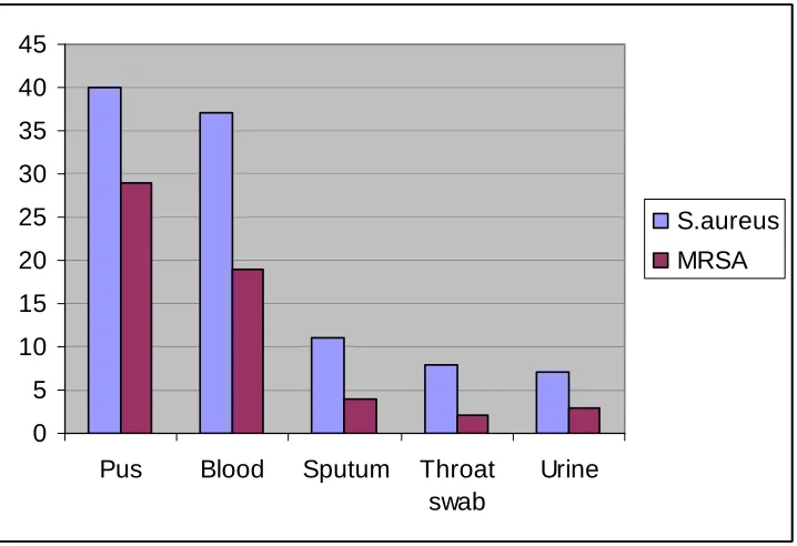
Among the 126 Staphylococcal species 103 were (42.91%) Staphylococcus aureus and 23 were (9.58%) Coagulase Negative Staphylococci (CoNS). Among the 103 Staphylococcus aureus, 40 were (38.83%) isolated from Pus specimen, 37 (35.92%) from Blood, 11 (10.67%) from Sputum, 8 (7.76%) from Throat swab and 7 (6.79%) were from Urine specimen.
Table no.2
Specimen wise isolation of Staphylococcus aureus n=103
From the above observation it was found that S.aureus
was isolated from all specimens and more number of S.aureus
Specimen No. of S.aureus isolates Percentage
Pus 40 38.83%
Blood 37 35.92%
Sputum 11 10.67%
Throat swab 8 7.76%
were isolated from Pus samples followed by Blood.
Out of 103 S.aureus isolates 57 (55.3%) were Methicillin Resistant S.aureus (MRSA). Sample wise analysis of MRSA showed that 29 out of 40 S.aureus strains were detected as MRSA from Pus specimens. 19 out of 37 from Blood, 4 out of 11 from Sputum, 2 out of 8 from Throat swab and 3 out of 7 S.aureus strains from Urine specimen were identified as MRSA. The number of S.aureus isolates and the percentage of MRSA from different specimens are given in the table-3.
Table.3
Sample wise identification of MRSA
Specimen S.aureus MRSA Percentage
Pus 40 29 28.15%
Blood 37 19 18.4%
Sputum 11 4 3.8%
Throat swab 8 2 1.94%
Urine 7 3 2.91%
Total 103 57 55.3%
From the above table it is inferred that MRSA was isolated from all specimens and the percentage of MRSA isolated from Pus samples (28.15%) was high followed by Blood (18.4%), Sputum (3.8%), Urine (2.91%) and Throat swab (1.94%) Specimens.
Analysis of age wise distribution of MRSA showed that among the MRSA isolates 16 (28%) were isolated from the age group of <1yr. 7 isolates (12.28%) from the age group of 1-15yrs, 13 (22.8%) from 16-50 yrs and 21 from the age group of >50yrs were isolated. The age wise distribution of MRSA is given in Table4.
TableNo.4
Agewise distribution of MRSA
n=57 Age group
No. of MRSA isolates Percentage <1 yr 16 28% 1-15 7 12.28% 16-50 13 22.8% >50 21 36.84%
From the above table it is inferred that the
Percentage of MRSA isolated from the age group of >50 yrs(36.84%) was the highest followed by <1yr of age (28%) showing that it is common in the extremes of age groups.
Among the 57 MRSA isolates 16 (28.06%) were associated with risk factors. Analysis of MRSA with associated risk factors showed that 10 patients (17.54%) with MRSA wound infection had Diabetes; 3 patients (5.26%) had Chronic Liver disease; 2 patients (3.5%)with MRSA Urinary infection had a history of intermittent catheterisation; 1 patient (1.75%) had a history of prolonged self medication with Quinolone antibiotic. The association of MRSA with risk factors is given in Table 5.
Table 5.
Percentage of MRSA associated with risk factors
n=16
Risk factors No. of MRSA Percentage of MRSA
Diabetes Mellitus 10 17.54%
Chronic Liver disease 3 5.26%
Urinary catheterisation 2 3.5%
Prolonged antibiotic intake 1 1.75%
Quinolone antibiotics.
All the 103 S,aureus isolates were subjected to Methicillin sensitivity testing by various phenotypic methods such as E-Test (Oxacillin), Oxacillin Resistance Screening Agar Base (ORSAB) , Disc diffusion method by using 5μg Oxacillin and 30μg Cefoxitin discs. By E-Test method 54 S.aureus strains among 103
isolates were (52.42%) identified as Methicillin resistant (MRSA) and 49 were Sensitive (47.57%) to Methicillin (MSSA) ORSAB
method detected 51 MRSA (49.51%) from 103 S.aureus isolates and 52 (50.48%) were MSSA. By disc diffusion method using Oxacillin disc 48 S.aureus (46.6%) isolates were identified as MRSA and 55 (53.39%) as MSSA. By disc diffusion method using Cefoxitin disc 56 (54.36%) isolates of S.aureus were identified as MRSA and 47 (45.63%) as MSSA. It is given in Table 6
TableNo.6 Detection of MRSA by various methods
n =103
TEST MRSA MSSA
E-Test 54(52.42%) 49(47.57%)
ORSAB 51(49.51%) 52(50.48%)
Oxacillin Disc 48(46.6%) 55(53.39%)
Cefoxitin Disc 56(54.36%) 47(45.63%)
the percentage of MRSA detected was high by Cefoxitin disc method (54.36%) followed by E.Test (52.42%), ORSAB method (49.51%) and Oxacillin disc method (46.6%).
PCR was done for all S.aureus isolates to identify the presence of mecA gene and to confirm geno typically the MRSA isolated by various phenotypic methods and the Specificity, Sensitivity, Positive Predictive value (PPV) , Negative Predictive Value (NPV) and Test of significance for each method were calculated as per the formulae. Among the 57 PCR positive MRSA strains, Cefoxitin disc identified one isolate as negative (false negative). E-Test identified 3, ORSAB 6 and Oxacillin disc 9 as false negatives. Among the 46 PCR negative isolates of S.aureus Cefoxitin disc falsely identified 1 as positive (false positive). The false positives identified by E-Test, ORSAB and Oxacillin disc were 2, 3 and 4 respectively. This is given in table 7. The Sensitivity, Specificity, Positive Predictive Value and Negative Predictive Value are given in table 8.
Table.7
Comparison of PCR with other phenotypic methods
Methods True positives False negatives False positives
PCR 57
E-Test 54 3 2
Cefoxitin disc 56 1 1
Oxacillin disc 48 9 4
ORSAB 51 6 3
It was found that Cefoxitin disc method showed only one false negative and one false positive thus very close to PCR in identifying true positives.
Table 8.
Sensitivity and Specificity of various phenotypic tests.
TEST SENSITIVITY SPECIFICITY PPV NPV
E-Test 94.73% 96% 96.42% 94.23%
ORSAB 91% 94.5% 94.44% 91.22%
Oxacillin disc 84% 93.12% 92.30% 85.93%
Cefoxitin disc 98.2% 97.91% 98.24% 98.27%
(97.91%) and the PPV and NPV are 98.24% and 98.27% respectively (Pvalue >0.05). Oxacillin disc method has the least sensitivity (84%) and specificity (93.12%) with the PPV and NPV of 92.30% and 85.93% respectively. E-Test showed 94.73% of sensitivity and 96% of specificity with 96.42% of PPV and 94.23% of NPV. ORSAB method has 91% of sensitivity and 94.5% of specificity with 94.44% of PPV and 91.22% of NPV.
Among all these methods PCR is the genotypic method which detects the mecA gene in S.aureus isolates resistant to Methicillin and is considered to be the gold standard test for detecting MRSA, but could not be adopted as a routine method of screening MRSA in small laboratories since it