0022-538X/84/030766-06$02.000
Copyright ©31984, American Society for Microbiology
Mapping of a Herpes Simplex Virus Type 2-Encoded Function That
Affects the
Susceptibility of
Herpes
Simplex
Virus-Infected
Target
Cells
to
Lysis by
Herpes
Simplex
Virus-Specific Cytotoxic
T
Lymphocytes
V. CELESTE CARTER,t STEPHEN R.JENNINGS, PATRICIA L. RICE, AND SATVIRS. TEVETHIA* Department ofMicrobiology and Cancer Research Center, The Pennsylvania State University College of Medicine,
Hershey, Pennsylvania 17033
Received 22August 1983/Accepted6 December 1983
Afunction(s) involved in the altered susceptibility of herpes simplex virus type 2 (HSV-2)-infected cells to specificlysis by cytotoxic T lymphocytes was mapped in the S component of HSV-2 DNA by using HSV-1 x HSV-2 intertypic recombinants (RH1G44, RS1G25, RSOBG10, A7D, and C4D) and HSV-1 MP. Target cellsinfected with RSOBG10, A7D,andC4D exhibited reduced levels ofcytolysis,asdid HSV-2-infected cells, whereas RH1G44 and RS1G25 recombinant-infected and HSV-1 MP-infected cells showed levels of lysis equaltothatofHSV-1 KOS-infectedcells. Theintertypicrecombinants
R50BG10,
RS1G25,RH1G44, andHSV-1MPinducedcross-reactive cytotoxicTlymphocytes. Coinfection of cells with HSV-1 KOS and eitherHSV-2 186 orR5OBG10recombinant also resulted in a decrease in the level ofspecificlysis by anti-HSVcytotoxic T lymphocytes.Previous studies from our laboratory (6) and others (11, 13) havefocusedontheinvolvement of herpes simplex virus (HSV)-specific glycoproteins in the T cell-mediated lysis of HSV-infected cells. Incomparing the susceptibility of HSV type 1 (HSV-1) and HSV type 2 (HSV-2)-infected cells to specific lysis cytotoxic T lymphocytes (CTLs) generated either to HSV-1 or HSV-2, it was discovered that HSV-2-infected cells were significantly less susceptible to lysis by CTLs (5). This lowered susceptibility of HSV-2-infected cells tospecific CTLs was neitherrelated to the replication of HSV-2 in mouse
(H-2b)
cells nor due to the lack of expressionof HSV-2 glycoproteins on the surface of HSV-2-infected target cells (5). In addition, HSV-2-infected cells were as efficient in blocking the activity of HSV-specific CTLs as were HSV-1-infected cells, although the degree of blocking achieved with both infectedcell types was low(5). As a first approach to determine the reasons behind this phenomenon, thepossibility wasconsideredthat theHSV-2 glycoproteins recognized as target antigens by anti-HSV CTLsareinvolved inthealteration ofsusceptibilityto CTL-mediated lysis of HSV-2-infected cells. The HSV-2 genome specifies five glycoproteins. Glycoproteins gA/B (2, 3, 16, 30) andgC, which was previously designated gF (2, 3, 20), have been mapped to the L component of the genome. Glycoproteins gD (16, 30), gE (2, 3, 19), and another glycoprotein ofmolecular weight92,000 to 120,000(92K to 120K) (15, 16) map in the S component of HSV-2 genome. The 92 to 120K glycoprotein has been designated gG (B. Roizman, personal communication). HSV-1 genome speci-fies four glycoproteins: gA/B, gC, gD, and gE (4, 18, 32). GlycoproteinsgA (32) has been reportedtobe antigenically related to gB (14), and the twoglycoproteinspecies havenot been differentiated serologically either by polypeptide-spe-cific antisera (8-10) or by monoclonal antibodies (21). The HSVglycoproteins have beenimplicated in theimmunoreg-*Correspondingauthor.
tPresent address: DepartmentofZoology, Universityof Califor-nia, Berkeley, CA 94720.
ulation of herpetic infections involving both humoral and cell-mediated mechanisms (1, 5, 7, 13, 23, 24, 28).
To resolve the mechanism of lowered susceptibility of HSV-2-infected target cells to CTL-mediated lysis, we made use of HSV-1 x HSV-2 intertypic recombinants which would allow us to identify regions on the HSV-2 genome which may be directlyinvolved in the lowered susceptibility of HSV-2-infected cells. The results of thesestudies demon-stratethat thefunction(s) determining the lowered suscepti-bility of HSV-2-infected cells maps in the S component of the HSV-2 genome, and the function(s) appears to be dominantbased on the reducedcytolysis of cells coinfected with HSV-1 and HSV-2.
MATERIALS AND METHODS
Cells and cell culture. Monolayer cultures of the human cellline HEp-2 were used for thepreparation of virus stocks. A continuous line of African green monkey kidney cells (Vero) was used for virus plaque assays. Both Hep-2 and Verocells weregrown at 37°C in Dulbecco modified Eagle medium supplemented with 10% fetal bovine serum and containing0.075or0.225%NaHCO3for cultures in closed or open vessels, respectively. C57BL/6 mouse embryo fibro-blasts transformed by simian virus 40(B6/WT-3) (26) were usedastargetcells inthe
5'Cr
release assay.Viruses andvirus assays. The 1 strain KOS and HSV-2 strain 186were kindly provided by Priscilla A. Schaffer, and HSV-1 MP and HSV-1 x HSV-2 recombinants RSOBG10, RH1G44, RS1G25, A7D, and C4D were kindly provided by Bernard Roizman. The origin of recombinants andtheparentalstrains usedtogeneratethese recombinants areshown in Table 1. Virus stockswereprepared inHEp-2 cells andtitrated in Verocellsby aplaqueassay
utilizing
a 2% methylcellulose overlay (31). All virus stocks were storedat -70°C.Generation of CTLs. CTLs capable ofspecifically killing syngeneic HSV-infectedcellsweregenerated by immunizing C57BL/6 (H-2b) mice (Jackson Laboratories, Bar Harbor, Maine) with
105
PFU of virus in each hind footpad. The 766on November 10, 2019 by guest
http://jvi.asm.org/
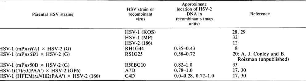
TABLE 1. HSV strains andintertypic recombinant viruses used
Approximate HSV strainor location of HSV-2
Parental HSVstrains recombinant DNAin Reference
virus recombinants(map
units)
HSV-1 (KOS) 28, 29
HSV-1 (MP) 32
HSV-2 (186) 12
HSV-1(mP)tsHAl x HSV-2 (G) RH1G44 0.35-0.43 8
HSV-1 (mP)tsSBl x HSV-2 (G) RS1G25 0.58-0.72 20;A. J. Conley andB.
Roizman (unpublished)
HSV-1 (mP)ts5OB x HSV-2(G) R5OBG10 0.82-1.0 33
HSV-1(17)tsJ(PAAr) x HSV-2 (GP6) A7D 0.78-1.0 17, 30
HSV-1(HFEM)tsN102(PAAr) x HSV-2 (186) C4D 0.0-0.28, 0.72-1.0 17, 30
draining lymph nodes were excised 5 days post-immuniza-tion, and lymphocyte suspensions were prepared by gently pressing the lymph nodes througha60-gauge stainless steel wire mesh. Viable cells were countedby trypan blue exclu-sion andsuspended at4 x 106 lymphocytesperml in RPMI 1640 mediumcontaining 2 x
10-5
M 2-mercaptoethanol, 20 mM HEPES(N-2-hydroxyethylpiperazine-N'-2-ethanesul-fonic acid) buffer, 100 U of penicillin per ml, 100 ,ug of streptomycin per ml, 0.03% glutamine, 0.225% NaHCO3, and 10% heat-inactivated (56°C, 30min) fetal bovine serum. Lymphocytes (2 x
107)
were addedto60-mmtissue culture dishes and incubated at 37°C in 5% CO, for 3 days. For controls, lymphocytesfrom nonimmunized micewere simi-larly prepared and cultured. The procedure has been de-scribedpreviously (5, 6).51Crreleaseassay. The 5tCr release assaywas performed
asdescribedpreviously(6).Briefly,confluentmonolayersof target cells growing in tissue culture flasks (75
cm2)
were infected with theappropriateHSVstrainorrecombinantatamultiplicity of infection (MOI) of 2.5, unless stated other-wise, and 200 ,uCi of
5tCr
(specific activity, 200 Ci/g; New England Nuclear Corp., Boston, Mass.) was addedto each flask. The cellswereplacedat 37°C for14to 16h. Then 2 x10⁴
cells in 0.1 mlwereaddedtoglassculture tubes(10by75 mm) with an equal volume of effectorlymphocytes,
at aneffector-to-target cell ratio of40:1 unless otherwise stated, andincubated at37°C for5 h.
RESULTS
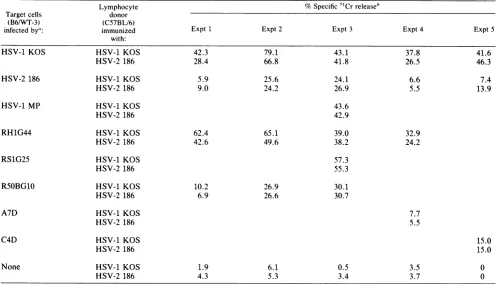
Susceptibility of HSV-1 x HSV-2 intertypic recombinant-infected B6/WT-3 cells to lysis by anti-HSV CTLs. CTLs generatedin C57BL/6 miceimmunized with HSV-1 KOS or HSV-2 186 were used in a
5'Cr
release assay to determine thesusceptibilitytolysisof target cellsinfectedwith HSV-1 x HSV-2 intertypic recombinant viruses. Recombinant vi-ruses(RH1G44, RS1G25, and R5OBG10)wereisolatedfrom cells cotransfected with HSV-1 DNA and restriction endo-nuclease digests of HSV-2 DNA (Table 1). Recombinants A7D and C4Dwere isolated by mixed infection of cells by HSV-1 and HSV-2 virions under selective pressure (Table 1). Recombinants express afull complement of HSV enve-lopeglycoproteins, but eachglycoprotein species is of only one parental type. Intertypic recombinants, therefore, pro-vide a useful tool for determining the region of type 2 parental DNA responsible for susceptibility to lysis by CTLs. The target cellsinfected with HSV-1 MP, RH1G44, and RS1G25 were as efficiently lysed as the HSV-1 KOS-infected target cells, but the R5OBG10-, A7D-, and C4D-infected target cells exhibitedalowered level oflysis,
asdidthe HSV-2 186-infected target cells (Table 2). The data indicatethat the function(s)that affects the HSV-2-infected target cell susceptibility is encoded within the HSV-2 se-quencesofR5OBG10, A7D,andC4D,andtherefore mapsto theS component of the HSV-2 genome. We havepreviously shown (5) that the parental viruses used to generate the intertypic recombinants(Table1) behaveinamannersimilar
tothatoftheHSV-1KOSand HSV-2 186strains used in this study.
Induction of CTLsinC57BL/6miceimmunizedwithHSV-1 MP, RSlG25,RHlG44, andR5OBGIO. AllHSV virus types examined generated CTLs in C57BL/6mice (Table 3). The HSV-1 MP-infected target cells exhibited slightly higher valuesofspecific lysiswhentestedin the
5tCr
release assay. Itisofinterest to note that HSV-1 MP causespolykaryocyte formation of the infected cell monolayer, and this may be involved in theincreased percent5Cr release observed with HSV-1 MP-infected target cells. The datastress the impor-tance of the right end of the HSV genome in the lowered susceptibility tolysis
observed with HSV-2-infected and RSOBG10-infectedtarget cells.Susceptibility of cells coinfected with HSV-1 KOS and intertypic recombinantR50BG1O tolysis byanti-HSV CTLs. Since HSV-2 and the recombinants RSOBG10, A7D, and C4D exhibited a lowered level of
lysis
by anti-HSV CTLs, coinfection experiments were performed with RSOBG10, HSV-2 186, and HSV-1 KOS.TheB6/WT-3targetcells were infected with either HSV-1 KOS, HSV-2 186, or RSOBG10,or werecoinfectedatacombined MOI of5with either
HSV-1 KOS and HSV-2 186or HSV-1 KOS andRSOBG10. Cells infected with RSOBG10exhibited asusceptibilityto
lysis
by anti-HSV CTLs similar to that of HSV-2 186-infected cells (Table4). Coinfectionof cells with HSV-1 KOSand HSV-2 186resulted in low levels of5'Cr
releasecomparabletothat of cells infected with HSV-2 186 alone. Coinfection ofcells with HSV-1 KOS and R50BG10 also resulted in reduced levelsoflysis
by anti-HSV CTLs,aswasobserved with cells coinfected with HSV-1 KOS and HSV-2 186 and cells infected with HSV-2 186 alone. The data suggest that the HSV-2-encoded function(s) affecting the susceptibility of HSV-infected cellstolysis
by HSV CTLs maps within the HSV-2 sequences of the HSV-1 x HSV-2 recombinant R50BG10,and further suggest thedominance of this HSV-2-codedfunctionoverHSV-1.We were interested in determining whether the reduction
of
lysis
of HSV-2-infected cells coinfected with HSV-2 couldbeovercomeby increasing the MOI of HSV-1 KOS relative to the MOI of HSV-2 186. The B6/WT-3 target cells were infected with HSV-1 KOSatincreasingMOIandwith
on November 10, 2019 by guest
http://jvi.asm.org/
[image:2.612.60.556.84.216.2]TABLE 2. Susceptibilityof HSV-1 x HSV-2 recombinant-infectedtargetcells tolysisbyanti-HSV CTLs
Lymphocyte %Specific 5'Crreleaseb
Target cells donor
(B6/WT-3) (C57BL/6)
infectedbya: immunized Expt 1 Expt 2 Expt 3 Expt 4 Expt5
with:
HSV-1 KOS HSV-1 KOS 42.3 79.1 43.1 37.8 41.6
HSV-2 186 28.4 66.8 41.8 26.5 46.3
HSV-2 186 HSV-1 KOS 5.9 25.6 24.1 6.6 7.4
HSV-2 186 9.0 24.2 26.9 5.5 13.9
HSV-1 MP HSV-1 KOS 43.6
HSV-2 186 42.9
RH1G44 HSV-1 KOS 62.4 65.1 39.0 32.9
HSV-2 186 42.6 49.6 38.2 24.2
RS1G25 HSV-1 KOS 57.3
HSV-2 186 55.3
RS0BG10 HSV-1 KOS 10.2 26.9 30.1
HSV-2 186 6.9 26.6 30.7
A7D HSV-1 KOS 7.7
HSV-2 186 5.5
C4D HSV-1 KOS 15.0
HSV-2 186 15.0
None HSV-1 KOS 1.9 6.1 0.5 3.5 0
HSV-2186 4.3 5.3 3.4 3.7 0
aTargetcells were infected
with
thedesignated virusstrainat aMOI
of2.5ormock infected,labeledwith200,uCiof51Cr,
andharvested14 hpostinfection.b
Lymphocyte cytotoxicity
wasdeterminedin
a5-h51Cr
release
assayat37°C,at aneffector-to-target
cell
ratio of40:1forexperiments
1, 2,and 4, 50:1forexperiment 3,and 20:1 forexperiment 5.
2186ataMOI of1. Even when theMOIof HSV-1KOSwas 10-foldhigherthan thatof HSV-2186,the resultant increase inlysis ofthecoinfectedtargetcellsbyanti-HSVCTLswas
negligible (datanot shown), indicatingthatthe HSV-2 func-tion(s) that affects the susceptibility to lysis by anti-HSV CTLs is dominant in cells coinfected with the type 1 virus and HSV-2 186.
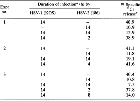
Effectofsuperinfection with HSV-2 of HSV-1-infected cells onthesusceptibilityof target cellstoCTLlysis.Weattempted todetermine the length of time of HSV-2 infection required for reducing the susceptibility of HSV-1-infected cells to
CTL lysis.Toaccomplish this,B6/WT-3cellswereinfected with HSV-1 and then with HSV-2 for 2, 4, 8, or 14 h, at which time the target cells were tested in a CTL assay. Infection of target cells with HSV-1 andHSV-2for theentire time (14 h), or the superinfection of HSV-1-infected cells with HSV-2 for 8 h, resulted in the reducedlysis oftarget cells by anti-HSV CTLs (Table 5). On the other hand, superinfectionofHSV-1-infected cells with HSV-2for 2or4 h did not result in the lowered lysis oftarget cells by anti-HSV CTLs, suggesting that HSV-2 viral functionsmust be expressedtoaffect thesusceptibilityof HSV-1-infected cells to lysis by anti-HSV CTLs. It should be pointed out that evidence does exist that the HSV-1-infected cells can be superinfected with HSV-2 (27).
We considered the possibility that the reduced level of lysisof cellscoinfectedwith HSV-1 and HSV-2 is duetothe coinfectioneventitself. However, B6/WT-3 cells coinfected with twoHSV-1 strains(KOS and 17) showednoreduction inlysis (datanotshown). Afurther explanation maybe that
HSV-2prevents theexpression of HSV-1glycoproteinsafter coinfection. However, cells coinfected with HSV-1 and HSV-2were found toexpressglycoproteinsof bothtypeson the cell surface, as tested by the indirect immunofluores-cence test (datanot shown), using type-specificmonoclonal antibody to HSV-1 gC (22) and to HSV-2 gC (2, 3), now tentatively designated gG (B. Roizman, personal
communi-cation),suggestingthat theloweredsusceptibilitytoCTLsof
TABLE 3. Induction ofCTLsinCS7BL/6mice with MP andthe HSV-1 x HSV-2recombinants
Target cells Lymphocytedonor immunizedwith': (B6/WT-3)
infectedby": KOS 186 MP RS1G25 R50BG10 RH1G44
HSV-1KOS 52.0 47.1 60.7 40.4 51.5 54.2 HSV-2 186 9.9 26.2 14.4 7.9 13.4 11.2 HSV-1 MP 70.6 68.6 74.8 68.7 69.3 71.2
RH1G44 47.2 44.5 57.6 39.4 46.4 51.5
RS1G25 50.7 52.6 52.9 47.2 52.9 50.3
RS0BG10 20.5 25.8 32.3 17.6 23.8 25.2
None 4.6 9.6 6.2 1.5 6.4 3.1
aTargetcells were
infected with
thedesignated
virus strain
at aMOI of 2.5 or mock infected, labeled with 200 C;iof51Cr, and harvested 14 h postinfection. Lymphocyte cytotoxicity was deter-minedina5-h51Crreleaseassayat 37°C, at aneffector-to-targetcell ratio of 40:1.
b Values represent thepercent specific 51Crrelease from target cells.
on November 10, 2019 by guest
http://jvi.asm.org/
[image:3.612.317.560.559.664.2]TABLE 4. Effect of coinfection ofB6/WT-3target cellswith HSV-1 KOS and either HSV-2186 orR50BG10ontheir
susceptibilitytolysisby anti-HSV CTLs Targetcells %Specific5"Crrelease'
(B6/WT-3) MOI
infectedbya: Expt 1 Expt 2 Expt 1 Expt 2
HSV-1 KOS 2.5 22.9 49.5 14.5 41.9
HSV-2 186 2.5 3.7 10.9 4.1 13.0
R5OBG10 2.5 10.6 22.7 8.1 16.0
HSV-1 KOS + 2.5 6.0 10.5 5.3 10.5
HSV-2186 2.5
HSV-1 KOS + 2.5 8.4 19.1 6.6 14.5
R50BG10 2.5
None 1.6 5.2 1.0 6.0
a Target cells were infected with the designated virus strain or mock infected, labeled with 200 ,uCi of51Cr, and harvested 14 h postinfection.
b Lymphocytecytotoxicity was determined in a 5-h5tCrrelease assay at37°C, at an effector-to-target cell ratio of 40:1.
HSV-1-infected cells uponsuperinfection withHSV-2is not dueto alack ofsynthesis of HSV-1 glycoproteins.
DISCUSSION
The results presented above have focused on the lower susceptibility of HSV-2-infected cells to lysisby anti-HSV CTLs. In our system, thelevelsoflysisby anti-HSV CTLs arelowerfor HSV-2 186-infected cells than for HSV-1 KOS-infected cells, even though similar levels of viral glycopro-teins were expressed on the surfaces of HSV-2-infected B6/WT-3 cells and HSV-1-infected cells (5). We therefore used HSV-1 x HSV-2 recombinantswith definedregionsof HSV-2 genome (Table 1) to determine which segment of HSV-2 DNA may code for the function(s) involved in the reduced susceptibility of HSV-2-infected cellsto lysis.
TheB6/WT-3targetcells infectedwith the HSV-1 x HSV-2 recombinants RH1G44 and RS1G25 were as sus-ceptible to lysis as HSV-1 KOS-infected cells. However, B6/WT-3 target cells infected with the HSV-1 x HSV-2 recombinantsR5OBG10, A7D,andC4D exhibitedlow levels oflysis by anti-HSV CTLs, as was observed with HSV-2 186-infected cells. These results indicate that the HSV-2 function(s) that affects cell susceptibility to lysis by anti-HSV CTLs maps within the HSV-2 DNA sequences of R50BG10, A7D, and C4D. The recombinant R5OBG10was isolated from cells transfected with HSV-1 cs5OB DNAand HSV-2 G DNA digested with the restriction enzyme HpaI (33) and contains HSV-2 sequencesto theright of0.82 map unitonthe HSV genome. TherecombinantA7D was gener-atedbyrescuing ats mutantofHSV-1 17 strainresistantto phosphonoacetic acid (PAA) with PAA-sensitive HSV-2 (GP6) virions,whereas therecombinant C4Dwasgenerated byrescuingats mutantof HSV-1 (HFEM)PAAr with HSV-2 (186)PAA'virions (17). The recombinantC4Dwasshown to contain HSV-2 DNA sequences from 0.0 to 0.28 and from 0.72 to 1.0 map unit, whereas the recombinant A7D con-tained HSV-2 DNA sequencesfrom 0.78 to 1.0 map unit. It isnotknown whetherA7Dand C4Dcontain additional HSV-2 DNAnotdetectable by the restriction enzyme analysis of theintertypic recombinant DNA. TwoHSV-specific glyco-proteinshavebeen identifiedwhich map in the S component of HSV-1 genome:gDandgE (19, 30). Glycoproteins gD and gE ofHSV-2,aswellasanadditional92KHSV-2 glycopro-tein,also map in theS component of HSV-2 genome (15, 16). This glycoprotein of HSV-2 has been designated gG (B.
Roizman, personal communication). However, the specific involvement ofgD, gE, orthe 92Kto 120K glycoproteinin thelowered susceptibilityof HSV-2-infected cellstolysis by anti-HSVCTLs couldnotbedetermined from thesestudies. Wealso cannot exclude thepossibility thatother functions affecting the target cell susceptibility are encoded in this region ofDNA.
The B6/WT-3 target cells infected with HSV-1 MP were
lysedasefficiently by anti-HSV CTLsaswereHSV-1 KOS-infected cells. The HSV-1 MP strain causes polykaryocyte formation of infected cells, and this fusion capability of HSV-1 MP may be involved in the substantially higher values ofpercent specific 51Crreleaseobtained with HSV-1 MP-infected cells seen in some experiments (Table 3). The results presented here demonstrate that gC isnot essential forthe induction ofhighlyreactive CTLs. The CTLs induced by HSV-1 MP were highly efficient at lysing target cells infected with HSV-1, HSV-2, or HSV-1 x HSV-2 recombi-nants. Since HSV-1 MP does not specify gC, the CTL population induced by the virus must be directed against cross-reactive determinantson gA/B, gD, orgE. As report-ed previously (5), this system for the induction of CTLs appears toinducepredominantlycross-reactivepopulations of CTLs. A secondary splenic CTL system has been de-scribed in which the secondary stimulationoflymphocytes by either homologous or heterologous viruses delineates both type-specificandcross-reactive CTLpopulations (11); the type-specific CTL population recognizes type-specific determinants ongC, whereasthecross-reactive CTL popu-lation recognizes determinants on other glycoprotein spe-cies. Theprimary systemused in thisstudydoesnotdetect significant levels oftype-specific CTLs, presumably reflect-ing the relative unimportance of gC in this system. This apparentdichotomybetween thetwosystemsmaybe dueto differences in the local response to HSV in the spleen and the popliteal lymph node. The HSV-1 x HSV-2 recombi-nants were also efficient at inducing CTLs, and these CTL populations contained predominantly cross-reactive CTLs. Recent studies (34), however, have shown that
HSV-1-TABLE 5. Effectof superinfection with HSV-2 of HSV-1-infected cells onthesusceptibilitytolysis by anti-HSV CTLs
ExptExptDuration ofinfectiona(h) by: %SpecificM~~~~~~~~~~~~~~5Cr
no. HSV-1(KOS) HSV-2(186) releaseb
1 14 - 40.9
- 14 10.9
14 14 12.9
14 2 38.9
2 14 - 41.1
- 14 11.8
14 14 19.1
14 4 41.6
3 14 - 40.4
- 14 10.8
14 14 7.5
14 2 37.8
14 8 14.0
aTarget cells (B6/WT-3) were infected with HSV-1, HSV-2, or both for various time periods, labeled with 200 ,uCi of51Cr, and reacted withCTLsgeneratedtoHSV-1.
bLymphocytecytotoxicity wasdetermined in a 5-h51Crrelease assayat 37°C,ataneffector-to-targetcell ratioof50:1.
on November 10, 2019 by guest
http://jvi.asm.org/
[image:4.612.318.557.511.683.2]coded gC possess antigenic determinants cross-reactive with gC of HSV-2.
In an attempt to determine the nature of the function(s) affecting the susceptibility of HSV-2-infectedcells to lysisby CTLs, B6/WT-3 target cells were coinfected with HSV-1 KOS and either HSV-2 186 or R50BG10. The coinfected B6/WT-3cells exhibited the HSV-2 level ofsusceptibility to lysis by anti-HSV CTLs. Varying the MOI of HSV-1 KOS and HSV-2 186 did not alter thesusceptibility of the coinfect-ed cells to lysis by HSV CTLs. In addition, increasing the MOI of HSV-1 KOS to a level 10-fold higher than that of HSV-2 186didnot significantlyincrease the susceptibility of the coinfected cells to lysis by anti-HSV CTLs. These results indicate that the HSV-2 function(s) is dominant in cells coinfected with HSV-1 KOS and HSV-2 186. It is possible that some differential alteration of membrane per-meability or an altered configuration of theHSV-2 glycopro-teins and cellular H-2 antigens could result in a reduced susceptibility to lysis by CTLs. Since the HSV-2-infected cells are as susceptible to lysis by antibody and complement as are the HSV-1-infected cells (5), and since glycoproteins of both types are synthesized during coinfection, the data provide a distinction between T cell-mediated cytolysis and antibody-dependent complement-mediated lysis of HSV-infected cells.
Theresultsreported here indicate that the reduced suscep-tibility of HSV-2-infected cells to lysis by anti-HSV CTLs maps to afunction contained within the S component of the HSV-2genome. Whether this reduction in susceptibility is associated with a particular glycoprotein species mapping within thisregion, or with some other HSV-2-specific func-tion, has not been determined. Studies are in progress to examine both the specificinvolvement of HSVglycoproteins on the susceptibility to lysis by anti-HSV CTLs and the relative effects of infection with HSV-1 and HSV-2 strains and recombinant viruses on thequalitative and quantitative expression of H-2 antigens at the cell surface.
ACKNOWLEDGMENTS
We are grateful to Bernard Roizman for providing us with HSV-1 x HSV-2 recombinants and L. Periera and W. Rawls for the monoclonal antibodytoHSV-1 and HSV-2glycoproteins.We thank CarolBuckfor herassistance in thepreparation of thismanuscript. This study was supported by Public Health Service research grants CA27503, CA 18450, and CA 10924 from the National Cancer Institute.
LITERATURE CITED
1. Adler, R., J. C. Glorioso, J. Cossman, and M. Levine. 1978. Possible role of Fc receptorsoncells infectedandtransformed byherpesvirus: escapefrom immunecytolysis. Infect. Immun. 21:442-447.
2. Balachandran, N., D. Harnish, R. A. Killington, S. Bacchetti, andW. E.Rawls.1981.Monoclonal antibodies to two glycopro-teins of herpes simplexvirus type 2. J. Virol. 39:438-446. 3. Balachandran, N., D. Harnish, W. E. Rawls, and S. Bacchetti.
1982.Glycoproteinsof herpes simplex virus type 2 as defined by monoclonal antibodies. J. Virol. 44:344-355.
4. Baucke, R. B., and P. G. Spear. 1979. Membrane proteins specified by herpes simplex viruses. V. Identification of an Fc-bindingglycoprotein. J. Virol. 32:779-789.
5. Carter, V. C., P. L. Rice, and S. S. Tevethia. 1982. Intratypic andintertypic specificity of lymphocytes involved in the recog-nition of herpes simplex virus glycoproteins. Infect. Immun. 37:116-126.
6. Carter, V. C., P. A. Schaffer, and S. S. Tevethia. 1981. The involvement of herpes simplex virus type 1 glycoproteins in cell-mediated immunity. J. Immunol. 126:1655-1660.
7. Cohen, G. H., M. Katze, C.Hydrean-Stern, and R. J. Eisenberg.
1978. Type-common CP-1 antigen ofherpes simplex virus is associated with a 59,000-molecular-weight envelope glycopro-tein. J. Virol. 27:172-181.
8. Conley, A. J., D. M. Knipe, P. C. Jones, and B. Roizman. 1981. Molecular geneticsof herpes simplexvirus.VII. Characteriza-tion of a temperature-sensitive mutant produced by in vitro mutagenesis and defective inDNAsynthesis and accumulation of-y polypeptides. J.Virol. 37:191-206.
9. Eberle, R., and R. J. Courtney. 1980.Preparation and character-ization of specific antisera to individual glycoprotein antigens comprising the major glycoprotein region of herpes simplex virus type 1. J. Virol. 35:902-917.
10. Eberle, R., and R. J. Courtney. 1980. gA and gBglycoproteins of herpes simplex virus type 1: twoforms ofasingle polypep-tide. J.Virol. 36:665-675.
11. Eberle, R., R. G. Russell, and B. T. Rouse.1981. Cell-mediated immunity to herpes simplex virus: recognition of type-specific and type-common surface antigens bycytotoxic T cell popula-tions. Infect. Immun. 34:795-803.
12. Esparza, J., D. J. M. Purifoy, P. A. Schaffer, and M. Benyesh-Melnick. 1974.Isolation, complementation andpreliminary phe-notypic characterization oftemperature-sensitive mutants of herpes simplex virus type2. Virology57:554-565.
13. Lawman, M. J. P., R. J. Courtney, R. Eberle, P. A. Schaffer, M. K.O'Hara, and B. T. Rouse. 1980.Cell-mediated immunity to herpessimplex virus: specificity of cytotoxic T cells. Infect. Immun. 30:451-461.
14. Machtiger, N. A., B. A.Pancake, R.Eberle, R. J. Courtney, S. S. Tevethia, and P. A.Schaffer. 1980.Herpes simplex virus glyco-proteins: isolation of mutantsresistant to immune cytolysis.J. Virol. 34:336-346.
15. Marsden, H. S., A. Buckmaster, J. W.Palfreyman, R. G. Hope, and A. C. Minson. 1983. Characterizationof anHSV-2-induced 92K glycoprotein. International Herpesvirus Workshop,p. 265. 16. Marsden, H. S., N. D. Stow, V. G. Preston, M. C. Timbury, and N. M. Wilkie. 1978. Physical mapping of herpessimplex virus-induced polypeptides. J. Virol. 28:624-642.
17. Morse, L.S., T. G. Buchman, B. Roizman, and P. A. Schaffer. 1977. Anatomy of herpes simplex virus DNA. IX. Apparent exclusionof some parental DNA arrangements in the generation of intertypic (HSV-1 x HSV-2)recombinants. J. Virol. 24:231-248.
18. Para, M. F., R. B. Baucke, and P. G.Spear. 1980. Immunoglob-ulin G(Fc) binding receptors on virions ofherpes simplex virus type 1 and transfer of these receptors to the cell surface by infection. J. Virol. 34:512-520.
19. Para, M. F., L. Goldstein, and P. G. Spear. 1982. Similarities and differences in the Fc-binding glycoprotein (gE) ofherpes simplex virus types 1 and 2 and tentative mappingof the viral gene for thisglycoprotein. J. Virol. 41:137-144.
20. Para, M. F., K. M. Zezulak, A. J. Conley, M.Weinberger, K. Snitzer, and P. G. Spear. 1983. Use ofmonoclonal antibodies against two75,000-molecular-weight glycoproteins specifiedby herpes simplex virus type 2 in glycoprotein identification and genemapping. J. Virol. 45:1223-1227.
21. Pereira, L., D. Dondero, B. Norrild, and B. Roizman. 1981. Differential reactivity and processing of glycoproteins gA and gB ofherpessimplexvirus types 1 and 2madeinVero and HEp-2 cells. Proc. Natl. Acad. Sci. U.S.A. 78:5202-5206.
22. Pereira, L.,T.Klassen, and J.R. Baringer. 1980. Type-common and type-specific monoclonal antibody to herpes simplex virus type 1. Infect. Immun. 29:724-732.
23. Pfizenmaier, K.,H. Jung, A. Starzinski-Powitz, M. Rollinghoff, and H. Wagner. 1977. The role of T cells inanti-herpes simplex virus immunity. I. Induction of antigen-specific cytotoxic T lymphocytes. J. Immunol. 119:939-944.
24. Pfizenmaier, K., H. Jung, A. Starzinski-Powitz, M. Rollinghoff, and H. Wagner. 1977. T cell mediated cytotoxicity against herpes simplex virus-infected target cells. Nature (London) 265:630-632.
25. Powell, K., and R. J. Courtney. 1975. Polypeptides synthesized in herpes simplex virus type 2 infected HEp-2 cells. Virology 66:217-228.
26. Pretell, J., R. S. Greenfield, and S. S. Tevethia. 1979.Biology of
on November 10, 2019 by guest
http://jvi.asm.org/
simian virus 40(SV40) transplantation rejection antigen (TrAg). V. In vitro demonstration of SV40 TrAg in SV40 infected nonpermissive mouse cells bythe lymphocytemediated
cyto-toxicity assay. Virology 97:32-41.
27. Purifoy, D.J. M., andK.L.Powell. 1977. Interference between
strains oftype 1 and type 2 herpes simplex virus. Virology 77:84-94.
28. Rawls,W. E.1973. Herpes simplex virus,p. 302-303. In A. S.
Kaplan (ed.), The herpes viruses. Academic Press,Inc., New York.
29. Rawls, W. E., D. Laurel, J. L. Melnick, H. H. Glickman, and R.H.Kaufman. 1968. A search for viruses insmegma,
premalig-nant and early malignant cervical tissues. The isolation of herpes viruseswithdistinctantigenic properties. Am. J. Epide-miol. 87:647-655.
30. Ruyechan,W.T., L.S. Morse, D. M.Knipe, and B. Roizman. 1979.Moleculargenetics of herpes simplex virus.II.Mapping of
themajorviralglycoproteinsand of thegenetic loci specifying
the social behavior of infected cells. J. Virol. 29:677-697. 31. Schaffer, P. A., V. C. Carter, and M. C. Timbury. 1978.
Collaborative complementation study oftemperature-sensitive
mutantsof herpes simplex virustypes1 and 2. J. Virol. 27:490-504.
32. Spear, P. G. 1976. Membrane proteins specified by herpes simplex viruses.I.Identification offourglycoproteinprecursors
andtheirproducts intype1-infected cells. J.Virol. 17:991-1008. 33. Tognon, M., D. Furlong, A. J. Conley, and B. Roizman. 1981. Molecular genetics of herpes simplex virus. V.Characterization ofamutantdefective in abilitytoformplaquesatlow tempera-tures and in a viral function which prevents accumulation of coreless capsids at nuclear pores late in infection. J. Virol. 40:870-880.
34. Zweig, M., S. D.Showalter, S. V. Bladen, C. J. Heilman, Jr., andB. Hampar. 1983. Herpessimplex virustype2glycoprotein gF andtype 1 glycoprotein gC have related antigenic
determi-nants. J. Virol. 47:185-192.