Copyright © 1997, American Society for Microbiology
Hexon-Only Binding of VP26 Reflects Differences between the
Hexon and Penton Conformations of VP5, the Major Capsid
Protein of Herpes Simplex Virus
PAUL T. WINGFIELD,
1STEPHEN J. STAHL,
1DARRELL R. THOMSEN,
2FRED L. HOMA,
2FRANK P. BOOY,
3BENES L. TRUS,
3,4ANDALASDAIR C. STEVEN
3*
Protein Expression Laboratory
1and Laboratory of Structural Biology,
3National Institute of Arthritis and
Musculoskeletal and Skin Diseases, and Computational Bioscience and Engineering Laboratory, Division of Computer
Research and Technology,
4National Institutes of Health, Bethesda, Maryland 20892, and Pharmacia & Upjohn Inc.,
Kalamazoo, Michigan 49007
2Received 12 June 1997/Accepted 26 August 1997
VP26 is a 12-kDa capsid protein of herpes simplex virus 1. Although VP26 is dispensable for assembly, the
native capsid (a T
5
16 icosahedron) contains 900 copies: six on each of the 150 hexons of VP5 (149 kDa) but
none on the 12 VP5 pentons at its vertices. We have investigated this interaction by expressing VP26 in
Escherichia coli and studying the properties of the purified protein in solution and its binding to capsids.
Circular dichroism spectroscopy reveals that the conformation of purified VP26 consists mainly of
b
-sheets
(
;
80%), with a small
a
-helical component (
;
15%). Its state of association was determined by analytical
ultracentrifugation to be a reversible monomer-dimer equilibrium, with a dissociation constant of
;
2
3
10
25M. Bacterially expressed VP26 binds to capsids in the normal amount, as determined by quantitative sodium
dodecyl sulfate-polyacrylamide gel electrophoresis. Cryoelectron microscopy shows that the protein occupies
its usual sites on hexons but does not bind to pentons, even when available in 100-fold molar excess.
Quasi-equivalence requires that penton VP5 must differ in conformation from hexon VP5: our data show that
in mature capsids, this difference is sufficiently pronounced to abrogate its ability to bind VP26.
The capsid of herpes simplex virus type 1 (HSV-1) is an
exceptionally large and elaborate viral particle. Approximately
1,250 Å in diameter, the shell of the mature capsid is made up
of
;
3,000 protein molecules, with a total mass of
;
200 MDa
(reviewed in references 28 and 34). In addition to a still
inde-terminate number of minor components, the shell contains
four abundant proteins; 960 copies of VP5 (149 kDa), the
major capsid protein, form the 150 hexons and 12 pentons that
pack together in the icosahedral surface lattice. At the 320 sites
of threefold symmetry are triplexes, which are heterotrimers—
probably with an
a2b
stoichiometry (24)— of VP23 (35 kDa)
and VP19c (50 kDa). These three proteins are all essential for
capsid assembly, as is an internal scaffolding protein,
pre-VP22a (35 kDa), of which
.
1,000 copies coassemble with the
shell proteins to form the procapsid (23, 37). This precursor
capsoid subsequently undergoes a structural transformation to
yield the mature shell. In vivo maturation involves proteolytic
processing of pre-VP22a to VP22a and its expulsion from the
capsid.
In addition to the three essential shell proteins—VP5,
VP19c, and VP23—the HSV-1 capsid contains a fourth
abun-dant protein (8, 17), now called VP26, of relatively low
molec-ular mass (12 kDa). Although VP26 is dispensable for
assem-bly (35, 36), it is incorporated into the capsid in large amounts,
approximately equimolar with VP5 (24). The sites that VP26
occupies have been determined by cryo-electron microscopy,
based on difference imaging. A comparison between
penton-less capsids with and without VP26 localized this protein to the
tips of hexons (7). From subtracting one-fifth of a penton from
one-sixth of a hexon from the same density map of intact
capsids, Zhou et al. concluded that VP26 is absent from
pen-tons (43). This proposal—which assumes that any structural
differences between the hexon and penton states of VP5 are
relatively slight—was corroborated by direct comparison
be-tween intact capsids with and without VP26 (38, 42). Thus, the
copy number of VP26 should be 900, if one assumes 100%
occupancy of the hexon binding sites.
While there is now consensus as to the location of VP26, the
nature of the molecular mechanism that excludes it from
pen-tons has been less clear. We have proposed that it is a
conse-quence of conformational differences between hexon VP5 and
penton VP5 (38). Such differences are manifested in other
ways, including dissimilar binding reactivities to hexons and
pentons of three monoclonal antibodies (39), differential
sen-sitivity to proteolysis (33), and differential susceptibility to
ex-traction from the capsid (7, 22). Alternatively, Zhou et al. (42)
have proposed a symmetry-mismatch mechanism, whereby
VP26 is envisaged to form hexamers in solution that fit on to
VP5 hexons but not on to pentons.
To investigate the properties of VP26 in greater depth, we
have expressed its gene—UL35 (13, 21)—in Escherichia coli,
purified the protein, and studied its properties, including its
propensity to oligomerize and to bind to capsids.
MATERIALS AND METHODS
Protein expression in E. coli.HSV VP26 was produced as a C-terminal fusion protein with glutathione S-transferase (GST), using vector pGEX-4T-3 (Phar-macia Biotech, Piscataway, N.J.). The DNA fragment encoding VP26 was gen-erated from pAcUL35 (36), using PCR as previously described (31), and cloned into the BamHI site of pGEX-4T-3. The resulting plasmid, pGEX-HSV-VP26, encodes the first 220 amino acids of GST, 6 amino acids containing the thrombin cleavage site (Leu-Val-Pro-Arg-✝-Gly-Ser, with✝indicating the cleavage site), followed by His and the 112 amino acids of VP26. Thrombin cleavage of the fusion protein should release the VP26 moiety with an additional Gly-Ser-His at the N terminus. E. coli RB 791 was transformed with the expression plasmid and
* Corresponding author. Mailing address: Bldg. 6, Room B2-34, 6
Center Dr., MSC 2717, National Institutes of Health, Bethesda, MD
20892-2717. Phone: (301) 496-0132. Fax: (301) 480-7629. E-mail: Alasdair
[email protected].
8955
on November 9, 2019 by guest
http://jvi.asm.org/
grown in 23LB (pH 7) at 37°C and 30% pO2in a 2-liter model MD fermentor (Braun Biotech, Melsungen, Germany). Protein expression was induced with 2 mM isopropylthiogalactoside (IPTG) at 37°C for 4 h.
Purification of the fusion protein.Pelleted cells (50 g) were resuspended in 100 ml of 100 mM Tris-HCl (pH 7.5) containing 5 mM dithiothreitol (DTT) and 5 mM EDTA (break buffer), lysed by passage through a French pressure cell at 16,000 lb/in2, and then sonicated at full power, 50% duty cycle, on ice for 5 min. The lysate was centrifuged in a JA-14 rotor (Beckman Instruments, Palo Alto, Calif.) at 13,000 rpm (26,0003g) at 4°C for 45 min. The pellet was washed once
by resuspension in 200 ml of break buffer containing 1% Triton X-100 and centrifugation at 26,0003g and then washed a second time with break buffer
alone. The pellet from the second wash was resuspended in 50 ml of 50 mM Tris-HCl containing 8 M guanidine-HCl (Gdn-HCl) and 10 mM DTT and then centrifuged in a Ti45 rotor at 35,000 rpm (95,0003g) for 30 min at 4°C. The
clear supernatant (50 ml) was applied to a Superdex 200 column (6 cm [diameter] by 60 cm [length]) equilibrated with 50 mM Tris-HCl containing 4 M Gdn-HCl and 5 mM DTT (S200 buffer). The column was loaded and eluted at 5 ml/min. Fractions were assayed for GST-VP26 by sodium dodecyl sulfate-polyacrylamide gel electrophoresis (SDS-PAGE) on 4 to 20% gels (Novex, San Diego, Calif.) following precipitation with 90% ethanol (26). The pooled fusion protein was stored at280°C until required.
Digestion of the fusion protein and refolding of VP26.GST-VP26 in S200 buffer was diluted to about 0.75 to 1.0 mg/ml with the same buffer and then dialyzed, first against 50 mM Tris-HCl (pH 8.0) containing 0.15 M NaCl, 2.5 M urea, 10 mM 3-[(3-cholamidopropyl)-dimethylammonio]-1-propanesulfonate (CHAPS; Calbiochem, La Jolla, Calif.), and 2 mM EDTA and then against the same buffer minus the urea. The slightly cloudy solution was clarified by centrif-ugation at 5,0003g for 20 min. Thrombin (catalog no. T6884; Sigma Chemical,
St. Louis, Mo.) was then added to the supernatant to a concentration of 0.5% (wt/wt; an A0.1%of 1.8 at 280 nm was used to estimate the protease concentra-tion). The solution was incubated for 1.5 h at;22°C, the same amount of thrombin was added, and the digestion continued for a further 1.5 h, whereupon it was quenched by adding 0.1 mM phenylmethylsulfonate fluoride from a 0.1 M stock in dry ethanol. Solid Gdn-HCl was added to 8 M, and the solution was applied to a column of Superdex 200 equilibrated in S200 buffer. VP26-contain-ing fractions were identified by SDS-PAGE, performed as described above. To the pooled fractions, 10 mM CHAPS was added, and the solution was dialyzed, first against 50 mM Tris-HCl (pH 7.5) containing 2.5 M urea, 10 mM CHAPS, and 0.25 M NaCl and then against same buffer minus the urea. As VP26 does not contain any cysteine residues, reductant was not included in the final folding buffer or in any of the buffers used in the studies described below. The folded protein was concentrated in a Centriprep-10 centrifugal ultrafiltration device (Amicon-Millipore, Bedford, Mass.) and sterile filtered through Millex-GV 0.22-mm-pore-size filters (Millipore).
The concentrations of purified GST-VP26 and VP26 were determined by measuring the absorbancies at 280 nm in a 1-cm-path-length cell, using a double-beam, diode array, UV/VIS spectrophotometer (model 8450A; Hewlett-Packard, Palo Alto, Calif.). Molar absorbance coefficients were calculated from the amino acid compositions according to Wetlaufer (41), yielding values of 48.3 mM21z cm21(A0.1%51.25) and 7.02 mM21zcm21(A0.1%50.57) for GST-VP26 and VP26, respectively.
Protein sequencing and mass spectrometry.Samples in solution were applied to polyvinylidene difluoride membranes by using a ProSpin preparation cartridge (Perkin-Elmer, Foster City, Calif.). Proteins resolved by SDS-PAGE were elec-troblotted onto ProBlott membranes as instructed by the manufacturer (Perkin-Elmer). Automated Edman degradation was performed by using a Blott car-tridge (Perkin-Elmer) and an Applied Biosystems model 477A protein sequencer. The molecular weight of VP26 was measured with a model G203AQ matrix-assisted laser desorption time-of-flight (MALDI-TOF) mass spectrome-ter (Hewlett Packard).
Circular dichroism.Spectra were recorded at 22°C on a J-720 spectropola-rimeter (Jasco Inc., Easton, Md.). Measurements in the far UV (180 to 260 nm) were made by using either 0.01- or 0.02-cm-path-length cells and a 1-nm band-width. The protein concentration was 0.5 to 1.0 mg/ml. The protein buffer was exchanged for 50 mM sodium phosphate (pH 7.2)–250 mM NaCl, plus or minus 10 mM CHAPS, using a PD-10 column (Pharmacia Biotech). Immediately prior to use, solutions were filtered with Millex-GV 0.22-mm-pore-size filter units and degassed. Secondary structure was estimated from the spectra by the CONTIN method (27).
Analytical ultracentrifugation.A Beckman Optima XL-A analytical ultracen-trifuge with an An-60Ti rotor and standard double-sector centerpiece cells was used. Centrifugation at 23,000 rpm was for 14 to 20 h at 20°C. The protein buffer used in most experiments was 50 mM Tris-HCl (pH 7.5)–0.25 M NaCl–10 mM CHAPS, but analyses were also performed with the NaCl concentration in-creased to 0.5 M or with the CHAPS concentration reduced to 5 or 0 mM. Baseline corrections were established by overspeeding at 45,000 rpm for 4 h. Data were analyzed with the Beckman Origin software. The partial specific volume of VP26 was calculated from its amino acid composition (9) to be 0.724 ml/g at 10°C (0.720 at 20°C). The solvent density was calculated as described previously (18). To calculate the dissociation constant for VP26, a monomer mass of 12,377 and the extinction coefficient given above were used.
Capsid production.Capsids were assembled by infecting Sf9 cells with recom-binant baculoviruses expressing HSV capsid genes and purified as previously described (36, 38). Briefly, combinations of baculoviruses expressing all six HSV-1 capsid genes (ALL) or expressing all except the UL35 gene [(ALL2 UL35)] were expressed in Sf9 insect cells. At 64 h postinfection, cells were harvested, and ALL and (ALL2UL35) capsids were purified (separately) by two cycles of sucrose gradient centrifugation.
Binding of VP26 to HSV-1 capsids.Purified (All2UL35) capsids at;1 mg/ml in phosphate-buffered saline (PBS) were mixed with various amounts of VP26 (see Results) in a final reaction volume of 100ml and incubated for 30 min at 22°C. Immediately beforehand, the VP26 was centrifuged at 100,0003g for 30
min, and the supernatant was taken for use in the binding reaction. After the incubation, the capsids were pelleted by centrifugation at 20,0003g for 30 min
at 4°C, washed twice by resuspension in 200ml of PBS and repelleting, and finally resuspended in 50ml of PBS. Binding reactions were also carried out in the presence of 7.5 mM CHAPS. Samples were run on 4 to 20% polyacrylamide gels (Novex) according to the manufacturer’s instructions. Protein bands stained with Coomassie blue were quantitated by using a PDI 325oe optical scanner and software (PDI, New York, N.Y.).
Cryo-electron microscopy.Grids bearing vitrified capsid-containing films were prepared from 5-ml drops of suspensions of capsids at 2 to 3 mg of protein per ml in PBS as described by Booy et al. (5). These specimens were observed in a Philips EM400RT electron microscope equipped with a liquid-N2-cooled Gatan 626 cryo-holder and modified anticontamination blades, using minimal-dose procedures at a magnification of336,000.
Three-dimensional image reconstruction.For digital analysis, we selected three micrographs which had suitably dense distributions of particles (e.g., Fig. 5a) and, as determined by optical diffraction, no significant astigmatism or drift. Their defocus values were such that the first zero of the contrast transfer function lay at frequencies of about (29.0 Å)21, (25.2 Å)21, and (29.0 Å)21, respectively. These images were scanned at 6.6 Å per pixel on a Perkin-Elmer PDS-1010MG microdensitometer. Since all three micrographs were recorded in the same se-ries, they were assumed to have the same magnification. Individual capsid images were extracted by means of the X3DPREPROCESS program (10). Their orien-tations were solved and refined by using the Polar Fourier Transform program (1), taking an earlier reconstruction (11) as the starting model. The 159 particles with the highest correlation coefficients (out of a total of 282) were used to calculate a density map to a resolution limit of 24 Å, by Fourier-Bessel synthesis (2, 3, 12, 15). The phases of the terms beyond the first contrast transfer function zero were inverted in the individual Fourier transforms prior to calculating the reconstruction.
RESULTS
Expression and purification of VP26.
Initially, we attempted
to express VP26 directly in E. coli but were unsuccessful.
How-ever, expression as a GST fusion resulted in substantial
syn-thesis (5 to 10% of cell protein) of a protein in the expected
molecular mass range, i.e.,
;
38 kDa. This protein was
insolu-ble and consequently was extracted in Gdn-HCl and then
frac-tionated to about 75% purity by gel filtration, also in Gdn-HCl
(Fig. 1, lane 2). Previous work had shown that VP26 extracted
from capsids with Gdn-HCl could be rebound to capsids—and,
by inference, refolded—upon removal of the denaturant (7,
22).
VP26 was detached from the 26-kDa GST by thrombin
di-gestion. Prior to digestion, the fusion protein was folded by
equilibrium dialysis, using a buffer containing the zwitterionic
detergent CHAPS (10 mM) and 0.15 M NaCl. In this buffer, a
compromise was achieved between maintaining the solubility
of the substrate and products (with CHAPS and salt) and
activity of the thrombin (inhibited by high salt concentrations).
VP26 was separated from the GST moiety and thrombin and
further purified by gel filtration under denaturing conditions
(Fig. 1). Purified VP26 was adjusted to
;
0.5 mg/ml in 8 M
Gdn-HCl and refolded by using buffers containing 10 mM
CHAPS and moderately high ionic strength (0.25 to 0.5 M
NaCl). Both components appeared necessary to prevent
grad-ual aggregation. The purity of the resulting material is
dem-onstrated by SDS-PAGE in Fig. 1 (lane VP26). The only
de-tectable contaminants are trace amounts of breakdown
products of slightly lower molecular weight (MW) (see the
legend to Fig. 1).
8956
WINGFIELD ET AL.
J. V
IROL.
on November 9, 2019 by guest
http://jvi.asm.org/
Identification of the expressed protein.
To confirm that the
protein isolated was VP26, its N-terminal amino acid sequence
was determined. The first five residues were
Gly-Ser-His-Met-Ala, indicating that processing had occurred as expected,
be-tween Arg and Gly of the thrombin cleavage site (see Materials
and Methods). The protein’s identity as VP26, extended by
three residues at its amino terminus, was further established by
MALDI-TOF mass spectrometry, which yielded a mass value
within 0.05% of that predicted from the DNA coding sequence
(i.e., 12,377).
Secondary structure.
The overall conformation of VP26 was
examined by far-UV circular dichroism (Fig. 2, spectrum 1). A
similar spectrum was obtained when CHAPS was omitted from
the buffer, indicating that the detergent has no apparent effect
on the overall structure of VP26. Comparison with the random
coil obtained after denaturation with Gdn-HCl (Fig. 2,
spec-trum 2) indicates that, as purified, VP26 has a well-defined
secondary structure and is therefore likely to be partly if not
completely folded. From these data, its secondary structure
was estimated to be mainly
b
-sheet (
;
80%), with some
a
-helical contribution (13 to 15%). These values concur with
those predicted (75%
b
-strand plus turn; 25%
a
-helix) by the
neural network-based PHD program (30). In the aromatic
region, i.e., 340 to 260 nm, its spectrum (not shown) is weak,
most likely due to the single Trp and Tyr residues being located
in flexible regions.
Oligomeric status of VP26.
The MW of VP26 was
deter-mined by sedimentation equilibrium. During the
centrifuga-tion runs (14 to 16 h at 10 to 20°C), the protein was equally well
behaved in 5 to 10 mM CHAPS with 0.25 to 0.5 M NaCl. At
equilibrium, the protein concentration gradient increased from
0.01 mg/ml at the top to approximately 2.5 mg/ml at the bottom
of the cell, depending on the concentration of the starting
sample. Conversion of these data to plots of MW versus
con-centration (not shown) indicated that the MW increased as a
function of radial position. The overall weight-average MW
was 18,000 to 20,000, intermediate between those of a
mono-mer (12,377) and a dimono-mer (24,754). Extrapolating to the
high-est concentrations (bottom of cell) gave MWs approaching
that of the dimer. These findings suggest that in the buffer
used, VP26 is a reversible monomer-dimer system. A good fit
to the data was obtained, using this model with a dissociation
constant of 2
3
10
25M (Fig. 3). It follows that VP26 was 40 to
60% monomeric at the concentrations used in the binding
studies described below, i.e., 4 to 16
m
M.
When the same data were fitted for alternative models such
as monomer-trimer, monomer-tetramer, monomer-pentamer,
or monomer-hexamer equilibria, or an ideal (noninteracting)
mixture of monomers and hexamers, poor agreement was
ob-tained, with higher residuals and systematic trends in their
residual plots (data not shown). In contrast, the residuals from
the monomer-dimer model scatter randomly about the zero
value (Fig. 3), which is a hallmark of a good fit. In particular,
the chi-square statistic for the monomer-hexamer model was
almost 20-fold higher than that for the successful
monomer-dimer model.
[image:3.612.74.269.69.329.2]Binding of purified VP26 to HSV-1 capsids.
To test our
bacterially expressed VP26 for competence to bind to capsids,
the purified protein was mixed with (All
2
UL35) capsids (35)
FIG. 1. Expression of HSV-1 VP26 in E. coli as a GST fusion protein and its [image:3.612.311.547.71.311.2]subsequent purification, as monitored by SDS-PAGE. Starting with;50 g (wet weight) of cells, about 850 mg of the GST-fusion protein (lane 2) was recovered, using a two-stage dialysis scheme (see Materials and Methods). Following throm-bin digestion, which was essentially complete (cf. lane 3), gel filtration followed by the second refolding yielded a 60 to 70% recovery of purified VP26 (lane 5), separated from GST (lane 4); 1.5 to 2.0 mg of VP26 was obtained per g (wet weight) of cells. Trace amounts of two contaminants of slightly lower MWs remain. They appear to arise from thrombin side reactions which generate small amounts of both N- and C-terminally truncated VP26 and a N-terminal fragment of the mature GST protein (data not shown). Interestingly, small amounts of lower-MW forms were also observed in VP26 extracted from native capsids (5), although in this case, they may represent phosphorylated forms (20).
FIG. 2. Circular dichroic spectrum of HSV-1 VP26 (spectrum 1). Protein incubated with Gdn-HCl (spectrum 2) evinces a loss of ellipticity at 220 nm and a large increase at 206 to 204 nm, typical of denatured proteins (40), thus emphasizing that the VP26 was originally folded. The spectra are shown in far-UV region to limits of reliable detection as monitored by photomultiplier voltage (usually less than 600 V). The units of the ordinates are mean residue ellipticity [u]mwrand have the dimensions degzcm2zdmol21.
on November 9, 2019 by guest
http://jvi.asm.org/
(Fig. 4). The molar ratios of VP26 to VP5 were 2:1 and 8:1,
respectively, in two parallel incubations. Unbound VP26 was
removed by two cycles of pelleting the capsids, discarding the
supernatants, and resuspending the pellets in buffer. Analysis
of the washed capsids by SDS-PAGE (Fig. 4) clearly indicated
binding of VP26. The molar ratios of VP5 to VP26 were
determined by densitometric analysis to be
;
1:1.16 for the
twofold incubation and 1:1.24 for the eightfold incubation. We
conclude that in both cases, VP26 bound to the capsids in
saturating amounts, close to the theoretical value for wild-type
capsids (i.e., 1:0.95 [see the introduction]). When these
exper-iments were repeated in CHAPS-containing buffer similar to
that used for analytical ultracentrifugation, essentially identical
binding of VP26 to capsids was observed (data not shown).
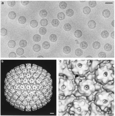
Visualization of bound VP26.
To determine the sites on the
capsid surface to which VP26 had bound, a sample of capsids
from the incubation at eightfold molar excess was examined by
cryo-electron microscopy (Fig. 5a) and three-dimensional
im-age reconstruction. The outer surface of the resulting density
map is shown in Fig. 5b. The six “horns” of density around the
rim of each hexon, associated with six VP26 monomers, are
clearly present, whereas these features are absent from
pen-tons. This conclusion was confirmed by inspecting sections
through the map, which show all levels of density coded as gray
levels (data not shown). These images contained no density
above background in the regions overlying the penton tips,
implying no detectable level of occupancy by VP26.
In principle, the absence of VP26 from the pentons of native
capsids might reflect a limited supply of VP26 in the nuclei of
infected cells, combined with a preference for hexons over
pentons, rather than complete inability of VP26 to bind to
pentons. That the latter proposition is correct is attested by this
experiment, in which, after saturation of the hexon sites, the
remaining VP26 did not bind to pentons despite being
avail-able in high excess, i.e., at a molar ratio of
;
100:1 relative to
penton VP5.
DISCUSSION
Now that the molecular anatomy of the herpesvirus capsid
has been defined—at least for its major constituents (34)—
attention is shifting to the interactions that control assembly
and maturation. These include the interactions responsible for
the import of capsid proteins from the cytoplasm to the
nu-cleus, the interactions whereby subunits associate into
assem-bly-competent building blocks, the morphogenetic interactions
that organize building blocks into procapsids, and the revised
set of interactions that stabilize the mature capsid. In this
context, we have addressed the question of how incorporation
of VP26 into the capsid is regulated.
Expression, purification, and properties of VP26.
Although
the main aims of expressing a protein as a fusion are usually to
increase its solubility and simplify purification, we used this
strategy only to stabilize the expressed protein, albeit in an
aggregated form. The simple purification and folding scheme
that we have developed should be suitable for handling other
insoluble GST fusion proteins.
[image:4.612.50.295.67.314.2]Isolated VP26 has a high isoelectric point (11.2) and low
FIG. 3. Sedimentation equilibrium analysis of HSV-1 VP26. Measurements [image:4.612.321.532.69.369.2]were made by using a Beckman Optima XL-A analytical ultracentrifuge, running at 23,000 rpm. The absorbance gradient in the centrifuge cell after attaining sedimentation equilibrium is shown in the bottom panel. The solid line is the result of the fit using reversible monomer-dimer model, and the open circles are the experimental values. The top panel shows the differences between the fitted and experimental values as a function of radial position (residuals).
FIG. 4. Binding of purified VP26 to HSV-1 capsids, as monitored by SDS-PAGE on a 4 to 20% gradient gel stained with Coomassie blue. These (All2 UL35) capsids (denoted here as HSV2UL35) lack VP26 (lane 1). After comple-mentation with twofold (lane 2) and eightfold (duplicates in lanes 3 and 4) molar excesses of VP26, the capsids were separated from unreacted VP26 by two wash cycles. The amount of VP26 retained was essentially the same in all three samples and in the same proportion relative to the major capsid protein, VP5, as in wild-type capsids (24). The purified VP26 is shown in lane 5. Stds., size standards.
8958
WINGFIELD ET AL.
J. V
IROL.
on November 9, 2019 by guest
http://jvi.asm.org/
intrinsic solubility. Although we have not made a
comprehen-sive investigation of solvent conditions, it was clear from the
outset that both GST-VP26 and VP26 have low solubility in
standard buffers, such as PBS. Aggregation can be partially
prevented by increasing the ionic strength with NaCl.
How-ever, the key to maintaining solubility was the inclusion of
CHAPS to at least 5 mM. The detergent did not affect either
the conformation of VP26 or its ability to bind to capsids.
However, even in the presence of detergent and high ionic
strength, the solubility was limited to
;
2 to 3 mg/ml. Thus
solubilized, VP26 consists of a mixture of monomers and
dimers. When studied in the absence of detergent, with
aggre-gates removed by high-speed centrifugation immediately
be-forehand, VP26 behaved in a similar manner (data not shown).
VP26’s mode of binding to capsids.
VP26 extracted from
purified wild-type capsids by heating binds to capsids that lack
this protein (22). We have now found that bacterially expressed
VP26 also binds faithfully to the hexons of acceptor capsids
while the pentons remain unoccupied, even with a high molar
excess of VP26. Its binding is unaffected by the three additional
amino acids present at its N terminus; in fact, the entire
GST-VP26 fusion protein appears to be capable of binding to
cap-sids (preliminary observations [not shown]).
Previously, we have visualized VP26 subunits around the
rims of VP5 hexons in two difference maps at
;
30-Å
resolu-tion: one from pentonless Gdn-HCl-extracted capsids with and
without VP26 (7), and the other between two kinds of
bacu-lovirus-derived capsids, All and (All
2
UL35) (38). In both
FIG. 5. (a) Cryo-electron micrograph of purified (All2UL35) HSV-1 capsids after in vitro complementation with an eightfold molar excess of purified VP26. Bar5 100 nm. (b) Three-dimensional structure of these capsids at 2.4-nm resolution, as viewed along a twofold axis of symmetry. Bar510 nm. (c) Blowup of the capsid structure around a penton site. Each hexon has six prominent horns of density around its outer rim (solid arrow), corresponding to six monomers of VP26. These features are absent from pentons (hollow arrow). Bar55 nm.on November 9, 2019 by guest
http://jvi.asm.org/
[image:5.612.71.527.67.527.2]cases, we found it difficult to decide whether the six VP26
monomers per hexon actually make contact with each other,
since this feature was acutely sensitive to the choice of
thresh-old used for contouring. From their difference map at
;
20 Å
resolution between wild-type B capsids and
baculovirus-de-rived (All
2
UL35) capsids, Zhou et al. (42) described the
VP26 monomer as consisting of a large and a small domain,
with six monomers forming a continuous ring. Since VP26 in
solution consists of monomers and dimers, it follows that such
contacts as exist between VP26 subunits on capsids are not
extensive enough to promote hexamerization in the absence of
the underlying template of a VP5 hexon. Alternatively, the
conformation of VP26 may change upon binding to the capsid,
to allow formation of intersubunit contacts.
Our sedimentation analysis indicates that purified VP26
ob-serves a monomer-dimer equilibrium, with no evidence for
hexamers or other oligomers higher than the dimer. We
con-clude, therefore, that its disinclination to bind to pentons
re-flects the absence of binding sites on pentons. It follows that
the surface residues of VP5 which form the VP26 binding site
on hexons must be conformed differently on pentons, so as to
abrogate binding.
VP26 binding in intracellular capsid assembly.
We note that
incorporation of VP26 may occur differently in vivo. VP26 has
no known nuclear import signal, and Rixon and coworkers
have reported that an association with VP5 is necessary for
VP26 to enter the nucleus (29). What happens next is unclear,
but we may consider several possibilities. The transported
complexes may enter the nucleus intact, or they may dissociate
upon traversing the nuclear envelope. In the nucleus, VP5 may
be present both with and without VP26, with the latter
mole-cules being selectively incorporated into nascent procapsids at
vertex sites. Alternatively, all VP5 molecules may be charged
with VP26, with VP26 molecules being released when their
VP5 partners adapt to the penton conformation. At present,
we are unable to distinguish between these (and other)
possi-bilities. However, as noted above, VP26 is not very soluble,
implying that in vivo it is likely to be engaged, for the most
part, in complexes of one kind or another.
Functional implications: a mechanism for diversifying the
binding sites presented on the capsid surface.
Although VP26
is not required for capsid assembly, other observations imply
that it is of some utility to the virus. These include its high copy
number (900) and evolutionary persistence. With the sole
ex-ception of the distant relative, channel catfish virus (6, 13), a
VP26 counterpart has been detected in every herpesvirus
stud-ied to date. Moreover, a recent preliminary report has been
made of a VP26-null mutant that is capable of replication in
cultured cells but is 30-fold less efficient than the wild-type
allele in propagating in trigeminal ganglia (14). Other
con-served properties include genetic setting—close to the major
capsid protein gene and in the opposite polarity, basic charge,
and generally low MW. However, compared with other
her-pesvirus capsid proteins (6), the size of VP26-like proteins is
not closely conserved, ranging from 75 amino acids for human
cytomegalovirus (16) to 176 for Epstein-Barr virus (19). VP26
of HSV-1 has 116 amino acids. Nor is there much sequence
similarity between VP26 and its counterparts in other
herpes-viruses, although homologies are evident between the
mem-bers of this family expressed by several gammaherpesviruses
(19).
As previously noted (6), its exposed site suggests that VP26
is well placed to couple the capsid to functional partners, such
as the tegument, in the final phase of virion assembly.
How-ever, the capsid must also engage in different interactions at
other stages of the replicative cycle, for instance, with
molec-ular motors in cytoplasmic transport subsequent to cell entry
(25, 32) and with nuclear pores, when discharging the viral
genome into the nucleus of an infected cell (4). In this context,
it is plausible that the device of leaving some capsomers
(pen-tons) bare and decorating others (hexons) with an adapter
protein, VP26, would confer the advantage of expanding the
range of functional binding sites on the capsid surface.
ACKNOWLEDGMENTS
We thank Ira Palmer and Josh Kaufman for expert help in protein
expression and purification, Pat Spinella for N-terminal sequencing,
and T. Baker and J. Conway for software.
REFERENCES
1. Baker, T. S., and R. H. Cheng. 1996. A model-based approach for determin-ing orientations of biological macromolecules imaged by cryoelectron mi-croscopy. J. Struct. Biol. 116:120–130.
2. Baker, T. S., J. Drak, and M. Bina. 1988. Reconstruction of the three-dimensional structure of simian virus 40 and visualization of the chromatin core. Proc. Natl. Acad. Sci. USA 85:422–426.
3. Baker, T. S., J. Drak, and M. Bina. 1989. The capsid of small papova viruses contains 72 pentameric capsomeres: direct evidence from cryo-electron-microscopy of simian virus 40. Biophys. J. 55:243–253.
4. Batterson, W., D. Furlong, and B. Roizman. 1983. Molecular genetics of herpes simplex virus. VIII. Further characterization of a temperature-sensi-tive mutant defectemperature-sensi-tive in release of viral DNA and in other stages of the viral replicative cycle. J. Virol. 45:397–407.
5. Booy, F. P., W. W. Newcomb, B. L. Trus, J. C. Brown, T. S. Baker, and A. C. Steven.1991. Liquid-crystalline, phage-like, packing of encapsidated DNA in herpes simplex virus. Cell 64:1007–1015.
6. Booy, F. P., B. L. Trus, A. J. Davison, and A. C. Steven. 1996. The capsid architecture of channel catfish virus, an evolutionarily distant herpesvirus, is largely conserved in the absence of discernible sequence homology with herpes simplex virus. Virology 215:134–141.
7. Booy, F. P., B. L. Trus, W. W. Newcomb, J. C. Brown, J. F. Conway, and A. C. Steven.1994. Finding a needle in a haystack: detection of a small protein (the 12 kDa VP26) in a large complex (the 200 MDa capsid of herpes simplex virus). Proc. Natl. Acad. Sci. USA 91:5652–5656.
8. Cohen, G. H., M. Ponce de Leon, H. Diggelmann, W. C. Lawrence, S. K. Vernon, and R. J. Eisenberg.1980. Structural analysis of the capsid polypep-tides of herpes simplex virus types 1 and 2. J. Virol. 34:521–531. 9. Cohn, E. J., and J. T. Edsall. 1943. Proteins, amino acids and peptides. Van
Nostrand-Reinhold, Princeton, N.J.
10. Conway, J. F., B. L. Trus, F. P. Booy, W. W. Newcomb, J. C. Brown, and A. C. Steven.1993. The effects of radiation damage on the structure of frozen hydrated HSV-1 capsids. J. Struct. Biol. 111:222–233.
11. Conway, J. F., B. L. Trus, F. P. Booy, W. W. Newcomb, J. C. Brown, and A. C. Steven.1996. Visualization of three-dimensional density maps reconstructed from cryoelectron micrographs of viral capsids. J. Struct. Biol. 116:200–208. 12. Crowther, R. A. 1971. Procedures for three-dimensional reconstruction of spherical viruses by Fourier synthesis from electron micrographs. Philos. Trans. R. Soc. Lond. Ser. B 261:221–230.
13. Davison, M. D., F. J. Rixon, and A. J. Davison. 1992. Identification of genes encoding two capsid proteins (VP24 and VP26) of herpes simplex virus type 1. J. Gen. Virol. 73:2709–2713.
14. Desai, P., N. A. DeLuca, and S. Person. The HSV-1 VP26 polypeptide is not essential for growth in cell culture but is important for virus pathogenesis. Submitted for publication.
15. Fuller, S. D. 1987. The T54 envelope of Sindbis virus is organized by interactions with a complementary T53 capsid. Cell 48:923–934. 16. Gibson, W., K. S. Clopper, W. J. Britt, and M. K. Baxter. 1996. Human
cytomegalovirus (HCMV) smallest capsid protein identified as product of short open reading frame located between HCMV UL48 and UL49. J. Virol. 70:5680–5683.
17. Heilman, C. J., Jr., M. Zweig, J. R. Stephenson, and B. Hampar. 1979. Isolation of a nucleocapsid polypeptide of herpes simplex virus types 1 and 2 possessing immunologically type-specific and cross-reactive determinants. J. Virol. 29:34–42.
18. Laue, T. M., B. D. Shah, T. M. Ridgeway, and S. L. Pelletier. 1992. Com-puter-aided interpretation of analytical sedimentation data for proteins, p. 90–125. In S. E. Harding, A. J. Rowe, and J. C. Horton (ed.), Analytical centrifugation in biochemistry and polymer science. Royal Society for Chem-istry, Cambridge, England.
19. Lin, S.-F., R. Sun, L. Heston, L. Gradoville, D. Shedd, K. Haglund, M. Rigsby, and G. Miller.1997. Identification, expression, and immunogenicity of Kaposi’s sarcoma-associated herpes virus-encoded small viral capsid an-tigen. J. Virol. 71:3069–3076.
20. McNabb, D. S., and R. J. Courtney. 1992. Posttranslational modification and subcellular localization of the p12 capsid protein on herpes simplex virus type 1. J. Virol. 66:4839–4847.
8960
WINGFIELD ET AL.
J. V
IROL.
on November 9, 2019 by guest
http://jvi.asm.org/
21. McNabb, D. S., and R. J. Courtney. 1994. Identification and characterization of the herpes simplex virus type 1 virion protein encoded by the UL35 open reading frame. J. Virol. 66:2653–2663.
22. Newcomb, W. W., and J. C. Brown. 1991. Structure of the herpes simplex virus capsid: effects of extraction with guanidine-HCl and partial reconsti-tution of extracted capsids. J. Virol. 65:613–620.
23. Newcomb, W. W., F. L. Homa, F. P. Booy, D. R. Thomsen, B. L. Trus, A. C. Steven, J. V. Spencer, and J. C. Brown.1996. Assembly of the herpes simplex virus capsid: characterization of intermediates observed during cell-free cap-sid formation. J. Mol. Biol. 263:432–446.
24. Newcomb, W. W., B. L. Trus, F. P. Booy, A. C. Steven, J. S. Wall, and J. C. Brown.1993. Structure of the herpes simplex virus capsid: molecular com-position of the pentons and triplexes. J. Mol. Biol. 232:499–511. 25. Penfold, M. E., P. Armati, and A. L. Cunningham. 1994. Axonal transport of
herpes simplex virions to epidermal cells: evidence for a specialized mode of virus transport and assembly. Proc. Natl. Acad. Sci. USA 91:6529–6533. 26. Pepinsky, R. B. 1990. Selective precipitation of proteins from guanidine
hydrochloride-containing solutions with ethanol. Anal. Biochem. 195:177– 181.
27. Provencher, S. W., and J. Gloeckner. 1981. Estimation of globular protein secondary structure from circular dichroism. Biochemistry 20:33–37. 28. Rixon, F. J. 1993. Structure and assembly of herpesviruses. Semin. Virol.
4:135–144.
29. Rixon, F. J., C. Addison, A. McGregor, S. J. Macnab, P. Nicholson, V. G. Preston, and J. D. Tatman.1996. Multiple interactions control the intracel-lular localization of the herpes simplex virus type 1 capsid proteins. J. Gen. Virol. 77:2251–2260.
30. Rost, B., and C. Sander. 1994. Combining evolutionary information and neural networks to predict protein secondary structure. Proteins 19:55–72. 31. Scharf, S. J., G. T. Horn, and H. A. Erlich. 1986. Direct cloning and sequence
analysis of enzymically amplified genomic sequences. Science 233:1076–1078. 32. Sodeik, B., M. W. Ebersold, and A. Helenius. 1997. Microtubule-mediated
transport of incoming herpes simplex virus 1 capsids to the nucleus. J. Cell Biol. 136:1007–1021.
33. Steven, A. C., C. R. Roberts, J. Hay, M. E. Bisher, T. Pun, and B. L. Trus.
1986. Hexavalent capsomers of herpes simplex virus type 2: symmetry, shape, dimensions, and oligomeric status. J. Virol. 57:578–584.
34. Steven, A. C., and P. G. Spear. 1997. Herpesvirus capsid assembly and envelopment, p. 312–351. In W. Chiu, R. M. Burnett, and R. L. Garcea (ed.), Structural biology of viruses. Oxford University Press, New York, N.Y. 35. Tatman, J. D., V. G. Preston, P. Nicholson, R. M. Elliott, and F. J. Rixon.
1994. Assembly of herpes simplex virus type 1 capsids using a panel of recombinant baculoviruses. J. Gen. Virol. 75:1101–1113.
36. Thomsen, D. R., L. L. Roof, and F. L. Homa. 1994. Assembly of herpes simplex virus (HSV) intermediate capsids in insect cells infected with re-combinant baculoviruses expressing HSV capsid proteins. J. Virol. 68:2442– 2457.
37. Trus, B. L., F. P. Booy, W. W. Newcomb, J. C. Brown, F. L. Homa, D. R. Thomsen, and A. C. Steven. 1996. The herpes simplex virus procapsid: structure, conformational changes upon maturation, and roles of the triplex proteins VP19c and VP23 in assembly. J. Mol. Biol. 263:447–462. 38. Trus, B. L., F. L. Homa, F. P. Booy, W. W. Newcomb, D. R. Thomsen, N.
Cheng, J. C. Brown, and A. C. Steven.1995. Herpes simplex virus capsids assembled in insect cells infected with recombinant baculoviruses: structural authenticity and localization of VP26. J. Virol. 69:7362–7366.
39. Trus, B. L., W. W. Newcomb, F. P. Booy, J. C. Brown, and A. C. Steven. 1992. Distinct monoclonal antibodies separately label the hexons or the pentons of herpes simplex virus capsid. Proc. Natl. Acad. Sci. USA 89:11508–11512. 40. Venyaminov, S. Y., I. A. Baikalov, Z. M. Shen, C.-S. C. Wu, and J. T. Yang.
1993. Circular dichroism analysis of denatured proteins: inclusion of dena-tured proteins in the reference set. Anal. Biochem. 214:17–24.
41. Wetlaufer, D. B. 1962. Ultraviolet spectra of proteins and amino acids. Adv. Protein Chem. 17:303–390.
42. Zhou, Z. H., J. He, J. Jakana, J. D. Tatman, F. J. Rixon, and W. Chiu. 1995. Assembly of VP26 in herpes simplex virus-1 inferred from structures of wild-type and recombinant capsids. Nat. Struct. Biol. 2:1026–1030. 43. Zhou, Z. H., B. V. V. Prasad, J. Jakana, F. J. Rixon, and W. Chiu. 1994.
Protein subunit structures in the herpes simplex virus capsid determined from 400-kV spot-scan electron cryomicroscopy. J. Mol. Biol. 242:456–469.