White Rose Research Online URL for this paper:
http://eprints.whiterose.ac.uk/130875/
Version: Accepted Version
Article:
Hoskere, A.A., Sreedharan, S., Ali, F. et al. (3 more authors) (2018) Polysulfide-triggered
fluorescent indicator suitable for super-resolution microscopy and application in imaging.
Chemical Communications, 54 (30). pp. 3735-3738. ISSN 1359-7345
https://doi.org/10.1039/c8cc01332b
[email protected]
https://eprints.whiterose.ac.uk/
Reuse
Items deposited in White Rose Research Online are protected by copyright, with all rights reserved unless
indicated otherwise. They may be downloaded and/or printed for private study, or other acts as permitted by
national copyright laws. The publisher or other rights holders may allow further reproduction and re-use of
the full text version. This is indicated by the licence information on the White Rose Research Online record
for the item.
Takedown
If you consider content in White Rose Research Online to be in breach of UK law, please notify us by
Journal Name
COMMUNICATION
a.
Organic Chemistry Division, CSIR-National Chemical Laboratory, Pune-411008, India.
b.Department of Chemistry, University of Sheffield, Sheffield, S3 7HF, UK, Email:
c.
Department of Biomedical Science, University of Sheffield, Sheffield, S10 2TN, UK.
d.
CSIR-Central Salt & Marine Chemicals Research Institute, Bhavnagar: 364002, Gujarat, India; E-Mail: [email protected].
Electronic Supplementary Information (ESI) available: See DOI: 10.1039/x0xx00000x
Received 00th January 20xx, Accepted 00th January 20xx
DOI: 10.1039/x0xx00000x
www.rsc.org/
Polysulfide-Triggered Fluorescent Indicator Suitable for
Super-Resolution Microscopy and Application in Imaging
Anila Hoskere A,a Sreejesh Sreedharan,b Firoj Ali,a Carl G. Smythe,c Jim A. Thomasb* and Amitava Dasa,d*
A new physiologically benign and cell membrane permeable BODIPY based molecular probe, MB-Sn specifically senses intracellular hydrogen polysulfides (H2Sn, n > 1) localized in the endoplasmic reticulum. This reagent is suitable for mapping the intracellular distribution of H2Sn by wide-field as well as super-resolution Structured Illumination Microscopy (SIM).
Hydrogen polysulfide H2Sn is one of the reactive sulphur
species (RSS) and is primarily produced in cells from
3-mercaptopyruvate by the enzyme 3-mercaptopyruvate
sulfurtransferase.1 It is also produced by the reaction of H2S
with NO.1 H2Sn influences many important biological functions
and activities associated with H2S. For example, it induces
sulfuration, wherein a sulphur atom is added to cysteine thiol groups within proteins, causing changes in conformation and
the activity of the affected protein.2 By activating transient
receptor potential (TRP) A1 channels, H2Sn induces Ca
2+
influx
in astrocytes more efficiently than H2S.
3,4a
It is also a potent signaling molecule that regulates the activity of tumor
suppressors and transcription factors.3
To develop a deeper understanding of the regulatory roles of
H2Sn, it is crucial to identify appropriate reagent and
methodology for efficient detection and tracking of H2Sn within
cells. Traditional spectroscopic methods for the detection of
H2Sn rely on measuring characteristic absorbances at 290 - 300
nm and 370 nm5- a methodology of limited application due to
its low sensitivity. Another methodology using mass spectroscopic technique requires derivatization of the sample with monobromobimane to produce chemically unstable
derivatives that often lead to inaccurate results.3 Moreover,
none of these methods are suitable for studying in-vivo
biological processes or mapping the intracellular distribution of
H2Sn. Thus, there is clear scope to develop an efficient
methodology for detection of H2Sn using a fluorescence-based
molecular probe.4c There are several literature reports for
detection of other biothiols such as cysteine, glutathione, and
H2S using this approach.
6
However, such reports are scarce for
H2Sn due to the lack of proper understanding about its
reactivity. Recent literature reports reveal that most polysulfide probes rely on a nucleophilic substitution reaction involving the polysulfide moiety and a 2-fluoro-5-nitro benzoate derivative, while the transient species undergo spontaneous cyclization to release the fluorophore in its “on
state”.7 However, such probes also participate in nucleophilic
substitution reaction with other biothiols, and this limits their
application as a specific reagent for H2Sn. Another approach
involves the use of H2Sn induced aziridine ring opening
reactions.8 Such probes show good selectivity for H2Sn,
however, their utility is largely limited to cell-free studies.
Barring some recent reports from Xian and co-workers, H2Sn
specific probes are still very sparse in the literature.9 However;
there is no report on an organelle specific H2Sn probe that is
suitable for Structured Illumination Microscopy (SIM).10a,b
Fluorescence microscopy has proved to be an invaluable tool for imaging studies as well as for studying the functions of
specific analytes in living systems.4b,c In many instances, the
applicability of this technique is limited by its relatively poor
spatial resolution.10a However, recent advances in
super-resolution microscopy (SRM) have broken the diffraction limit of conventional optical-based techniques providing hitherto
inaccessible imaging capabilities.10b,d One such SRM technique
is SIM, which provides an appreciable increase in resolutions
(100 - 120 nm)10a,b with minimal disruption of data acquisition
rate. SIM requires considerably lower illumination intensities compared to other super-resolution microscopies like STORM
and STED,10c,d making it particularly suited to live cell imaging.
With these facts in mind, we have developed a BODIPY based
probe MB-Sn that could exclusively detect H2Sn in physiological
conditions as well as it could be used for mapping endogenous
H2Sn localized in the endoplasmic reticulum of cells.
COMMUNICATION Journal Name
(A)
5 8 0 6 3 8 6 9 6 0
1 x 1 06
2 x 1 06
In te n s it y
W a v e le ng th (nm )
0 2 0µM
1 2 34 5 67 89 1 0 1 1 1 2 1 3 1 4 1 5 1 6 0 .0
5 .0 x 1 05
1 .0 x 1 06
1 .5 x 1 06
2 .0 x 1 06
In te n s it y
D iffe re nt A na ly te s
(B)
We chose difluoroborondipyrromethene (commonly known as BODIPY), as a fluorescent reporter functionality owing to its excellent photophysical properties such as high molar absorptivity, tuneable emission, excellent photostability,
narrow emission bandwidth and high quantum yield.6c,10d,11
The dual-reactivity of H2Sn was exploited in designing this
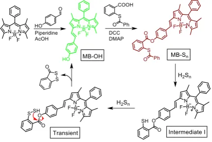
molecular probe using a BODIPY moiety functionalized with benzothioester as the polysulfide recognition site. Detailed synthetic procedures and all relevant characterization data for the molecular probe and the intermediates are provided in the supporting information (Fig. S14-17). Esterification of the
phenolic functionality of MB-OH was achieved by reacting with
thiobenzoate in the presence of DCC to yield the final
chemodosimetric reagent MB-Sn. The extended conjugation of
one the pyrrole arm (Scheme 1) helped in achieving shifts in the absorption and emission bands maxima toward the red region of the spectrum.
Scheme 1. The synthetic route towards MB-Sn and its reaction with H2Sn.
With the probe in hand, we first checked its absorption and emission response in 20 mM phosphate buffer medium, pH 7.4
(9:1 Phosphate buffer: CH3CN) containing 50 µM CTAB. Na2S2 is
known to be highly unstable and readily decomposes in the
buffer. CTAB is used to ensure the stability of Na2S2 for our
studies.8,9a Absorption spectra of MB-Sn showed a band in the
red region with a maximum ~570 nm (Fig. S2A & S3). Earlier reports revealed that modulation of the hydroxyl group of styryl BODIPYs results in significant quenching of the BODIPY-based fluorescence through an efficient photo-induced
electron transfer.6c,10e Accordingly, MB-Sn is expected to be
poorly fluorescent. Steady state emission studies confirmed this assumption and revealed a poor emission quantum yield
(Φf
MB-Sn
= 0.007, Rhodamine B is used as reference).Upon
addition of Na2S2 (a H2Sn donor), a significant increase in the
fluorescence intensity (Figure 1A) is observed accompanied by a slight shift in the emission maximum to the longer
wavelength (λMax
Ems
~ 584 nm). The new emission is
characteristic of MB-OH (Fig. S2B) indicating the release of the
free probe, MB-OH upon reaction of MB-Sn with H2Sn.
Fluorescence intensities gradually increased upon concomitant
increase in the concentration of Na2S2 (0-20 µM, Figure 1A),
resultant from a remarkable elevation in quantum yield to Φf =
0.125. Time-dependent luminescence studies reveal that the maximum emission intensity is attained within 10 minutes of
the initial mixing time, signifying the fast response of the
probe to H2Sn (Fig. S4). Fluorescence intensity was found to
increase linearly with the concentration of Na2S2 (0 - 10 µM)
and the detection limit of the probe is evaluated as 26.01 nM following 3σ method (Fig. S7). This confirms the high sensitivity
[image:3.595.46.262.277.422.2]of the chemodosimetric probe MB-Sntowards H2Sn.
Figure 1. (A) Emission response of MB-Sn (10 µM) in the presence of varying
concentration of Na2S2 (0-20 µM). (B) Emission response of MB-Sn (10 µM) in the
presence of various analytes (1) MB-Sn only, (2) H2O2 (200 µM), (3) HOCl (50 µM), (4) .OH (200 µM H2O2 + 50 µM Fe
II
), (5) 1O2 (200 µM H2O2 + 50 µM OCl
-), (6) O2¯ (50 µM), (7) NO2¯ (50 µM), (8) NO3¯ (50 µM), (9) Cys/ Hcy (100 µM), (10) GSH, (100 µM), (11) Na2S, (100 µM), (12)
.
OH + Na2S (50 µM + 100 µM), (13) 1
O2 + Na2S (50 µM + 100 µM), (14) H2O2+ Na2S (200 µM + 100 µM), (15) HOCl + Na2S (50 µM + 100 µM), (16) Na2S2 (20 µM). Excitation-530 nm and emission was monitored at 584 nm. Each spectrum was recorded after 10 minutes of incubation with MB-Sn.
Generation of MB-OH from MB-Sn, along with the turn-ON
emission response, is attributed to the cleavage of the
thioester group following a nucleophilic attack by H2Sn. The
resulting thiolate (Scheme 1) functionality reacts with another
H2Sn to generate a transient species that undergoes a
spontaneous cyclization reaction to release benzodithiolone and MB-OH (Scheme 1). To confirm this, we have carried out a
control reaction between MB-Sn and Na2S2, which reveals a
distinct change in solution colour from pale pink to purple (Fig.
S1). Analytical and spectroscopic (1H NMR and HRMS) data for
the isolated product confirmed the formation of MB-OH
(Scheme 1 & Fig. S20, S21).
In order to evaluate the specificity of this reagent towards
H2Sn, the emission response on formation of MB-OH produced
in situ is compared with the emission response of MB-Sn
towards various reactive oxygen species (ROS) (e.g. ¥OH, H2O2,
1
O2, O2¯, HOCl), anionic analytes (e.g. NO2¯, NO3¯, SO4
2
¯, SO3¯,
S2O3¯) and biothiols like H2S, cysteine (Cys)/ homocysteine
(Hcy), Glutathione (GSH) (Figure 1B). Figure 1B shows that the chemodosimetric probe shows excellent selectivity towards
H2Sn as all other analytes failed to induce any detectable
change in the observed fluorescence.
Literature reports also reveal that H2Sn can be generated in situ
from the reaction of H2S with HOCl.
3,12
To verify this fact, a solution containing different ROS was added to an aqueous
solution of H2S (100 µM), and subsequently, emission spectra
were recorded in the presence of MB-Sn. A significant
enhancement in fluorescence intensity at ~ 584 nm was observed for a solution containing HOCl as ROS. This confirms
the in situ generation of H2Sn by the reaction of HOCl with H2S
(Figure 1B). The effect of pH was also examined, the results
Journal Name COMMUNICATION
physiological pH whereas its reactivity is decreased both under acidic as well as basic conditions (Fig. S5 & S6).
In general, thioester groups are known to react with the
biothiols,13 and one would expect biothiols like Cys, Hcy and
GSH to compete and interfere with the H2Sn detection process.
These biothiols are present in a relatively higher concentration in human blood plasma, and a reaction with such biothiols
would lead to the undesired consumption of MB-Sn. Figure 1B
also confirms that biothiols fail to interfere with the detection
and estimation of H2Sn, even when present at 10 fold higher
concentrations than H2Sn. It may be argued that biothiols (Cys,
Hcy, GSH) could react with MB-Sn to yield the intermediate 1
(see scheme 1) and fail to yield MB-OH eventually. To nullify
such a possibility, we have evaluated the emission response of
MB-Sn(10 µM) with higher concentrations of Cys and GSH (1
mM each) in the presence or absence of S8 (50 µM), as
reaction intermediate 1 should further react with elemental
sulphur (S8)to yield MB-OH with an associated fluorescence
ON response. Gratifyingly, negligible change in the emission was observed (Fig. S8) when the reaction is carried out in
presence and absence of S8. This further, confirms the
specificity of the chemodosimetric probe towards H2Sn even in
the presence of competing biothiols.
After ensuring the specificity and high sensitivity of MB-Sn
towards H2Sn, theefficacy of this probe molecule as an imaging
reagent for mapping the intracellular H2Sn in living cells was
examined.
The cytotoxicity of MB-Sn towards RAW 264.7 macrophages,
evaluated using a conventional MTT assay, revealed negligible toxicity, even when present at higher doses (Fig. S9). Next,
widefield fluorescence microscopy experiments were
performed to check whether MB-Sn could be used for
visualizing exogenous H2Sn in the RAW 264.7 macrophages.
Cells are first incubated with MB-Sn (10 µM) for 25 minutes,
and no significant fluorescence was observed from these cells (Fig. S10). Then, these cells were further incubated with
different concentrations of Na2S2 (2.5, 5 and 12.5 µM,
respectively) and widefield microscopic images were recorded. While a bright intracellular fluorescence is observed and this
indicates that MB-Sn is cell permeable and capable of
detecting H2Sn inside the living cells (Fig. S10). This result
motivated us to explore the possibility of using MB-Snin
super-resolution imaging, particularly, SIM. SIM experiments with
MB-Sn for cells treated with Na2S2 produce clear-cut images
(Figure 2). Interestingly, the emission intensities from the cells
found to vary linearly as a function of [H2Sn]. This signifies that
MB-Sn is capable of monitoring the relative change in H2Sn
levels inside the RAW 264.7 macrophages (Figure 2). These
studies also confirm that MB-Sn is stable enough to withstand
the relatively prolonged laser irradiation needed for SIM
studies and suggest that MB-Sn has the potential for being
used as an efficient chemodosimetric probe for super-resolution microscopy.
Intracellular luminescence due to the in-situ generation of
MB-OH on the reaction of MB-Sn and H2Sn as a function of
exposure time is also examined. Results reveal that bright intracellular fluorescence is observed within 2 minutes after
incubating the pre-treated cells with Na2S2 (Fig. S13) and this
further indicates the fast response of the chemodosimetric probe.
We then explored whether MB-Sn could be used to map
endogenous H2Sn. Literature reports have demonstrated that
over-expression of cystathionine γ-lyase (CSE) in cells leads to
significant elevation in polysulfide levels,4a accordingly, we
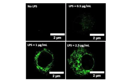
stimulated the cells with lipopolysaccharides (LPS) to induce the over-expression of CSE. Living cells are incubated with different concentration of LPS (0 - 2.5 µg/mL) and maintained for 16 hours in DMEM culture medium with 10% FBS, and then
these pre-treated cells are further incubated with MB-Sn (10
[image:4.595.308.524.230.343.2]µM) for 25 minutes.
Figure 2. SIM images of exogenous H2Sn in RAW 264.7 macrophages. Cells were incubated with MB-Sn(10 µM) for 25 minutes and then incubated with different Na2S2 concentrations for 20 minutes. Bottom row- corresponding 3D profile plots.
Figure 3. Endogenous H2Sn detection by SIM. RAW 264.7 cells were treated with different concentration of LPS for 16 hours followed by incubation with MB-Sn (10 µM)
for 25 minutes.
SIM images show bright intracellular fluorescence, and this
confirms the generation of polysulfides and the efficacy of
MB-Sn in detecting endogenous H2Sn (Figure 3). Figures 2 and 3
also reveal that MB-Sn is not localized in the nucleus. To
ascertain its exact localization, co-staining experiments are carried out. Results of the microscopic studies reveal a wide distribution of the probe in the endoplasmic reticulum of RAW 264.7 cells. Co-staining experiment with the commercial ER tracker green probe was also performed. Further analysis helps us to evaluate Pearson’s correlation coefficient of 0.944
confirming the precise localization of MB-Snwithin the ER of
the cells (Figure 4). We have further utilized MB-Snfor 3D-SIM
imaging - see figure 4 and video provided in the ESI.
[image:4.595.307.536.385.519.2]COMMUNICATION Journal Name
organelles.14 We have explored the possibility of using MB-Sn
[image:5.595.42.280.99.215.2]as a probe molecule for dual colour imaging.
Figure 4. Colocalization experiments (Cells were pre-treated with LPS, 2.5 µg/mL
before MB-Snand ER-Tracker incubation): (A) Wide-field (WF) images of MB-Sn,
ER-tracker green, merged image and Pearson’s profile plot indicating maximum colocalization (0.944), confirming the preferential colocalization in the Endoplasmic reticulum. (B) Corresponding SIM images of MB-Sn, ER-tracker green, merged image
and 3D-SIM image.
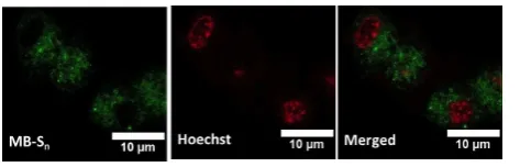
Figure 5.Dual colour SIM using MB-Snin the presence of LPS and Hoechst-33342
To realize this, we performed both widefield (Fig. S12) and SIM imaging (Figure 5) with a nuclear staining dye Hoechst-33342 in LPS treated cells. Images shown in Figure 5, confirm the usefulness of the probe molecule for dual-colour imaging.
In summary, we have designed a BODIPY based
chemodosimetric probe MB-Snthat specifically detects H2Sn in
physiological condition. H2Sn mediated thioester cleavage
followed by spontaneous cyclization led to the generation of
free MB-OH with a turn-ON emission response. Its non-toxic
nature and cell membrane permeability allowed us to utilize it
for imaging exogenous and endogenous H2Sn in living cells.
Co-localization studies with ER tracker green confirmed the precise localization of the probe in the ER region of cells. We also demonstrated the utility of the probe in SIM, 3D-SIM, and dual-colour imaging. We believe this method will open up a new window for developing SIM compatible probes for other biologically important analytes.
A.D. acknowledges SERB (India) Grants (SB/S1/IC-23/2013, &JCB/2017/000004 and DBT (India) grant BT/PR22251/NNT/ 28/1274/2017) for funding. AHA, FA, acknowledge UGC &CSIR for their research fellowships. SS and JAT are grateful to the Imaging Life initiative of the University of Sheffield, and MRC funding (Grant No: MR/K015753/1) for SIM facilities. References
1 H. Kimura, Proc. Jpn. Acad., Ser. B, 2015, 91, 131.
2 A. K. Mustafa, M. M. Gadalla, N. Sen, S. Kim, W. Mu, S. K.
Gazi, R. K. Barrow, G. Yang, R. Wang and S. H. Snyder, Sci.
Signal., 2009, 2, ra72.
3 Y. Kimura, Y. Mikami, K. Osumi, M. Tsugane, J.-i. Oka and H.
Kimura, FASEB J., 2013, 27, 2451.
4 (a) T. Ida, T. Sawa, H. Ihara, Y. Tsuchiya, Y. Watanabe, Y.
Kumagai, M. Suematsu, H. Motohashi, S. Fujii, T. Matsunaga,
M. Yamamoto, K. Ono, N. O. Devarie-Baez, M. Xian, J. M.
Fukuto and T. Akaike, Proc. Natl. Acad. Sci. U.S.A, 2014, 111,
7606; (b) A. S. Klymchenko, Acc. Chem. Res., 2017, 50, 366;
(c) D. Wu, A. C. Sedgwick, T. Gunnlaugsson, E. U. Akkaya, J.
Yoon and T. D. James, Chem. Soc. Rev., 2017, 46, 7105.
5 R. Greiner, Z. Pálinkás, K. Bäsell, D. Becher, H. Antelmann, P.
Nagy and T. P. Dick, Antioxid. Redox Signal., 2013, 19, 1749.
6 (a) V. S. Lin, W. Chen, M. Xian and C. J. Chang, Chem. Soc.
Rev., 2015, 44, 4596; (b) Y. Niu, Y.-Z. Chen, H.-R. Zheng,
L.-Z. Wu, C.-H. Tung and Q.-L.-Z. Yang, Chem. Soc. Rev., 2015, 44,
6143; (c) F. Ali, A. H. A, N. Taye, R. G. Gonnade, S.
Chattopadhyay and A. Das, Chem. Commun., 2015, 51,
16932; (d) A. H. A, F. Ali, S. Kushwaha, N. Taye, S.
Chattopadhyay and A. Das, Anal. Chem., 2016, 88, 12161; (e)
A. H. A, U. R. G, F. Ali, N. Taye, S. Chattopadhyay and A. Das,
Chem. Commun., 2015, 51, 15592; (f) H. S. Jung, X. Chen, J. S.
Kim and J. Yoon, Chem. Soc. Rev., 2013, 42, 6019.
7 (a) C. Liu, W. Chen, W. Shi, B. Peng, Y. Zhao, H. Ma and M.
Xian, J. Am. Chem. Soc., 2014, 136, 7257; (b) W. Chen, X. Yue,
H. Zhang, W. Li, L. Zhang, Q. Xiao, C. Huang, J. Sheng and X.
Song, Anal. Chem., 2017, 89, 12984; (c) M. Gao, R. Wang, F.
Yu, J. You and L. Chen, Analyst, 2015, 140, 3766; (d) M. Gao,
F. Yu, H. Chen and L. Chen, Anal. Chem., 2015, 87, 3631; (e)
L. Zeng, S. Chen, T. Xia, W. Hu, C. Li and Z. Liu, Anal. Chem.,
2015, 87, 3004; (f) Q. Han, Z. Mou, H. Wang, X. Tang, Z.
Dong, L. Wang, X. Dong and W. Liu, Anal. Chem., 2016, 88,
7206; (g) Y. Huang, F. Yu, J. Wang and L. Chen, Anal. Chem.,
2016, 88, 4122; (h) K.-B. Li, F.-Z. Chen, Q.-H. Yin, S. Zhang, W.
Shi and D.-M. Han, Sens. Actuators, B, 2018, 254, 222; (i) J.
Ma, J. Fan, H. Li, Q. Yao, F. Xu, J. Wang and X. Peng, J Mater
Chem B, 2017, 5, 2574; (j) H. Shang, H. Chen, Y. Tang, R. Guo
and W. Lin, Sens. Actuators, B, 2016, 230, 773.
8 W. Chen, E. W. Rosser, D. Zhang, W. Shi, Y. Li, W.-J. Dong, H.
Ma, D. Hu and M. Xian, Org. Lett., 2015, 17, 2776.
9 (a) W. Chen, A. Pacheco, Y. Takano, J. J. Day, K. Hanaoka and
M. Xian, Angew. Chem., Int. Ed., 2016, 55, 9993; (b) W. Chen,
E. W. Rosser, T. Matsunaga, A. Pacheco, T. Akaike and M.
Xian, Angew. Chem., Int. Ed., 2015, 54, 13961; (c) W. Chen, C.
Liu, B. Peng, Y. Zhao, A. Pacheco and M. Xian, Chem. Sci.,
2013, 4, 2892; (d) Y. Fang, W. Chen, W. Shi, H. Li, M. Xian and
H. Ma, Chem. Commun., 2017, 53, 8759; (e) J. Guo, S. Yang,
C. Guo, Q. Zeng, Z. Qing, Z. Cao, J. Li and R. Yang, Anal.
Chem., 2018, 90, 881.
10 (a) M. Gustafsson, Proc. Natl. Acad. Sci. U.S.A, 2005, 102,
13081; (b) D. Li, L. Shao, B.-C. Chen, X. Zhang, M. Zhang, B. Moses, D. E. Milkie, J. R. Beach, J. A. Hammer, M. Pasham, T. Kirchhausen, M. A. Baird, M. W. Davidson, P. Xu and E.
Betzig, Science, 2015, 349, aab3500; (c) S. Sreedharan, M. R.
Gill, E. Garcia, H. K. Saeed, D. Robinson, A. Byrne, A. Cadby, T. E. Keyes, C. Smythe, P. Pellett, J. Bernardino de la Serna
and J. A. Thomas, J. Am. Chem. Soc.,2017, 139, 15907; (d) E.
Sezgin, F. Schneider, V. Zilles, E. Garcia, D. Waithe, A. S. Klymchenko and C. Eggeling, bioRxiv; (e) F. Ali, S. Sreedharan, A. H. Ashoka, H. K. Saeed, C. G. W. Smythe, J. A. Thomas and
A. Das, Anal. Chem., 2017, 89, 12087.
11 N. Boens, V. Leen and W. Dehaen, Chem. Soc. Rev., 2012, 41,
1130.
12 P. Nagy and C. C. Winterbourn, Chem. Res. Toxicol., 2010, 23,
1541.
13 D. Macmillan, Angew. Chem. Int. Ed. 2006, 45, 7668.
14 (a) F. Hu, X. Cai, P. N. Manghnani, K. Kenry, W. Wu and B. Liu,
Chem. Sci., 2018, DOI: 10.1039/C7SC04585A; (b) A. K. Tong, Z. Li, G. S. Jones, J. J. Russo and J. Ju, Nat. Biotechnol., 2001,
[image:5.595.44.281.293.369.2]