EVALUATION OF BRAINSTEM AUDITORY
EVOKED POTENTIAL AND SERUM ZINC
LEVELS IN CHILDREN WITH ATTENTION
DEFICIT HYPERACTIVITY DISORDER
Dissertation submitted to
THE TAMIL NADU DR. MGR MEDICAL UNIVERSITY In partial fulfillment of the regulations
for the award of the degree of M.D. PHYSIOLOGY
Branch V
INSTITUTE OF PHYSIOLOGY & EXPERIMENTAL MEDICINE
Madras Medical College and Government General Hospital
Chennai – 600003
The Tamil Nadu Dr. MGR Medical University Chennai – 600 032
CERTIFICATE
This is to certify that the dissertation entitled “EVALUATION
OF BRAINSTEM AUDITORY EVOKED POTENTIAL AND
SERUM ZINC LEVELS IN CHILDREN WITH ATTENTION
DEFICIT HYPERACTIVITY DISORDER” by Dr.J. Anitha Ponmalar
for M.D Physiology is a bonafide record of the research done by her
during the period of study (2012-2015) in the Institute of Physiology and
Experimental Medicine, Madras Medical College, Chennai – 600 003.
DEAN
Madras Medical College Chennai - 600 003.
DIRECTOR AND PROFESSOR
ACKNOWLEDGEMENT
I express my sincere thanks to Dr.R.Vimala, M.D.,Dean, Madras
Medical College, Chennai for allowing me to conduct this study in this
institution.
I express my profound gratitude and sincere thanks to
Prof. Dr. K. Padma, M.D., Director, Institute of Physiology and
Experimental medicine, Madras Medical College, Chennai for providing
insightful discussions about the research and opportunity to develop my
individuality and immense support without whom this work would not
have been completed successfully.
I am thankful to Dr.Shanti Nambi, Head of Department of Child
Psychiatry, Child Guidance Clinic, Institute of Child Health, Chennai for
granting me permission to recruit cases from the department. I also thank
Dr. Vimal Doshi, MD Psychiatry for his valuable suggestions and help
through out this study.
I am greatly indebted to Dr. A. Parimala, MD, whose enthusiastic
supervision provided me the necessary momentum to bring this work into
I wish to express my sincere thanks to Prof. Dr.R. Vijayalakshmi,
M.D. Professor of our department for her suggestions and support .
I thank our professors Dr.Thirupathi, M.D, and Dr. P. Sathya,
MD for their valuable support during the study.
I sincerely thank Prof. Dr. K.Pramila, M.D Department of
Biochemistry, Institute of Child Health, Chennai for granting me the
permission to avail the laboratory facilities.
I thank Dr.Poonguzhali Gopinath, M.D, Department of
Biochemistry, ICH, Chennai for her great help in lab procedures.
I thank Assistant Professors Dr.Ratna manjushree, M.D.,
Dr. Kanmani Karthikeyan, M.D, Dr. Janet Sugantha, M.D,
Dr.T.N.Vijayalakshmi, M.D., Dr.Anantha subramaniam, M.D,
Dr. Shantimalar , M.D and all other Assistant professors of our
department for their help and timely suggestions.
I thank Dr. Sathya Narayanan, Msc Physiology, PhD Madras
Medical College, Chennai , for helping me in statistical analysis.
I am grateful to my co-PG,S and all other post graduates who
I am greatly indebted to all my patients without whose cooperation
this study would not have been possible.
I thank my parents, Husband Mr. D. Paul, Son John Samuel for
their constant support, help and encouragement.
CONTENTS
LIST OF TABLES
LIST OF PHOTOGRAPHS AND FIGURES
LIST OF GRAPHS ABBREVIATIONS
SL.NO Title Page No
1. INTRODUCTION 1
2. REVIEW OF LITERATURE 15
3. AIM AND OBJECTIVES 66
4. MATERIALS AND METHODS 67
5. RESULTS 105
6. DISCUSSION 114
7. CONCLUSION 124
8. SUMMARY 125
BIBLIOGRAPHY
ANNEXURES ( i – v)
(i) ETHICAL COMMITTEE APPROVAL
(ii) CONSENT FORM
(iii) PROFORMA
(iv) MASTER CHARTS
SL.NO LIST OF TABLES Page No
1. Generators of BAEP waveforms B/w 50 & 51 2. Normal waveforms of BAEP 92 3. Gender differences in BAEP waveforms 100 4. Comparison of Age, Ht, Wt and BMI between
Normal and ADHD children
106
5. Comparison of Absolute wave latencies between Normal and ADHD children – Right ear
108
6. Comparison of Interpeak latencies between Normal and ADHD children – Right ear
108
7. Comparison of Absolute wave latencies between Normal and ADHD children – Left ear
109
8. Comparison of Interpeak latencies between Normal and ADHD children – Left ear
109
9. Comparison of serum zinc levels between Normal and ADHD children
110
10. ANOVA for serum zinc levels among three ADHD subtypes
111
11. ANOVA for IPL I – III among three ADHD subtypes
111
12. Correlation of BAEP variables of Right ear with serum zinc levels
112
13. Correlation of BAEP variables of Left ear with serum zinc levels
LIST OF PHOTOGRAPHS
PHOTO NO TITLE BETWEEN PAGES
1. Computerised Neurostim Medicaid System for Recording BAEP
73 & 74
2. BAEP Recording in a Normal Child 81 & 82
3. Colorimeter, Serum Samples and Zinc Kit
102 & 103
LIST OF FIGURES
FIGURE NO TITLE BETWEEN
PAGES
1. Afferent connections of Reticular Formation
5
2. Efferent connections of Reticular formation
5
3. Auditory Evoked Potentials 45 & 46
4. Auditory Pathway 47 & 48
5. BAEP waves 50
6. Origin of BAEP 50 & 51
7. Apparatus for BAEP 74
LIST OF GRAPHS
GRAPH NO TITLE BETWEEN
PAGES
1. Comparison of Age between normal and ADHD children
105 & 106
2. Comparison of BMI between normal and ADHD children
105 & 106
3 Distribution of ADHD children among different socio-economic classes
107
4. Comparison of absolute latencies of BAEP waveforms in right ear of normal and ADHD children
108 & 109
5. Comparison of IPLs of right ear between normal and ADHD children
108 & 109
6. Comparison of absolute latencies of BAEP waveforms in left ear of normal and ADHD children
109 & 110
7. Comparison of IPLs of left ear between normal and ADHD children
109 & 110
8. Comparison of serum zinc levels between normal and ADHD children
ABBREVIATIONS
ADHD - Attention Deficit Hyperactivity Disorder
ADD - Attention Deficit Disorder
ABR - Auditory Brainstem Response
BAEP - Brainstem Auditory Evoked Potential
BERA - Brainstem Evoked Response Audiometry
AVCN - Anterior Ventral Cochlear Nucleus
SLR - Short Latency Response
MLR - Middle Latency Response
LLR - Late Latency Response
PCVN - Posterior Ventral Cochlear Nucleus
DCN - Dorsal Cochlear Nucleus
SPL - Sound Pressure Level
dB - Decibel
IPL - Inter Peak Latencies
EEG - Electro Encephalography
ERP - Event Related Potentials
Hz - Hertz
MRI - Magnetic Resonance Imaging
PET - Positron Emission Tomography
ABSTRACT
EVALUATION OF BRAINSTEM AUDITORY EVOKED POTENTIAL AND SERUM ZINC LEVELS IN CHILDREN WITH
ATTENTION DEFICIT HYPERACTIVITY DISORDER
Degree for which submitted : Doctor of Medicine(MD) in Physiology
Supervisor and guide : Prof. Dr. K. Padma
Director and Head of the Department
Department : Institute of Physiology and Experimental Medicine
College : Madras Medical College, Chennai – 600003.
University : The Tamilnadu Dr. M.G.R.
Medical University, Chennai-600032.
Year : 2014
BACKGROUND
Attention Deficit Hyperactivity Disorder is a behavioural and
neurocognitive condition characterized by developmentally inappropriate
and impairing levels of gross motor overactivity, inattention and
impulsivity. Brainstem Auditory Evoked Potential (BAEP) is an
important neurophysiological tool which can be used to assess the
functional integrity of the auditory pathway and its neurotransmission. It
auditory stimuli in the auditory pathway. Using BAEP, we can easily
detect the early changes occurring in the auditory pathway in children
with ADHD and detect hearing impairment which may contribute to
inattention even before clinical manifestation.
AIM OF THE STUDY
· To determine the functional integrity of auditory pathway in
children with ADHD by recording brainstem auditory evoked
potential.
· To assess serum zinc levels in these children.
· To find the correlation between serum zinc levels and Brainstem
auditory evoked potential in children with ADHD.
MATERIALS AND METHODS
30 boys with ADHD in the age group 6 - 11 years without any
clinical evidence of hearing impairment were included in the study.
Controls were age and BMI matched healthy children. Both the controls
and ADHD children were subjected to BAEP and serum zinc levels were
RESULTS
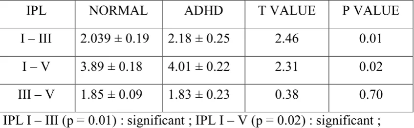
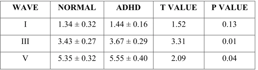
ADHD children showed significant prolongation of wave III, V
absolute latencies and I-III, I-V IPLs of BAEP. The serum zinc levels
were significantly decreased in ADHD children when compared to
controls suggesting the contribution of zinc in the pathogenesis of
ADHD.
CONCLUSION
There was significant prolongation of central conduction time in
ADHD children even though there was no clinical evidence of hearing
impairment assessed by pure tone audiogram prior to the study. Hence
BAEP can be utilized as an objective electrophysiological tool to evaluate
the functional integrity of auditory pathway from the external ear to
lower brainstem.
KEY WORDS
Attention Deficit Hyperactivity Disorder, Brainstem auditory
1
1.
INTRODUCTION
“The slightest deviation in brain activity can be felt in the body and
small electrical imbalances can become amplified into bigger health
problems”
- Dr. Rodolfo Linas
Attention is the patient’s ability to perceive a specific stimulus
without distraction by other stimuli (environmental or internal)
Vigilance is the capacity to maintain attention over a prolonged
period (sustained attention)
Hyperactivity is the state in which there is increased motor
activity with aggressiveness, over-talkativeness and uncoordinated
physical activity.
Impulsivity is defined as failure to ignore appealing stimuli other
than target stimuli which leads to loss of sustained attention to a
particular target task hindering successful completion of that task.
Attention is maintained by Ascending Reticular Activating
2
nuclei (intralaminar and midline reticular nuclei) and then to the
neocortex especially the prefrontal (dorsolateral frontal cortex area 45
and 46, orbitofrontal cortex area 11 and 47) and anterior cingulate
(limbic) cortex corresponding to brodmann area 24, 32 and 33. RAS
sends a strong facilitatory drive to the cortical neurons raising their
background excitability (EEG activity).
The functional importance of reticular formation remained
unrecognized until 1946 when Magoun and Moruzzi103 found that
electrical stimulation of ventromedial part of medullary reticular
formation caused inhibition of cortically induced and reflexly produced
movement. Magoun in 1949 showed that high frequency stimulation of
midbrain reticular formation produces EEG alerting response and arouses
a sleeping animal. Damage to this area produces a comatose state.
Electrical stimulation of posterior hypothalamus also produces arousal.
The ascending reticular activating system is responsible for the awake
state recorded in EEG.
Jasper and Hambery66 found that stimulation of non specific
projection system of thalamus which consists of midline nuclei,
3
in widespread rhythmic activity and concluded that nonspecific
projection system of thalamus is the rostral end of reticular activating
system(discovered by Moruzzi and Magoun)
The reticular formation is the phylogenetically old reticular core
of the brain which occupies the central portion of medulla and midbrain
surrounding the fourth ventricle and cerebral aqueduct. It contains the
cell bodies and fibres of aminergic systems such as dopaminergic, nor
adrenergic and serotoninergic systems and also cholinergic systems and
project to widespread areas of central nervous system where they
influence motor functions, consciousness, attention, sleep and
wakefulness, motivation, emotion, reward processing and addiction. All
systems in reticular formation are influenced by projections from other
brain areas and can inturn influence the functions of those areas. Thus
reticular formation is “THE INTEGRATOR IN CENTRAL
NERVOUS SYSTEM”
The brainstem reticular formation comprises of medullary,
pontine and midbrain reticular formation. Structurally it is classified into
1. Neuronal aggregates
4 a. Afferent connections
b. Efferent connections.
NEURONAL AGGREGATES OF RETICULAR FORMATION
Nuclei of median column:
Lie in the midline and called as raphe nuclei
Nuclei of medial column:
Lie lateral to nuclei of median column and called as magnocellular
nuclei as these are made of large cells.
Nuclei of lateral column:
Lie lateral to medial column and called as parvocellular nuclei as
these are made of small neurons.
Functional neuronal aggregates:
They are not anatomical entities but have fairly well defined
5
RETICULAR PATHWAYS
Afferent connections:
1. From spinal cord – spinoreticular tract
2. From cranial nerves – brainstem afferent
3. From Tectum( superior and inferior colliculi) – tectoreticular tract
4. From cerebellum – cerebello reticular tract
6
6. Neocortex – corticoreticular fibres from motor and sensory cortex,
orbital , prefrontal, parietal and temporal lobes, cingulated gyrus
and collaterals from corticofugal fibres.
7. Limbic lobe including amygdala and hippocampus.
Efferent connections:
1. To spinal cord – reticulospinal tract
2. To brainstem – reticulobulbar fibres
3. To cerebellum
4. To red nucleus, substantia nigra and tectum in midbrain
5. To thalamus, subthalamic nuclei and hypothalamus
6. To corpus striatum, neocortex and limbic lobe indirectly through
thalamus and hypothalamus.
FUNCTIONS OF RETICLAR FORMATION
1. Through functional neuronal aggregates influences respiratory,
circulatory ,gastrointestinal and visceral functions.
2. The descending fibres influence motor control and stretch reflexes,
7
3. The ascending fibres which constitutes the ASCENDING
RETICULAR ACTIVATING SYSTEM controls the levels of
cortical activity(attention, alertness,- wakefulness) and electrical
activity of the cortex (EEG)
Reticular Activating System is a complex polysynaptic pathway
arising from Brainstem reticular formation which projects to intralaminar
and reticular nuclei of thalamus which inturn projects diffusely and
non-specifically to wide regions of cortex including frontal, parietal, temporal
and occipital cortices.
Collaterals funnel into ARAS not only from long ascending
sensory tracts but also from trigeminal, auditory, visual and olfactory
systems. The complexity of neuronal network and degree of convergence
abolishes modality specificity and most reticular neurons are activated
with equal facility by different sensory stimuli.
NEUROTRANSMITTER SYSTEMS OF RAS
Dopaminergic system
Dopaminergic neurons are located in two anatomically and
8
area. Dopaminergic cell bodies in substantia nigra projects to caudate
nucleus and putamen and play a role in the control of movement. The
ventral tegmental area in the rostral midbrain has widespread projections
to various CNS areas and plays a role in circuitry involved in reward,
motivation, emotion, impulse control and decision making.
Nor-adrenergic system:
Neurons using norepinephrine as the principal neurotransmitter
are clustered in pons next to fourth ventricle in locus coeruleus and also
scattered throughout the lateral tegmentum of brainstem. Activities of
these neurons are either tonic (constant and continuous) or
phasic(increases periodically and temporarily).The main function of these
neurons is to modulate attention, arousal, mood and pain in conjuction
with serotoninergic and dopaminergic systems.
Tonic firing of noradrenergic neurons contributes to general
levels of arousal and attention whereas phasic firing helps us to focus
attention onto a specific task while suppressing distracting stimuli. But
too much of activation of noradrenergic neurons decreases our ability to
9
Serotoninergic system:
Located in raphe nuclei which is a collection of neurons in the
midline along the entire length of brainstem and spinal cord and
modulates regulation of mood, appetite, sleep, state of wakefulness,
modulation of pain and some cognitive functions.
Cholinergic system:
Located in the tegmentum of pons and plays a neuromodulatory
role by enhancing functioning of synapses. Cholinergic projections to the
thalamus strengthens the excitatory glutaminergic output from thalamus
to cortex and plays a role in arousal and motor functions.
Histaminergic system:
Located in tegmentum of midbrain and functionally related to
cluster of histaminergic neurons located in the posterior hypothalamus.
Projections of these neurons play a role in general arousal and alertness.
When activity of noradrenaline and serotonin containing neurons
is dominant, there is reduced level of activity in acetylcholine containing
10
awake state. Phasic bursts of noradrenaline release causes arousal state
and focusing of attention.
Thus attention is mediated by a complex interaction of limbic,
neocortical and ascending reticular activating systems. Damage to many
brain areas can disrupt attention. For eg: metabolic disturbances, drug
intoxication, extensive bilateral cortical damage, bilateral lesions of
frontal lobe or limbic system.
Attention Deficit Hyperactivity Disorder is the most common
neurobehavioral disorder of childhood affecting school aged children
characterized by inattention, increased distractibility and difficulty
sustaining attention, poor impulse control, decreased self-inhibitory
capacity and motor restlessness inappropriate for that particular age
( Durston.S) 45. They have poor school performance ( Barkley, RA)19
due to difficulty in focusing their attention efficiently and easy
distractibility (Kennemans JJ 72 et al) (Vanderstelt O 163 et al) (Van
Mourik Rooster 162 et al)
In addition to poor school performance, they have problems like
11
have low self esteem. ADHD often co-exists with emotional, behavioral,
language and learning disorders.
DSM-IV (4th edition of American Psychiatric Association’s
Diagnostic and statistical manual) states that the prevalence of ADHD is
5-8% (Polanczyk G 115 et al) and identifies three subtypes of ADHD
according to (Chhabildas N35 et al) namely
1. Predominantly inattentive type (30%)
2. Hyperactive-impulsive type(9%)
3. Combined type(61%) which is the commonest type.
ADHD is 3-4 times more common in males (9.2% in males and
2.9% in females), but the inattentive subtype is more common in females.
There is no single causal factor in the aetiology of ADHD. Genetic
and environmental factors acting during foetal and postnatal
development such as maternal smoking and toxins such as lead may
play a role. It commonly occurs following damage to CNS (prematurity,
TBI) ,toxin exposure or as a sequelae of CNS infection. Attention is
mediated by multiple neuronal circuits involving many neurotransmitters
12
M138 et al). Morphologic and functional brain dysfunction including
moderate reduction in size of dorsolateral prefrontal cortex, corpus
callosum, basal ganglia and cerebellum(Makris N 90 et al) and
hypoperfusion of frontal-striatal dopamine pathways may be
implicated(Sillitoe RV 137 et al)
Molecular genetic studies reveals strong genetic component such
as abnormalities in dopamine transporter gene, norepinephrine transporter
gene and choline transporter gene. Patients with predominantly
inattentive ADHD had changes in norepinephrine transporter gene ,
predominantly hyperactive-impulsive type had altered dopamine
transporter gene and combined type had altered choline transporter gene.
Alterations in Dopamine Transporter Gene (DAT1) and D4 Dopamine
Receptor Gene (DRD4) are more likely to have functional significance
(Faraone SV49 et al).
Evoked potentials are electrical potentials recorded in response to
an external stimulus which may be visual, auditory or somatosensory.
Brainstem Auditory Evoked Potential is produced by presenting
auditory stimuli in the form of clicks to the ear which results in sequence
13
scalp. These waveforms are specific for different structures in the
auditory pathway and so provides specific localization of pathology .
Brainstem Auditory Evoked Potentials are markers which
provide precise information about the delay in the transmission of
auditory impulses from the periphery to the brainstem (Hood LJ 1998)61.
BAEP in the first 10 milliseconds (Early Latency Response) provides
information about the functional integrity of the auditory pathway from
the vestibulo-cochlear nerve upto midbrain inferior colliculus.(Jacobson
J.T.1985)65
Brainstem Reticular Activating system is responsible for
formation of evoked potentials and neurotransmitter imbalance in the
fronto-striatal network which is the hallmark of ADHD will lead to
changes in the amplitude and latency of waveforms of BAEP. It acts as
an important neurophysiological tool for assessing the delay in
transmission of auditory impulses and assists in the early diagnosis of
ADHD.
Zinc is a cofactor for metabolism of many neurotransmitters and
fatty acids. It is necessary for 100 different metalloenzymes and complex
14
also regulates dopamine metabolism which is involved in the
pathogenesis of ADHD. Dopamine transporter has an endogenous zinc
binding site and zinc acts as a potent non-competitive blocker and
prevents dopamine binding to the dopamine transporter and thus
increases the extracellular availability of dopamine by decreasing its
degradation (Lepping P. Haber M)82
Published reports of the role of zinc in ADHD shows low levels
of zinc in serum, red cells, hair, nails and urine of affected
children(Arnold LE, Di silvestro RA) 11. Zinc can be considered as an
adjuvant in treating ADHD along with approved pharmacological
stimulant therapies (Akhondzadeh S et al)4.
Hence considering these above factors,the present study was
undertaken to evaluate the functional integrity of auditory pathway in
children with ADHD by performing BAEP and to find out the role of
zinc in the pathogenesis of ADHD children by comparing serum zinc
15
2. REVIEW OF LITERATURE
Attention Deficit Hyperactivity Disorder is a behavioral and
neurocognitive condition characterized by developmentally inappropriate
and impairing levels of gross motor over activity, inattention and
impulsivity (APA 2000 ) which results in deficits of executive functions.
It involves impaired ability to plan a work and to execute that plan. The
prevalence of ADHD has been increasing in the recent years probably
due to formation of nuclear families rather than joint families and also
poor parenting techniques.
BAEP is a neurophysiological test which can be used to assess the
integrity of auditory pathway and to localize defective neurotransmission
in the pathway and can be used to identify hearing impairment in ADHD
which may contribute to attention deficit in these children. Only few
BAEP studies have been reported in ADHD children and these studies
have shown increase in latency of BAEP waves and so with this
background , the present study has been taken up to assess the integrity
of auditory pathway and its neurotransmission in ADHD children.
As several researches now-a-days point out to nutritional factors
16
zinc levels in children with ADHD to identify if zinc deficiency has
played a role in the pathogenesis of ADHD.
ATTENTION DEFICIT HYPERACTIVITY DISORDER
Early in the mid-nineteenth century the problems of inattentiveness
and overactivity in children were recognized by Heinrich and Hoffman
(struwwel peter 149 et al) in the moralistic children’s book Slovenly
Peter which featured the characters Fidgety Phil and Harry. In 1902,
ADHD was discovered by George Still (Still. G145 et al) as the behavioral
sequelae of viral encephalitis called as Still’s disease. He described about
the overactivity and impulsivity of these children and he believed to have
a combination of organic and environmental factors resulting in lack of
inhibitory control(impulsivity) and inattention in these children.
After the influenza pandemic in 1919 to 1920, children who
survived developed severe behavioral problems similar to that described
in Still’s monograph. The flu survivors were thought to have organic
brain damage and so the condition was termed as Minimal Brain Damage
Syndrome.
In 1937, C.Bradley published a report that d,l-amphetamine
17
behavior problems in a residential treatment centre. This concept was
ignored for about 30 years when Keith Conners and Leon evaluated the
efficacy of d- Amphetamine in a double blind placebo controlled trial for
children with learning disability and behavior problems. In the early
1960’s the condition was renamed as Minimal Brain Dysfunction which
implied specific anatomic location which was not proved till then.
Genesis of the terms ADHD and Hyperkinetic Disorder
Initially ICD-9 (International Classification of Diseases and
Related Health Problems) and DSM (Diagnostic and Statistical Manual
of Mental Disorders) adopted the same descriptive definition for this
condition termed hyperkinetic syndrome of childhood. This term believed
that hyperactivity was the core feature of this disease. But further
research suggested that the main disability associated with this condition
is the difficulty in maintaining sustained attention and impulsivity
whereas motor overactivity is only secondary. This resulted in 1980
version of APA (American Psychiatric Association) classification which
renamed the condition as Attention Deficit Disorder (ADD). This
included three symptom categories as diagnostic criteria in which three
18
and two out of five overactivity symptoms was proposed. Based on this
three subtypes of ADD were described namely Attention Deficit
Hyperactivity Disorder, Attention Deficit Disorder without Hyperactivity
and a Residual type that included adults with ADD symptoms but no
longer met childhood ADHD criteria.
In 1987, DSM version named as DSM III-R, which included a
single criterion list requiring 8 of the 14 possible symptoms of
hyperactivity, impulsivity and inattention was published. Duration
criteria was added stating that the behavior should be present since age 7
years and for atleast 6 months. This version had no sub groups.
In 1994, APA published the 4th edition of DSM and in 2000, a Text
Revision, DSM IV-TR which is the current version.
Diagnostic Definition of ADHD
There are two main approaches to define disorders of
inattentiveness, hyperactivity and impulsiveness namely DSM IV-TR
(APA 2000) which recognizes the term “Attention Deficit Hyperactivity
Disorder” and ICD-10 (WHO , 1992) which uses the term “Hyperkinetic
Disorder”. Both are based on the same descriptions of behavior but the
19
Hyperkinetic Disorder (ICD – 10) requires all three components to
be present and that the diagnostic criteria must be met in more than one
situation(both home and school) and excluded by presence of other
disorders such as autism and anxiety states.
ADHD (DSM-IV) is divided into 3 subtypes
1) Combined type
2) Inattentive type
3) Hyperactive/impulsive type
With five main diagnostic criteria
1. Onset before 7 years.
2. Duration greater than 6 months.
3. An 18- item symptom list of which 6 of 9 inattention (or ) 6 of 9
hyperactive/impulsive symptoms have persisted for atleast 6 months to a
degree that is maladaptive and inconsistent with developmental level.
20
5. Symptoms that donot occur exclusively during the course of
pervasive developmental disorder, schizophrenia or other psychotic
disorder.
The practical consequence of these different diagnostic
definitions is that the ICD-10 category of Hyperkinetic Disorder is a
subgroup of ADHD described in DSM IV criteria (Santosh,
Taylor,Swanson et al) 130. Diagnosis is usually clinical using history
taken from parent and atleast one other adult such as teacher or coach.
There is no simple objective test such as blood test to diagnose ADHD.
EPIDEMIOLOGY
ADHD is the most common childhood psychiatric disorder
affecting 5- 12% of children worldwide. Polancyzyk et al 115 estimated
this prevalence using a meta-analysis which included about one lakh
patients and 100 articles.
Prevalence rates vary in various cultural settings according to
diagnostic criteria used to classify the disease and reported in a range of
1 -20%. (Jensen 2005) 67 . The male female ratio is 2:1 in community
samples in contrast to 5:1 or 9:1 in mental health clinic sample (Szatmari
21
(breaking rules) such as interference with teacher and classmates,
distractability and both physical and verbal aggression than females
(Abikoff 2002)3. Females present more often with less disruptive
symptoms, more attention problems and problems such as depression and
anxiety than males with ADHD . Male sex, low socioeconomic status
and young age are associated with high prevalence of ADHD
(Biederman J )26
Recent studies prove that ADHD symptoms continue into teenage
and adulthood although previously it was proposed that ADHD
symptoms wane off as the child reaches adolescence (Pierce EW113 et al)
In India, few epidemiological studies are available for ADHD
which report prevalence rates ranging from 10-20% . They have also
shown that ADHD is more prevalent in boys than in girls. ADHD is more
prevalent in lower socioeconomic class. (Malhi P, Singh P et al) 91. It is
also more common in nuclear families and behavioral problems are high
in children from nuclear families(Verghese A, Beig A)164
AETIOLOGY
The aetiology of ADHD is complex and involves interaction
22
development which creates a neurobiological susceptibility to this
disorder. The expression of disease is mediated by alteration in different
neural networks and neurotransmitters involved in mediating attention
and executive functions(Sonuga- Barke EJ)140.
Prenatal, perinatal and post natal factors play a role in the
pathogenesis of ADHD. Prenatal factors are associated with maternal
lifestyle during pregnancy such as smoking and alcohol intake ( Linnet,
Dalsgaard et al) 86.
Maternal smoking has the strongest evidence among environmental
factors and it increases the risk for ADHD by 2.7 fold.
( Milberger S et al) 98. There is a dose- response relationship between
maternal smoking and ADHD. (Thapar, Fowler, Rice et al)157.This may
be due to the effect of smoking on nicotinic receptors which modulate
dopamine which is involved in pathogenesis of ADHD(Potter AS,
Newhouse PA, Bucci)117 .Prenatal exposure to alcohol may lead to
structural anomaly of cerebellum which is implicated in the pathogenesis
of ADHD (Coffin JM et al)39.Exposure to cocaine may lead to
23
Seasonal predilection is associated with some cases of ADHD
children who are born commonly in the month of September(winter)
which may due to viral infections triggering the disease in genetically
predisposed individuals(Seeger G et al).132 Matenal stress during
pregnancy may lead to ADHD due to increased cortisol secretion in
stress( Kapoor, Dunn, Kostaki et al)71
Perinatal factors have also been implicated in ADHD. There is a
two fold increase in ADHD in low birth weight children(Bhutta, Cleves,
Casey et al)25. This effect is due to subtle lesions in frontosriatal
circuit involved in pathogenesis of ADHD ( Carmody, Bendersky,
Dunn et al)31. Pregnancy and birth complications have been reported in
mothers of children with ADHD( Ben Amor, Grizenko, Schwartz et al,
2005) 21.
Post natal factors include social and biological factors. Biological
factors include a role for dietary deficiencies.
There is an imbalance of essential fatty acids such as omega-3
and omega-6 fatty acids which has been suggested in the pathogenesis
(Richardson, Montgomery, 2005et al )119. Iron deficiency has been
24
2004 )74. A role for artificial food additives in the aetiology of ADHD
have been proposed in a randomized control trial but remains
controversial ( Mccann, Barrett, Cooper et al 2007) 96 . Idiosyncratic
allergies and specific food allergies have been reported by some parents .
Exposure to lead is associated with risk of hyperactive and inattentive
behavior (Levitt M 1999)84.
Social factors are also implicated in the aetiology in ADHD.
Children experiencing parental neglect and abuse may be at increased
risk of ADHD (Glod and teicher, 1996).52 An English and Roman
adoptees study has shown that these children were persistently
hyperactive and inattentive even after adoption into another family before
the age of 4 yrs (Kreppner, O’Connor & Rutter, 2001)77.
Genetic factors play an important role in the pathogenesis of
ADHD. The mechanism by which they exert influence is not known
(Asherson, Kuntsi, Taylor 2005)16. Family, adoption and twin studies
show that ADHD is a highly heritable disorder(Rietveld, Hudziak,
Bartels et al)120. Siblings of ADHD children show an eight fold increase
in risk of ADHD (Faraone and Biederman, 2000)48. Biological relatives
25
Crawford et al) 141. According to twin studies, ADHD is one of the most
heritable conditions accounting about 60 to 90% (Thapar A et al)156
Genes regulating neurotransmitter systems have been proposed to
be involved in ADHD .Specific genes involved in the pathogenesis of
ADHD were studied and these studies showed significant pooled effects
for three polymorphisms of dopamine genes, the D4 and D5
receptors(DRD4 AND DRD5) and the dopamine transporter (Faraone
et al 2005)49 ( Thapar A et al )156. DAT 1 and DRD4 are more likely to
have functional significance out of these. There is no evidence for
nor-epinephrine gene variants as of now but are under research. But DAT I
association has been contradicted in a recent meta analysis( Wohl,
Purper- Ouakil, mouren et al 2005)169.
Based on animal knock out models an association of functional
polymorphisms of serotonin transporter and receptor genes SLC6A4 and
HTR 1B with ADHD has been suggested (Faraone et al , 2005)49.
Genome scan studies on potential alleles for ADHD demonstrate linkage
26
GENE ENVIRONMENT INTERACTIONS
Genes may interact with each other and with environmental risk
factors to increase risk of ADHD in a non-linear manner so that genes of
small effect show disproportionately larger manifestation of the disease
when interacting with environmental factors (Rutter, Moffitt &
Capsi)125.
Studies show that gene variants such as DRD4 and DAT 1 interacts
with prenatal substance exposures in specific subtypes of ADHD.
Smoking is associated with combined ADHD in genetically
susceptible children. (Khan, Khoury, Nicholas et al)70. Alcohol
consumption during pregnancy is also associated with specific
genes(Brookes, Mill, Guindalini et al)30, DAT 1 and DRD4 genes
interacts with season of birth( Seeger, Schloss, Schimidt et al)132.
PATHOGENESIS OF ADHD
The pathogenesis of ADHD involves three main concepts namely
genetic aetiology, frontostriatal or executive dysfunction disorder and a
catecholamine disorder. A change in concept of ADHD from a primary
27
neuro cognitive defects as the core pathology of the disease
( Castellanos, Sonuga- barke et al)34.
The concept of fronto-striatal/executive function disorder was
proposed based on the similarities between people with ADHD and
frontal lobe lesions (Denckla, 2002)42.The circuit involved in this
executive cognitive function is the Thalamo – cortico –striatal loop
which consists of projections from the dorsolateral prefrontal cortex to
the caudate nucleus of neostriatum which pass via a complex set of direct
and indirect basal ganglia pathways through the thalamus and back to the
prefrontal cortex (Alexander &crutcher et al, 1990)5. This circuit has
connections to other regions such as the cerebellum, the parietal cortex,
the dorsal anterior cingulated cortex, corpus callosum and the frontal
motor cortex (Timmann, D et al) 159 (Emond V, Joyal C, Poissant H)47.
Activity within this circuit is mediated by GABA and glutamate which is
responsible for the background thalamo-cortical EEG activity which is
modified by other neurotransmitters such as dopamine, norepinephrine
and acetyl choline during stimulus related evoked responses and arousal
28
The prefrontal cortex functions in thinking and planning
( Mc.Geer et al, 1987) 95. It is described as attention association area and
integrates body movements and behavior associated with attaining goals
(Posner & Peterson1990)116. It operates in focusing the mind on specific
tasks by screening out distracting sensory inputs in order to concentrate
on a goal.
Basal Ganglia are masses of grey matter that lie lateral to the
thalamus and involved in regulation of motor activity. Thalamus
communicates with basal ganglia through neurotransmitter dopamine,
brainstem relays information to basal ganglia via serotonin and acetyl
choline mediates transmission from cerebral cortex to basal ganglia.
Basal ganglia is involved in planning of movements and organization of
goal directed behaviors. Basal Ganglia is found to be less active in
children with ADHD (Gabrieli, Brewer et al)50 .
Other than fronto-striatal executive circuit several extra-executive
circuits have been implicated in the neurobiology of ADHD.
Toplak, dockstader & tannock 160 et al, 2006 has implicated
29
Sagvolden, johansen, aaseet al.,(2005) 126; SonugaBarke et al(2005)140
has implicated orbitofrontal – ventral striatal circuitry in reward and
motivation.
Banaschewski, Brandeis, Heinrich et al.,200417 has implicated
posterior parietal network in attention, orientation and alerting response.
Anterior cingulate cortex is implicated in affective component
of ADHD. Thus compensatory networks like insula and cerebellum
are implicated in lower cognitive tasks in ADHD .(Castellanos FX,
Lee PP et al)33
The evidence of catecholamine (dopamine and nor-epinephrine)
dysfunction is indirectly shown by three observations.
1. ADHD symptoms are reduced by DA & NE agonists which act by
different mechanisms to increase extracellular DA & NE
(Pliszka,2005)114
2. There are a number of polymorphisms in gene affecting
30
3. Lesions and knock out of genes related to catecholamines in animal
models produce symptoms that mimics ADHD(Arnsten and
Li,2005)15.
The neurotransmitter dopamine is involved in motor control and
feeling of well being. Dopamine transmission is found to be reduced in
children with ADHD (Castellanos et al)34
EVIDENCES FAVOURING FRONTO-STRIATAL/EXECUTIVE
FUNCTION HYPOTHESIS
Performance of children with ADHD is worse than controls in
cognitive and executive functions such as response inhibition (ability to
inhibit a response which is already initiated), delay aversion(a preference
between a small immediate and a large delayed reward), interference
control, planning and working memory (Huang-Pollock&Nigg)63.
Nigg et al, 2005106 conducted a study which showed that half of the
children with ADHD showed a deficit in atleast one executive function.
MRI studies in children with ADHD have reduction in brain
31
Castellanos, Lee, Sharp et al33 has shown reduction particularly
in the dorsolateral prefrontal cortex and neostriatum (caudate nucleus and
putamen).
Emond.V, Joyal.C & Poissant H et al 47 has shown reduction in
volumes of and also corpus callosum ,dorsal anterior cingulate cortex and
cerebellum.
Shaw, lerch, Greenstein et al., 2006 133 have shown that cortical
thinning is especially marked in prefrontal regions associated with
executive control.
Shaw P, Rabin C et al134 has shown a marked delay in cortical
maturation in children with ADHD. The delay was associated with
decreased cortical thickness in all regions of brain, especially Prefrontal
cortex and right posterior parietal cortex. Children who continued to have
ADHD symptoms in adulthood had thinner grey matter in the prefrontal
cortex which persisted. Children who grew out of the disorder showed
normalization of right posterior parietal cortex thickness which was
32
Diffusion tensor imaging(DTI) is a modality of MRI which
provides information about the direction and integrity of neural tracts in
the brain.
D’Agati E, Caserelli et al41 have conducted DTI studies in
ADHD children and has shown developmental changes such as defective
myelination in the white matter pathways surrounding basal ganglia and
cerebellum. These changes cause a decrease in the speed of neural
communication.
White matter abnormalities such as reduction in volume has been
reported in ADHD children. Silk et al136 2008 in his study in 15 young
males found white matter abnormalities in several regions underlying
inferior parietal, occipito-parietal, Inferior parietal and inferior temporal
cortex.
Durston, Tottenham, Thomas et al.,(2003) 45 have conducted
functional MRI ( f MRI ) studies which showed reduced activity in
ventrolateral and dorsolateral prefrontal cortex and Rubia et al (1999) 124
has shown reduced activity in neostriatum which includes caudate and
33
PET and SPECT show dynamic measures of brain metabolism at
rest and during cognitive tasks and several studies have shown
abnormalities in cerebral activation in ADHD with a hypo perfusion in
frontal and striatal areas. Imaging studies using ligands highly selective
for dopamine transporter which is involved in the pathogenesis of ADHD
has shown that there is increase in DAT binding in the striatum of ADHD
children.
Volkow ND et al165 has done PET studies showing that
methylphenidate hydrochloride blocks DAT and the extracellular
dopamine increase is directly proportional to the level of blockade. This
leads to increased perception of the external sensory stimuli in ADHD
children.
CLINICAL FEATURES
Hyperactivity
This is the increase in activity which does not change with the
set demands of the situation. So, children with ADHD are more active in
the classroom and less active in the playground as compared to normal
children. The level of gross motor activity decreases with age, but
34
difficulties include problems of sensory motor coordination such as poor
hand writing, clumsiness and delay in achieving motor milestones. Motor
impairment is associated in 50% of ADHD children. Motor problems are
related to abnormalities in structure or function of cerebellum and basal
ganglia.
Attentional Difficulties
ADHD children show several attentional difficulties. Cognitive
functions such as problem solving, planning, orientation, alertness,
response inhibition and working memory are impaired. They exhibit easy
distractability, inability to sustain attention on a task, failure to follow
instructions, inability to complete a task without constant supervision and
forgetfulness in daily routines. Other domains involving affective
components such as motivation , response inhibition and delay aversion
are also involved (Castenellos et al)34 (Nigg JT et al) 106.
Impulsivity
These children engage in dangerous activity, yells out in class,
interrupts or intrudes on others during games or conversations. Impulsive
behavior results in trouble with parents, teachers or other children
35
thinking leading to decreased academic performance. This is due to
internal pressure persuading the child to respond quickly.
Cognitive aspects
They have difficulty in time management and donot develop
internal sense of pace in planning tasks. This poor time sense leads to
problems in estimating actual difficulty waiting in a line and planning
how much time a task requires.
Behavioral aspects
Children with ADHD lack persistence. They become bored and
leave games early before they are finished. Other disabilities include
temper outbursts, mood lability, greater intra individual variability of
reaction time and cerebellar associated defects in motor timing .
Comorbidity
Considerable comorbidity in children occurs with conduct
disorder, oppositional defiant disorder, mood and anxiety disorders and
36
Course of ADHD
Between 30 – 70% of children diagnosed as ADHD will continue
to show symptoms as adults. In a study of adult ADHD, both genders had
manifestation but females unlike in childhood situation were in majority
and had higher rates of depression, anxiety disorders and conduct
disorder than controls.(Biederman et al 1996)26
DIAGNOSIS
Since 1994, the diagnosis of ADHD has been most commonly
made based on DSM IV criteria. Most children are referred because of
impairment in academic performance, family or peer relationship.
Although symptoms of gross motor overactivity declines with age,
impairment due to inattention and impulsive behavior may continue into
adulthood as adult ADHD. The presence of psychiatric comorbidity
increases with age further complicating the clinical picture. ADHD is a
diagnosis primarily made by history and various rating scales such as the
Parent Teacher Rating scales for ADHD(Du paulGJ), the SNAP
(Swanson) and Conner’s revised long form(Conners). Additional
diagnostic information can come from both positive and negative family
37
Mental status examination is done to rule out other mental
disorders. Physical examination identifies soft neurological signs which
include non focal motor deficits such as deficits in balance, motor
planning and control and also deficits in sensory integration. Gustaffson
et al, 2000 found a significant correlation between soft neurological signs
and decreased cerebral blood flow measures in the frontal lobes
bilaterally in children with ADHD. Inquiry about dietary intake and food
allergy should also be done.
Electrophysiological studies such as EEG and Evoked Potentials
are simple tools which aid in the diagnosis of ADHD. Several studies in
ADHD children has reported changes which pose them as an important
diagnostic tool.
EEG provide information about the background electrical activity
of the brain. This study was first done in ADHD patients in 1938 by
Jasper HH 66, Solomon P, Bradley when they mostly included
qualitative EEG. With advancement in technology QEEG(Quantitative
EEG) is used now not only for diagnostic purposes but also for
38
Monastra (2000)102 found that diagnosis of ADHD can be
accurately made by the use of Q EEG procedures which measures
electrophysiological activity in specific brain regions . The ratio of slow
wave to fast wave activity (theta/beta ratio) is a developmental marker.
Young children have high ratio which declines with age.
In ADHD children this ratio doesn’t decrease with age and it is
typically greater during tasks that demand focus.
Several QEEG studies have been done which have shown high
theta/beta ratio in areas such as prefrontal cortex which is specifically
involved in ADHD with high specificity and sensitivity.
EVOKED POTENTIALS
Evoked potentials are electrical potentials that occur in a group of
neurons in response to stimulation of a sense organ which can be
recorded by surface electrodes over the corresponding cortical areas. EPs
are reliable measures of objective function of specific sensory pathways.
They are very sensitive to detect abnormality even in a clinically normal
subject and are highly specific in localizing the lesion in the particular
39
Event Related Potentials (Long latency potentials)
These are long latency evoked potentials evoked by endogenous
stimuli which provide information about brain activity during cognitive
tasks in response to auditory or visual stimuli and requires subject’s
attention and cooperation. They have a longer latency and higher
amplitude and are not influenced by the intensity and frequency of
stimulus. Due to the use of different types of performance indicators most
of the studies related to ERP s are difficult to interpret.
Stimulus Related Potentials(short latency potentials)
These are short latency potentials evoked by exogenous stimuli
which provide information about the integrity of neural pathways and
neurotransmitter systems involved in interpretation of sensory stimuli
presented to the individual which may be auditory, visual or tactile
stimuli and they are independent of subject’s level of attention and can be
recorded even during sleep or anaesthesia. But they are influenced by
frequency and intensity of the stimuli as opposed to Event related
40
ADHD and BAEP
Brainstem Auditory Evoked potentials may provide useful
information about the integrity of auditory pathway and neurotransmitter
systems involved in auditory transmission. It has been postulated that
asymmetrical conduction of acoustic stimuli in the brainstem plays a role
in the pathogenesis of ADHD (Lahat E et al)79. Defective auditory
transmission may lead to defective attention in ADHD.A few studies
have been conducted using BAEP in ADHD children and shows increase
in amplitude and latency of several waveforms showing defective
auditory transmission. BAEP can be used as a simple and cost effective
physiological tool to assess auditory transmission.
Hannan Azzam et al 56 conducted an ABR study using click
stimulus as well as speech stimulus and MMN on 15 Arabic speaking
ADHD children and 15 age matched controls in the child/adolescent
psychiatry clinic, Ain Shams University and found increased absolute
latency of wave III and inter-peak latencies I-III & I-V in both ears in
33%(5/15) of ADHD children. Abnormal speech ABR was observed in
87% (13/15) ADHD children. The peak MMN latencies were prolonged
41
A Puente et al 2conducted a study in 18 school children
diagnosed with ADD and compared them with 18 normal school children
by recording both short latency auditory evoked response and long
latency auditory evoked response and showed that brainstem conduction
was significantly abnormal in children with ADHD. He found prolonged
latencies of waves III & V and prolonged I-III & I-V IPL in ADD
children as compared to controls. The latency of P300 was increased and
mean amplitude was significantly decreased in children with ADHD
compared to controls. He concluded that school children with ADD show
significant abnormality in both Brainstem Auditory Evoked Response
and Long Latency Auditory Evoked Response and so these
electrophysiological procedures involving auditory pathway can be useful
in diagnosis of children with ADHD.
Lahat et al79 conducted a study in which he found prolonged wave
III latency in females and prolonged wave V latency in both males and
females. There was a prolonged I-III IPL in females and prolonged I-V
IPL in both male and female subgroups. This result showed disturbance
in the neural transmission in the auditory pathway. The latency of wave I
42
neural conduction in ADHD children is not due to abnormality in inner
ear or eighth nerve.
NeelamVaney, MD et al 105conducted a study on 20 male ADHD
children and 20 age and sex matched controls in the Electrophysiology
Laboratory of the Department of Physiology, University College of
Medical Sciences, Delhi and showed that there were no statistically
significant differences in the absolute peak latencies and amplitude as
well as the IPLs of ABR waves in ADHD children as compared to
controls.
Schochat E et al131 conducted a study in 21 ADHD children and
found that all children had normal ABR and normal latency for wave V.
Some delayed P300 was found, some small and some absent. Among 42
ears combined 52.38% did not have P300. Medicated group had more
presence of P300 than non-medicated group.
Ana Carla Leite Romero et al9 in his article published in
Brazilian Journal of Otorhinolaryngology has shown that when long
latency auditory evoked potentials were measured in ADHD children
43
LE which was higher for ADHD children and N2 amplitude and latency
was abnormal in ADHD group.
Hilary Gomes et al 59conducted a study to characterize the deficits
in attention allocation in ADHD children. Findings for Nd, P3b and Ta in
these children suggest that deficits in auditory selective attention in
children with ADHD may be attributed to reduced information during
early auditory processing. Nd waveforms were not elicited in children
with ADHD and they exhibit significantly lesser auditory responses at
100ms (Ta).
The development of electrophysiological techniques such as EEG
and Evoked potentials have passed through a series of interesting
historical aspects after the introduction of electrodiagnosis based on
faradic and galvanic current by Erb in 1861.
Richard Caton118, a lecturer at Liverpool, UK had an idea that
as the nerve impulse flows in and out of the brain, it may be detected at
the surface and reported the findings in a meeting in British Medical
Association in 1875. He demonstrated the effects of anoxia and
anaesthesia on these potentials using optical magnification of movement
44
not available at that time. He described EEG(Background electrical
activity) as well as cerebral potential changes evoked by sensory
stimuli(Evoked Potentials) and published his reports in British Medical
Journal in 1877.
In 1929, Hans Berger, a German psychiatrist was the first to
record EEG and documented brain waves using two scalp electrodes
on his own son and named this spontaneous ongoing activity as
“Des Electrenkephalogram”.
In 1939 Davis recorded the first auditory evoked potential but the
amplitude of this evoked potential was very small and it was George
Dawson who discovered an innovative method of extracting the evoked
potential from the ongoing EEG activity. First he used a photographic
superimposition averaging technique to bring out EP wave form from
back ground EEG activity and later he used an electronic summation
technique which subsequently became the computerized averaging
technique used today.
In 1967, Sohmer and Feinmesser were the first to publish ABR
45
structures and showed that cochlear potentials could be obtained
non-invasively.
The concept of far field potential was first developed by Jewett
and in 1971, Jewett and Williston gave a clear description of human
ABR wave forms and correctly interpreted the generation of the waves
from brainstem. This short latency auditory response was first called as
Jewett bump and now known as BAEP.
In 1974, Hecox and Galambos showed that ABR could be used for
threshold estimation in adults and infants.
In 1977, Setters and Brackman published landmark findings on
prolonged IPL in tumor cases.
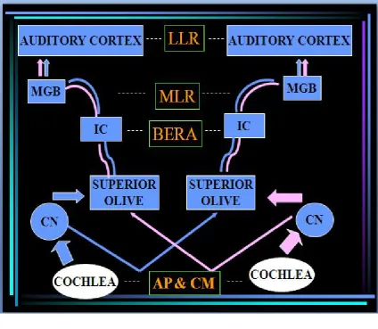
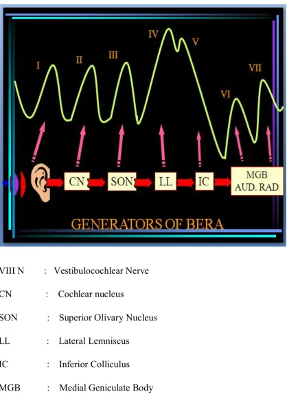
BRAINSTEM AUDITORY EVOKED POTENTIAL
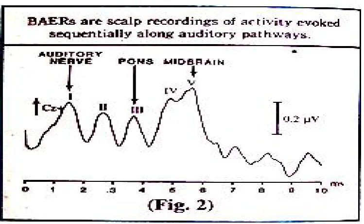
BAEP is an objective test of auditory brainstem function in
response to auditory click stimuli which consists of seven positive waves
recorded during the first 10 milliseconds after a click stimulus labeled as
waves I to VII. The synonyms of BAEP include Auditory Brainstem
FIGURE 3. GENERATORS OF VARIOUS AUDITORY EVOKED POTENTIALS
AP & CM - Action potential and Cochlear microphonics
CN - Cochlear nucleus
IC - Inferior colliculus
MGB - Medial Geniculate Body
MLR - Middle Latency Response
46
Brainstem Evoked Potential(BSEP), ABR audiometry, and Brainstem
Evoked Response Audiometry (Jiang et al) 68.
The auditory evoked potential of first 10 millisecond after the
acoustic stimulus is called the short latency response (SLR). The SLR is
popularly known as brain stem auditory evoked potentials (BAEP), since
it records the auditory evoked potential when the auditory stimulus is
traversing through the brain stem region. The middle latency response
(MLR) is the transient response that occurs in the 10- 50 milliseconds
post-stimulus time and the late latency response (LLR) is the response
recordable in the 50-300 milliseconds post-stimulus period
(Picton TW et al)112.
ANATOMICAL AND NEUROPHYSIOLOGICAL BASIS OF BAEP83
The sound pressure wave produces displacement of the tympanic
membrane which is transmitted to the oval window through the inner
ossicles. This produces movement of the perilymph (contained in the
scalavestibuli and scala tympani, which are joined at the apex of the
cochlea by the helicotrema) and then secondarily of the endolymph,
which is present in the ductus cochlearis or scala media which includes
47
spiral organ contains hair cells that, when displaced by the movement of
the tectorial membrane, produces auditory potentials. The receptor
potentials leads to the release of the neurotransmitters, which trigger
action potentials in the dendrites of the afferent nerve fibers of the
cochlear nerve. There are 20,000-30,000 hair cells distributed over a
distance of approximately 31.5mm in the 2.5 spirals of the cochlea.
High-frequency sounds activate the basal portion of the basilar membrane
where as low-frequency sounds generate receptor potentials in the apical
portion of the cochlea. The click, which is the most commonly used
stimulus to produce Brain stem auditory evoked potentials (BAEPS),
contains mainly high-frequency components and mainly activates the
basal portion of cochlea.
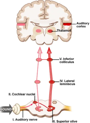
The cochlear nerve neurons are bipolar, situated in spiral ganglia
and their dendrites go to hair cells and axons to the cochlear nucleus.
The cochlear nucleus has three sub-nuclei:
1. Anterior ventral cochlear nucleus (AVCN)
2. Posterior ventral cochlear nucleus (PCVN)
48
The output of AVCN is through the ventral acoustic striae forming
the bulk of trapezoid body to terminate in the superior olivary nuclei and
inferior colliculus. The neurons in the AVCN discharge at short latency
to acoustic stimuli with a pattern like that of cochlear nerve.
The output of PVCN mostly goes through the ventral and middle
acoustic striae to terminate in the superior olivary nuclei and inferior
colliculus.
DCN output terminates in the superior olivary nucleus and
contralateral inferior olivary nucleus through dorsal striae, The
discharges from these neurons are different from those of AVCN by
having longer latency.
The cochlear nuclear complex thus terminates in the superior
olivary nuclear complex, which has medial and lateral components at the
base of the pons. The medial superior olivary nucleus receives the input
from both ipsilateral and contralateral AVCN which are excitory. The
lateral superior olivary nucleus also receives ipsilateral excitatory inputs
from AVCN and PVCN and inhibitory inputs from contralateral AVCN
and PVCN via trapezoid body. From the olivary nuclear complex, the
49
colliculi. The olivary nuclei are the first in the auditory pathways where
the neurons are affected in a non-linear manner to binaural stimulation.
The inferior colliculi and the lateral lemnisci converge the input
from contralateral cochlear nucleus and superior olivary nucleus. The
impulse from inferior colliculus travels to medial geniculate body.
Neurons in the medial geniculate body form the acoustic radiation of the
internal capsule, synapsing in the Heschl gyrus of the primary auditory
cortex (superior temporal gyrus and upper bank of sylvian fissure
including the frontal and parietal opercula). High frequency tones, such
as clicks, activate mainly the deeper most mesial portion of the heschl
gyrus.
The orderly orientation of the neurons in dorsal cochlear, medial
and lateral superior olivary nuclei results in summation of synaptic
potentials which results in high amplitude electrical fields.
As the auditory impulses travels through the different components
of the auditory pathway, it undergoes some degree of processing at each
level. Passage of the impulse through this pathway generates an electrical
activity which can be monitored by placing a surface (far –field)
50
activity presents a waveform with discrete peaks, the character of which
is dependent upon the structural and functional integrity of the above
mentioned auditory pathway.
BAEP is produced by presenting auditory stimuli to each ear
which results in a sequence of waveforms that bear a close relationship to
the auditory pathway structures (peripheral, pontomedullary, pontine and
midbrain) and so allows specific localization of pathology in the auditory
pathway. BAEPs are recorded within 10 milliseconds of the acoustic
[image:67.595.131.506.415.646.2]stimulus
FIGURE : 6
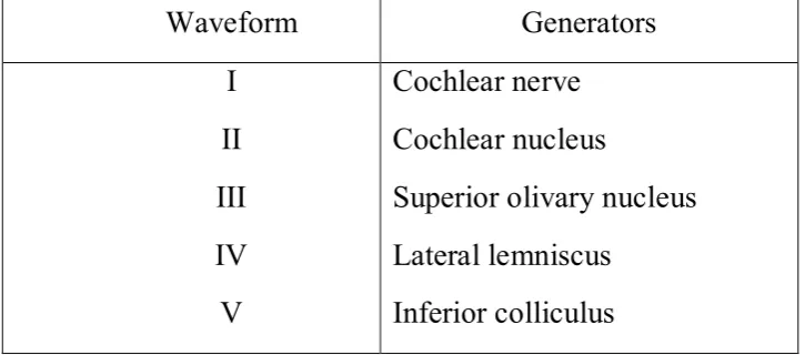
TABLE NO.1
Generators of BAEP waveforms (Chiappa 1990)36
Waveform Generators
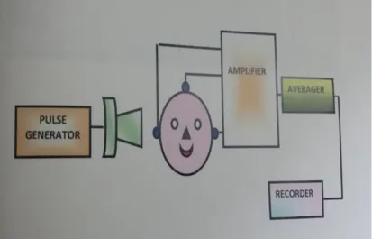
I
II
III
IV
V
Cochlear nerve
Cochlear nucleus
Superior olivary nucleus
Lateral lemniscus
[image:68.595.128.491.578.738.2]