Copyright © 2003, American Society for Microbiology. All Rights Reserved.
Nuclear Export of Vpr Is Required for Efficient Replication of Human
Immunodeficiency Virus Type 1 in Tissue Macrophages
Michael P. Sherman,
1,2Carlos M. C. de Noronha,
1Lauren A. Eckstein,
1Jason Hataye,
1Pamela Mundt,
1Samuel A. F. Williams,
1Jason A. Neidleman,
1Mark A. Goldsmith,
1,2and Warner C. Greene
1,2,3*
Gladstone Institute of Virology and Immunology
1and Departments of Medicine
2and of Microbiology and
Immunology,
3University of California, San Francisco, California
Received 9 December 2002/Accepted 4 April 2003
Retroviruses must gain access to the host cell nucleus for subsequent replication and viral propagation.
Human immunodeficiency virus type 1 (HIV-1) and other primate lentiviruses are distinguished from the
gammaretroviruses by their ability to infect nondividing cells such as macrophages, an important viral
res-ervoir in vivo. Rather than requiring nuclear membrane breakdown during cell division, the HIV-1
preinte-gration complex (PIC) enters the nucleus by traversing the central aqueous channel of the limiting nuclear
pore complex. The HIV-1 PIC contains three nucleophilic proteins, matrix, integrase, and Vpr, all of which
have been implicated in nuclear targeting. The mechanism by which Vpr can display such nucleophilic
prop-erties and yet also be available for incorporation into virions assembling at the plasma membrane is
unre-solved. We recently characterized Vpr as a nucleocytoplasmic shuttling protein that contains two novel nuclear
import signals and an exportin-1-dependent nuclear export signal (NES). We now demonstrate that mutation
of this NES impairs the incorporation of Vpr into newly formed virions. Furthermore, we find that the Vpr NES
is required for efficient HIV replication in tissue macrophages present in human spleens and tonsils. These
findings underscore how the nucleocytoplasmic shuttling of Vpr not only contributes to nuclear import of the
HIV-1 PIC but also enables Vpr to be present in the cytoplasm for incorporation into virions, leading to
enhancement of viral spread within nondividing tissue macrophages.
Human immunodeficiency virus type 1 (HIV-1) and other
primate lentiviruses are able to infect nondividing cells,
nota-bly terminally differentiated macrophages (41), an important
viral reservoir within the infected host (31, 34, 53). This
bio-logical feature distinguishes the lentiviruses from the
oncoret-roviruses (or gammaretoncoret-roviruses), in which cell division
asso-ciated with nuclear membrane dissolution is required for
infection (33, 42, 62). HIV-1 is also able to infect resting,
nondividing T cells in lymphoid tissues (13). These nondividing
T cells may contribute to the establishment of protected
res-ervoirs in the host, undermining attempts to eradicate
virus-producing cells in the long term (5, 6, 15, 56, 77).
After entry by fusion and uncoating, the viral reverse
scriptase complex traverses the cytoplasm while reverse
tran-scribing the two strands of RNA into DNA (20), forming the
viral preintegration complex (PIC). The nuclear envelope
forms a barrier that the PIC must negotiate. The nuclear
en-velope is studded with nuclear pore complexes (NPCs) that
form a conduit with a central aqueous channel mediating
bi-directional transport of many macromolecules. The NPC
cor-responds to a 125-MDa structure comprising 50 to 100
polypeptides. Many of these proteins are members of the
nucleoporin family characterized by FG repeats (46). During
active transport, the central aqueous channel accommodates
protein complexes as large as 25 nm in diameter. However, the
HIV-1 PIC exhibits a Stokes diameter of 56 nm and represents
one of the largest known cargoes successfully transported
across the NPC (48). How HIV-1 performs this feat of
“mo-lecular gymnastics” remains unknown (66).
All retroviruses contain three major open reading frames,
including
gag
(generates the viral core after intravirion
pro-cessing of the p55
gagprecursor polypeptide),
pol
(encodes the
reverse transcriptase, integrase, and protease enzymes), and
env
(directs the production of the transmembrane and surface
glycoproteins). In addition, the primate lentiviruses contain
genes for regulatory (
rev
and
tat
) and accessory (
vpr, vpx, vpu,
vif,
and
nef
) proteins. Viral protein R (Vpr) is highly conserved
in vivo (47, 65) and serves many functions in the viral life cycle
yet is frequently lost during in vitro propagation of the virus
(25, 80), highlighting an experimental limitation of such in
vitro culture systems.
Vpr induces G
2cell cycle arrest in proliferating human cells
when it is overexpressed (1, 25, 28, 60) or in the context of
infection with a recombinant vector (37). This effect correlates
with the production of herniations in the nuclear envelope
(10). Arrest in the G
2phase of the cell cycle enhances viral
replication, in part by increasing the activity of the long
termi-nal repeat (25). Other studies suggest that the prolonged G
2arrest induced by Vpr promotes apoptosis of the infected cell,
perhaps leading to increased virion release and enhanced viral
burden (57, 70–72, 78). Although it has been suggested that
Vpr inhibits apoptosis early and promotes apoptosis late
dur-ing the course of HIV-1 infection (9), we have not observed a
consistent effect of Vpr on T-cell depletion in infected
lym-phoid histocultures (14).
* Corresponding author. Mailing address: Gladstone Institute of
Virology and Immunology, P.O. Box 419100, San Francisco, CA
94141-9100. Phone: (415) 826-3800. Fax: (415) 826-1817. E-mail:
7582
on November 8, 2019 by guest
http://jvi.asm.org/
within the arginine-rich carboxy-terminal segment and a
sec-ond that depends on highly conserved leucines present in the
two
␣
-helical regions (35, 64). The distal leucine-rich helix also
contains a nuclear export signal (NES) (64). This NES utilizes
the chromosome maintenance region 1 protein (CRM1) (17,
52), which binds to the leucine-rich NES directly and mediates
export through the NPC in a leptomycin B-sensitive manner
(51, 76). However, the biological significance of the
nucleocy-toplasmic shuttling properties of Vpr remains unknown.
Vpr is predominantly found in the nuclei of HIV-1-infected
cells (43), probably reflecting the strength of its two nuclear
targeting signals (64). This nucleophilic property of Vpr,
cou-pled with its presence in the viral PIC, led to the observation
that Vpr facilitates more efficient HIV-1 replication in
nondi-viding monocyte-derived macrophages (4, 8, 22, 29). In vitro
assays further supported a direct role for Vpr in PIC import
(58, 59). However, more recent studies have questioned the
role of Vpr in the nuclear uptake of the HIV-1 PIC (2) and
have also shown that Vpr is not required for HIV infection of
nondividing T cells (14). Further, it is unclear how this
nucleo-philic protein is incorporated into virions, which are assembled
in the cytoplasm at or near the plasma membrane.
In this study, we investigated whether the nuclear export
function of Vpr contributes to virion incorporation and
whether virion Vpr contributes to viral replication in tissue
macrophages. For these studies, we have employed HIV
mo-lecular clones containing a mutation (L67A) in the distal helix
of Vpr that selectively compromises the nuclear export
pheno-type of Vpr while maintaining its nuclear import and G
2cell
cycle-arresting functions in HIV-1-infected peripheral blood
mononuclear cells (PBMCs). We have evaluated the growth of
wild-type and mutant viruses in both the T-cell and
macro-phage compartments of lymphoid histocultures produced with
human tonsillar or splenic tissue. HIV infection in the ex vivo
lymphoid histoculture system is likely to more closely
approx-imate the conditions encountered in vivo than does infection of
mitogen-stimulated PBMCs (23, 24). This tissue-based system
is composed of a mixture of HIV-1 targets, including
lympho-cytes, macrophages, and the supporting cellular network. It
requires no addition of cytokines or activating or
differentiat-ing agents like those used in more homogeneous primary
cel-lular systems.
MATERIALS AND METHODS
Plasmids.A hemagglutinin (HA) epitope was introduced at the amino termi-nus of NL4-3 Vpr to form HA-Vpr as previously described (65). For subcellular localization studies, we used a green fluorescence protein (GFP)-pyruvate kinase (PK)-Vpr chimera (64). This GFP-PK-Vpr fusion protein or relevant Vpr mu-tants allowed subcellular localization by fluorescence microscopy. Since the
back-bone is larger (⬇90 kDa) than the passive diffusion size of the NPC (⬇60 kDa),
its import and export occur by active transport (52). The nuclear localization sequence (NLS)-GFP-PK-Vpr construct has been characterized (64), and an L67A mutation was derived by cloning the NLS (PKKKRKV) from the simian
construction of the HIV-1 NL4-3⌬Vpr strain has been described previously (14).
Cell cultures, transfections, and microscopic analysis.Expression vector DNA was transfected into HeLa or 293T cells with calcium phosphate. Cells were cultured in Dulbecco modified Eagle medium (GIBCO BRL, Gaithersburg,
Md.) supplemented with 10% fetal bovine serum, 2 mML-glutamine, penicillin
G (100 U/ml), and streptomycin (100g/ml). All plasmids were transfected with
either 4g of DNA per well of six-well plates or 3g in experiments
incorpo-rating 1g of an expression vector encoding the red fluorescent protein (RFP)
(pDsRed1-N1) (Clontech, Palo Alto, Calif.). Cells plated on coverslips for mi-croscopic analyses were washed with phosphate-buffered saline, fixed for 10 min in 1% paraformaldehyde, and rinsed in water. The coverslips were then inverted and mounted on glass slides using Gel Mount (Biomeda Corp., Foster City,
Calif.). Nuclei were visualized with Hoechst 33342 stain (10g/ml; Molecular
Probes, Eugene, Oreg.) added to the paraformaldehyde. Cells were analyzed with a Nikon TE 300 Quantum fluorescence microscope and a Hamamatsu Orca II charge-coupled device camera.
Heterokaryon analyses.Heterokaryons were generated as described previ-ously (64, 69). Briefly, transfected 293T cells were cultured for 24 h, washed, trypsinized, and replated overnight at a 1:10 ratio with excess untransfected
HeLa cells to achieve a total cell concentration of 1.5⫻106per well. The cells
were exposed to cycloheximide (25g/ml) for 1 h to prevent de novo protein
synthesis and then subjected to membrane fusion by the addition of 50% poly-ethylene glycol for 3 min. The cells were washed with phosphate-buffered saline (PBS) and incubated for 1 h with cycloheximide. The pDsRed1-N1 vector ex-pressing RFP was also included in the transfections. RFP localizes in the nucleus and cytoplasm of the donor cell and diffuses into the entire cytoplasm of the newly formed heterokaryon, thus delineating its boundaries. However, while RFP is present in the donor nucleus of the heterokaryon, it does not enter the newly fused nuclei (recipient nuclei) during this 2-h procedure. Thus, it is pos-sible to discern readily whether the test protein linked to the GFP shuttles from the donor nucleus (red) to the recipient nuclei (unstained) within the hetero-karyon.
Western blot and coimmunoprecipitation analyses.Coimmunoprecipitation experiments were performed by cotransfecting the HA-Vpr vector with
Pr55⌬MA-GFP (11), which contains the p6 domain that mediates Vpr binding.
Pr55⌬MA-GFP lacks the RNA retention signal in MA that inhibits RNA export
and subsequent expression (63). An equivalent number of 293T cells (600,000
cells) were transfected and harvested 48 h later in 500l of lysis buffer (50 mM
HEPES [pH 7.9], 250 mM NaCl, 0.5% NP-40 detergent, 0.5 mM EDTA
sup-plemented with protease inhibitor [Roche] [1 tablet/ml], 100M
phenylmethyl-sulfonyl fluoride). Lysates were aliquoted for direct analysis or incubated with monoclonal mouse antibody HA.11 immobilized on Sepharose Fast Flow beads (Covance) for 15 h at 4°C and then washed three times with lysis buffer. The beads were boiled for 5 min in loading buffer to dissociate any bound proteins before analysis by sodium dodecyl sulfate-polyacrylamide gel electrophoresis. Western blotting was performed with monoclonal anti-HA (Boehringer
Man-heim), polyclonal anti-GFP (Clontech), monoclonal anti-p24gag(NEN) or
poly-clonal rabbit anti-Vpr (35) antiserum diluted 1:2,000.
PBMC isolation and infection.HIV-seronegative blood was obtained by leu-kopheresis, and mononuclear cells were isolated on Histopaque-1077, washed with PBS, and activated with phytohemagglutinin (Sigma, St. Louis, Mo.) at
50g/ml. After 24 h, the cells were washed and cultured in RPMI supplemented
with interleukin-2 (10 U/ml; Roche). Cultures were infected with HIV-1 by
resuspending 107cells and 200 ng of viral stocks in 1 ml of medium for 2 h before
washing them and culturing them at 106cells/ml.
Cell cycle analyses.Cell cycle experiments were performed with pEGFP ex-pression vector (Clontech) cotransfected into 293T cells at a 1:7 ratio with the indicated HA control or HA-Vpr expression vector. After culture for 24 to 48 h, the cells were trypsinized and fixed for 30 min in 2% formaldehyde. The cells were next washed with PBS and treated with 1 mg of RNase A per ml plus 0.01 mM To-Pro-3 iodide (Molecular Probes) in PBS for 30 min. Cellular DNA
content in the transfected (GFP⫹) and untransfected (GFP⫺) cells was assessed
with a FACScan flow cytometer. DNA profiles were analyzed with FlowJo
on November 8, 2019 by guest
http://jvi.asm.org/
software (Treestar). PBMCs, after 5 days of infection, were processed similarly except that fixation and permeabilization were performed with a solution of 1% paraformaldehyde, 1 mg of human immunoglobulin G per ml, and 0.1% Tween 20 in fluorescence-activated cell sorter buffer (PBS with 2% fetal calf serum). Lymphocytes were analyzed by first gating on live, cycling cells as determined by cell size (forward scatter) and granularity (side scatter) and by DNA content with mock-infected controls. This cycling gate was then interrogated identically be-tween the different infections for intracellular Gag polyprotein expression with the KC57 monoclonal anti-p24 antibody (1:50 dilution) (Coulter).
Preparation of viral stocks.Molecular clones of HIV-1 proviruses were trans-fected into 293T cells as described above, and the culture supernatants were collected after 48 h. These viral preparations were centrifuged for 10 min at 5,000
rpm in a Beckman GH 3.7 rotor before being aliquoted and frozen. The p24gag
concentration was determined by a standard enzyme-linked immunosorbent
as-say (NEN), and the 50% tissue culture infective dose (TCID50) was determined
by limiting dilution using pooled, phytohemagglutinin-activated PBMC cultures
as targets (23, 55). Virus (300 ng of p24gag) was collected by ultracentrifugation
at 40,000⫻gfor 90 min to assess virion incorporation of Vpr.
Culture and infection of human lymphoid tissues ex vivo.The ex vivo lym-phoid histoculture system was used to more closely approximate HIV infection events occurring in vivo. This system supports viral infection and replication in the absence of exogenous cytokine stimulation and provides the diverse array of cells present in normal lymphoid tissues (23, 26, 55). Noninflammatory spleen or tonsil tissue (obtained from the National Disease Research Interchange) was sectioned into 2- to 3-mm blocks and cultured as described previously (23). Six
blocks per well were inoculated with HIV-1 by dropwise addition of 50 TCID50.
At the indicated times, the medium was collected from the wells to monitor the replication kinetics or the tissue was mechanically disrupted and subjected to flow cytometric analysis as described above for PBMCs. In addition, cells derived from lymphoid tissue were immunostained with various antibodies, including anti-CD3, anti-CD4, anti-CD14, and anti-CD68. Replicates represent data col-lected from three separate wells.
RESULTS
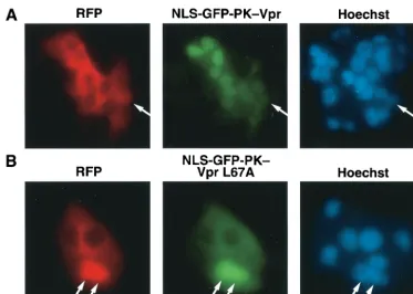
Segregation of the Vpr NES from the overlapping nuclear
import signal.
We previously mapped an NES to the
leucine-rich domain (LQQLL) of the distal helix spanning amino acids
64 to 68 of Vpr (64). Our mutational analysis further revealed
that the L67A analogue was effectively imported into the
nu-cleus but that L64A and L68A Vpr analogues were not. We
next investigated the nuclear export properties of the L67A
mutant. Wild-type and L67A Vpr were fused at the carboxy
terminus of the NLS-GFP-PK chimera, and heterokaryon
shuttling studies were performed (Fig. 1). The fusion protein
containing wild-type Vpr effectively relocalized into the
accep-tor nuclei in the heterokaryons, indicating effective shuttling.
However, a chimera containing the VprL67A mutant failed to
exit the donor nucleus. GFP-PK-VprL67A localizes to the
nu-cleus after addition of the export inhibitor leptomycin B (64)
but appears predominantly in the cytoplasm at baseline,
indi-cating that some degree of the nuclear export function is
re-tained (data not shown). However, when measured against the
import strength of the SV40 NLS, as shown in these
experi-ments, the nuclear export properties of the L67A Vpr mutant
were significantly compromised.
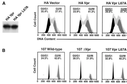
The VprL67A export mutant causes G
2cell cycle arrest.
To
assess whether the L67A mutant interferes with the G
2 [image:3.603.106.479.66.332.2]-arrest-ing properties of Vpr, an HA-VprL67A expression vector was
FIG. 1. Identification of a Vpr mutation that segregates nuclear import from nuclear export. The SV40 NLS was cloned into the GFP-PK-Vpr
fusion protein to direct this predominantly cytoplasmic shuttling protein into the nucleus. Cells were fused with polyethylene glycol in the presence
of cycloheximide (to prevent de novo production of Vpr in the cytoplasm) to examine whether the fusion protein, although on average appearing
nuclear, was actually shuttling as indicated by exit from the donor nucleus and accumulation in the recipient nucleus. RFP was included to
demarcate the boundaries of the heterokaryon and to mark the transfected (donor) nucleus red (arrows; also shown in the Hoechst staining panel)
but not the recipient nuclei (devoid of RFP) introduced by cell fusion. Note that the NLS-GFP-PK-Vpr was able to exit the donor nucleus and
accumulate in the recipient nuclei (A) while the L67A mutant was defective for such shuttling (B).
on November 8, 2019 by guest
http://jvi.asm.org/
prepared and introduced into 293T cells. Western blotting
revealed that wild-type Vpr and the VprL67A mutant were
expressed at comparable levels (Fig. 2A). Cell cycle analysis
showed that the levels of G
2cell cycle arrest were similar in
cells transfected with wild-type and L67A export mutant Vpr.
To verify that this mutant causes cell cycle arrest at levels
achieved during HIV-1 infection of primary cells, we generated
proviral molecular clones containing the single VprL67A point
mutation or lacking Vpr altogether. Stocks of wild-type 107,
107L67A, and 107
⌬
Vpr generated after transfection of 293T
cells were equally infectious as determined by TCID
50/p24
gagcontent (data not shown). PBMCs were infected with these
viruses, and the cell cycle profiles were assessed in the p24
gag-positive lymphocytes (those productively infected and making
the p55
gagprecursor of p24
gag) and p24
gag-negative lymphocytes
by analyzing their DNA contents (Fig. 2B). The 107- and
107VprL67A-infected lymphocytes displayed similar increases
in the percentage of cells with a 4
N
complement of DNA
compared with 107
⌬
Vpr-infected lymphocytes, indicating
reten-tion of G
2-arresting properties by the L67A mutant in these
primary cells.
Compromise of Vpr export results in decreased
incorpora-tion of Vpr into virions.
One possible function for Vpr nuclear
export is to ensure that there are adequate amounts of this
protein in the cytoplasmic compartment to allow its
incorpo-ration into new virions. Since 107, 107VprL67A, and 107
⌬
Vpr
were equally infectious in PBMC cultures, similar amounts of
viral particles, as measured by p24
gag, were pelleted and
exam-ined for virion-associated Vpr (Fig. 3A). Markedly less (
⬍
3%)
VprL67A than wild-type Vpr was associated with virions.
How-ever, these findings do not exclude the possibility that the
L67A mutation interfered with the binding of Vpr to the p6
component of the p55
gagprecursor and, as a result, was not
effectively recruited to the virion. Although previous mapping
studies indicated that the p6-binding domain of Vpr is located
in the helix-turn-helix domain at the amino terminus (44, 45),
we performed immunoprecipitation experiments to directly
test whether the L67A mutant binds as well as p6 wild-type Vpr
does (Fig. 3B). These studies revealed indistinguishable levels
of binding of VprL67A and wild-type Vpr to Gag as measured
with the Pr55
⌬
MA-GFP fusion protein. Immunoprecipitation
and immunoblotting confirmed that similar amounts of
HA-tagged Vpr and Gag proteins were expressed under each of
these conditions.
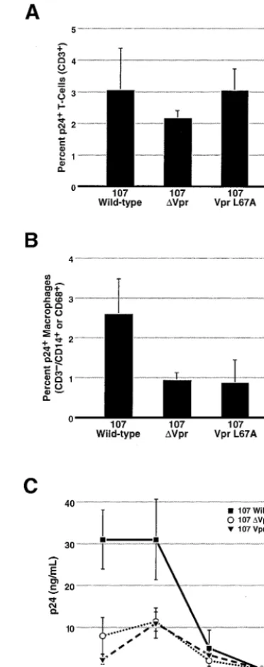
Vpr export is required for efficient infection of macrophages
present in human lymphoid tissues.
Spleen and tonsil tissue
offer an important tool to study the replication of HIV-1 in
what appears to be a more physiologically relevant system. As
is true for activated PBMC infection, we observed no
depen-dence on Vpr for replication of CXCR4-dependent viruses
(14). Consistent with this observation, examination of the
num-ber of infected T cells by intracellular anti-p24
gag[image:4.603.81.501.66.343.2]
immunostain-ing showed no dependence on Vpr for CCR5-dependent viral
infection of CD4
⫹T lymphocytes (Fig. 4A). However, when
FIG. 2. The VprL67A mutant induces cell cycle arrest. Cells were analyzed for DNA content, and cell cycle profiles were determined with
FlowJo software using the Watson Pragmatic model. Primary data are represented by the gray background. (A) Both wild-type Vpr and the
L67A-Vpr export mutant were expressed at comparable levels (left panel) in 293T cells and induced a similar accumulation of cells at the G
2/M
interface, characterized by a 4
N
complement of DNA. (B) The Vpr L67A export mutant was introduced as a single amino acid change in the HIV-1
CCR5-dependent 107 proviral backbone. PBMCs were infected for 5 days and stained to assess intracellular p24 production, and the infected
lymphocytes (anti-p24
gag⫹) were analyzed for DNA content. Note the significant accumulation of cells paused or arrested at the G
2
/M phase of
the cell cycle for lymphocytes infected with the wild-type and VprL67A mutant viruses, compared with the control 107⌬Vpr virus.
on November 8, 2019 by guest
http://jvi.asm.org/
the number of HIV-producing macrophages was assessed, a
60% reduction was consistently observed with the
CCR5-de-pendent virus containing the Vpr export mutant (L67A)
com-pared to wild-type virus. Viruses lacking Vpr displayed an
equivalent decrease. Since both nuclear import and G
2cell
cycle arrest are intact with VprL67A, these results suggest that
nuclear export of Vpr is important for viral spread to
macro-phages present in lymphoid tissues. Because nuclear export
appears to be important for incorporation of Vpr into virions,
these data imply that Vpr is required during the initial phase of
infection of target macrophages in these tissues. Of note,
al-though macrophages represent only about 1% of the lymphoid
tissue, we observed a 67% reduction in the amount of p24
gagpresent in the media of these lymphoid histocultures after
infection with virus containing the VprL67A export mutant or
lacking Vpr altogether. These findings support the notion that
macrophages are an important reservoir that
disproportion-ately contribute to viral burden in these lymphoid tissues (14).
DISCUSSION
This study shows that the nuclear export property of the
nucleocytoplasmic shuttling protein Vpr is required for
effi-cient incorporation of Vpr into virions, which is required for
efficient HIV-1 replication in tissue macrophages. We studied
a Vpr mutant (L67A) that is compromised in its nuclear export
property but retains wild-type nuclear import and causes G
2 [image:5.603.323.509.71.541.2]cell cycle arrest. Using this mutant in the context of a
molec-ular HIV clone to infect lymphoid histocultures derived from
human tonsil or spleen tissue, we found that the limited
incor-poration of the Vpr L67A mutant into virions reduced the
number of infected tissue macrophages by 60%. A similar
reduction was obtained with viruses lacking the entire Vpr
gene. In contrast, the L67A Vpr export mutant had no effect
on levels of HIV infection of T cells within the same lymphoid
FIG. 3. The Vpr L67A nuclear export mutant is less efficiently
incorporated into newly formed virions yet retains the ability to bind to
Gag. (A) Virions were collected by centrifugation and analyzed by
sodium dodecyl sulfate-polyacrylamide gel electrophoresis. Levels of
p24 Gag are shown in the lower panel, and levels of intravirion Vpr are
shown in the upper panel. Note the substantial decrease (⬎97%) in the
amount of VprL67A associated with the virion compared with
wild-type Vpr (top row). (B) HA-tagged Vpr constructs were cotransfected
with p55⌬MAGFP. This vector encodes a large segment of Gag,
in-cluding the p6 domain, but does not require Rev for its expression.
Immunoprecipitation (IP, top two rows) of exogenously expressed HA
fusion proteins and subsequent immunoblotting (WB) were used to
evaluate the interaction of wild-type Vpr and the Vpr L67A analogue
with Gag. Expression levels of the Vpr proteins and the Gag fusion
protein are shown in the lower two panels. Note the equivalent
inter-action of VprL67A and wild-type Vpr with the Gag fusion protein (top
panel).
FIG. 4. An HIV-1 molecular clone containing the Vpr L67A export
mutant can infect T cells efficiently but is as defective as
⌬Vpr HIV-1
for infection of tissue macrophages. Lymphoid histocultures prepared
from tonsil and spleen (represented here) were infected with 50
TCID
50per block of tissue with the indicated CCR5-dependent 107
virus. After 7 to 10 days, the blocks were mechanically dispersed and
examined by fluorescence-activated cell sorting using the indicated cell
surface markers and intracellular anti p24
gagstaining. There were six
blocks per well, and error bars represent the standard deviation for
three wells. (A) There was no difference in the number of infected T cells
(CD3
⫹lymphocytes) in the presence or absence of Vpr. (B) A 60%
reduction in the number of infected macrophages (CD3
⫺CD14
⫹and/or CD68
⫹) was observed for the
⌬Vpr and the VprL67A mutants
compared with viruses expressing wild-type Vpr. (C) Supernatants of
these cultures were colleted over the course of the experiment and
analyzed for p24
gaglevels by enzyme-linked immunosorbent assay.
There was a 67% decrease in the amount of secreted p24
gagin the
cultures infected with the
⌬Vpr or the VprL67A virus compared with
the wild-type virus. These results were reproduced in more than four
independent experiments with human spleen and tonsil tissue.
on November 8, 2019 by guest
http://jvi.asm.org/
HIV-2/simian immunodeficiency virus SM Vpx protein aids in
PIC import into the cell nucleus (16) and also enhances
infec-tions of macrophages in lymphoid tissues (14). These data also
add support to the proposal that infected macrophages
signif-icantly contribute to the viral load of HIV-1-infected patients
(14, 26, 31, 34, 53).
Vpr is incorporated into the HIV-1 PIC and is thought to
facilitate its nuclear import in cooperation with the matrix
(MA) (3, 22, 27, 75) and integrase (2, 21) proteins as well as the
central DNA flap (81); however, the actual involvement of
integrase and the central DNA flap has recently been
ques-tioned (12). The specific role of MA in PIC import has also
been questioned (19, 50, 61) but may be partially explained by
recognition that p55
gagis a nucleocytoplasmic shuttling protein
(11). Finally, even the role of Vpr during PIC import has been
controversial. Vpr facilitates replication in monocyte-derived
macrophages, but only when virus is added at a low multiplicity
of infection (21). Impaired viral DNA import requires
muta-tions in both Vpr and MA, suggesting at least some
redun-dancy in their functions (29). Others have concluded that Vpr
does not contribute to HIV infection of monocyte-derived
macrophages at all independently of MA (27) or even with the
loss of both these nuclear import signals (40). However, in the
more biologically relevant context of lymphoid tissue, Vpr
clearly facilitated replication in tissue macrophages (14), and
in contrast to other studies (32), its presence in the virion is
required for this effect. It is clear that Vpr does not facilitate
replication in artificially arrested T cells (2, 8, 27). Further, in
our previous work utilizing the CXCR-4-dependent HIV strain
NL4-3, we demonstrated that Vpr does not facilitate HIV
infection of resting T cells infected in ex vivo lymphoid
his-tocultures (14). Vpr also does not contribute to overall viral
production when macrophages are excluded from infection, as
seen with NL4-3 infection of lymphoid tissues (14). These
findings suggest that Vpr exerts its effects in a cell
type-re-stricted manner.
Jenkins et al. have confirmed the nucleocytoplasmic
shut-tling properties of Vpr (36). Curiously, in this study, Vpr
ex-port was insensitive to leptomycin B even though mutations in
the predicted leucine-rich, CRM-1-binding domain abrogate
the phenotype (36, 64). Our prior studies demonstrated that
leptomycin B impairs nuclear export of Vpr (64). Jenkins et al.
also suggested that nuclear export of Vpr was not required for
virion incorporation when virions were produced in the
pres-ence of cotransfected plasmids encoding a Vpr export mutant.
While Vpr is specifically incorporated into the virion through
interaction with the p6 portion of the p55
gagprecursor protein
(7, 39, 54, 79), the stoichiometry, once thought to be 1:1 with
p55
gagin the virion, is now estimated to be as low as 1:7 with
capsid (49) or even as low as 14 to 18 Vpr molecules per virion
(67). More importantly, the quantity of Vpr in the virion can be
greatly enhanced (up to ca. 40-fold) by cotransfecting cells with
tion. Under these conditions, the Vpr NES is key for full
incorporation of Vpr into virions.
The absence of Vpr does not completely prevent the
infec-tion of tissue macrophages. Vpr enhances such infecinfec-tions
ei-ther by acting synergistically with the oei-ther import factors or by
providing a redundant signal for more efficient import of the
PIC across the NPC. It has been suggested that Vpr acts like an
importin-

homologue through its direct binding to
nucleo-porins within the NPC, although this appears to be context
dependent (18, 58, 74). While MA and integrase utilize the
importin-
␣
/importin-

-dependent pathway of nuclear import,
Vpr possesses noncanonical NLSs and is not imported
exclu-sively through these classical mechanisms (21, 22, 35, 38, 64).
Thus HIV-1 may have adapted a novel strategy to bypass
cellular defense mechanisms targeted at excluding viruses from
the nucleus. Importantly, Vpr does not facilitate the infection
of resting, nondividing T cells in the same tissue context where
macrophage infection is enhanced (13, 14). Therefore,
cell-specific factors must dictate whether Vpr is required for or is
active in PIC import. In view of the unexpectedly large
contri-bution of infected macrophages to the viral burden in
lym-phatic tissues, interrupting macrophage-dependent growth by
compromising Vpr action in vivo could lead to a sharp decline
in the viral burden in infected patients.
ACKNOWLEDGMENTS
We thank Robin Givens for assistance in the preparation of the
manuscript; John Carroll, Jack Hull, Stephen Gonzales, and Chris
Goodfellow for assistance with graphics; and Stephen Ordway and
Gary Howard for editorial assistance.
M.P.S. was supported by NIH/NIAID grant K08-AI01866. L.A.E.
was supported by the Biomedical Sciences Graduate Program (BMS)
and the National Institutes of Health (NIH) Medical Scientist Training
Program (MSTP) at University of California, San Francisco (UCSF).
This work was supported by NIH grant R01-AI45234 (W.C.G.) and
the UCSF-GIVI Center for AIDS Research (NIH P30-MH59037)
(W.C.G.) and in part by NIH grant R01-AI43695 (M.A.G.), the
UCSF-California AIDS Research Center (CC99-SF-001) (M.A.G. and
W.C.G.), and the J. David Gladstone Institutes (M.A.G. and W.C.G.).
REFERENCES
1. Bartz, S. R., M. E. Rogel, and M. Emerman.1996. Human immunodeficiency
virus type 1 cell cycle control: Vpr is cytostatic and mediates G2
accumula-tion by a mechanism which differs from DNA damage checkpoint control.
J. Virol.70:2324–2331.
2. Bouyac-Bertoia, M., J. D. Dvorin, R. A. Fouchier, Y. Jenkins, B. E. Meyer, L. I. Wu, M. Emerman, and M. H. Malim.2001. HIV-1 infection requires a
functional integrase NLS. Mol. Cell7:1025–1035.
3. Bukrinsky, M. I., S. Haggerty, M. P. Dempsey, N. Sharova, A. Adzhubel, L. Spitz, P. Lewis, D. Goldfarb, M. Emerman, and M. Stevenson.1993. A nuclear localization signal within HIV-1 matrix protein that governs
infec-tion of non-dividing cells. Nature365:666–669.
4. Bukrinsky, M. I., N. Sharova, M. P. Dempsey, T. L. Stanwick, A. G. Bukrin-skaya, S. Haggerty, and M. Stevenson.1992. Active nuclear import of human immunodeficiency virus type 1 preintegration complexes. Proc. Natl. Acad.
Sci. USA89:6580–6584.
5. Chun, T. W., R. T. Davey, Jr., M. Ostrowski, J. Shawn Justement, D. Engel, J. I. Mullins, and A. S. Fauci.2000. Relationship between pre-existing viral reservoirs and the re-emergence of plasma viremia after discontinuation of
highly active anti-retroviral therapy. Nat. Med.6:757–761.
on November 8, 2019 by guest
http://jvi.asm.org/
6. Chun, T. W., L. Stuyver, S. B. Mizell, L. A. Ehler, J. A. Mican, M. Baseler, A. L. Lloyd, M. A. Nowak, and A. S. Fauci.1997. Presence of an inducible HIV-1 latent reservoir during highly active antiretroviral therapy. Proc. Natl.
Acad. Sci. USA94:13193–13197.
7. Cohen, E. A., G. Dehni, J. G. Sodroski, and W. A. Haseltine.1990. Human immunodeficiency virus vpr product is a virion-associated regulatory protein.
J. Virol.64:3097–3099.
8. Connor, R. I., B. K. Chen, S. Choe, and N. R. Landau.1995. Vpr is required for efficient replication of human immunodeficiency virus type-1 in
mono-nuclear phagocytes. Virology206:935–944.
9. Conti, L., P. Matarrese, B. Varano, M. C. Gauzzi, A. Sato, W. Malorni, F. Belardelli, and S. Gessani.2000. Dual role of the HIV-1 vpr protein in the
modulation of the apoptotic response of T cells. J. Immunol.165:3293–3300.
10. de Noronha, C. M., M. P. Sherman, H. W. Lin, M. V. Cavrois, R. D. Moir, R. D. Goldman, and W. C. Greene.2001. Dynamic disruptions in nuclear
envelope architecture and integrity induced by HIV-1 Vpr. Science294:
1105–1108.
11. Dupont, S., N. Sharova, C. DeHoratius, C. M. Virbasius, X. Zhu, A. G. Bukrinskaya, M. Stevenson, and M. R. Green.1999. A novel nuclear export
activity in HIV-1 matrix protein required for viral replication. Nature402:
681–685.
12. Dvorin, J. D., P. Bell, G. G. Maul, M. Yamashita, M. Emerman, and M. H. Malim.2002. Reassessment of the roles of integrase and the central DNA
flap in human immunodeficiency virus type 1 nuclear import. J. Virol.76:
12087–12096.
13. Eckstein, D. A., M. L. Penn, Y. D. Korin, D. D. Scripture-Adams, J. A. Zack, J. F. Kreisberg, M. Roederer, M. P. Sherman, P. S. Chin, and M. A. Gold-smith.2001. HIV-1 actively replicates in naive CD4(⫹) T cells residing
within human lymphoid tissues. Immunity15:671–682.
14. Eckstein, D. A., M. P. Sherman, M. L. Penn, P. S. Chin, C. M. De Noronha, W. C. Greene, and M. A. Goldsmith.2001. HIV-1 Vpr enhances viral burden
by facilitating infection of tissue macrophages but not nondividing CD4(⫹)T
cells. J. Exp. Med.194:1407–1419.
15. Finzi, D., M. Hermankova, T. Pierson, L. M. Carruth, C. Buck, R. E. Chaisson, T. C. Quinn, K. Chadwick, J. Margolick, R. Brookmeyer, J. Gal-lant, M. Markowitz, D. D. Ho, D. D. Richman, and R. F. Siliciano.1997. Identification of a reservoir for HIV-1 in patients on highly active
antiret-roviral therapy. Science278:1295–1300.
16. Fletcher, T. M., III, B. Brichacek, N. Sharova, M. A. Newman, G. Stivahtis, P. M. Sharp, M. Emerman, B. H. Hahn, and M. Stevenson.1996. Nuclear import and cell cycle arrest functions of the HIV-1 Vpr protein are encoded
by two separate genes in HIV-2/SIV(SM). EMBO J.15:6155–6165.
17. Fornerod, M., M. Ohno, M. Yoshida, and I. W. Mattaj.1997. CRM1 is an
export receptor for leucine-rich nuclear export signals. Cell90:1051–1060.
18. Fouchier, R. A., B. E. Meyer, J. H. Simon, U. Fischer, A. V. Albright, F. Gonzalez-Scarano, and M. H. Malim.1998. Interaction of the human im-munodeficiency virus type 1 Vpr protein with the nuclear pore complex.
J. Virol.72:6004–6013.
19. Fouchier, R. A., B. E. Meyer, J. H. Simon, U. Fischer, and M. H. Malim.
1997. HIV-1 infection of non-dividing cells: evidence that the amino-termi-nal basic region of the viral matrix protein is important for Gag processing
but not for post-entry nuclear import. EMBO J.16:4531–4539.
20. Frankel, A. D., and J. A. Young.1998. HIV-1: fifteen proteins and an RNA.
Annu. Rev. Biochem.67:1–25.
21. Gallay, P., T. Hope, D. Chin, and D. Trono.1997. HIV-1 infection of nondividing cells through the recognition of integrase by the
importin/karyo-pherin pathway. Proc. Natl. Acad. Sci. USA94:9825–9830.
22. Gallay, P., V. Stitt, C. Mundy, M. Oettinger, and D. Trono.1996. Role of the karyopherin pathway in human immunodeficiency virus type 1 nuclear
im-port. J. Virol.70:1027–1032.
23. Glushakova, S., B. Baibakov, L. B. Margolis, and J. Zimmerberg.1995. Infection of human tonsil histocultures: a model for HIV pathogenesis. Nat.
Med.1:1320–1322.
24. Glushakova, S., B. Baibakov, J. Zimmerberg, and L. B. Margolis.1997.
Experimental HIV infection of human lymphoid tissue: correlation of CD4⫹
T cell depletion and virus syncytium-inducing/non-syncytium-inducing phe-notype in histocultures inoculated with laboratory strains and patient isolates
of HIV type 1. AIDS Res. Hum. Retroviruses13:461–471.
25. Goh, W. C., M. E. Rogel, C. M. Kinsey, S. F. Michael, P. N. Fultz, M. A. Nowak, B. H. Hahn, and M. Emerman.1998. HIV-1 Vpr increases viral expression by manipulation of the cell cycle: a mechanism for selection of
Vpr in vivo. Nat. Med.4:65–71.
26. Grivel, J. C., M. L. Penn, D. A. Eckstein, B. Schramm, R. F. Speck, N. W. Abbey, B. Herndier, L. Margolis, and M. A. Goldsmith.2000. Human im-munodeficiency virus type 1 coreceptor preferences determine target T-cell
depletion and cellular tropism in human lymphoid tissue. J. Virol.74:5347–
5351.
27. Haffar, O. K., S. Popov, L. Dubrovsky, I. Agostini, H. Tang, T. Pushkarsky, S. G. Nadler, and M. Bukrinsky.2000. Two nuclear localization signals in the HIV-1 matrix protein regulate nuclear import of the HIV-1 pre-integration
complex. J. Mol. Biol.299:359–368.
28. He, J., S. Choe, R. Walker, P. Di Marzio, D. O. Morgan, and N. R. Landau.
1995. Human immunodeficiency virus type 1 viral protein R (Vpr) arrests
cells in the G2phase of the cell cycle by inhibiting p34cdc2 activity. J. Virol.
69:6705–6711.
29. Heinzinger, N. K., M. I. Bukinsky, S. A. Haggerty, A. M. Ragland, V. Kewal-ramani, M. A. Lee, H. E. Gendelman, L. Ratner, M. Stevenson, and M. Emerman.1994. The Vpr protein of human immunodeficiency virus type 1 influences nuclear localization of viral nucleic acids in nondividing host cells.
Proc. Natl. Acad. Sci. USA91:7311–7315.
30. Henklein, P., K. Bruns, M. P. Sherman, U. Tessmer, K. Licha, J. Kopp, C. M. de Noronha, W. C. Greene, V. Wray, and U. Schubert.2000. Func-tional and structural characterization of synthetic HIV-1 Vpr that transduces cells, localizes to the nucleus, and induces G2 cell cycle arrest. J. Biol. Chem.
275:32016–32026.
31. Hockett, R. D., J. M. Kilby, C. A. Derdeyn, M. S. Saag, M. Sillers, K. Squires, S. Chiz, M. A. Nowak, G. M. Shaw, and R. P. Bucy.1999. Constant mean viral copy number per infected cell in tissues regardless of high, low, or
undetectable plasma HIV RNA. J. Exp. Med.189:1545–1554.
32. Hrimech, M., X. J. Yao, F. Bachand, N. Rougeau, and E. A. Cohen.1999. Human immunodeficiency virus type 1 (HIV-1) Vpr functions as an
imme-diate-early protein during HIV-1 infection. J. Virol.73:4101–4109.
33. Humphries, E. H., and H. M. Temin.1974. Requirement for cell division for
initiation of transcription of Rous sarcoma virus RNA. J. Virol.14:531–546.
34. Igarashi, T., C. R. Brown, Y. Endo, A. Buckler-White, R. Plishka, N. Bischof-berger, V. Hirsch, and M. A. Martin.2001. Macrophages are the principal reservoir and sustain high virus loads in rhesus macaques after the depletion
of CD4⫹T cells by a highly pathogenic simian immunodeficiency virus/HIV
type 1 chimera (SHIV): implications for HIV-1 infections of humans. Proc.
Natl. Acad. Sci. USA98:658–663.
35. Jenkins, Y., M. McEntee, K. Weis, and W. C. Greene.1998. Characterization of HIV-1 vpr nuclear import: Analysis of signals and pathways. J. Cell Biol.
143:875–885.
36. Jenkins, Y., P. V. Sanchez, B. E. Meyer, and M. H. Malim.2001. Nuclear export of human immunodeficiency virus type 1 Vpr is not required for virion
packaging. J. Virol.75:8348–8352.
37. Jowett, J. B., V. Planelles, B. Poon, N. P. Shah, M. L. Chen, and I. S. Chen.
1995. The human immunodeficiency virus type 1 Vpr gene arrests infected T
cells in the G2⫹M phase of the cell cycle. J. Virol.69:6304–6313.
38. Karni, O., A. Friedler, N. Zakai, C. Gilon, and A. Loyter.1998. A peptide derived from the N-terminal region of HIV-1 Vpr promotes nuclear import in permeabilized cells: elucidation of the NLS region of the Vpr. FEBS Lett.
429:421–425.
39. Kondo, E., F. Mammano, E. A. Cohen, and H. G. Gottlinger.1995. The p6gag
domain of human immunodeficiency virus type 1 is sufficient for the
incor-poration of Vpr into heterologous viral particles. J. Virol.69:2759–2764.
40. Kootstra, N. A., and H. Schuitemaker.1999. Phenotype of HIV-1 lacking a functional nuclear localization signal in matrix protein of gag and Vpr is
comparable to wild-type HIV-1 in primary macrophages. Virology253:170–
180.
41. Lewis, P., M. Hensel, and M. Emerman.1992. Human immunodeficiency
virus infection of cells arrested in the cell cycle. EMBO J.11:3053–3058.
42. Lewis, P. F., and M. Emerman.1994. Passage through mitosis is required for oncoretroviruses but not for the human immunodeficiency virus. J. Virol.
68:510–516.
43. Lu, Y. L., P. Spearman, and L. Ratner.1993. Human immunodeficiency virus type 1 viral protein R localization in infected cells and virions. J. Virol.
67:6542–6550.
44. Mahalingam, S., V. Ayyavoo, M. Patel, T. Kieber-Emmons, and D. B. Weiner.1997. Nuclear import, virion incorporation, and cell cycle arrest/ differentiation are mediated by distinct functional domains of human
immu-nodeficiency virus type 1 Vpr. J. Virol.71:6339–6347.
45. Mahalingam, S., S. A. Khan, M. A. Jabbar, C. E. Monken, R. G. Collman, and A. Srinivasan.1995. Identification of residues in the N-terminal acidic
domain of HIV-1 Vpr essential for virion incorporation. Virology207:297–
302.
46. Mattaj, I. W., and L. Englmeier.1998. Nucleocytoplasmic transport: the
soluble phase. Annu. Rev. Biochem.67:265–306.
47. Meyers, G., S. Wain-Hobson, L. E. Henderson, B. T. Korber, K.-T. Jeang, and G. N. Pavlakis (ed.).1994. Human retroviruses and AIDS. Los Alamos National Laboratory, Los Alamos, N.M.
48. Miller, M. D., C. M. Farnet, and F. D. Bushman.1997. Human immunode-ficiency virus type 1 preintegration complexes: studies of organization and
composition. J. Virol.71:5382–5390.
49. Muller, B., U. Tessmer, U. Schubert, and H. G. Krausslich.2000. Human immunodeficiency virus type 1 Vpr protein is incorporated into the virion in significantly smaller amounts than gag and is phosphorylated in infected
cells. J. Virol.74:9727–9731.
50. Nie, Z., D. Bergeron, R. A. Subbramanian, X. J. Yao, F. Checroune, N. Rougeau, and E. A. Cohen.1998. The putative alpha helix 2 of human immunodeficiency virus type 1 Vpr contains a determinant which is respon-sible for the nuclear translocation of proviral DNA in growth-arrested cells.
J. Virol.72:4104–4115.
51. Nishi, K., M. Yoshida, D. Fujiwara, M. Nishikawa, S. Horinouchi, and T.
on November 8, 2019 by guest
http://jvi.asm.org/
human immunodeficiency virus type 1 virions: requirement for the p6 region
of gag and mutational analysis. J. Virol.67:7229–7237.
55. Penn, M. L., J. C. Grivel, B. Schramm, M. A. Goldsmith, and L. Margolis.
1999. CXCR4 utilization is sufficient to trigger CD4⫹T cell depletion in
HIV-1-infected human lymphoid tissue. Proc. Natl. Acad. Sci. USA96:663–
668.
56. Pierson, T., J. McArthur, and R. F. Siliciano.2000. Reservoirs for HIV-1: mechanisms for viral persistence in the presence of antiviral immune
re-sponses and antiretroviral therapy. Annu. Rev. Immunol.18:665–708.
57. Poon, B., K. Grovit-Ferbas, S. A. Stewart, and I. S. Y. Chen.1998. Cell cycle arrest by Vpr in HIV-1 virions and insensitivity to antiretroviral agents.
Science281:266–269.
58. Popov, S., M. Rexach, L. Ratner, G. Blobel, and M. Bukrinsky.1998. Viral protein R regulates docking of the HIV-1 preintegration complex to the
nuclear pore complex. J. Biol. Chem.273:13347–13352.
59. Popov, S., M. Rexach, G. Zybarth, N. Reiling, M. A. Lee, L. Ratner, C. M. Lane, M. S. Moore, G. Blobel, and M. Bukrinsky.1998. Viral protein R regulates nuclear import of the HIV-1 pre-integration complex. EMBO J.
17:909–917.
60. Re, F., D. Braaten, E. K. Franke, and J. Luban.1995. Human
immunode-ficiency virus type 1 Vpr arrests the cell cycle in G2by inhibiting the
activa-tion of p34cdc2-cyclin B. J. Virol.69:6859–6864.
61. Reil, H., A. A. Bukovsky, H. R. Gelderblom, and H. G. Gottlinger.1998. Efficient HIV-1 replication can occur in the absence of the viral matrix
protein. EMBO J.17:2699–2708.
62. Roe, T., T. C. Reynolds, G. Yu, and P. O. Brown.1993. Integration of murine
leukemia virus DNA depends on mitosis. EMBO J.12:2099–2108.
63. Schwartz, S., B. K. Felber, and G. N. Pavlakis.1992. Distinct RNA
se-quences in thegagregion of human immunodeficiency virus type 1 decrease
RNA stability and inhibit expression in the absence of Rev protein. J. Virol.
66:150–159.
64. Sherman, M. P., C. M. de Noronha, M. I. Heusch, S. Greene, and W. C. Greene.2001. Nucleocytoplasmic shuttling by human immunodeficiency
vi-rus type 1 Vpr. J. Virol.75:1522–1532.
65. Sherman, M. P., C. M. de Noronha, D. Pearce, and W. C. Greene.2000. Human immunodeficiency virus type 1 Vpr contains two leucine-rich helices that mediate glucocorticoid receptor coactivation independently of its effects
on G2cell cycle arrest. J. Virol.74:8159–8165.
66. Sherman, M. P., and W. C. Greene.2002. Slipping through the door: HIV
entry into the nucleus. Microbes Infect.4:67–73.
nodeficiency virus type 1 Vpr induces apoptosis following cell cycle arrest.
J. Virol.71:5579–5592.
71. Stewart, S. A., B. Poon, J. B. Jowett, Y. Xie, and I. S. Chen.1999. Lentiviral delivery of HIV-1 Vpr protein induces apoptosis in transformed cells. Proc.
Natl. Acad. Sci. USA96:12039–12043.
72. Stewart, S. A., B. Poon, J. Y. Song, and I. S. Chen.2000. Human
immuno-deficiency virus type 1vpr induces apoptosis through caspase activation.
J. Virol.74:3105–3111.
73. Toohey, K., K. Wehrly, J. Nishio, S. Perryman, and B. Chesebro.1995. Human immunodeficiency virus envelope V1 and V2 regions influence
rep-lication efficiency in macrophages by affecting virus spread. Virology213:
70–79.
74. Vodicka, M. A., D. M. Koepp, P. A. Silver, and M. Emerman.1998. HIV-1 Vpr interacts with the nuclear transport pathway to promote macrophage
infection. Genes Dev.12:175–185.
75. von Schwedler, U., R. S. Kornbluth, and D. Trono.1994. The nuclear local-ization signal of the matrix protein of human immunodeficiency virus type 1 allows the establishment of infection in macrophages and quiescent T
lym-phocytes. Proc. Natl. Acad. Sci. USA91:6992–6996.
76. Wolff, B., J. J. Sanglier, and Y. Wang.1997. Leptomycin B is an inhibitor of nuclear export: inhibition of nucleo-cytoplasmic translocation of the human immunodeficiency virus type 1 (HIV-1) Rev protein and Rev-dependent
mRNA. Chem. Biol.4:139–147.
77. Wong, J. K., M. Hezareh, H. F. Gunthard, D. V. Havlir, C. C. Ignacio, C. A. Spina, and D. D. Richman.1997. Recovery of replication-competent HIV
despite prolonged suppression of plasma viremia. Science278:1291–1295.
78. Yao, X. J., A. J. Mouland, R. A. Subbramanian, J. Forget, N. Rougeau, D. Bergeron, and E. A. Cohen.1998. Vpr stimulates viral expression and in-duces cell killing in human immunodeficiency virus type 1-infected dividing
Jurkat T cells. J. Virol.72:4686–4693.
79. Yao, X. J., R. A. Subbramanian, N. Rougeau, F. Boisvert, D. Bergeron, and E. A. Cohen.1995. Mutagenic analysis of human immunodeficiency virus type 1 Vpr: role of a predicted N-terminal alpha-helical structure in Vpr
nuclear localization and virion incorporation. J. Virol.69:7032–7044.
80. Yedavalli, V. R., C. Chappey, and N. Ahmad.1998. Maintenance of an intact human immunodeficiency virus type 1 vpr gene following mother-to-infant
transmission. J. Virol.72:6937–6943.
81. Zennou, V., C. Petit, D. Guetard, U. Nerhbass, L. Montagnier, and P. Charneau.2000. HIV-1 genome nuclear import is mediated by a central
DNA flap. Cell101:173–185.
on November 8, 2019 by guest
http://jvi.asm.org/