Copyright © 1997, American Society for Microbiology
Proteolytic Processing in African Swine Fever Virus: Evidence
for a New Structural Polyprotein, pp62
CARMEN SIMO´ N-MATEO,† GERMA´N ANDRE´S, FERNANDO ALMAZA´N,ANDELADIO VIN˜ UELA*
Centro de Biologı´a Molecular “Severo Ochoa” (Consejo Superior de Investigaciones Cientı´ficas-Universidad Auto´noma de Madrid), Facultad de Ciencias, Universidad Auto´noma de Madrid, Cantoblanco, 28049 Madrid, Spain
Received 5 March 1997/Accepted 22 April 1997
We have identified an open reading frame (ORF), CP530R, within the EcoRI C* fragment of the African
swine fever virus (ASFV) genome that encodes a polyprotein of 62 kDa (pp62). Antisera raised against different regions of ORF CP530R recognized a polypeptide of 62 kDa in ASFV-infected cells during the late phase of virus replication, after the onset of viral DNA synthesis. Pulse-chase experiments showed that polyprotein pp62 is posttranslationally processed to give rise to two proteins of 35 kDa (p35) and 15 kDa (p15). This proteolytic processing was found to take place at the consensus sequence Gly-Gly-X through an ordered cascade of proteolytic cleavages like that which also occurs with ASFV polyprotein pp220 (C. Simo´n-Mateo, G. Andre´s, and E. Vin˜uela, EMBO J. 12:2977–2987, 1993). Immunofluorescence studies showed that polyprotein pp62 is localized in the viral factories. In addition, immunoprecipitation analysis of purified virus particles showed that mature products p35 and p15 are major structural proteins. According to these results, polyprotein processing represents an essential strategy for the maturation of ASFV structural proteins.
African swine fever virus (ASFV) is a large, DNA-contain-ing virus responsible for a highly lethal disease of domestic pigs (for reviews, see references 6 and 27); it also infects soft ticks (Ornithodorus sp.), which act as vectors for the virus. Animal viruses with very large DNA genomes include families of ico-sahedral viruses (Herpesviridae and Iridoviridae) and brick-shaped viruses (Poxviridae). However, ASFV does not fit well into any of these groups since, although its genomic structure is similar to that of poxviruses (hairpin loop structures and terminal inverted repetitions at the DNA ends) (11, 24), its icosahedral morphology is similar to that of iridoviruses (5). It is the only member of the new genus “African swine fever-like virus” (8). The complexity of ASFV, with a genome length of 170 kbp, is evidenced by the large number (about 100) of virus-induced polypeptides detected in infected cells by two-dimensional (2-D) gel electrophoresis analysis (10, 21). In ad-dition, the ASFV particle contains more than 50 proteins with molecular masses ranging from 10 to 150 kDa (4, 10), including the enzymatic machinery required for the synthesis and pro-cessing of early mRNA (20). Recently, the nucleotide se-quence of the genome of ASFV strain BA71V has been re-ported and 151 open reading frames (ORFs) have been identified (28).
A striking feature of this complex DNA virus is the use of polyprotein processing as a mechanism of gene expression. Thus, we previously showed that ASFV encodes a polyprotein, named pp220, which after proteolytic processing gives rise to four major structural proteins: p150, p37, p34, and p14 (23). Interestingly, all of the proteolytic cleavages were shown to take place at the consensus sequence Gly-Gly-X (19, 23). This report extends the study of proteolytic processing in the mat-uration of some ASFV proteins by describing a new polypro-tein, named pp62. Polyprotein pp62 was identified as a late protein which, after proteolytic processing at Gly-Gly-X se-quences, produces two major structural proteins: p35 and p15.
According to our results, polyprotein processing is an essential mechanism for the generation of major components of the virus particle, sharing this gene expression strategy with posi-tive-strand RNA viruses and retroviruses (12, 15).
MATERIALS AND METHODS
Cells and viruses.Vero cells (CCL 81), obtained from the American Type Culture Collection, were cultured in Dulbecco’s modified Eagle’s medium (DMEM) supplemented with 10% newborn calf serum, the concentration of which was reduced to 2% during viral infection. ASFV strain BA71V, adapted to grow in Vero cells, has been previously described (9). Highly purified ASFV was obtained by Percoll equilibrium centrifugation as described by Carrascosa et al. (4).
Computer analysis.Computer analyses of DNA and protein sequences were performed by using the software package of the University of Wisconsin Genetics Computer Group (7).
Antisera.The complete CP530R ORF was cloned in plasmid KS (Stratagene) in two steps. The nucleotide sequence coding for the first 14 amino acids was obtained by synthesizing two complementary oligonucleotides containing a PstI site, which were paired and cloned into the pGMT7 vector. This recombinant plasmid was digested with XbaI-PstI to obtain a fragment (100 bp) which also contained thef10 promoter and the Shine-Dalgarno sequence. The remainder of the CP530R ORF (2.5 kbp) was obtained from the restriction fragment SD/RC9of the ASFV genome (strain BA71V) (18) by digestion with the restric-tion enzymes PstI and HindIII. Both fragments were ligated into plasmid KS (Stratagene) to produce recombinant plasmid KS-CP530R.
From this plasmid, we obtained plasmids KS-p35C9and KS-p15C9, containing different regions of ORF CP530R. Plasmid KS-p35C9was obtained by cutting plasmid KS-CP530R with the restriction enzyme PstI and then blunt ending it with the mung bean enzyme. Thereafter, the linearized plasmid was digested with
MluI, blunt ended with Klenow, and finally religated to obtain plasmid KS-p35C9,
which contains the 39region of ORF CP530R (see Fig. 4B). To obtain plasmid KS-p15C9, containing the 59region of ORF CP530R, plasmid KS-CP530R was digested with the restriction enzyme AccI and religated (see Fig. 4B).
Escherichia coli BL21DE3 (25), which contains in its genome the T7 RNA
polymerase gene under the control of an isopropyl-b-D-thiogalactopyranoside
(IPTG)-inducible promoter, was transformed with the recombinant plasmids. The 16-kDa (KS-p15C9, p15 region) and 46-kDa (KS-p35C9, p35 region) proteins which were used as immunogens were obtained by inducing the cultures for 2 h with 1 mM IPTG (data not shown). The cultures were further incubated for 2 h with rifampin (0.2 mg/ml). The bacterial pellet (from a 100-ml culture) was lysed in 5 ml of phosphate-buffered saline (PBS) by freezing at220°C and thawing and sonicated twice for 2 min each time. Bacterial lysates were subjected to centrif-ugation (10 min at 12,0003g). In all cases, the recombinant proteins were the
main component present in the pellet. About 100mg of each protein was injected subcutaneously into rabbits in complete Freund’s adjuvant. At intervals of 2 weeks, rabbits were boosted subcutaneously three times, once in incomplete
* Corresponding author. Phone: 34 1 397 84 36. Fax: 34 1 397 84 90. E-mail: [email protected].
† Present address: Department of Genetics, University of Gent, 9000 Gent, Belgium.
5799
on November 9, 2019 by guest
http://jvi.asm.org/
luted at least 10-fold in radioimmunoprecipitation assay buffer (0.01 M Tris-HCl [pH 7.5], 0.15 M NaCl, 1% sodium deoxycholate, 1% Nonidet P-40, 0.1% SDS) and incubated with a 1:20 dilution of the specific antisera for 2 h at 4°C. A 25% suspension of protein A-coated Sepharose CL-4B (Sigma Chemical Co., St. Louis, Mo.) was then added, and incubation was continued for 30 min at 4°C. The immune complexes were isolated by centrifugation, washed four times with cold radioimmunoprecipitation assay buffer, and finally solubilized by boiling in Laemmli sample buffer (62.5 mM Tris-HCl [pH 6.8], 2% SDS, 100 mM dithio-threitol, 10% glycerol) for 3 min.
Gel electrophoresis.High-resolution 2-D gel electrophoresis was performed as described by Simo´n-Mateo et al. (23). SDS-polyacrylamide gradient (7 to 20%) gels (16) were used for both 2-D and conventional gels.
Protein sequencing.To determine the N-terminal sequence of protein p35, ASFV proteins from Percoll-purified virions were resolved by 2-D gel electro-phoresis and then transferred to a polyvinylidene difluoride membrane. After Coomassie blue staining, protein p35 was directly subjected to N-terminal se-quencing by using an ABI 473A pulse-liquid phase protein sequencer (Applied Biosystems) and the modified cycles described in the manufacturer’s instructions.
Indirect immunofluorescence.Vero cells were infected at a multiplicity of infection of 1 PFU/cell. At 12 hpi, the cells were rinsed with PBS and fixed with methanol at220°C for 5 min. For staining, fixed cells were incubated with a 1:100 dilution of the different antisera and then with a 1:200 dilution of Texas red-conjugated goat anti-rabbit antibodies (Amersham). Nuclear and viral DNAs were visualized by staining with 5mg of bis-benzimide (Hoechst 33258; Sigma) per ml of PBS for 5 min. Cells were examined under an Axiovert fluorescence microscope (Carl Zeiss, Inc., Oberkochen, Germany) and photographed with Kodak TMAX (ASA 400) film.
RESULTS
Identification of ORF CP530R. One strategy used in our
laboratory to identify genes coding for viral proteins has been the screening of an ASFV expression library (pEX) in which viral proteins are synthesized as fusion products withb -galac-tosidase (1). To identify the gene encoding structural protein p35, a serum raised against the 35-kDa proteins of purified virus particles isolated from SDS-polyacrylamide gels was used. Nine different clones were selected with this serum and divided by DNA hybridization into two groups, one with six clones (group I) and the other with three clones (group II). A representative clone of each group was hybridized to a genomic library of viral DNA to localize the corresponding genes in the viral genome of the BA71V strain. Both groups were found to hybridize specifically with EcoRI fragment C9 (RC9) (data not shown).
The sequencing at the insertion point of the viral DNA of the clones from group I confirmed that all of them expressed part of ORF CP2475L as a fusion protein withb-galactosidase (data not shown). This ORF has been reported to encode polyprotein pp220, which is posttranslationally processed to give rise to structural proteins p150, p37, p34, and p14 (23). Specifically, all clones contained the region corresponding to protein p37.
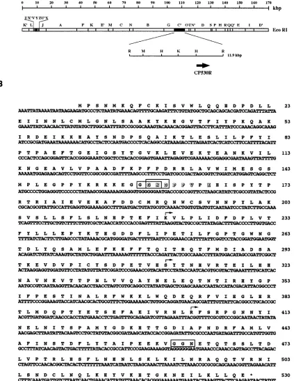
The sequencing of the clones from group II revealed that they contained part of ORF CP530R (27) (Fig. 1). This ORF codes for a protein of 530 amino acids with a predicted
mo-cleavage at this position could give rise to a protein with a predicted molecular mass of 8 kDa.
A computer search of the databases for ORF CP530R (7) showed similarity with an ASFV ORF, PIG1, reported by Hingamp et al. (13). ORF PIG1 is located in the RC9 restric-tion fragment of the BA71V isolate, like ORF CP530R, and encodes a putative polypeptide of 225 amino acids. Despite its similarity, the PIG1 sequence significantly differs from that reported here. Thus, several nucleotide changes and frame-shifts in ORF PIG1 would produce amino acid changes and, what is more important, a striking difference in the length of this ORF. The results presented below support the conclusion that our sequence is correct and indeed codes for a protein of 530 amino acids.
ORF CP530R codes for a protein of 62 kDa.To characterize
the protein coded for by ORF CP530R, two polyclonal rabbit antisera against different regions of its sequence were ob-tained. The different polypeptides used as immunogens were expressed in bacteria from the appropriate recombinant plas-mids as described in Materials and Methods. For a schematic representation of the localization of the CP530R regions used as immunogens, see Fig. 4B.
Both sera were used in immunoprecipitation experiments with extracts of infected Vero cells metabolically labeled with [35S]methionine-cysteine along the infection cycle. Figure 2
shows the results obtained after immunoprecipitation of unin-fected (U) and inunin-fected (I) cells labeled for 2 h at 12 hpi. Only one protein with an apparent molecular mass of 62 kDa, named pp62, was specifically detected with both sera in the extract of ASFV-infected cells (lanes I2). The molecular mass of this protein was similar to that predicted from the amino acid sequence of ORF CP530R (60.5 kDa). It should be noted that protein pp62 was not detected in extracts obtained in the presence of cytosine arabinoside (AraC), an inhibitor of viral DNA replication (Fig. 2, lanes I1). This result indicates that protein pp62 is expressed after the onset of viral DNA synthe-sis and therefore belongs to the class of late ASFV proteins.
Characterization of the mature proteins produced by
pro-teolytic processing of protein pp62. To identify the proteins
that might be derived from protein pp62 by proteolytic pro-cessing, infected cells were pulse-labeled with [35
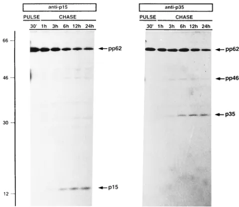
S]methionine-cysteine for 30 min at 12 hpi and chased for 1, 3, 6, 12, and 24 h in the presence of a 10-fold excess of methionine-cysteine. Figure 3 shows an immunoprecipitation analysis with the dif-ferent antisera raised against protein pp62. In the pulse, the antiserum specific for the p35 region recognized protein pp62, the level of which decreased during the chases, coinciding with the accumulation of a 35-kDa protein (named p35) (Fig. 3, anti-p35). The cleavage to produce protein p35 was detected 3 h after the pulse and continued in the following chase peri-ods.
on November 9, 2019 by guest
The antiserum against the NH2-terminal region (Fig. 4B) of ORF CP530R also recognized protein pp62 during the pulse, and the level of this protein declined from 3 h of chase in parallel with an increase of a 15-kDa protein (named p15) (Fig.
[image:3.612.99.522.83.630.2]3, p15). Interestingly, immunoprecipitation with the anti-serum against the protein p35 region detected a faint 46-kDa protein (named pp46) during the chase periods (Fig. 3, anti-p35). This protein could be an intermediate precursor in which FIG. 1. Nucleotide and predicted amino acid sequences of ORF CP530R. (A) Localization in the ASFV genome of restriction fragment RC9containing ORF CP530R. The position and orientation of ORF CP530R are indicated by the arrow. (B) Sequence of ORF CP530R. The numbers on the right are amino acid positions. Potential cleavage sites (Gly-Gly-X) are indicated by open boxes. The arrows indicate the sites of insertion into the pEX vector of the different clones selected in the screening with anti-p35 serum. The NH2-terminal sequence determined for protein p35 is indicated by shadowed letters.
on November 9, 2019 by guest
http://jvi.asm.org/
the Gly-Gly-X at positions 463 and 464 has not yet been cleaved to produce the mature protein p35. The processing at this putative cleavage site could give rise to an 8-kDa protein besides protein p35. However, this protein could not be de-tected in this experiment.
Considering that the 8-kDa protein has 13 leucines and only one cysteine (Fig. 1B), an attempt was made to detect it by pulse-labeling with [3H]leucine. However, no protein with the
expected molecular weight was detected after immunoprecipi-tation or immunoblotting with antiserum against the p35 re-gion (data not shown). One possible explanation for these results might be the nonimmunogenic properties of the pro-tein, since a specific antiserum against polyprotein pp62 using the region spanning amino acids 464 to 530 as an antigen could not be obtained. Another possible explanation is that the pro-tein is rapidly degraded after the cleavage, since the NH2
terminus consists of an Asn residue. This amino acid is con-sidered destabilizing and would confer on this protein a half-life of 3 min (26).
Despite the inability to detect the 8-kDa protein, there are several reasons to believe that the 46-kDa protein is an inter-mediate precursor. First, immunoprecipitation with the anti-serum against the protein p35 region detected a 46-kDa pro-tein which was not detected with antiserum against the propro-tein p15 region. Second, the pp46 intermediate precursor appeared 3 h after the pulse, at the same time as mature protein p35. Finally, the size of the polypeptide expressed in bacteria span-ning amino acid residues 176 to 530 and that of the 46-kDa protein coincided (data not shown), as did the 35-kDa protein detected during the chase periods and that predicted from the region including amino acids 160 to 463.
In summary, protein pp62 is a polyprotein that after proteo-lytic processing gives rise to two mature proteins, p15 and p35, and most likely to an 8-kDa protein. Interestingly, the presence of the intermediate precursor pp46 indicates that polyprotein pp62 is processed through an ordered cascade of cleavages like that which occurs with ASFV polyprotein pp220 (23). Figure 4A shows a scheme of the proteolytic cleavages involved in the generation of the mature proteins. The initial event in the proteolytic cascade would be recognition of one of the two overlapping cleavage sites located at positions 158 to 160. This cleavage takes place 2 to 3 h after the synthesis of polyprotein pp62, giving rise to mature protein p15 and preprotein pp46. The last processing step would be cleavage at positions 463 and 464 within preprotein pp46, producing mature protein p35 and an 8-kDa protein corresponding to the COOH-terminal frag-ment of preprotein pp46.
Polyprotein pp62 processing gives rise to two major
struc-tural proteins.To determine whether the proteins generated
from polyprotein pp62 are structural, immunoprecipitation analyses of 35S-labeled, highly purified ASFV particles were
presence (I1) of 40mg of the DNA replication inhibitor cytosine arabinoside per ml and labeled with [35S]methionine-cysteine for 2 h at 12 hpi. Cell extracts were immunoprecipitated with anti-p15 and anti-p35 sera and then analyzed by SDS-polyacrylamide gel electrophoresis. Protein pp62 is indicated.
[image:4.612.57.296.460.667.2]FIG. 3. Proteins derived from ASFV polyprotein pp62 by proteolytic pro-cessing. Infected Vero cells were pulse-labeled with [35S]methionine-cysteine for 30 min (PULSE) at 12 hpi and then chased for 1, 3, 6, 12, and 24 h (CHASE). The cell extracts were immunoprecipitated with anti-p15 and anti-p35 sera and analyzed by SDS-polyacrylamide gel electrophoresis. The proteins detected in both pulses and chases, as well as the positions of molecular weight markers (103), are indicated.
FIG. 4. Schematic representation of the cascade of proteolytic cleavages occurring during ASFV polyprotein pp62 processing. (A) Triangles indicate the positions of the Gly-Gly-X cleavage sites in polyprotein pp62. Precursors (pp62 and pp46) are represented by empty boxes. Mature products (p15, p35, and p8) are represented by hatched boxes. (B) Scheme of polyprotein pp62 with the regions used to obtain the different antisera (anti-p15 and anti-p35) against the polyprotein. The numbers of the amino acids spanning the regions and the restriction sites used in the cloning of these regions are indicated.
on November 9, 2019 by guest
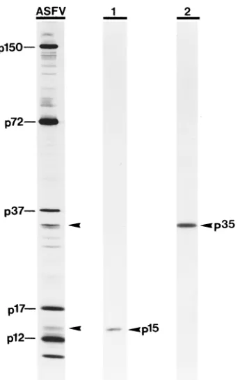
performed with antisera against the different regions of polyprotein pp62. Both antisera recognized the mature pro-teins derived from polyprotein pp62 in ASFV particles (Fig. 5), indicating that they are incorporated into the viral particle. Analysis of highly purified virions by 2-D gel electrophoresis revealed that proteins p35 and p15 are among the major struc-tural proteins (data not shown).
To ascertain that protein p35 was produced after the cleav-age of one of the two overlapping Gly-Gly-X sequences at positions 157 to 160 (Fig. 1B), we analyzed the amino-terminal sequence of structural protein p35 recovered from 2-D gels of purified virions. The sequence obtained, GNDPPVXT, dem-onstrates that the Gly-Gly-Gly sequence at positions 157 to 159 is the cleavage signal that gives rise to structural protein p35. Next, we investigated the subcellular localization of polypro-tein pp62 and its derived mature propolypro-teins. For this, indirect immunofluorescence analyses were carried out with ASFV-infected cells processed at late times of infection by using antisera against polyprotein pp62. Specific labeling was de-tected with both antisera in discrete cytoplasmic areas close to the nucleus (large arrows in Fig. 6A and B), which were iden-tified by bis-benzimide staining as viral factories (arrows in Fig. 6C and D). Additionally, we detected a punctate labeling spreading throughout the cytoplasm (small arrows in Fig. 6A and B), which most probably represents single virus particles.
DISCUSSION
Proteolytic processing of viral proteins is a common event during the replication of many viruses and can be considered to play two general roles: (i) a mechanism for gene expression in which a viral polyprotein is proteolytically processed to yield
the mature proteins and (ii) protein processing involved in the maturation of virus particles during viral morphogenesis (for reviews, see references 12 and 15). A previous study revealed that four of the major ASFV structural proteins (p150, p37, p34, and p14) were primarily synthesized as a polyprotein (pp220) and then proteolytically processed to produce the ma-ture proteins (23). In this report, we have extended the study of ASFV structural proteins by showing that proteins p35 and p15 are also synthesized as a polyprotein (pp62) which, after two proteolytic cleavages, gives rise to the mature proteins.
Both ASFV polyproteins are cleaved after the second Gly residue of the consensus sequence Gly-Gly-X that is also rec-ognized in adenovirus structural proteins and in some cellular proteins such as polyubiquitin (19). A similar cleavage site, Ala-Gly-X, is used in the maturation of some core protein precursors of poxviruses such as vaccinia virus and fowlpox virus (17). The similarity in the cleavage motifs of all of these viruses might indicate that they use similar mechanisms for the processing of their structural protein precursors. In the case of ASFV, only some of the putative cleavage sites (Gly-Gly-X) present in the polyproteins are cleaved, suggesting that addi-tional residues are involved in determining the secondary and tertiary structures of the precursor proteins necessary for the cleavage to occur. Comparison of the amino acid sequences surrounding the sites cleaved in pp220 and pp62 showed no homology. In general, it has been suggested that residues lo-cated in the negative positions (N terminal to a cleavage site) are usually the important ones in determining the specificity of the proteolytic processing of viral polyproteins (15). This could explain the strict Gly-Gly conservation found at the22 and21 positions of the polyprotein pp220 and pp62 cleavage sites.
Polyprotein pp62 is processed by an ordered cascade of proteolytic cleavages like that which occurs in the case of polyprotein pp220 (23). The initial event in the proteolytic cascade would be the cleavage at positions 158 and 159 by which structural protein p15 is separated from preprotein pp46. This proteolytic maturation does not occur immediately after the synthesis of the precursor, but rather there is a delay of about 3 h before any appreciable proteolysis is observed. This delay might be due to a requirement for conformational changes resulting from any posttranslational modification,
[image:5.612.94.265.67.340.2]ac-FIG. 5. Proteins p35 and p15 are major structural proteins. Highly purified virus particles labeled with [35S]methionine-cysteine were analyzed by SDS-polyacrylamide gel electrophoresis before (lane ASFV) or after immunoprecipi-tation with anti-p15 (lane 1) or anti-p35 (lane 2) serum. The positions of p15 and p35, as well as other major ASFV structural proteins, are indicated.
FIG. 6. Immunofluorescence detection of ASFV polyprotein pp62 in in-fected cells. ASFV-inin-fected Vero cells were fixed at 16 hpi and incubated with anti-p35 (A) or anti-p15 (B) serum and then stained with Texas red-conjugated goat anti-rabbit antibodies and a fluorescent DNA dye (Hoechst 33258). Both sera labeled the viral factories (large arrows in A and B) as defined by DNA staining (arrows in C and D). Additionally, strong labeling of punctate structures spread throughout the cytoplasm (small arrows in A and B), which most likely correspond to virus particles, was detected.
on November 9, 2019 by guest
http://jvi.asm.org/
step of ASFV assembly like that which occurs with polyprotein pp220 (2). This has been shown to be the case for the matu-ration of structural polyproteins in RNA viruses (12) and of single proteins in some complex DNA viruses (3, 14). In this context, the order of the recognition of the different cleavage sites in both polyproteins could be a way to regulate viral assembly; i.e., a certain cleavage can only take place after another cleavage has been carried out. These ordered series of proteolytic events may ensure that only correctly folded viral proteins enter the assembly pathway and may provide a mech-anism for rendering the process irreversible.
The synthesis of at least six ASFV major structural proteins as polyproteins could serve to cluster genes whose products are somehow related, allowing them to be synthesized as the result of a single transcription-translation event and, if necessary, allowing their transport to a specific subcellular and subviral site. Furthermore, their initial synthesis as a polyprotein pre-cursor may ensure a 1:1 molar ratio of the mature gene prod-ucts, which could be necessary for establishing a certain or-dered structure. This is the case for the four nonidentical polypeptides produced by the proteolytic processing of poly-protein P1 in picornavirus (12). In relation to this, we have recently shown that ASFV structural proteins p150, p37, p34, and p14, produced after the proteolytic processing of polypro-tein pp220, are present in the virus particle in essentially equimolar amounts (2). Furthermore, all of these proteins share the same location in the virus particle, the “core shell,” a thick protein layer surrounding the DNA-containing nucle-oid.
In summary, we have provided evidence that polyprotein processing in ASFV-infected cells is an essential mechanism for the maturation of a significant proportion of the major ASFV structural proteins.
ACKNOWLEDGMENTS
We thank S. Baars for technical assistance in the screening of the expression library and A. L. Carrascosa for preparation of anti-p35 serum. We thank M. L. Salas and J. Salas for critical reading of the manuscript.
This work was supported by grants from the Comisio´n Interminis-terial de Ciencia y Tecnologı´a (PB93-061-C02-01), the European Eco-nomic Community (AIR-CT93-1332), and the Junta de Extremadura and by an institutional grant from the Fundacio´n Ramo´n Areces. C.S.-M., G.A., and F.A. were recipients of fellowships from the Min-isterio de Educacio´n y Ciencia.
cles. Arch. Virol. 10(Suppl.):92–94.
9. Enjuanes, L., A. L. Carrascosa, M. A. Moreno, and E. Vin˜uela. 1976. Titra-tion of African swine fever virus. J. Gen. Virol. 32:471–477.
10. Esteves, A., M. I. Marques, and J. V. Costa. 1986. Two-dimensional analysis of African swine fever virus proteins and proteins induced in infected cells. Virology 152:181–191.
11. Gonza´lez, A., A. Talavera, J. M. Almendral, and E. Vin˜uela. 1986. Hairpin loop structure of African swine fever virus DNA. Nucleic Acids Res. 14: 6835–6844.
12. Hellen, C. U. T., and E. Wimmer. 1992. The role of proteolytic processing in the morphogenesis of virus particles. Experientia 48:201–215.
13. Hingamp, P. M., M. L. Leyland, J. Webb, S. Twigger, R. J. Mayer, and L. K.
Dixon.1995. Characterization of a ubiquitinated protein which is externally located in African swine fever virions. J. Virol. 69:1785–1793.
14. Katz, E., and B. Moss. 1970. Formation of a vaccinia virus structural polypeptide derived from a high molecular weight precursor: inhibition by rifampicin. Proc. Natl. Acad. Sci. USA 66:677–684.
15. Kra¨usslich, H. G., and E. Wimmer. 1988. Viral proteinases. Annu. Rev. Biochem. 57:701–754.
16. Laemmli, U. K. 1970. Cleavage of structural proteins during the assembly of the head of bacteriophage T4. Nature 227:680–685.
17. Lee, P., and D. E. Hruby. 1995. Analysis of the role of the amino-terminal peptide of vaccinia virus structural protein precursors during proteolytic processing. Virology 207:229–233.
18. Ley, V., J. M. Almendral, P. Carbonero, A. Beloso, E. Vin˜uela, and A.
Talavera.1984. Molecular cloning of African swine fever virus DNA. Virol-ogy 133:249–257.
19. Lo´pez-Otı´n, C., C. Simo´n-Mateo, L. Martinez, and E. Vin˜uela. 1989. Gly-Gly-X, a novel consensus sequence for the proteolytic processing of viral and cellular proteins. J. Biol. Chem. 264:9107–9110.
20. Salas, M. L., J. Kuznar, and E. Vin˜uela. 1981. Polyadenylation, methylation and capping of the RNA synthesis in vitro by African swine fever virus. Virology 101:169–175.
21. Santare´n, J. F., and E. Vin˜uela. 1986. African swine fever virus-induced polypeptides in Vero cells. Virus Res. 5:391–405.
22. Sanz, A., B. Garcı´a-Barreno, M. L. Nogal, E. Vin˜uela, and L. Enjuanes. 1985. Monoclonal antibodies specific for African swine fever virus proteins. J. Vi-rol. 54:199–206.
23. Simo´n-Mateo, C., G. Andre´s, and E. Vin˜uela. 1993. Polyprotein processing in African swine fever virus: a novel strategy of gene expression for a DNA virus. EMBO J. 12:2977–2987.
24. Sogo, J. M., J. M. Almendral, A. Talavera, and E. Vin˜uela. 1984. Terminal and internal inverted repetitions in African swine fever virus DNA. Virology
133:271–275.
25. Studier, F. W., A. H. Rosenberg, J. J. Dunn, and J. W. Dubendorff. 1990. Use of the T7 RNA polymerase to direct expression of cloned genes. Methods Enzymol. 185:60–89.
26. Varshavsky, A. 1992. The end rule of protein turnover. Cell 69:725–735. 27. Vin˜uela, E. 1987. African swine fever virus, p. 31–49. In Y. Becker (ed.),
African swine fever. Martinus Nijhoff Publishing, Boston, Mass.
28. Yan˜ez, R., J. M. Rodrı´guez, M. L. Nogal, L. Yuste, C. Enrı´quez, J. F.
Rodrı´guez, and E. Vin˜uela.1995. Analysis of the complete nucleotide se-quence of African swine fever virus. Virology 208:249–278.