STIMULI-RESPONSIVE NANOMATERIALS FOR
CONTROLLED DELIVERY BY LIGHT, MAGNETIC
AND ELECTRICAL TRIGGERS
Meng Bi Zhang
December 2017
A thesis submitted for the degree of Doctor of Philosophy of
The Australian National University
Declaration
I certify that this thesis does not include any material that has previously been submitted for a degree in any university. To the best of my knowledge, it does not contain any material previously published or written by another person except where due and clear reference is made in the text. The work in this thesis is my own, except for the contributions made by others as described in the Acknowledgements.
Meng Bi Zhang
12th of December, 2017
This accomplishment does not belong to me alone. I owe much and more to the
following fine folk.
First and foremost, I thank the many wiser minds who aided me along the
way. The road was often long and winding, and some days it seemed to go uphill on
both sides. In particular, Dr Antonio Tricoli, thank you for giving me the freedom
and support to pursue my many ideas, even the mad ones. Especially the mad ones.
In the end, we did some fascinating science together.
Secondly, I thank the many collaborators who made this work possible. It
was a privilege to have worked with you. Special thanks must go to the many
technicians who helped along the way. Many of the most interesting results presented
here would have been impossible without your time, expertise and patience.
Thirdly, I thank my immediate colleagues in the Nanotechnology Research
Group. Your company made the good times much more enjoyable and the bad times
almost tolerable. Special mention goes to Noushin Nasiri who was there since the
beginning. We battled many dragons together.
I would like to acknowledge the Australian government for giving me food to
eat and a roof to sleep under for the duration. Without such funding, the work may
indeed have happened, but I would surely be poorer, colder, thinner, and sadder.
To my friends good and true, thank you for keeping me sane with tea, cake,
crafts and laughs. You know who you are.
And finally, to the ones who supported me from coast-to-coast, thank you for
your patience in the whole affair. As the days weathered in weeks and months and
years, your resolve remained strong even as my own began to crumble, and that
made all the difference in the end. Your love stretched wider and warmer than the
desert between us, and I am grateful every day for it.
The use of nanomaterials for biomedical applications is an emerging and important
field. This is particularly true of advancements in targeted and controlled drug
delivery, which offer several important improvements over traditional drug
administration. The clinical efficacy of small-molecule therapeutics is currently
limited by many factors, including: poor solubility, inefficient cellular uptake, overly
rapid renal clearance and an inability to target only desired locations such as diseased
tissues. The use of nanocarriers for drug delivery may greatly improve the efficacy
over traditional therapeutics by lowering the total dosage, limiting the exposure to
affected areas only, and giving greater temporal control over drug elution. These
materials often make use of both organic and inorganic components, exploiting the
unique and useful properties of each constituent to achieve novel, synergistic
functions.
This dissertation presents a study of nanocomposites comprising the three
most important materials in this field: titania, iron oxides and polypyrrole. Titania is
a strong photocatalyst, iron oxides provide useful responses to applied magnetic
fields, and polypyrrole is a polymer with unique electrochemical properties. Studies
in this dissertation were aimed at combining these three materials to create a novel
structure that is responsive towards light, magnetic fields and electrical stimulation to
serve as an enabling platform for the loading and release of biologically interesting
compounds.
These nanomaterials have been paired with amino acids lysine and
L-glutamic acid, two organic molecules of interest due to their ability to bind to DNA
released on these carriers: Ketoprofen, an important anti-inflammatory that is
traditionally hindered by its limited cellular uptake levels; and fluorescein
isothiocyanate, a fluorescent dye molecule that is a common tool used in this field for
nanocarrier location and easy visualisation of release-related kinetics.
First, an investigation into the effect of pH on the binding of amino acids to
titania, iron oxide and polypyrrole is presented with a view towards optimising the
functionalised material for subsequent loading and release of the model drugs (in this
case, amine-reactive molecules). The release mechanism of photo-activated TiO2 is
studied in detail with a particular focus on the competition between the cleavage of
bonds versus organic degradation on the catalyst’s surface. Both mechanisms are
currently reported in literature and studies were aimed at identifying the more
dominant pathway in the system developed alongside understanding the crucial role
of reaction time scales on this photochemistry.
Then, the pH-tuneable flocculation of the amino acid-functionalised
nanoparticles via electrostatic attractions is exploited to create a novel, anisotropic
assembly of iron oxides. These filaments display a dynamic and unique response
towards a rotating magnetic field by creating local microscale vortices. This motion
is used to enhance local delivery rate of molecules through magnetic-field triggered
microscale mixing.
Finally, this anisotropic iron oxide structure is combined with polypyrrole to
create a unique, novel material that possesses directional conductivity, a
photothermal response, and magnetic field-triggered release of loaded molecules at
Thesis Organisation
A detailed review of literature is given in Chapter 1 Background and Literature
Review. This comprises an introduction to metal oxide nanomaterials and their role
in nanomedicine (Section 1.2), magnetic nanomaterials and their unique magnetic field responsive character (Section 1.3), synthesis of metal oxide nanomaterials (Section 1.4), functionalisation strategies (Section 1.5), intrinsically conductive polymers (Section 1.6), and biocompatibility (Section 1.7). The key research questions arising from this literature review and associated research aims are
presented in Section 1.8. The results of experiments on the effect of pH on amino acid functionalisation of TiO2 and Fe3O4 will be presented in Chapter 2. Chapter 3
investigates the flocculation effect of this functionalised material and presents a
method to exploit this for self-assembly of Fe3O4 under a magnetic field to create
novel, anisotropic structures. An analogous functionalisation of PPy with amino
acids is presented in Chapter 4. Finally, the findings of previous chapters are
combined to produce a composite material of amino acid-functionalised,
light-responsive, conductive and magnetically responsive material that is presented in
Chapter 5. Conclusions and recommendations for future work will be presented in
Chapter 6. Methods and materials are given at the end of each chapter where
Journal Papers
T. Zhang, P. Costigan, N. Varshney, A. Tricoli, RSC Advances 2016, 6, 33843-33850.
T. Zhang, M. A. Go, C. Stricker, V. R. Daria, A. Tricoli, J. Mater. Chem. B 2015, 3, 1677-1687.
J. Ou, F. Wang, Y. Huang, D. Li, Y. Jiang, Q.-H. Qin, Z. Stachurski, A. Tricoli, T. Zhang, Colloids and Surfaces B: Biointerfaces 2014, 117, 466-472.
Conference Proceedings
T. Zhang, M. A. Go, C. Stricker, V. R. Daria, A. Tricoli, Journal of Materials Chemistry B 2015, 3, 1677-1687.
In Submission
K.Bruggeman, T.Zhang, A.Tricoli, D.Nisbet, Pharmaceutical Research, “Reverse
UV-Triggered Growth Factor Delivery from TiO2 Nanoparticles in a
Nomenclature
3D Three-dimensional
AA Amino Acid
APS Ammonium persulfate
ATF Amino-terminal fragment
BDNF Bovine-derived nerve factor
BET Brunauer, Emmett and Teller
BSA Bovine serum albumin
DMSO Dimethyl sulfoxide
EtOH Ethanol
Fe2O3 Iron oxide, hematite phase
Fe3O4 Iron oxide, magnetite phase
FeCl3 Iron (III) chloride
FSP Flame Spray Pyrolysis
FTIR Fourier Transform Infrared
Glut (L)-Glutamic Acid
H2O Water
HCl Hydrochloric Acid
HFMF High Frequency Magnetic Field
HSA Human serum albumin
IEP Isoelectric Point
Lys (L)-Lysine
NaOH Sodium Hydroxide
NIR Near Infrared
NPs Nanoparticles
PAni Polyaniline
PEDOT Poly(3,4-ethyl-enedioxthiophene)
pKa Acid dissociation constant
PPy Polypyrrole
PTX Paclitaxel
SEM Scanning Electron Microscopy
SiO2 Silica, Silicon Dioxide
SnO2 Tin Dioxide
SP Superparamagnetic
TEM Transmission Electron Microscopy
TiO2 Titanium Dioxide
TMX Tamoxifen
UV Ultraviolet
Vis Visible
Table of Contents
Chapter 1 Background and Literature Review ...1
1.1 Overview ...2
1.2 Ceramic Nanomaterials for Drug Release ...4
1.3 Magnetic nanomaterials for drug release ...15
1.4 Synthesis of Metal Oxide Nanomaterials ...31
1.5 Functionalizing metal oxide nanomaterials ...36
1.6 Intrinsically Conductive Polymers ...47
1.7 Biocompatibility ...56
1.8 Research Questions ...60
Chapter 2 Amino acid functionalisation of metal oxide nanoparticles ...63
2.1 Introduction ...65
2.1 Synthesis of MOx nanoparticles and their material properties ...65
2.2 Adsorption of amino acids ...73
2.3 Activity of –NH2 termination ...79
2.4 Response of AA-functionalised TiO2 nanoparticles towards UV light ...82
2.5 Loading and release of model drugs ...89
2.6 Materials and Methods ...94
Chapter Three Self-assembly of anisotropic iron oxide nanostructures ...99
3.1 Lysine-assisted assembly of Isotropic structures ...101
3.2 Magnetic-field assisted assembly of anisotropic structures ...102
3.3 Modelling magnetic interactions...105
3.4 Motion of anisotropic structures under an applied magnetic field ...108
3.5 Using vortexing motion to improve microscale mixing ...110
3.6 Materials and Methods ...113
Chapter 4 Doping polypyrrole with amino acids ...116
4.1 Amino acid doping of polypyrrole during chemical oxidative polymerisation ...118
4.2 Nanoparticles as seeds during polypyrrole polymerisation ...126
4.3 Materials and Methods ...135
Chapter 5 Polypyrrole-coated iron oxide filaments ...138
5.1 Synthesis of PPy-Fe3O4 filament structures ...140
5.2 Electrical properties of anisotropic structures ...142
5.3 Electrochemical dedoping of glutamate ...143
5.4 Magnetic-rotation assisted delivery ...145
5.5 Materials and Methods ...148
Chapter 6 Conclusions and Future Work ...150
Figure 1. Overview of different endogenous and exogenous stimuli for stimulation of nanomaterials. ... 3
Figure 2. (a) Bandgaps of rutile, anatase and brookite.[19]. (b) General scheme for photocatalytic reactions at TiO2 nanoparticle surface in aqueous conditions. (c)
Detailed hole-attack pathways for anatase and rutile. [26] ... 6
Figure 3. Example of Mesoporous SiO2 electron micrographs. [79] ... 14
Figure 4. Heterogeneous composite nanostructures of magnetite ... 21
Figure 5. Known structures of PPy-Fe3O4 nanocomposites. (a) core-shell[147], (b)
aggregated[147], (c) multiple core-shell[148], (d) raspberry (PPy core, Fe3O4
decorations)[149] ... 22
Figure 6. Transmission electron microscopy images of a single magnetotactic bacterium (A) of chains of magnetosomes extracted from whole magnetotactic bacteria (B) of individual magnetosomes detached from the chains by heat and SDS treatment (C).[152] ... 25
Figure 7. Schematic illustrating various known 1D structures resulting from synthesis under an applied magnetic field. (a) single crystal nanowire, (b) multi-crystal
nanowire, (c) bundled nanowire, (d) nanochain of isotropic nanoparticles, (e)
aggregated chain. ... 25
Figure 8. Formation of aggregated nanochains under an applied magnetic field.[169] 29
Figure 9. Flexible motion of soft-encapsulated nanochains[169] ... 29
Figure 10. Irreversible formation of spheroid bundles upon applying a rotating
magnetic field.[169] ... 30
Figure 12. (a) Schematic representation of a typical FSP reactor. (b) TEM showing typical TiO2 product and (c) SEM showing top view of porous film of the same. .... 33
Figure 13. A nanoparticle of 5 nm with different ligand molecules. Left to right: trioctylphosphine oxide (TOPO), triphenylphosphine (TPP), dodecanethiol (DDT), tetraoctylammonium bromide (TOAB) and oleic acid (OA). [198] ... 37
Figure 14. Phosphonate interaction with TiO2 surface. From left to right: mono-, bi-
and tridentate anchorage. Adapted from Neouze and Shubert.[201] ... 38
Figure 15. Carboxylate interaction with TiO2 surface. From left to right: physical
adsorption, monodentate coordination, bridging chemisorption, chelating
chemisorption.[201] ... 39
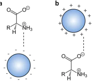
positively charge metal oxide nanoparticle surfaces. ... 46
Figure 19.Chemical structure of polypyrrole (PPy). ... 47
Figure 20. Chemical structure of polyaniline (PANI) ... 48
Figure 21. Chemical structure of PEDOT ... 48
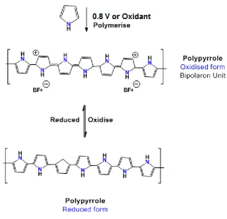
Figure 22. Polymerisation of polypyrrole by either electrochemical or chemical pathway. ... 49
Figure 23. Evolution of optical absorbance of polypyrrole upon successive doping content.[254] ... 51
Figure 24. Chapter three overview. (a) FSP-synthesis of metal oxide nanoparticles. (b) Functionalisation using amino acids at different pH resulting in different coating morphology. (c) Exploiting –NH2 termination for attachment of FITC dye or ketorprofen. (d) Exposure of photocatalytic TiO2 to UV light for release of the functional molecules. ... 64
Figure 25. TEM showing (a) SiO2 and (b) TiO2 NPs synthesised at 5/5/5 flame conditions. ... 66
Figure 26. (a) XRD Spectrum of flame-made TiO2 nanoparticles showing Anatase (A) and Rutile (R) phases. (b) Optical absorbance of TiO2 and (c) Tauc plot showing bandgap at ca. 390nm... 67
Figure 27. Particle size of FSP-made iron oxide nanoparticles as a function of [Fe]/O2 (mol/L) at constant 5bar pressure. ... 68
Figure 28. XRD spectra of FSP-made iron oxide NPs as a function of [Fe]/O2 at constant 5 bar pressure. ... 69
Figure 29. Magnetite weight content of FSP made iron oxide NPs as a function of [Fe]/O2 at constant 5 bar pressure. ... 69
Figure 30. Magnetic hysteresis of flame-made iron oxides at various [Fe]/O2 concentration and fixed 5bar pressure drop (coloured lines), compared against commercial Fe3O4 NPs (black line)... 71
Figure 31. TEM and size distributions for (a) flame-made at [Fe]/O2 = 0.0003 mol/L, (b) flame-made at [Fe]/O2 = 0.0009 mol/L, and (c) commercial iron oxides. ... 72
Figure 32. Evolution of (a) zeta potential and (b) hydrodynamic diameter of unmodified TiO2 (black circles) and lysine-functionalised TiO2 synthesised at acidic (red circles), neutral (green circles) and alkaline (blue circles). ... 76
1.5 (blue), pH 9 (pink) and pure L-lysine powder (black). ... 77
Figure 35. (a) Absorbance spectra of FITC conjugated onto bare TiO2 (black), and
lysine-TiO2 prepared with excess L-lysine at pH 1.5 (pink) and pH 9 (blue). (b)
Absorbance spectra of FITC conjugated onto lysine-TiO2 prepared with low
concentration of L-lysine at pH 1.5 (red) and pH 9 (blue) ... 81
Figure 36. nFITC/ Surface Ti-binding site ratio computed for lysine-TiO2 at low
concentration pH 1.5 across a long term study. ... 81
Figure 37. (a) Zeta potential and (b) hydrodynamic diameter of lys-TiO2 after
increasing exposure to UV-A light. ... 83
Figure 38. hydrodynamic diameter distribution of lys-TiO2 before (black line) and
after 30s (blue line) exposure to UV-A light. ... 83
Figure 39. (a) Reaction between primary amine and fluorescamine producing a fluorescent adduct. (b) Fluorescamine assay of free lysine concentration in solution after exposure to UV light. ... 84
Figure 40.Photocatalytic cyclization of lysine into pipecolinic acid isomers. Adapted from [333] ... 85
Figure 41. Mass spectrometry of particle-free solution after successive UV
illumination showing (a,b) L-lysine and (c,d) pipecolinic acid, its radical cyclisation product. ... 86
Figure 42. Fluorescamine assay of particle-free solution for glutamate-TiO2 after
exposure to UV. ... 87
Figure 43. (a) Mass spectrometry results for particle-free solution after exposing glutamate-TiO2 to UV light showing evolution of (b,c) free glutamate alongside (d,e)
pyroglutamic acid. ... 88
Figure 44. (a) Reaction pathway to synthesise ketoprofen-lysine prodrug.[337] (b, c,d) mass spectrometry results showing release of ketoprofen and absence of (e)L-lysine and (f) pipecolinic acid peaks. ... 91
Figure 45. Chapter four overview.(a) Assembly of magnetic filaments, (b) dynamic response of filaments under rotating magnetic field, (c) micro-vortex-enhanced diffusion under a rotating magnetic field. ... 100
Figure 46. (a) SEM and (b) TEM images of flame-made magnetite ([Fe]/O2 = 0.9
mmol/L) encapsulated in polylysine (PLL) at pH 8. 8 showing a nanorough surface morphology. ... 102
Figure 47. Aspect ratio distributions of encapsulated PLL-FexOy as computed from
([Fe]/O2 = 0.3 mmol/L), and (g) commercial Fe3O4 nanoparticles. ... 104
Figure 48. Modelling interaction agglomerate size and magnetic susceptibility on the collision frequency. ... 108
Figure 49. Magnetically assembled filaments (a) without and (b) under the influence of a magnetic field... 110
Figure 50. Schematic representation of magnetic filaments undergoing (a)
translational, (b) rotational and (c) orbital motion under applied magnetic fields. . 110
Figure 51. Flow of dye and water streams through a multichannel microfluidic chip (a) without and (b) with vortexing from magnetic filaments. ... 111
Figure 52. Chapter 5 overview. (a) Synthesis of nanocomposites with Fe3O4 cores
and glutamic acid-doped polypyrrole coating (b) photothermal response of
nanocomposites. ... 117
Figure 53. UV-vis absorption spectra of polymerisation products of (a) pure
polypyrrole, (b) glutamic acid doped polypyrrole, and (c) lysine doped polypyrrole at
pH 2, 7 and 10. (d) Calculation of absorbance band ratio λ290nm/ λ460nm. ... 121
Figure 54. Scanning Electron Microscopy of (a) polypyrrole synthesised at pH 2, (b) polypyrrole synthesised at pH 7, (c) glutamic acid doped polypyrrole synthesised at pH 2, and (d) glutamic acid doped polypyrrole synthesised at pH 7. ... 123
Figure 55. FTIR of PPy (black lines) and glutamate-doped PPy (red and green lines) polymerised at (a) pH 2 and (b) pH 7. ... 125
Figure 56. Fluorescamine assay of glutamate-doped PPy synthesised at pH 2 (red line) and pH 7 (green line) ... 126
Figure 57. Transmission electron micrographs Fe3O4 NPs (dark phase) coated in PPy
(light phase) via APS oxidant route at (a) 10:1, (b) 5:1, (c) 2:1, (d) 1:1 and (e) 1:10 pyrrole:Fe3O4 mass ratio. ... 128
Figure 58. FTIR for Fe3O4 NPs (red line) and PPy-Fe3O4 composites created using
APS synthesis route at 10:1 pyrrole: Fe3O4 mass ratio. ... 129
Figure 59. Transmission electron micrographs Fe3O4 NPs (dark phase) coated in PPy
(light phase) via FeCl3.6H2O oxidant route at (a) 10:1, (b) 2:1, (c) 1:1, (d) 1:2 and (e)
1:10 pyrrole:Fe3O4 mass ratio. ... 130
Figure 60. (a) TEM of glutamate-doped PPy granules in raspberry-like structure around iron oxide cores. (b) Optical absorbance spectra of PPy-Fe3O4 (black line)
and Glut-PPy-Fe3O4 (green line)... 131
Figure 61. Photothermal effect of PPy-Fe3O4 nanocomposites in raspberry-like (red
circle). ... 132
Figure 62. Chapter 6 overview. (a) Synthesis of glutamic acid-doped polyrrole-coated Fe3O4 filaments, (b) directional conducitivity, (b) electrochemical dedoping of
glutamate. ... 139
Figure 63.Optical micrograph showing (a) isotropic product typical of synthesis in the absence of magnetic field and (b) filament-like structures aligned in applied magnetic field direction. (b) Length distribution and (c, d) transmission electron micrographs of the same. ... 141
Figure 64. Length distribution of PPy-Fe3O4 microfilaments. ... 142
Figure 65. Micrographs showing alignment and corresponding resistance of PPy-Fe3O4 microfilaments in (a) parallel, (b) 45° oblique and (c) perpendicular to
electrode array. ... 143
Figure 66. Recorded resistance of PPy-Fe3O4 microfilaments arranged perpendicular
to the electrode array at various concentrations. ... 143
Figure 67. (a, b) Schematic illustrating electrochemical dedoping of bound glutamate anions by applying a negative potential. (c) Fluorescamine assay of particle-free solution for perpendicular aligned glut-PPy-Fe3O4 filaments in PBS after 2 min with
no applied potential (black line) and -5V (red line). ... 144
Figure 68.Timelapse of eltuion of FITC from PPy-Fe3O4 filaments at variable
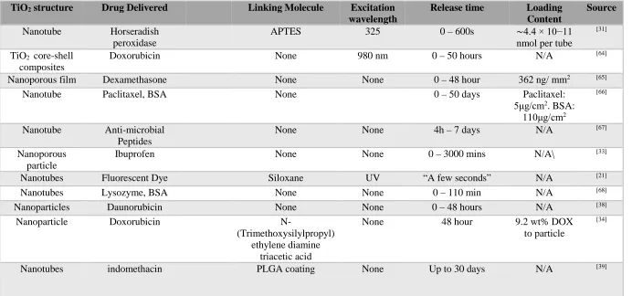
Table 1 Selected results on TiO2 for light-activated release of drugs ... 12
Table 2. Magnetite-containing nanomaterials for biomedical purposes ... 19
Table 3. Magnetically directed synthesis of anisotropic materials ... 27
Table 4. A brief comparison of different chemical routes towards MOx nanomaterial synthesis ... 34
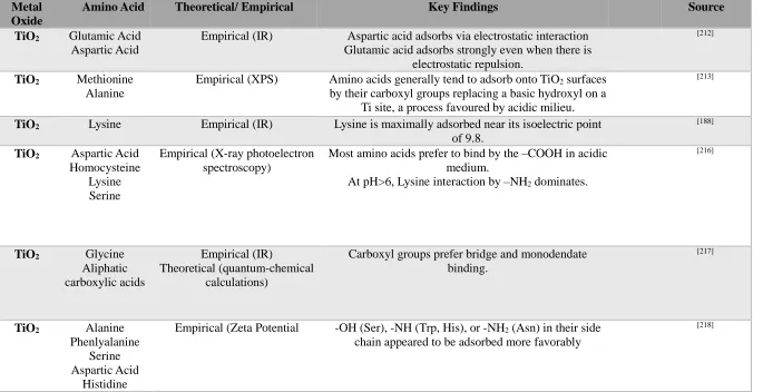
Table 5. Key studies on interaction of metal oxide surfaces with amino acids ... 40
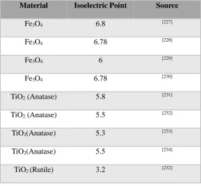
Table 6. Isoelectric point of iron and titanium oxides ... 45
Table 7. Polypyrrole for biomedical applications ... 54
Table 8 Remanence and magnetisation at 1T for flame-made and commercial
magnetite. ... 71
Table 9. Evolution in zeta potential and hydrodynamic diameter of metal oxide
nanoparticles after adsorption of L-lysine from aqueous (pH 7) solution. ... 74
Table 10. Evolution of zeta potential and hydrodynamic diameter between pure TiO2
and lysine-functionalized TiO2 ... 79
Table 11. Initial observations for polypyrrole and doped polypyrrole synthesis after 24 hours at various reaction pH. ... 119
Table 12. Absorption bands of pyrrole derivatives and their assignation ... 120
C
HAPTER
O
NE
Background and Literature Review
1.1 Overview Literature
R
The application of nanomaterials for biomedical applications is a field that has been
gaining increasing importance in the past decade. This scientific discipline involves the
design, synthesis and application of precisely engineering structures in the
nano-to-microscale for novel therapeutic and diagnostic functions. This enabling technology
makes use of both organic and inorganic components, exploiting the unique and useful
properties of each constituent to novel, synergistic functions.
In particular, the development of such nanomaterials that are designed to undergo
predetermined responses towards anticipated or applied stimuli has gained increasing
importance in medicine. Such structures make use of endogenous or exogenous stimuli
(or combination thereof) and translate these triggers into desirable and useful actions of
some medical benefit.
Endogenous stimuli (that is, a stimulus that originates from the body itself) that
are currently known include pH[1] and temperature[2] changes, or response towards
particular enzymes[3]. Endogenous stimulation has the advantage of being naturally
occurring, meaning no addition technology is required and there is therefore no
additional health risk arising from the application of the stimulus. Additionally, changes
in local pH, temperature and enzymes are often signals of important changes in
biological function that may be a direct symptom of disease and is an indicator that the
intervention offered by the nanomaterial is required.
On the other hand, exogenous (externally applied) stimuli are currently popular
due to the greater degree of control and predictability that is afforded by such systems.
Popular exogenous stimuli include light, magnetic field and ultrasound[4] (Figure 1).
These methods differ on several terms, including depth of penetration within human
tissues, cost of operation, and potential side effects.
The nanocarrier is typically designed to undergo useful responses to stimulation
by undergoing physical changes such as by swelling or shrinking[5] and opening or
closing[6], or chemical changes such as by oxidising or reducing[7]. These responses may
be used for applications such as drug release[8], photo[9] /chemo[10]/magnetotherapies[11]
and directed cell growth[12].
The potential of such materials towards drug delivery is particularly significant.
The clinical efficacy of traditionally administered medicine is currently limited by many
factors, including: poor solubility[13], inefficient cellular uptake[14], and an inability to
target only at the desired location of diseased tissues[15]. The use of nanocarriers for
drug delivery may greatly improve the efficacy of treatment by addressing these issues.
Tethering a pharmaceutical to a nanomaterial that can be directed to an affected site
such as a tumour using, for example, a magnetic field, and then being able to control the
release time and rate of the drug for maximum efficiency would represent a significant
advancement.
Figure 1. Overview of different endogenous and exogenous stimuli for stimulation of nanomaterials.
Over the past decade, the field has exploded with solutions of ever-increasing
sophistication to achieve these aims. Here, a survey of relevant literature has been
provided with a focus towards the following:
• Material type: Metals, ceramics, polymers and various composites of all three material classes have been proposed for these applications. This thesis
will focus primarily on ceramics and polymers.
• Synthesis strategies: The most prominent strategies for the synthesis of the above materials are outlined with a particular emphasis on flame-spray
pyrolysis, a high-throughput solution for production of ceramic
nanoparticles.
• Functionalisation strategies: The key methods for modifying the nanomaterial surfaces with functional organic molecules will be studied. The
importance of amino acids in this role and the challenges of using them are
discussed.
• Biocompatibility issues: Finally, a literature survey is conducted to ascertain the biocompatibility of the materials used in this thesis.
1.2 Ceramic Nanomaterials for Drug Release
Ceramic nanomaterials, particularly nanostructured metal oxides, have emerged as
an important class of materials for bio-applications due to several key desirable
properties. The most prominent and important ceramic nanomaterials are titanium
dioxide (TiO2), silicon dioxide (SiO2) and iron oxides (FexOy). TiO2, particularly in the
Anatase phase, is a strong photocatalyst that has been used for light-triggered drug
release[16]. SiO2 is commonly used as an inert coating or carrier for functional
moieties[17]. Magnetic phases of iron oxides, such as magnetite (Fe3O4), maghemite (γ
-Fe2O3) and hematite (α-Fe2O3) provide useful magnetic functionality while being
inherently less toxic than other magnetic materials that contain heavy metal species (e.g.
Co, Ni)[18]. The following section will discuss in detail the state-of-the-art in terms of
the design, synthesis and application of TiO2 and SiO2. Due to the prominence of iron
oxides, they are discussed separately in Section 1.2 Magnetic Nanomaterials.
1.2.1 Titanium dioxide the photocatalyst
Titanium dioxide is of key interest in applications such as controlled drug delivery as it
is one of the strongest photocatalysts that is currently known. The photocatalytic
behaviour of TiO2 is reasonably well-understood. Upon illumination with ultraviolet
light corresponding to the bandgap of TiO2, ca. 3.05eV for anatase and 2.98eV for
rutile,[19] an electron is excited from the valence band to the conductance band,
generating charge carriers (electron and hole). The photogenerated holes react with
water to create hydroxyl radicals and together begin to oxidise the nearby organic
molecules at the particle surface (Figure 2). At the same time, electrons in the
conduction band will be involved in reduction reactions with dissolved oxygen to
generate peroxide.[20] This a property that has been exploited in other disciplines to great
effect (e.g. solar cells, water splitting, self-cleaning coatings) and has recently been
exploited in the context of drug delivery systems.[21]
𝐻𝐻2𝑂𝑂+ ℎ+ → 𝑂𝑂𝐻𝐻 ∙ +𝐻𝐻+ (Equation 1)
𝑂𝑂2+ 2𝑒𝑒−+ 2𝐻𝐻+ → 𝐻𝐻2𝑂𝑂2 (Equation 2)
Designs that use TiO2 for drug delivery typically involve first the adsorption of
functional molecules onto the TiO2 surface (either a drug directly or via a linking
molecule), then release of this bound molecule is triggered by exploiting the
photocatalytic redox events at the nanoparticle surface[22]. The mechanism behind this is
not yet entirely understood. For example, although it has been widely observed that
whole, undamaged organic molecules such as pharmaceuticals[23] may be delivered by
this method, there have been studies suggesting that adsorbed molecules, e.g. oxalic
acid, are likely to be photocatalytically decomposed when TiO2 is exposed to light[24].
At the same time, other authors hypothesise that the redox events prefer to cleave the
TiO2-organic ligand bond first and there is therefore little decomposition recorded at
shorter time frames such as is typically required for drug release.[25]
Figure 2. (a) Bandgaps of rutile, anatase and brookite.[19]. (b) General scheme for photocatalytic reactions
at TiO2 nanoparticle surface in aqueous conditions. (c) Detailed hole-attack pathways for anatase and
rutile. [26]
[image:33.595.101.446.333.684.2]This area has been heavily researched in the past decade and remains an important topic
today. A summary of key results to date have been presented in Table 1. The literature
has been reviewed with respect to the following criteria:
(i) Nanostructure
Currently known TiO2 nanostructures include simple nanoparticle[27], porous
nanoparticle[28], nanotube[29], and porous film[30]. Porous structures, particularly
mesoporous structures have the advantage of high surface area that can be used for the
binding of the target drugs[31]. However, simple nanoparticles have the advantage of
smaller size and therefore ease of injection/ diffusion at the desired sites[32]. Porous
films are suitable for implants designed for longer term application.
(ii) Drug loaded/ delivered
An attempt has been made to catalogue the range of currently known drugs that can be
delivered using the photocatalytic response of TiO2. However, it should be noted that
the identity of the delivered molecule is arguably one of the less important parameters.
Given a good understanding of the mechanism behind attachment and release
mechanism, it is possible to engineer the material to deliver any molecule of interest.
(iii) Functional molecule (if present)
Studies differ in the strategy used for loading the target drug onto the TiO2. Some
adsorb the target molecule directly onto the metal oxide surface[33], while others use a
linking functional molecule[34]. The use of a tethering molecule may result in a more
robust binding of the drug, reducing unintended passive leaching of the drug out of the
nanoparticle. However, this also introduces an additional complexity to the structure.
For example, upon UV illumination, bonds may break between the TiO2 surface and the
linking molecule and/or between the linking molecule and the target drug. Furthermore,
many drug molecules are highly specific and cannot be modified by a linker without
losing efficacy, with the exception of modifications that result in easily metabolised
prodrugs such as lysine-modified Ketoprofen, a transformation that in this case
increases the effectiveness of the anti-inflammatory in question[35].
(iv) Excitation wavelength
The excitation wavelength is significant as UV light has high attenuation within
tissue[36]. This means the penetration depth is typically low and damage to the
surrounding tissue may be expected as the light energy dissipates. Light at longer
wavelengths / lower energy is preferable in this respect.
(v) Illumination time
Similarly, the time scale over which the drug release happens is important both in terms
of limiting the damage to healthy tissues[37] and determining the time required for
effective elution of the drug away from the nanocarrier. For these reasons, it is desirable
to minimise the time required for the release, or to consider pulsed release profiles.
(vi) Drug loading content
The final payload content as calculated as a weight ratio to the nanoparticle carrier is an
important parameter as it serves as an efficiency metric for the drug delivery system.
Due to the unknown toxicity of nanoparticles in general (in part due to their novelty and
therefore lack of long-term studies), it is important to maximise this parameter.
A survey of literature has been presented below (Table 1) on the use of TiO2 for
release of drugs. There are two points of interest in the research to date. Firstly, it
should be noted that, despite the photocatalytic character of TiO2, the majority of papers
found did not make use of this directly. Instead, porous forms of TiO2 (commonly the
nanotubes formed by anodic oxidation of Ti metal) were used as a passive base for the
loading and diffusion-limited release of the loaded drug out of the pore spaces. In such
cases, the photocatalytic character of the TiO2 carrier has no bearing on the
effectiveness of the platform, and a similar effect can be expected for any base material
of similar surface area and porosity. As I will discuss in Section 1.2.3, this is the basis behind the popular use of mesoporous SiO2. In these porous structures based on passive
release, the time-frame for delivery of the payload molecule is usually in the range of
several hours[38], and even up to the month scale[39] for full elution. On the other hand,
photocatalysis is fast-acting. It should be noted that Shrestha et al. were able to achieve
a remarkable release “within a few seconds” due to their use of a Siloxane linker that
bound covalently to the TiO2 surface and then cleaved by the action of UV light
illumination.[21] However, Shrestha et al.’s work is in the minority here, and highlights
an opportunity for further studies of a photocatalytic-driven drug release system.
Perhaps part of the hesitation in the use of light-activated release systems based
on TiO2 is due to the inherent limitation of using UV in a biomedical context. The
excitation wavelengths required to excite TiO2 are usually in the ultraviolet B (UV-B)
region, the band gap of pure Anatase and Rutile being ca. 3.2 eV (387 nm) and 3.0 eV
(413 nm) respectively[40]. Ultraviolet light is cytotoxic, causing inflammatory and
immunosuppressant effects even after short-term exposure[41]. Furthermore, it attenuates
quickly within tissues with low penetration past the epidermis[36] and is therefore
unsuitable for triggering any internal devices. This is undesirable and must be improved
if TiO2-based structures are to have a future in drug release. Strategies have emerged to
improve this property. For example, X-rays may also be used to excite photochemical
reactions at the TiO2 surface to trigger photocatalytic drug release.[42, 43] Although
X-rays carry the inherent risk of high energy ionising radiation, this may be justified by
the benefits of targeted drug delivery and partially mitigated by the short exposure times
typically required for photocatalysis to occur in a similar risk management strategy to
the current use of X-rays for diagnosis.
Alternatively, the band gap of TiO2 may be shifted towards the lower energy /
longer wavelength side of the electromagnetism spectrum by applying some suitable
modifications. Visible light presents the advantage of being less toxic than ultraviolet[44]
and penetrates more deeply in human tissues than ultraviolet. However, near-infrared
light is superior in this regard, having the best penetration in tissue[45] when compared
with both visible[46] and ultraviolet[36]. It is for this reason that NIR-triggered
nanomaterials (usually coupled with a thermoresponsive component) have become an
important area of research.
For example, doping titanium oxides with metal ions such as Ru[47], V[48] and
Fe[49] or non-metals such as C[50], N[51], S[52] and B[53] may shift the wavelength into the
visible range[54] and even leading into the NIR[55] range. Alternatively, a sensitising dye
may be coupled with TiO2 nanoparticles to extend the wavelength into the visible range
(typically < 750 nm). This strategy is widely used for improving the absorption of
natural light in dye-sensitised solar cells[56] and water-splitting[57]. The most prominent
dyes for this purpose Rhodamine B[58], Coumarin[59] and Ruthenium complexes[60] are
all well-known for their toxicity, a disadvantage that cannot easily be overcome.
Finally, TiO2 nanoparticles may be coupled with plasmonic Au[61] and Ag[62]
nanoparticles. Plasmonic Au in particular is a popular choice for drug delivery due to
the tuneable excitation wavelength and the chemically and biologically inert nature of
gold[63].
Despite these limitations, TiO2 remains a popular study subject in drug release.
However, perhaps it should be noted that TiO2 is better suited for application in external
applications such as wound dressings, and that while its optical properties can be
improved, it is worthwhile considering that other materials may be better candidates for
internal applications.
Table 1 Selected results on TiO2 for light-activated release of drugs
TiO2 structure Drug Delivered Linking Molecule Excitation
wavelength
Release time Loading Content
Source
Nanotube Horseradish peroxidase
APTES 325 0 – 600s ∼4.4 × 10⁻¹¹ nmol per tube
[31]
TiO2 core-shell
composites
Doxorubicin None 980 nm 0 – 50 hours N/A [64]
Nanoporous film Dexamethasone None None 0 – 48 hour 362 ng/ mm2 [65]
Nanotube Paclitaxel, BSA None 0 – 50 days Paclitaxel:
5μg/cm2. BSA:
110μg/cm2
[66]
Nanotube Anti-microbial Peptides
None None 4h – 7 days N/A [67]
Nanoporous particle
Ibuprofen None None 0 – 3000 mins N/A\ [33]
Nanotubes Fluorescent Dye Siloxane UV “A few seconds” N/A [21]
Nanotubes Lysozyme, BSA None None 0 – 110 min N/A [68]
Nanoparticles Daunorubicin None None 0 – 48 hours N/A [38]
Nanoparticle Doxorubicin
N-(Trimethoxysilylpropyl) ethylene diamine
triacetic acid
None 48 hour 9.2 wt% DOX to particle
[34]
Nanotubes indomethacin PLGA coating None Up to 30 days N/A [39]
1.2.2 SiO2 the inert carrier
Although nanoporous TiO2 structures have been used for drug loading and release, it is
SiO2 that is more popular in this role. The almost ubiquitous popularity of SiO2 arises
from its relative chemical inertia, a property that makes it an ideal passive carrier with
long-term stability and low impact on its biological environment.
In addition to the spherical particles, a mesoporous form of amorphous SiO2 has
been known since the 1970s[69]. Mesoporous SiO2 nanoparticles have extremely high
surface area (> 900 m2/g) is particularly useful for drug release[70] and gene
transfection.[71] In the simplest designs, the porous SiO
2 acts as a sponge whose many
nanoscale pores may be loaded with the medical cargo, typically at levels exceeding
those of other common drug delivery carriers such as liposomes, non-porous
nanoparticles, or polymers.[17] The surface chemistry of SiO2 allows for electrostatic[72],
hydrogen-bonding[73] and van der Waals interaction with the cargo, ultimately allowing
for loading capacities that surpass the solubility limit of a solution or osmotic gradient
loading. This is particularly effective for drugs that have low solubility in aqueous
media but may form stable complexes with the nanoparticles, for example ibuprofen[74],
itraconozole[74], and highly hydrophobic anti-cancer drug Tamoxifen[72].
In this simplest scenario, drug elution is ongoing out of the pores with a rate that
is typically diffusion limited[75]. Modifications such as a polymer coating and gating
molecules at the silica surface allow greater control over the delivery. For example,
such components may be sensitive to endogenous triggers for the release of the payload.
This includes local pH[76] and salt concentration.[77] With appropriate design, porous
silica based nanocarriers may also be selectively activated by exogenous triggers. One
example is the use of labile, heat-sensitive gates[78]. Many strategies have been
developed to functionalise SiO2 surfaces with organic molecules suitable as tethering
ligands or gate-keeping molecules, just as for TiO2. These will be discussed in in
Section 1.5.
Figure 3. Example of Mesoporous SiO2 electron micrographs. [79]
In addition to mesoporous particles, SiO2 is currently important as a coating or
matrix material for other functional nanoparticles. For example, it is commonly used for
coating superparamagnetic iron oxides (SPIONs)[80], quantum dots[81], and plasmonic
Ag[82] and Au[83]. The chemical stability of SiO
2 makes it an excellent barrier that can
improve the cytotoxicity[84] and long-term stability of the functional nanoparticle cores
while being simultaneously optically[85] and magnetically transparent so that it does not
effect the functionality of the nanoparticle core. Finally, it is has a well-established
Stöber synthesis route[86] that results in particles of well-controlled monodisperse
morphology[87] at a large scale and low-cost, which has contributed in no small way to
its currently ubiquitous popularity.
1.3 Magnetic nanomaterials for drug release
Of the inorganic nanomaterials, none have attracted more interest for biomedical
applications than magnetic materials. This is due to the importance of magnetism in
biomedical applications that allow for ‘at-a-distance’ manipulation by the application of
external magnetic fields[88]. Magnetic forces, unlike other stimuli such as light and
x-rays, have the advantage of deep penetration with relatively low attenuation or harm to
living tissues[89]. This can be used to direct their location within biological systems[90],
allowing for targeted therapies[91]. Secondly, magnetic nanoparticles may be used as
MRI contrast enhancement agents[92], which allows easy monitoring of their location
and action using existing technology[93], unlike other materials that effectively become
invisible once they enter the biological system. A dynamic magnetic field may be also
to induce magnetothermal responses[94] in the nanoparticles that can then be used for
direct hyperthermia therapy[95] or for thermal-responsive drug delivery[94].
Unsurprisingly, this class of materials has been extensively studied over the past
decade. This is particularly true for iron oxides such as magnetite (Fe3O4) and hematite
(α-Fe2O3), due to their biocompatibility over other magnetic materials[96]. As a result of
research efforts to date, the ability to control their chemical and physical properties is
already advanced. Various synthesis methods have been documented, and the ability to
control their size, composition, shape and surface chemistries are well understood.
Here, I have presented a survey of the key findings of the past two decades
concerning the most biomedically important magnetic nanoparticles, their synthesis and
resulting structures, methods for their functionalisation towards drug delivery, and
finally some recent results in magnetic-field assisted synthesis.
1.3.1 Ferromagnetic versus superparamagnetic
It is useful to categorise magnetic nanomaterials into ferromagnetic and
superparamagnetic as these classes have distinct behaviours. While the development of
nanoscale ferromagnetic structures is known[97, 98], there has been far more work
devoted to superparamagnetic nanoparticles. This is due to the challenge of
guaranteeing good dispersion of the ferromagnetic nanoparticles compared with
superparamagnetic materials. Due to the high surface area of nanomaterials, they
usually exhibit strong attraction due to the van der Waals[99] and electrostatic[100]
interactions that cause the formation of large, flocculated assemblies. In the case of
ferromagnetic materials, an additional flocculation pathway by attraction of permanent
magnetic poles exists and may not be easily reversed.[101] The formation of such
aggregates is a disadvantage as it undermines the desirable small size and
correspondingly large surface area nanomaterials. The overall smalls size is critical as it
allows for injection into the blood stream[102], penetration through the
blood-brain-barrier (BBB)[103], and uptake by cells[104]. The high surface area is crucial as it provides
the platform for interaction with the system, such as through drug loading and
release[105]. The formation of agglomerates in vivo may cause adverse effects such as
thrombosis, although the critical size scale for this condition remains in debate[106].
On the other hand, superparamagnetic materials only develop a magnetic
moment when in the presence of an external, inducing magnetic field. Materials that are
ordinarily ferromagnetic in the bulk may take on superparamagnetic properties as the
particle size decreases below that of the domain size, meaning that they become single
rather than multiple domain[107, 108]. This is one of the key examples where material
properties do not directly scale down to the nano regime. Materials with documented
size-dependant superparamagnetism include cobalt ferrite[109] and magnetite (Fe
3O4).[108]
1.3.2 Magnetite
A brief review of the literature shows that of the magnetic nanomaterials,
superparamagnetic forms of magnetite, Fe3O4, are overwhelmingly preferred for
biomedical applications. This may be in part due to its convenient and well-established
superparamagnetic character, so important for applications of drug release. At particle
diameters of 10 – 20 nm, Fe3O4 becomes superparamagnetic in nature[110], existing in
single magnetic domains. This size scale of magnetite has been well-studied, and
numerous efficient, facile, scalable routes of synthesis exist for producing such particles
with highly specific size and shape control. Above this size Fe3O4 NPs will consist of
multiple domains and show an open hysteresis value typical of ferromagnetic materials.
Despite this, research towards biomedical applications of Fe3O4 has been conducted on
a range of iron oxide sizes from 10 – 100 nm. Some key results of the past decade on
superparamagnetic iron oxide nanoparticles (SPIONs) for biomedical purposes, their
mode of synthesis, and their relevant material characteristics have been presented in the
following table (Table. 2).
1.3.3 Passive Versus Activated Release
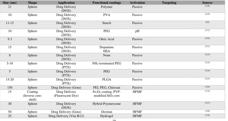
It is noteworthy that most drug release studies to date make use of a passive and
sustained release mechanism wherein the payload is allowed to diffuse out gradually
from the magnetically guided nanocarrier. However, in recent years, rapid rate drug
release promoted by an active agent has gained much attention. An activated system
allows for a local and temporary enhancement in release rate. This ‘on-off’ action
allows for pulsatile release profiles that is allows, additional control over therapy
compared to a sustained, passive system.
Release rate may be accelerated depending on the pH of the transfer medium
and the relative solubility of the drug at these conditions.[111] The magnetothermal
properties of SPIONs[112] have been well-studied and its use in hyperthermia therapy
using the heat generated by magnetically vibrating SPIONs to directly destroy cancer
cells[113] and bacteria has been proposed. However, this has not translated to a
significant body of work on using a high frequency magnetic field (HFMF) to remotely
trigger the release of drugs. An exception is the work of Hu et al.[114] who present a
drug-loaded SiO2 core surrounded by a Fe3O4 coating layer that is designed to rupture
upon applying HFMF.[115] Meanwhile, Satakar et al.’s study makes use of the
magnetothermal response combined with a thermally-responsive hydrogel coating.[116]
Work on thermally-responsive hydrogel coatings for drug delivery are relatively
well-established[117], but as yet combining this with the magnetothermal effect has not been
reported.
It is possible to envision a system that combines magnetically activated delivery
with MRI visibility to create a synergistic system. It is particularly popular in current
research to functionalize the surface of magnetite and magnetite-composite materials
with a targeting moiety for cancer-specificity towards the development of so-called
‘theranostic’ systems that simultaneously allow visualisation of the affected areas and
therapy through either drug or direct hyperthermia treatment.
Table 2. Magnetite-containing nanomaterials for biomedical purposes
Size (nm) Shape Application Functional coatings Activation Targeting Source
21 Sphere Drug Delivery (DOX)
Polymer Passive [118]
10 Sphere Drug Delivery (DOX)
PVA Passive [119]
11-15 Sphere Drug Delivery (DOX)
Starch Passive [92]
10 Sphere Drug Delivery (DOX)
PEG pH [111]
9.3 Sphere Drug Delivery (DOX)
Oleic Acid Passive [120]
15 Sphere Drug Delivery (DOX)
Dopamine HSA
Passive [121]
8 Sphere Drug Delivery (DOX)
None Passive [122]
5-10 Sphere Drug Delivery (PTX)
NH2-terminated PEG Passive [123]
5 Sphere Drug Delivery (PTX)
PEG Passive [124]
15-20 Sphere Drug Delivery (PTX)
PLGA Passive [125]
150 Sphere Drug Delivery (Gene) PEI, PEG, Chitosan Passive [126]
15 Coating (Inverse
core-shell)
Drug Delivery (Fluorescent Dye)
Fe3O4 coating,
PVP-modified SiO2 core
HFMF [114]
30 Sphere Drug Delivery (DOX)
Hybrid Poymersome HFMF [127]
50 Sphere Drug Delivery (Gene) Dextran HFMF [128]
25 Sphere Drug Delivery (Vita B12) Hydrogel HFMF [116]
8 Sphere Drug Delivery (TMX) Folic Acid, β -cyclodextran
HFMF [129]
10 Sphere Drug Delivery Eicosane HFMF [130]
2000 Cluster Drug Delivery (Budesonide)
Lipid HFMF [131]
7 - 9 Sphere Hyperthermia Therapy (Cancer)
PEG, Folic Acid
HFMF [113]
10000 Cluster Hyperthermia Therapy (Antibacterial)
None HFMF [132]
23600 Cluster Hyperthermia Therapy (Cancer)
Silica HFMF [133]
6-20 Sphere Hyperthermia Therapy (Cancer)
Silica, hydrogel HFMF [134]
60 – 80 Cluster Hyperthermia Therapy (Cancer)
PEG, Folic Acid HFMF [135]
13 – 40 Cube Hyperthermia Therapy None HFMF [136]
5 Sphere Hyperthermia Therapy Hyaluronic acid, PEG HFMF [137]
45 Magnetosomes Hyperthermia Therapy Citrate, PEG HFMF [138]
12 Sphere MRI Autofluorescent Albumin - ATF [139]
80 – 160 Cluster MRI Citrate - - [140]
7.5 Sphere MRI Mercaptopropionic acid, dextran
- - [141]
6 Sphere MRI Folic Acid, PEG, PDLLA, - Folate [142]
10 – 15 Sphere MRI Fluorescent Dextran Protamine [143]
10 Sphere MRI Targeting ligand 2-aminoethyl-trimethyl
ammonium
[144]
MRI Glial fibrillary acidic
protein
[145]
16 Sphere MRI Carboxylic manna Mannose receptor [146]
1.3.4 Fe3O4-containing nanocomposites
Composites nanostructures containing Fe3O4 are useful as they may add additional
functionality and useful properties. Most notably, coating the iron oxide in a material
such as silica or a suitable polymer may further improve the water-stability of the
nanoparticle as well as its chemical inertness in biological conditions. For this reason,
most composites of iron oxide focus on core-shell like structures, although other
structures do exist. The synthesis of composite structures may be performed in two
steps, whereby the Fe3O4 NPs are then combined with the second material or through a
single-step co-synthesis where the two materials are formed simultaneously. The main
structures currently known in literature have been illustrated below (Figure 4).
Figure 4. Heterogeneous composite nanostructures of magnetite
1.3.5 Composites with polypyrrole
In this thesis, Fe3O4 is combined with polypyrrole to create structures that have
magnetic functionality combined with the electrical, optical and thermal properties of
polypyrrole. The known structures to date are simple core-shell, multiple core-shell,
aggregated and raspberry-like structures (Figure 5).
Figure 5. Known structures of PPy-Fe3O4 nanocomposites. (a) core-shell[147], (b) aggregated[147], (c)
multiple core-shell[148], (d) raspberry (PPy core, Fe
3O4 decorations)[149]
Composites of PPy and Fe3O4 may be synthesised by including suspended Fe3O4
NPs in the chemical polymerisation mixture for PPy, in a process of seeded
polymerisation. However, the resulting structures differ significantly depending on the
particulars of the process. There are several important concepts to consider during such
a two-step synthesis. Firstly, the degree of aggregation of the nanoparticles is important.
For uniform core-shell structures, a highly desirable structure, the nanoparticles must be
well-dispersed during synthesis. Secondly, both the location and type of nucleation of
the PPy particles is important. For example, a homogeneous nucleation that is not
concentrated at the Fe3O4 nanoparticle surfaces will result in randomly aggregated
structures.
Indeed, it has been reported that when synthesised without surfactants to prevent
the aggregation of Fe3O4 NPs, an aggregated structure comprising polypyrrole NPs that
nucleates away from the Fe3O4 surface results (Fig. 4 b) whereas the inclusion of a
surfactant such as sodium dodecyl sulfonate will promote a core-shell structure
comprising a homogenous polypyrrole layer over the nanoparticle cores.[147] The use of
a copolymer such as PVA may have a similar effect in promoting the growth of the
core-shells[148]. Wuang et al. combined typical oxidation route with PVA-based
microemulsions to create PPy particles with multiple encased Fe3O4 cores.[150] Tian et
al.[149] was able to create the reverse, raspberry-like structure of PPy serves surrounded
by Fe3O4 decorations on the structure surface by applying using an FeCl3.6H2O oxidant
in excess and applying a second step to convert excess Fe3+ into Fe3O4 nanoparticles.
The oxidant used will also have an effect on the structure. While both APS and
FeCl3.6H2O are commonly used in oxidation of polypyrrole, in the synthesis of
PPy-Fe3O4 composites the use of FeCl3.6H2O may be advantageous due to the common ion
effect. It is hypothesised that Fe3+
(aq) adsorbs on the Fe3O4 nanoparticle surfaces, thus
promoting the polymerisation of PPy at the nanoparticle surface.[148]
1.3.6 Magnetically-driven synthesis and self-assembly
One concept that is unique to magnetic nanomaterials is the use of a magnetic field
during their synthesis to promote the growth and/or assembly of 1-dimensional,
anisotropic nanostructures along the direction of the field lines. This process involves
the transport and arrangement of matter under an applied magnetic field. This may be
follow by or occur simultaneously with stabilization of the assembled structure by
chemical means. Before discussing the recent progresses in this area, it is important to
understand the fundamental underlying theories governing this process.
Firstly, the movement of transition metal ions in a solution is governed by the
magnetic force, F(z) defined as
𝐹𝐹 (𝑧𝑧) = 𝜒𝜒𝜒𝜒𝐻𝐻(𝑧𝑧)𝜕𝜕𝜕𝜕𝜕𝜕𝑧𝑧(𝑧𝑧) (Equation 3)
Where χ is the molar magnetic susceptibility of metal ions with mole number n at the position z within a magnetic field H(z). Hence, it has been observed that larger movement under the same field gradient occurs for ions of larger susceptibility[151]. At
the same time, a larger overall ion concentration induced larger movement, which
indicates that the ions move as a group and not as independent single particles. The drift
velocity of these ions, v(z), has the following simple relationship to the magnetic force.
𝑣𝑣(𝑧𝑧) ∝ 𝐹𝐹(𝑧𝑧) (Equation 4)
The movement of ions is in line with the magnetic field, with overall migration towards
the centre of the field. This results in the formation of 1D structures along the field
lines. These two equations govern the transport component. The energy component is
governed by the following equation for the magnetic energy E of an object of volume V
under a magnetic field B through a space of permittivity μ0:
𝐸𝐸 = 2𝜇𝜇1 0𝜒𝜒𝜒𝜒𝐵𝐵
2 (Equation 5)
If the magnetic energy is greater than that of the thermal energy for a system, the
objects will orient to the applied field. Since magnetic properties are anisotropic within
a crystalline grain, they will do so by orienting in the crystal direction that will
minimise the energy of the system. This direction is called the ‘easy direction’. In
Fe3O4, the easy magnetic axes are along [111] and [110] directions.
These fundamental forces are utilized in nature within magnetotectic bacteria.
This class of bacteria form chains of magnetic nanomaterials within their bodies. It is
currently understood that these anisotropic magnetic nanostructures, called
magnetosomes, aid the bacteria in finding their orientation within the earth’s magnetic
field (Figure 6). Some attempts have been made to harvest these magnetosomes and
apply them subsequently to biomedical applications[152] such as delivery of DOX[153]
and magnetothermal therapy.[138] However, due to the need for culturing, harvesting and
purification of these magnetosomes, there has emerged a recent interest in artificially
synthesising analogous materials.
Figure 6.Transmission electron microscopy images of a single magnetotactic bacterium (A) of chains of magnetosomes extracted from whole magnetotactic bacteria (B) of individual magnetosomes detached
from the chains by heat and SDS treatment (C).[152]
Figure 7. Schematic illustrating various known 1D structures resulting from synthesis under an applied magnetic field. (a) single crystal nanowire, (b) multi-crystal nanowire, (c) bundled nanowire, (d)
nanochain of isotropic nanoparticles, (e) aggregated chain.
The current work on the synthesis of anisotropic magnetic materials under an
applied magnetic field may be divided into several categories depending on the resulting
morphology. The two main structures are rod-like (Figure 7 a,b) and chain-like (Figure
7 c). Of course, as for all nanomaterials, the tendency to form aggregates (Figure 7 c,e)
is ever present.
Rod-like or wire-like structures are typically synthesised in one step, coupling
an applied magnetic field with a typically liquid phase synthesis of iron oxides. For
example, Wang et al. have reported the fabrication of single crystal Fe3O4 nanowires by
hydrothermal process by applying a field of up to 0.35T. [154] The nanowire that resulted
was found to grow primarily along the [110] direction and is in good agreement with the
theory discussed above of ion migration along magnetic field lines during synthesis.
Similar results were observed for sonochemical[155] and thermal decomposition[156]
synthesis. It is similarly possible to create multi-crystalline nanowires and rods by such
one-step methods under certain, kinetic driven conditions.[157]
In addition to rod or wirelike materials, application of a magnetic field can also
be used to assemble individual isotropic (usually spheroid) nanoparticles into 1D,
chainlike forms. These structures may be understood to form in a two-step process.
First, the nanoparticles are formed without the influence of a magnetic field, and then
arranged under the aligning magnetic field. This is typically accomplished in the liquid
phase with suitable surface treatment of the magnetic nanoparticles for either colloidal
stability or as an adhesive coating to stabilize the assembled anisotropic structures. For
example, Chong et al. were able to create end-on-end, well-dispersed nanochains with
tuneable size by encapsulating Fe3O4 NPs with SiO2 in a microfluidic reactor.[158]
The key publications of the past decade in magnetic field directed synthesis have
been summarized in the following table (Table 3).
Table 3. Magnetically directed synthesis of anisotropic materials
Method Material Field Strength
(T) Source L iqui d Pha se
Hydrothermal Single crystal Fe3O4 nanowire 0.25 [154]
Hydrothermal Fe3S4 and FeS2 multi-crystal
microrods
0.045 [159]
Hydrothermal Co3O4 nanowire 0.24 [160]
Sol-gel TiO2 nanowires 9.4 [161]
Solvothermal Bi single crystal nanowires 8 [162]
Solvothermal Ni-Co alloy nanowires 0.08 [115]
Sonochemical Fe3O4 nanorods 0.5 [155]
Microwave Fe3O4/C/CdS coaxial
nanochains
0.2 [163]
Wet chemistry Ni nanochains 0.5 [164]
Wet chemistry PEDOT:PSS encapsulated iron oxide NPs nanochains
0.065 [165]
Wet chemistry microfluidic
SiO2 encapusulated Fe3O4 NPs
nanochains
Unreported [158]
Wet chemistry nanocapillary
coating
Fatty acid encapsulated Fe3O4
NPs nanochains
0.0012 [166]
V a po r Pha se Chemical Vapor Deposition
GaN nanowire 0.25 – 0.8 [167]
Arc Plasma SWCNT 0.02- 0.2 [168]
Flame synthesis Co multi-crystal nanowire 1 [157]
This present work is particularly interested in the synthesis of chainlike and
aggregated chainlike structures. Unlike the rod-like counterparts, these chains can be
flexible when a suitably soft material is chosen as the encapsulating agent. For example,
Ho et al. was able to create conductive, flexible filaments of iron oxides by using
conductive polymer PEDOT:PSS as the coating material. [165] Of course, when a rigid
material such as SiO2 is used as the coating instead, the resulting nanoparticle chains are
as rigid as expected.[158] Additionally, the assembly of magnetic nanochains may be
engineered to be reversible. Bharti et al. coated iron oxide chains with fatty acids to
create a thermally-responsive material that would be reconfigurable when the fatty acid
was in liquid state and exploited this for magnetically directed self-healing of the
filaments.[166]
In addition to end-on-end chains of single particles, it is possible to create large,
needle-like structures that result from the aggregation of large numbers of nanoparticles
in the direction of the magnetic field.[169] These structures are termed ‘needle-like’ due
to the profile of the structure’s width dimension, comprising a middle section of several
nanoparticles’ width that tapers to a fine, needle-like point (Figure 8). At present, it is
unclear what experimental conditions cause these two vastly different reported
morphologies, and how conditions may be selectively and controllably exploited to
favour one over the other.
Finally, it is important to consider the dynamic behaviour of the chainlike
materials, in particular the soft, flexible materials. Bharti et al.[169] have reported a
flexible, curling motion (Figure 10) that is unique to soft-coated nanochains and is not,
for example, observable in the SiO2-encapsulated chains presented by Chong et al.[158]
Although the irreversible formation of circular bundles reported by Bharti may be a
disadvantage as it destroys the anisotropy of the structure, it is nonetheless an
interesting and unique material property. At the very least, this work indicates the
importance of tracking the dynamic behaviour of such structures which can be
demonstrably irreversible.
Figure 8.Formation of aggregated nanochains under an applied magnetic field.[169]
Figure 9. Flexible motion of soft-encapsulated nanochains[169]
Figure 10. Irreversible formation of spheroid bundles upon applying a rotating magnetic field.[169]
To date, the synthesis and assembly of 1D nanomaterials under a magnetic field
remains relatively novel, despite the potential of such materials in numerous areas. For
the purpose of this work, it is the potential of such materials towards drug delivery that
is important. Anisotropic materials that may be magnetically actuated have numerous
potentially useful properties. Firstly, they may be arranged or patterned by external
magnetic field alignment. The ability to create aligned structures for drug delivery has
been of interest recently, in particular in scaffolds for neuron growth. The ability to
promote neurite extension in one direction by using a suitably patterned nano- or micro-
vehicle loaded with a neural growth factor is an area of high novelty and high impact
research. 1D magnetic nanomaterials have a strong advantage over scaffold and coating
system as they have the potential for dynamic response. While scaffolds are fixed,
nanowires and chains can be manipulated at will by applying a simple change to the
magnetic field strength or direction. To date, there have been no attempts to study what
![Figure 2. (a) Bandgaps of rutile, anatase and brookite.[19]. (b) General scheme for photocatalytic reactions at TiO2 nanoparticle surface in aqueous conditions](https://thumb-us.123doks.com/thumbv2/123dok_us/8081060.228920/33.595.101.446.333.684/figure-bandgaps-brookite-general-photocatalytic-reactions-nanoparticle-conditions.webp)
![Figure 10. Irreversible formation of spheroid bundles upon applying a rotating magnetic field.[169]](https://thumb-us.123doks.com/thumbv2/123dok_us/8081060.228920/57.595.55.467.63.290/figure-irreversible-formation-spheroid-bundles-applying-rotating-magnetic.webp)
![Figure 17. Association of amino acids in zwitterionic form (Adapted from [223]).](https://thumb-us.123doks.com/thumbv2/123dok_us/8081060.228920/71.595.113.439.67.312/figure-association-amino-acids-zwitterionic-form-adapted.webp)