Tracking in atomic detail the functional
specializations in viral RecA helicases
that occur during evolution
Kamel El Omari
1, Christoph Meier
1, Denis Kainov
2,3, Geoff Sutton
1,
Jonathan M. Grimes
1,4, Minna M. Poranen
5, Dennis H. Bamford
5,6, Roman Tuma
7,
David I. Stuart
1,4and Erika J. Mancini
1,*
1
Division of Structural Biology, The Wellcome Trust Centre for Human Genetics, University of Oxford, Headington, Oxford OX3 7BN, UK, 2Institute for Molecular Medicine Finland (FIMM), University of Helsinki, 00290 Helsinki, Finland, 3Department of Environmental Research, Siauliai University, Vilniaus gatve_88, 76285 Siauliai, Lithuania, 4Diamond Light Source Limited, Harwell Science and Innovation Campus, Didcot,
Oxfordshire OX11 0DE, UK, 5Department of Biosciences, University of Helsinki, Biocenter 2, PO Box 56, 00014 Helsinki, Finland, 6Institute of Biotechnology, University of Helsinki, Biocenter 2, PO Box 56, 00014 Helsinki, Finland and7Astbury Centre for Structural Molecular Biology and School of Cellular and Molecular Biology, University of Leeds, Leeds LS2 9JT, UK
Received June 3, 2013; Revised July 18, 2013; Accepted July 19, 2013
ABSTRACT
Many complex viruses package their genomes into empty protein shells and bacteriophages of the
Cystoviridae family provide some of the simplest
models for this. The cystoviral hexameric NTPase, P4, uses chemical energy to translocate single-stranded RNA genomic precursors into the procapsid. We previously dissected the mechanism of RNA translocation for one such phage, r12, and have now investigated three further highly diver-gent, cystoviral P4 NTPases (from r6,r8 and r13). High-resolution crystal structures of the set of P4s allow a structure-based phylogenetic analysis, which reveals that these proteins form a distinct subfamily of the RecA-type ATPases. Although the proteins share a common catalytic core, they have different specificities and control mechanisms, which we map onto divergent N- and C-terminal domains. Thus, the RNA loading and tight coupling of NTPase activity with RNA translocation inr8 P4 is due to a remarkable C-terminal structure, which wraps right around the outside of the molecule to insert into the central hole where RNA binds to coupled L1 and L2 loops, whereas in r12 P4, a C-terminal residue, serine 282, forms a specific hydrogen bond to the N7 of purines ring to confer purine specificity for ther12 enzyme.
INTRODUCTION
Viruses protect their genome by condensing it into a com-partment, the virion. Many complex viruses rely on rapid encapsidation by energy-dependent transport of the nucleic acid into an empty preformed capsid (procapsid). This process requires the presence of portal complexes, which are conduits for nucleic acid molecules, and molecu-lar motors that convert the chemical energy gained from nucleoside triphosphate (NTP) hydrolysis into mechanical movement, resulting in nucleic acid translocation.
Some viruses, including herpesvirus and tailed double-stranded DNA (dsDNA) bacteriophages, package their genome using a multi-protein packaging motor (terminase) that transiently assembles at a single vertex (1–4). These complexes are relatively elaborate, consisting of a large dodecameric portal that is an integral part of the capsid and an oligomeric transiently associated terminase, neither of which can work in the absence of the other. The ATPase-nuclease terminase subunit is responsible for re-cruiting the viral DNA to the procapsid. Compacting rela-tively stiff dsDNA into a small volume of the procapsid has a high energy cost. Single-molecule experiments have revealed that viral packaging proteins can exert forces as high as 110 pN on dsDNA, making them some of the strongest known biological motors (5).
Similarly, dsRNA bacteriophages of the Cystoviridae
family (bacteriophages f6 through to f14, and f2954) encapsidate single-stranded RNA (ssRNA) genomic pre-cursors into procapsids (6). However, their packaging
*To whom correspondence should be addressed. Tel: +44 1865 287560; Fax: +44 1865 287549; Email: [email protected]
ßThe Author(s) 2013. Published by Oxford University Press.
This is an Open Access article distributed under the terms of the Creative Commons Attribution License (http://creativecommons.org/licenses/by/3.0/), which permits unrestricted reuse, distribution, and reproduction in any medium, provided the original work is properly cited.
at University of Leeds on March 24, 2015
http://nar.oxfordjournals.org/
machinery is less complex, consisting of a hexamer that is at the same time the physical portal and the active genome translocating motor (7,8). Although this motor shares the same function of translocating the genomic nucleic acid into the procapsid, the challenges differ between ssRNA and dsDNA. ssRNA is significantly more flexible (persist-ence length lp1–2 nm) than dsDNA (lp50 nm) (9), and the packaging densities are less than those found for dsDNA viruses (10); therefore, high forces are probably not required. However, naturally occurring ssRNAs, such as the genomic precursors, exhibit extensive local second-ary structure (11,12), and thus the packaging motor has to exhibit helicase activity.
The lipid-enveloped bacteriophages of theCystoviridae
family infect Gram-negative bacteria, mainly plant-patho-genicPseudomonasspecies (13) and share similarities with the members of the Reoviridae family, including blue-tongue virus and rotavirus (14). Their genome of14 kb consists of three dsRNA segments small (S), medium (M) and large (L), which are sequentially encapsidated as ssRNA precursors into the icosahedrally symmetric procapsid by the packaging NTPase P4 (15–23).
P4 NTPases are structural components of the procapsid, built by co-assembly of 120 copies of the major structural protein P1 with10 copies of the viral RNA-dependent RNA polymerase P2, 10 hexamers of P4 and 12 trimers of the assembly cofactor P7 (24) (Figure 1). In bacteriophage f6, P4 hexamers nucleate procapsid assembly in vitro (7,25), are essential for genome packaging (21) and also have a role in transcription (21,26). Up to 12 P4 hexamers lie on the 5-fold symmetry axes of facets of the procapsid (16,24,27), creating a symmetry mismatch. Although the P4 hexamer constitutes the packaging motor, the specificity for viral RNA is mediated by RNA-binding sites on the P1 shell, which recognize three distinct packaging signals on the genomic precursors (28,29).
[image:2.612.60.301.563.679.2]Previous studies have revealed the structure and mech-anism of f12 P4 (30–32). P4 is a protein of 35 kDa, which can assemble into a hexameric ring. NTP-binding sites are located on the external perimeter of the ring at the interfaces between adjacent subunits, whereas the nucleic acid binding sites are found in the central channel (31) (Figure 1). P4 proteins are the only known RNA-specific
helicases belonging to helicase Superfamily 4 (SF4) (33). SF4 encompasses mainly DNA helicases and is char-acterized by five conserved sequence motifs (H1, H1a, H2, H3 and H4) (34). Motifs H1, H1a and H2 are involved in nucleotide binding and hydrolysis, whereas H3 is involved in the coupling of NTP hydrolysis to nucleic acid translocation, and H4 in oligonucleotide binding. Crystal structures off12 P4 at different key cata-lytic states of the protein unveiled a power stroke mech-anism by which a conformational change associated with sequential NTP hydrolysis is responsible for RNA trans-location (31,35,36).
P4 NTPases show little sequence similarity; however, they are believed to share a common architecture and mechanism of action. When recombinant P4 proteins are studied in iso-lation, they show variation in their in vitro biochemical properties (Table 1):f8 andf13 P4 NTPases form stable complexes with RNA and their ATPase activities are strongly stimulated by RNA (f8 has no detectable ATPase activity in absence of RNA), whereasf6 andf12 P4s bind RNA transiently and are only weakly stimulated; the isolated P4 hexamers off8 andf13 have measurable helicase activitiesin vitroin contrast tof6 P4, which only acquires processive helicase activity in the context of the procapsid (30); thef12 P4 hexamer has low translocation processivity and lacks helicase activity (36); the NTPase activity off12 P4 is specific to purine bases (26), whereas the other P4s can also accept pyrimidine bases (8,40). These differences in biochemical properties are presumably re-flected in the hexamer architecture and structural details of different domains. To gain further insights into RNA loading, interaction and translocation mechanisms and the structural evolution of these packaging enzymes, we have solved the crystal structures of three additional P4 proteins, fromf8,f13 and from the prototype virus of the cystoviral family,f6. We also report here the structural and/or bio-chemical characterization off12 P4 mutants to explain nu-cleotide specificity and RNA recognition. We compare these structures with that of wild-typef12, whose structure has already been reported (31), creating a series of structurally related viral packaging motors.
MATERIALS AND METHODS
Cloning, expression and purification
Recombinant full-length P4 from f8,f13 and C-termin-ally truncatedf8 P4281 (missing residues 281–321) and
f6 P4310 (missing residues 310–331) were expressed from plasmids pSJ1b (41), pDK3 (8), pDK10 (42) and pJTJ7.3/7 (43), respectively. Point mutations were introduced into f12gene 4using plasmid pPG27 (32) as a template to introduce amino acid substitutions S252Q, R272A, Q278A, S292A, Y288A and TTS202-204 by site-directed mutagenesis (QuikChange, Stratagene) following the manufacturer recommendations. The corresponding plasmids were designated as pDK33, pDK35, pDK30, pDK31, pDK29 and pDK249 respectively. The insertion of LKK instead of TTS (residues 202–204) was introduced by amplifying the N-terminal portion of the P4 gene with primers 1 and 2 (Supplementary Table S1) and the
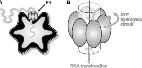
Figure 1. The cystovirus P4 protein, a molecular packaging motor. (A) Cartoon showing the position of the P4 hexamer (grey) on the empty cystovirus procapsid (black) while packaging ssRNA. (B) Cartoon model of the mechanism of RNA translocation by P4. The energy derived from the hydrolysis of ATP is mechanically converted to the translocation of single-stranded ssRNA.
at University of Leeds on March 24, 2015
http://nar.oxfordjournals.org/
C-terminus part with primers 3 and 4. PCR products were digested with NdeI/AflII (N-terminal part) and AflII/ EcoRI (C-terminal part) and ligated into pT7-7 vector at NdeI-EcoRI sites. Sequencing was used to confirm the mutations.
Recombinant P4 proteins were expressed inEscherichia coli BL21(DE3) or B834(DE3) and purified to homogen-eity as previously described (31,32,42). Briefly,E. colicells were grown at 37C in Luria-Bertani medium until
OD540nm reached 0.5–0.6. Cultures were then chilled on ice and induced with 1 mM isopropyl-b -thiogalacto-pyranoside. Induced cells were further incubated for 12–14 h at 17–18C, harvested by centrifugation and
lysed with a French pressure cell. P4 proteins were purified by chromatography: Heparin and Q-sepharose columns (GE Healthcare) followed by size exclusion chro-matography (Superdex 200, GE Healthcare).
Cloning, expression, purification and characterization of C-terminally His-tagged f8 P4 (f8 P4His), which exhibits full RNA-induced ATPase activity, was described previously (44).
Crystallization
Crystallization conditions of the P4 proteins have been pre-viously described (32,42). In brief, crystals off6 P4310 proteins were grown at 24C from a 3.5 mg/ml protein
solution in 20 mM HEPES (pH 8.0), 5 mM MgCl2, 2 mM CaCl2, 5 mM adenosine diphosphate (ADP) and 100 mM NaCl, and they appeared after 9 months in drops in which
3ml of protein had been mixed with 3ml of a reservoir
solution consisting of 6% PEG 4000 and 90 mM sodium acetate (pH 4.5). Crystals were cryo-protected by transferring them into reservoir solution with a final glycerol concentration of 25% before freezing in a nitrogen-gas stream at 173C.
From a 12 mg/ml protein solution,f13 P4 crystals were grown at 20C using 100 mM Tris–HCl (pH 7.0), 900 mM
trisodium citrate and 200 mM NaCl as precipitant. Crystals were cryo-protected as f6 P4310, but using a final glycerol concentration of 20%.
The f8 P4 crystals were grown at 24C in 100 mM
sodium acetate (pH 4.6) and 2.2 M ammonium sulphate as a precipitant. Drops consisted of 0.9ml of protein at a concentration of 3 mg/ml, 0.9ml of reservoir solution and
0.4ml of 100 mM dithiothreitol (DTT). Crystals of f8
P4281 obtained from a protein solution concentrated to 5 mg/ml appeared in 100 mM Tris (pH 8.0) and 18% PEG 1000. Crystals were cryo-protected following the protocol forf6 P4310.
Crystals off12 P4 mutants were obtained in a solution composed of 10% PEG 1500 in 100 mM sodium acetate (pH 4.8) and 5 mM AMPcPP. Crystals of wild-type f12 P4 with UTP were obtained with the same precipitant and 5 mM UTP.
Data collection and structure determination
Data collection was performed as previously detailed (32,42), and all data were indexed, integrated and scaled using HKL2000 (45). Crystallographic statistics for the data are detailed in Supplementary Table S2.
Table 1. Biochemical properties of wild-type and mutant P4 proteins P4 f 6 f 8 f 12 f 13 f 12 S292A f 12 Q278A f 12 Y288A f 12 TTS202-204 f 12 TTS202-204LKK Nucleotide binding RNA binding L1 loop Mol weight (kDa) 35 34.1 35.1 37.6 35.1 35.1 35.1 35.1 35.1 Quaternary struc-ture and stability Hexamer 7 (+ ATP/ADP) Hexamer (8) Hexamer (26) Hexamer (8) Hexamer Hexamer Hexamer Hexamer Hexamer Controlled ring opening (37) Frequent ring opening (38) KM (ATP) mM 0.19 ± 0.03 (8) N A 1.50 ± 0.04 (36) 0.40 ± 0.05 (8) 2.20 ± 0.5 kcat s -1 0.19 ± 0.06 (8) 0.00 ± 0.05 (39) 0.84 ± 0.12 (36) 1.60 ± 0.05 (8) 0 0.25 ± 0.13 0 0 0 KM (ATP ) mM with 1 mM polyC ND 0.17 ± 0.01 (39) 0.49 ± 0.02 (36) ND 2.10 ± 0.1 kcat s -1 with 1 mM polyC ND 7 6.40 ± 0.20 (39) 2.52 ± 0.07 (36) ND 0 0.96 ± 0.05 0 0 0 NTP specificity All (8) All (8) Purine base (26) All (8) ssRNA binding Kd > 1 Mm (8) Kd < 1 m M (8, 39) Kd > 1 m M (26,36) Kd < 1 m M( 8 ) ssRNA translocation Weak (7 ) Strong Weak (36) Strong (8) Processive (36,39) COD (helicase) activity Only in PC (8) Strong (8 ) None (8) Weak (8) RNA binding site ND L1 (LKK) (37) L2 (K241) (30) ND
at University of Leeds on March 24, 2015
http://nar.oxfordjournals.org/
Structures off12 P4 mutantsf12 P4-Q278A andf12 P4-S292A were solved by molecular replacement using the program PHASER (46) with wild-typef12 P4 (PDB code 1W4B) as the search model.
The structure of f13 P4 was solved by single-wave-length anomalous dispersion as described elsewhere (47). The substructure was determined using the program SHELX (48), and phases were refined using SHARP (49). After 6-fold non-crystallography symmetry averaging using General Averaging Program (unpublished program available from D. I. Stuart or J. M. Grimes), an interpretable electron density map was obtained into which the structure could be built.
The structure off6 P4 was solved by molecular replace-ment with the crystal structure of thef13 P4 as a search model. The search model included one hexamer in which each chain was truncated to the conserved ATPase core of the protein. A weak molecular replacement solution comprising two truncated hexamers was found by the program AMoRe (50). The preliminary phases were greatly improved by 12-fold non-crystallographic symmetry averaging and phase extension from low reso-lution using General Averaging Program. The last 34 residues of the f6 P4310 construct were not visible in the electron density; their absence might be due to prote-olysis, which would explain the long crystallisation period. The structure of f8 P4 was initially solved by single-wavelength anomalous dispersion from crystals of the selenomethione labelled protein in space groupP622 con-taining one monomer in the asymmetric unit. HKL2MAP (48) was used to identify the selenium sites, which were then fed into PHENIX AUTOSOL (51), resulting in an inter-pretable electron density map for the ATPase core domain. The electron density corresponding to the rest of the protein was not interpretable owing to the statistically dis-ordered crystal reported previously (42). The hexameric P4 was formed by applying the crystallographic symmetry and used as search model for molecular replacement with the program PHASER (46) to find a solution forf8 P4 (R32 space group) andf8 P4281 (P21212 space group).
Manual building was performed with the program COOT (52) and restrained refinement (with TLS) with either AUTOBUSTER (53) or REFMAC5 (54). The final models were validated with MolProbity (55). Refinement statistics are provided in Supplementary Table S2; in summary; the resolution (A˚)/R-factor(%)/ R-free(%) for the structures were f6 P4: 2.8/21.7/24.4,
f8 P4-His: 3.1/29.6/30.9, f12 P4 UTP: 1.9/19.4/20.4,
f13 P4: 1.7/16.4/18.8.
Hydrogen-deuterium exchange mapping
Previously published hydrogen-deuterium exchange (HDX) data for f8 P4 were used (37) and mapped onto the high-resolution structure presented in this work using average rate colouring as described (37).
ATPase activity of mutants
ATPase activity of f12 P4-binding site mutants was assayed using the EnzChek phosphate assay kit (Invitrogen) (39).
Evolutionary analysis of structures
The coordinates of the ATPase core of P4 from f8 (residues 104–261) were submitted to the DALI Server (56), a program that identifies and ranks proteins by struc-tural similarity. The DALI search returned 47 proteins, which have significant structural similarity to P4. All these proteins were then truncated to their core ATPase domains, and using the program SHP superimposed onto one another, and a matrix of structural relationships was calculated (57).
RESULTS AND DISCUSSION
Overall fold
All P4 proteins form a hexameric ring with a central channel varying in size from 13 to 21 A˚ (30 A˚ for f8 P4281) and external diameter of 100 A˚ (Figure 2). However, the hexamers have different charge distributions on their surfaces (Supplementary Figure S1) and different outline shapes:f6 P4,f8 P4 andf13 P4 form hexagonal notched rings, whereas f12 P4 has a smoother contour. The subunit interface within hexamers varies in size from
1500 to 1900 A˚2, and the number of hydrogen bonds,
salt bridges and hydrophobic interactions shows substan-tial variation (Supplementary Table S3). The interfaces within the P4 hexamers are more polar than expected for a stable oligomer. This is because rings of hexameric helicases are generally required to open to load the nucleic acid strand into the central cavity (Table 1) (58,59). The rounderf12 P4 subunits bury the biggest surface area and form the highest number of hydrogen bonds and salt bridges, whereas the interaction area is least for f8 P4, which harbours fewer hydrogen bonds and only three salt bridges. The buried area does not correlate with P4 ring stability. For example, f12 P4 has been shown to exhibit frequent ring opening unless it is bound to the procapsid (38), leading to low translocation processivity (36). On the other hand, f8 P4 is a processive translocase and opens only during loading a new RNA strand into the central channel (37). Ring stability correlates instead with the fraction of buried polar interactions (hydrogen bonds and salt bridges) per buried area. The less stable f6 and
f12 hexamers have 0.016 and 0.018 polar contacts per A˚2 respectively, whereas the more stablef8 andf13 exhibit values of 0.13 and 0.15, respectively.
ATPase core domain
Within the hexamer, the different P4 monomers adopt similar orientations and can be divided into three domains: an N-terminal region (110–150 residues), a central core NTPase domain of 160 residues and a smaller C-terminal domain (40–50 residues) (blue, grey and red, respectively, in Figure 2). Strikingly, despite low overall sequence conservation ranging from 9 to 21% amino acid sequence identity, the key structural features of the ATPase core domain (motifs H1, H1a, H2, H3 and H4) are well-conserved (Figure 3A andB). The ATPase domain is a Rossmann-type nucleotide-binding domain consisting of a twisted seven-stranded b-sheet with
at University of Leeds on March 24, 2015
http://nar.oxfordjournals.org/
Figure 2. The overall fold of cystoviral P4 proteins. (A) The P4 hexamers of bacteriophagesf6,f8,f12 andf13 (left to right) are viewed from the top and coloured by chain. (B) Side view of the P4 hexamers. (C) The panel shows structures of monomeric P4 in two orientations, the upper orientation of the monomer corresponding to the one depicted in cyan in (B); the lower one has undergone a rotation of 140C to show the
C-terminal domains. The core domain is coloured in grey, the N-C-terminal domain in blue and the C-C-terminal domain in red. Nucleotides, if present, are depicted as sticks with carbon, oxygen, nitrogen and phosphorus atoms coloured in yellow, red, blue and orange, respectively. Dotted lines represent the disordered region of the proteins.
at University of Leeds on March 24, 2015
http://nar.oxfordjournals.org/
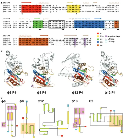
Figure 3. Structural conservation between P4 proteins. (AandB) Sequence and structural conservation of the helicase motifs in P4 proteins. Motifs H1, H1a and H2 are involved in nucleotide binding and hydrolysis, H3 is involved in the coupling of NTP hydrolysis to nucleic acid translocation, and H4 in oligonucleotide binding. Motifs H1, H1a, H2, H3, H4 are coloured in red, yellow, green, blue and brown, respectively; the arginine fingers are coloured purple, whereas the L1 and L2 loops are black and cyan, respectively. (A) Structure-based acid sequence alignment of the ATPase core domain off6,f8,f12 andf13 P4. Functionally important residues that are conserved amongst the different cystoviruses are indicated by stars, whereas a sphere marks the lysine in loop L2 (K241 inf12 P4), which is not conserved inf8 P4. (B) Cartoon representations off6,f8,f12 and
f13 P4 structures in equivalent orientations. The arginine fingers and the nucleotides are shown in a ball-and-stick representation. The colour coding is the same as in (A). (C) Topology diagrams of the N-terminal domains off6,f8,f12 andf13 P4. Secondary structural elements are coloured in green (strands) and yellow (helices). Topologically similar domains are shaded in pink (f6 andf8) and orange (f8 and C2). The topology for C2 was derived from PDB entry 2ENP.
at University of Leeds on March 24, 2015
http://nar.oxfordjournals.org/
mixed parallel and antiparallel topology flanked by five helices. Residues previously demonstrated to be critically important in the mechano-chemically coupling of ATP hydrolysis to RNA translocation inf12 P4 (35) are struc-turally conserved in other P4s (Figure 3A andB, Table 2), except for one residue in motif H4 (residue K241 inf12 P4), which has no equivalent inf8 P4 (see explanation for this later in the text). It is therefore likely that all cystoviral P4 NTPases use an RNA translocation mech-anism similar to that described forf12 P4 (31), although details may vary, especially for f8 P4 where a tight coupling between ATPase activity and RNA binding is observed (Table 1).
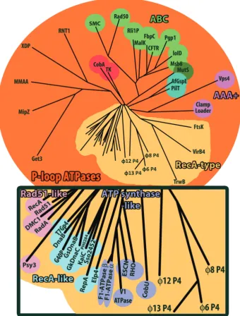
Structural classification based on the ATPase core domain shows that cystovirus P4 proteins are closely related to each other and only distantly related to other P-loop ATPases (Figure 4 and Supplementary Figure S2). They most closely resemble RecA-type ATPases (35), such as ATP synthase-like proteins (RHO, F1-ATPase, etc.), RecA-like proteins (RepA, T7 gp4, etc.) and Rad51-like protein (Rad51, RecA, etc.). Many of these proteins are involved in nucleotide repair and recombination and have similar functional properties to P4 proteins. This indicates that the cystoviral P4 proteins form a distinct subfamily of RecA-type ATPases.
N-terminal domain
The structural conservation across P4 proteins of the central ATPase core domains does not extend to the N-and C-terminal domains. Most of the N-terminal domain residues of P4 from f6 and f8 are visible in our crystal structures (starting from amino acid residues 2 and 12, respectively), whereas f13 P4 lacks the first 32 residues [which are predicted to be disordered (60)]. In all P4 struc-tures, the N-terminal domain covers the apical part of the hexamer (Figure 2), and inf12 P4, an N-terminal domain
a-helix projects from one subunit to the adjacent one, giving the hexamer a more rounded appearance. f6 P4 lacks such a helix and might stabilize the hexamer by strengthening subunit interfaces with nucleotides. f6 P4 is the only P4 that needs nucleotides and divalent cations to form hexamers (7). It is also conceivable that NTP binding triggers a conformational change in the f6 P4 subunits allowing them to form hexamers. Interestingly,
f8 andf13 P4s also lack such a stabilizing helix; however, the first 12 and 31 residues, respectively, are not visible
in the crystal structures and might play such a stabilizing role.
The N-terminal domains of cystoviral P4s are highly divergent (Figures 2, 3B and C). However in f6 and
f13, more than half of their residues can be superimposed with a root-mean-square deviation of 2.1 A˚, including two parallel helices and two small anti-parallel b-sheets, creating a topologically identical sub-domain (Figure 3C). In f8 and f12, the N-terminal domains have higher secondary structure content but are com-pletely unrelated to each other and to those in f6 and
f13. In f12 P4, the N-terminal domain is composed of two orthogonal a-helices and three anti-parallel b-sheets (Figure 3C). The f8 P4 N-terminal domain is composed of two helices separated by a four-stranded antiparallelb -sheet (Figure 3C). Structural alignment searches against the PDB database returned no significant matches for any of the N-terminal domains, aside from a weak structural similarity (43 of 87 residues within 3.7 A˚) off8 P4 to one half of a C2 domain (domain involved in targeting proteins to cell membranes; Figure 3C). Intriguingly, f8 lacks the P8 nucleocapsid protein layer present in other cystoviruses so that P4 proteins (together with P1 shell) interact directly with the viral lipid membrane (10).
C-terminal domain
The C-terminal domain of P4 comprises 40–50 amino acid residues downstream of the ATPase core (Figure 2) expected to be located at the bottom of the hexamer and to be essential for binding to the capsid protein P1 (38,61). The C-terminal domains of P4 proteins diverge substan-tially. In f6 andf13, the C-termini are predicted to be disordered with little secondary structure (60), and indeed, no density for these domains could be found in our crystal structures. In contrast, the corresponding regions in f8 and f12 are predicted to be mostly ordered (60) with a C-terminal helix preceded by a flexible loop. In P4 f12, the strand following the arginine finger motifs extends back into the ATP-binding site contributing two residues (Y288 and S292), which help position the nucleotide ring (see later in the text). The density for the amino acid chain then disappears to re-emerge into a C-terminal helix stacked at the bottom of the hexamer (Figure 2). In P4
[image:7.612.44.560.85.211.2]f8, the strand following the arginine fingers motifs does not extend as far as the ATP-binding site but instead climbs back along the side of the hexamer (partially
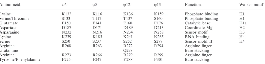
Table 2. Conserved residues and their function withinj6,j8,j12 andj13 P4 proteins
Amino acid j6 j8 j12 j13 Function Walker motif Lysine K132 K116 K136 K159 Phosphate binding H1
Serine/Threonine S133 T117 T137 S160 Phosphate binding H1 Glutamate E150 E141 E160 E176 Catalytic base H1a Aspartate D187 D171 D189 D213 Coordinate Mg H2 Asparagine N232 N216 N234 N258 Sensor motif H3 Lysine K239 K185 K241 K265 RNA binding H4 Serine S250 S237 S252 S277 Sensor motif II H4 Arginine R268 R263 R272 R294 Arginine finger
Glutamine Q278 Base stacking Arginine R273 R266 R279 R299 Arginine finger Tyrosine/Phenylalanine F275 F247 Y288 F301 Base stacking
at University of Leeds on March 24, 2015
http://nar.oxfordjournals.org/
Figure 4. Structure-based phylogenetic tree of ATPase enzymes. The matrix of evolutionary distances was calculated with SHP (56). The rectangle corresponds to a close-up view of the members of the RecA family. Abbreviations (In alphabetical order; Protein Data Bank accession codes are quoted in brackets): AfGspE, archaeal secretion ATPase, (2Oap); CFTR, Cystic Fibrosis Transmembrane Conductance Regulation, (1Xmi); Clamp Loader, eukaryotic clamp loader, (1Sxj); CobA, corrinoid adenosyltransferase, (1G64); CobU, adenosylcobinamide kinase/adenosylcobinamide phosphate guanylyltransferase, (1Cbu); DMC1, meiotic recombination protein, (2Zjb); DnaB,Thermus aquaticus DNAb, (2Q6t); Elp4, elongator complex protein 4, (4A8j); ESCN, prototypical T3ss ATPase EscN, (2Obl); F1-ATP Synthase-a, ATP synthase subunit-aheart isoform, (2Jj1); F1-ATPase-b, bovine mitochondrial F1-ATPase, (1E1r); FbpC, Fe(3+) ions import ATP-binding protein FbpC, (3Fvq); FtsK, DNA translocase FtsK, (2Iut); G40P, ATPase domain of G40P, (3Bh0); Get3, ATPase Get3, (3Sja); GkDnaC,Geobacillus kaustophilus DnaC, (2Vyf); GsDnaB,
Geobacillus stearothermophilusDnaB, (2R6c); IoID,Aquifex AeolicusABC transporter, (2Pcj); KaiC, Circadian clock protein kinase KaiC, (3K0e); MalK, maltose/maltodextrin import ATP-binding protein, (2Awn); MipZ, bacterial cell division regulator protein MipZ, (2Xit); MMAA, methylmalonic aciduria type A protein, (2Www); Msb8, Thermotoga maritima Abc transporter ATPp-binding protein, (1Vpl); MutS, DNA mismatch repair protein MutS, (1Ewq); P-gp, multidrug resistance protein Pgp-1, (4F4c); PH0284, Upf0273 Protein Ph0284, (2Dr3); PilT, twitching motility protein PilT, (2Gsz); Psy3, Platinum sensitivity protein 3, (4Dt1); Rad50, Dna Double-Strand Break Repair Rad50 Atpase, (3Qku); Rad51, DNA repair protein Rad51, (1Szp); RadA, DNA repair and recombination protein RadA, (4Dc9); RecA, Recombinase A, (1Mo4); RepA, regulatory protein RepA, (1G8y); Rho, transcription termination factor Rho, (3Ice); Rli1p, translation initiation factor, (3J16); RNT1, regulator of nonsense transcripts 1, (2Wjy); SMC, chromosome partition protein, (4I99); Sso2452, putative uncharacterized protein, (2W0m); T7Gp4, T7 DNA Primase/ Helicase, (1Cr1); TK, thymidine kinase, (2Ja1); TrwB, conjugal transfer protein TrwB, (1E9r); V1-ATPase, V-Type sodium ATPase, (3VR4); VirB4, type IV secretory pathway Virb4 components-like protein, (4Ag6); Vps4, vacuolar protein sorting-associated protein 4, (3Eih); XDP, Xpd/Rad3 related DNA helicase, (3Crv).
at University of Leeds on March 24, 2015
http://nar.oxfordjournals.org/
disordered) to re-emerge into as C-terminal helix at the top of the hexamer (Figure 2B), followed by a loop that dives into the central channel restricting its diameter by more than half (see later in the text for more discussion on the C-terminal domain).
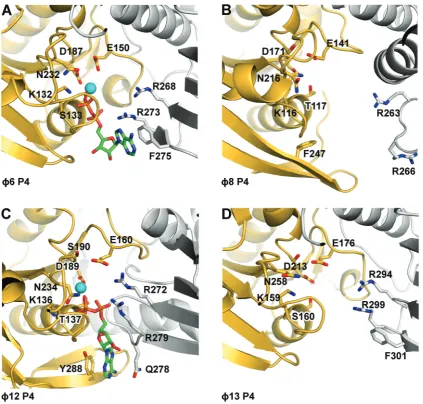
Nucleotide binding site
Thef6 P4 was crystallised with ADP-Mg2+bound in the nucleotide binding site, whereas P4 fromf8 andf13 were crystallized in their apo form. As for f12 P4, and other hexameric NTPases, the nucleotide binding sites inf6 P4 are located at the interfaces between neighbouring subunits. The ADP phosphate groups are bound via the conserved Walker A (H1) motif residues (K132, S133) (Figure 5); a conserved glutamate E150 (H1a) is pos-itioned to catalyse the nucleophilic attack on the
g-phosphate, whereas D187, a conserved aspartate in the
Walker B motif (H2), co-ordinates the magnesium ion. A sensor motif detecting the presence or absence of the
g-phosphate of NTP and modulating allosteric transitions of the RNA binding loop L2 in response to ATP binding and hydrolysis was identified in P4 fromf12 (N234) (31). The equivalent residue in f6 P4, N232, is positioned to contact theg-phosphate of the NTP (Figure 5) and might fulfil the same role. As the mechanism of NTP binding and hydrolysis is similar, it is likely that the equivalent conserved residues in P4 fromf8 andf13 (Figure 5 and Table 2) play analogous roles.
[image:9.612.89.513.66.470.2]It has been shown that f12 P4 possesses two essential ‘arginine fingers’ (35). We find that all P4 proteins follow this unusual pattern (Figure 5 and Table 2). Arginine fingers can contact the g-phosphate of the tri-phosphate from a neighbouring subunit, and the inser-tion of this residue in a catalytic site is believed to stabilize the transition state, thus facilitating ATP
Figure 5. Cartoon representation of the nucleotide binding sites off6 (A),f8 (B),f12 (C) andf13 (D) P4s. Within hexamers, adjacent monomers are coloured in yellow and grey. Nucleotides (ADP), if present, are depicted as sticks with carbon atoms coloured in green. Oxygen, nitrogen and phosphorus atoms are coloured in red, blue and orange, respectively, and the position of Mg2+(f12 P4) or Ca2+(f6 P4) is indicated with a cyan sphere.
at University of Leeds on March 24, 2015
http://nar.oxfordjournals.org/
hydrolysis. Arginine fingers in P4 proteins are all contributed from the same region (a loop between two strands in the C-terminal region) but display different conformations (Figure 5). In P4 from f6, f12 and
f13, the arginine fingers are pointing towards the cata-lytic sites, making the subunits competent and primed for hydrolysis. However, in f8 P4, these residues are displaced >8 A˚ from that position and therefore cannot contribute to catalysis. This suggests that in f8 P4, extensive conformational changes occur as a conse-quence of nucleotide and/or oligonucleotide binding, which render the enzyme competent for catalysis. Indeed, nucleotide binding kinetics revealed a first-order rate limiting step, which is consistent with a con-formational change associated with ATP binding (39,62). In RecA-like ATPases, bound nucleotides are stabilized by stacking of the adenine moiety between side chains, but these side chains are not conserved and are contributed from different regions. In RepA and T7 helicases, the ATP base stacks against residues belonging to the subunit carrying the catalytic site. In f12 P4 (31), as in RepA (63), the nucleotide base is sandwiched between Y288 from the catalytic subunit and Q278 from the neighbour-ing subunit. Inf6 P4, a much looser stacking of the nu-cleotide base is observed, with only one side chain (F275) stabilizing the adenine ring (Figure 5). From our struc-tures, we predict similar loose arrangements in P4 from
f8 andf13 where F247 (from the same subunit) and F301 (from a neighbouring subunit) seem to be in the correct orientation to stack the nucleotide base. The difference in the arrangement of the nucleotide binding motifs is likely to explain the mechanism of base-specific hydrolysis in different P4s. Of the P4s, only f12 is purine specific, with pyrimidines also being accepted by f16, f18 and
f13 (Table 1).
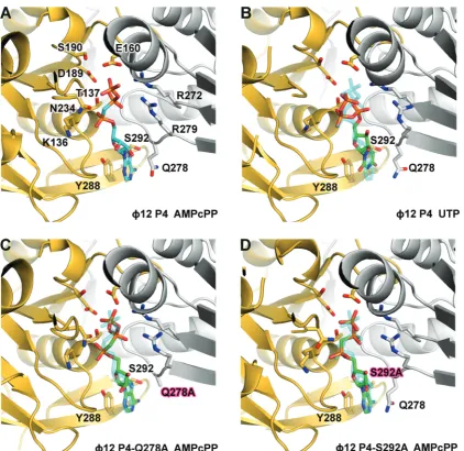
To understand this catalytic mechanism in detail, we performed side-directed mutagenesis of the residues in
f12 P4 involved in binding the nucleotide ring and analysed the mutants structurally and biochemically. In
f12 P4, the stacking interaction is critical for nucleotide binding, as replacement of the tyrosine with alanine (Y288A) completely abolished ATP binding and ATPase activity (Table 1) so that the apoprotein structure is found even in the presence of ATP (data not shown). However, the mutation Q278A had only a moderate effect on ATPase activity and virtually no effect on the structure of the bound ATP analogue AMPcPP when compared with the wild-type (Figure 6A and C), primarily increasing the KMas a result of reduced nucleotide affinity (Table 1). Hence, the stacking interactions primarily determine nu-cleotide affinity but not specificity. A specific feature in
f12 P4 is a hydrogen bond between the hydroxyl of S292 and N7 of the purine ring. The substitution S292A did not prevent ATP binding but completely abolished ATPase activity owing to misplacement of the triphos-phate moiety in the active site (Figure 6D). A displace-ment is also seen when the AMPcPP bound wild-type structure is compared with that of UTP bound hexamer (Figure 6A and B). This confirms that pyrimidine triphos-phates can bind the hexamer without being hydrolysed (36) and should act as competitive inhibitors. Indeed, we
find that UTP effectively competes with ATP and inhibits hydrolysis (data not shown). Hence, purine specificity is achieved by locking the base by hydrogen bonding to the N7 site of a purine. The correct coordination of the base results in the precise alignment of the nucleotide that is essential for catalysis so that UTP is misaligned and not hydrolysed. This is probably the mechanism underpinning the dependence of helicase efficiency on the type of nu-cleotide. For example, T7 gp4 helicase activity is optimal in presence of dTTP (58).
Nucleic acid binding site
It has been proposed that P4 hexamers bind nucleic acid through their central channel via two protruding loops named L1 and L2 (31) (Figure 3A andB, Supplementary Figure S3). Mutagenesis studies confirmed that these loops are essential for nucleic acid binding and transloca-tion (30,35,37). Structurally homologous loops were reported to bind ssDNA and ssRNA, respectively, in crystals of the E1 helicase of bovine papilloma virus and Rho ofE. coli(59). The L1 loops in P4 are rich in residues that contribute to flexibility (in f12 P4 they are dis-ordered), whereas the L2 loops are mainly composed of hydrophilic residues, amongst them a lysine, which inf12 P4 (K241) was shown to be essential for RNA binding (35). The structures of P4 from f6 and f13 show ordered L1 loops, which line the central channel and contact the L2 loops (Supplementary Figure S2). The L2 loops are found with lysine residues (K239 and K265, re-spectively) projecting towards the centre of the channel, in the same position as K241 inf12, suggesting a conserved mechanism for binding and translocating RNA. Although the L2 loop of f8 P4 contains hydrophilic residues (DDENVD), it does not project a lysine side chain towards the central channel. Nevertheless, the L1 loop contains a motif (LKK) that has been shown to be crucial for RNA binding (35). The first lysine of this motif (K185) is found in the equivalent position to K241 of f12 P4 and is also seen interacting with D220 of loop L2. We therefore postulate that K185 (loop L1) inf8 P4 plays the same role in RNA binding as K241 (loop L2) in
f12 P4, and that the coupling of the movement of the L1 and L2 loops to ATP hydrolysis via motion of helix 6, as proposed forf12, may be a general feature of all P4 mol-ecules (Supplementary Figure S2). The importance of the L1 loop is further supported by mutational analysis inf12 P4: deleting L1 loop central residues T202-T203-S204 or mutating them into the equivalent residues of f8 P4 (LKK) completely abolishes the ATPase activity (Table 1). This demonstrates that the integrity of the L1 loop is essential for ATP hydrolysis, despite being distal to the ATP active site.
RNA loading inr8 P4 and the structural basis of
processive translocation
The f8 P4 ATPase activity is tightly coupled to ssRNA translocation, as it will only hydrolyse ATP in the presence of ssRNA. As noted earlier in the text, the RNA binding motif LKK in loop L1 is located in the middle of the central channel (37). Nucleic acids are likely to bind in
at University of Leeds on March 24, 2015
http://nar.oxfordjournals.org/
the channel, ensuring topological enclosure of the strand and processive translocation.
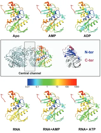
Based on transient cooperative exposure of subunit interfaces to HDX on RNA binding (residues 198–209 in Figure 7), it was suggested that RNA enters the central channel via a transient ring opening (37). The deletion of the C-terminal portion of the protein (residues 282–321) more than doubles the diameter of the central channel (from 13 to 30 A˚), as the C-terminus wraps upwards from the base of the hexamer, along the inter-subunit cleft, to stick down into the central channel (Figure 8). As the C-terminal domain is (i) necessary for ATP hydrolysis (data not shown), (ii) restricts the diameter of the central channel and (iii) blocks the inter-face through which RNA is thought to be loaded, we pos-tulate that the C-terminal region needs to be displaced by
RNA for ring opening and subsequent ATP hydrolysis to occur. To verify this hypothesis, previous HDX experi-ments (37) were further analysed by mapped to the f8 P4 structure.
The C-terminal region exhibits the fastest HDX within the protein (Figure 7). However, the distal C-terminal portion that extends into the central channel is marginally protected in the absence of RNA and becomes fully exposed only on addition of RNA, implying that this region becomes further exposed presumably by expulsion from the central channel (Figure 8B). Thus, it appears that
[image:11.612.90.513.64.474.2]f8 P4 has developed a specific mechanism to regulate ATPase activity and couple it with ssRNA binding such that RNA displaces the C-terminal domain, to allow ATP hydrolysis to occur. This would explain the tight coupling observed between ATP hydrolysis and translocation.
Figure 6. Cartoon representation of the nucleotide binding site off12 P4. (A) Wild-typef12 P4 bound to non-hydrolysable ATP analogue AMPcPP (PDB: 1W48) or (B) to UTP. (C) Q278A mutant bound to AMPcPP. (D) S292A mutant bound to AMPcPP. Within hexamers, adjacent monomers are coloured in yellow and grey. AMPcPP bound to wild-typef12 P4 is depicted in sticks, and the carbon atoms are coloured cyan (A), whereas carbon atoms in the UTP bound tof12 P4 (B) and AMPcPP bound to the P4 mutants Q278A and S292A (C andD) are coloured in green. Oxygen, nitrogen and phosphorus atoms are coloured in red, blue and orange, respectively. (B–D) The position of the AMPcPP bound to wild-type P4 is represented in transparent for comparison.
at University of Leeds on March 24, 2015
http://nar.oxfordjournals.org/
CONCLUSION
The current study broadens our understanding of the mechanism used by dsRNA bacterial viruses to package RNA genome during assembly. Interestingly, P4 proteins are only remotely related to packaging ATPases of dsDNA viruses such as gp17 from bacteriophage T4 (64) or pUL15 from Herpex Simplex virus 1 (65), which have more complicated portal complexes. Recently, however, it has been suggested that the ATPase of the phi29 DNA packaging motor is a member of the hexameric AAA+ superfamily (66), indicating that the mechanism of nucleic acid packaging might be similar.
A structure-based phylogeny (Figure 4) suggests that the RecA-like proteins may be the closest cellular relatives
of the P4, withf12 being the most similar to the cellular proteins,f8 being rather divergent andf6 andf13 rather similar to each other and intermediate in terms of diver-gence from the cellular proteins. These structural vari-ations map onto the various functional specializvari-ations of the molecules so that although the motors have a common catalytic mechanism, they have developed somewhat dif-ferent specificity and control mechanisms. We identify a specific hydrogen bond (serine 292 and N7 of the purine ring) responsible for the purine specificity of f12 P4 catalysed NTP hydrolysis reaction and find that an extra-ordinary insertion of the C-terminal peptide into the central channel of the hexamer explains the tight coupling of ATPase activity and RNA translocation in
[image:12.612.144.483.65.511.2]f8. Furthermore, the f8 P4 structure revealed a novel
Figure 7. Mapping of HDX data on thef8 P4 structure. HDX rates are coloured from slow-exchange (blue) to fast-exchange rates (red). Previously measured HDX rates (53) forf8 P4 in the presence/absence of AMP, ADP, ATP and RNA (as indicated) were mapped onto thef8 P4 monomer structure. The central box shows on the left, the orientation of all the monomers of the figure within the hexamer, and on the right, the same monomer in which the N- and C-terminal domains are coloured in blue and red, respectively.
at University of Leeds on March 24, 2015
http://nar.oxfordjournals.org/
mechanism of power transduction to the RNA in which RNA is engaged with the L1 loop, which, in turn, is coupled to the L2 loop. Comparison between the P4 struc-tures suggest that coupling between the two loops may be a general mechanistic feature of P4 and perhaps other SF4 helicases. Overall, the P4 machine represents a remarkable test bed where, by virtue of high mutational rates over long periods of time, nature has been able to devise a range of functional variations on the basic theme of regulated RNA translocation, resulting in an array of systems where although the molecular engine remains largely similar, the ignition and transmission systems have diverged markedly.
ACCESSION NUMBERS
Coordinates and structure factors of ADP-bound f6 P4310, f8 P4, f8 P4281, UTP-bound f12 P4, AMPcPP-bound f12 P4-Q278A and AMPcPP-bound
f12 P4-S292A and f13 P4 have been deposited in the Protein Data Bank under accession codes 4BLO, 4BWY, 4BLQ, 4BLR, 4BLS, 4BLT and 4BLP, respectively.
SUPPLEMENTARY DATA
Supplementary Data are available at NAR Online.
ACKNOWLEDGEMENTS
The authors thank Maria Harkiolaki and Martin Walsh for assistance with data collection. They thank the staff of the European Synchrotron Radiation Facility (ESRF, France) and the Diamond Light Source (DLS, UK) for technical support. They are indebted to Jiri Lisal for help
with the characterization of the enzymatic activity of the
f12 P4 mutants.
FUNDING
UK Medical Research Council (MRC); Academy of Finland [255342 and 256518 to D.H.B. as well as 250113 and 256069 to M.M.P.]; Sigrid Juselius Foundation (to DH.B. and M.M.P.); European Union Structural Funds programme [VP1-3.1 -SˇMM-07-K-03-069 to D.K.]; Academy of Finland Centre of Excellence in Virus Research 2006–2011 (to R.T.); The Wellcome Trust [075491/Z/04]; a Royal Society University Research Fellow (to E.J.M.); a Junior Research Fellow at Oriel College, Oxford (to K.E.O.). Funding for open access charge: Medical Research Council, UK.
Conflict of interest statement. None declared.
REFERENCES
1. Catalano,C.E. (2000) The terminase enzyme from bacteriophage lambda: a DNA-packaging machine.Cell. Mol. Life Sci.,57, 128–148.
2. Moore,S.D. and Prevelige,P.E. Jr (2002) DNA packaging: a new class of molecular motors.Curr. Biol., 12, R96–R98.
3. Simpson,A.A., Tao,Y., Leiman,P.G., Badasso,M.O., He,Y., Jardine,P.J., Olson,N.H., Morais,M.C., Grimes,S., Anderson,D.L.
et al. (2000) Structure of the bacteriophage phi29 DNA packaging motor.Nature,408, 745–750.
4. Feiss,M. and Rao,V.B. (2012) The bacteriophage DNA packaging machine.Adv. Exp. Med. Biol.,726, 489–509.
5. Smith,D.E., Tans,S.J., Smith,S.B., Grimes,S., Anderson,D.L. and Bustamante,C. (2001) The bacteriophage straight phi29 portal motor can package DNA against a large internal force.Nature,
413, 748–752.
6. Poranen,M.M. and Bamford,D.H. (2012) FamilyCystoviridae. In: King,A.M.Q., Carstens,E., Adams,M. and Lefkowitz,E.J. (eds),Virus Taxonomy, Ninth Report of the International Committee on Taxonomy of Viruses. Elsevier, London, UK, pp. 515–518.
7. Juuti,J.T., Bamford,D.H., Tuma,R. and Thomas,G.J. Jr (1998) Structure and NTPase activity of the RNA-translocating protein (P4) of bacteriophage phi 6.J. Mol. Biol., 279, 347–359. 8. Kainov,D.E., Pirttimaa,M., Tuma,R., Butcher,S.J.,
Thomas,G.J. Jr, Bamford,D.H. and Makeyev,E.V. (2003) RNA packaging device of double-stranded RNA bacteriophages, possibly as simple as hexamer of P4 protein.J. Biol. Chem.,278, 48084–48091.
9. Chen,H., Meisburger,S.P., Pabit,S.A., Sutton,J.L., Webb,W.W. and Pollack,L. (2012) Ionic strength-dependent persistence lengths of single-stranded RNA and DNA.Proc. Natl Acad. Sci. USA,
109, 799–804.
10. Jaalinoja,H.T., Huiskonen,J.T. and Butcher,S.J. (2007) Electron cryomicroscopy comparison of the architectures of the enveloped bacteriophages phi6 and phi8.Structure,15, 157–167.
11. Pirttimaa,M.J. and Bamford,D.H. (2000) RNA secondary structures of the bacteriophage phi6 packaging regions.RNA,6, 880–889. 12. Thomas,G.J. Jr, Prescott,B., McDonald-Ordzie,P.E. and
Hartman,K.A. (1976) Studies of virus structure by laser-Raman spectroscopy. II. MS2 phage, MS2 capsids and MS2 RNA in aqueous solutions.J. Mol. Biol.,102, 103–124.
13. Mindich,L. (1999) Precise packaging of the three genomic segments of the double-stranded-RNA bacteriophage phi6.
Microbiol. Mol. Biol. Rev.,63, 149–160.
14. Bamford,D.H., Burnett,R.M. and Stuart,D.I. (2002) Evolution of viral structure.Theor. Popul. Biol.,61, 461–470.
[image:13.612.42.285.65.300.2]15. Butcher,S.J., Dokland,T., Ojala,P.M., Bamford,D.H. and Fuller,S.D. (1997) Intermediates in the assembly pathway of the double-stranded RNA virus phi6.EMBO J.,16, 4477–4487.
Figure 8. The C-terminal domain off8 P4. (A) Surface presentation of thef8 P4281 (left) and the full-length protein (right). The C-terminal domain is coloured in red. (B) A model for ssRNA induced displace-ment of the C-terminal domain inf8 P4 hexamer.
at University of Leeds on March 24, 2015
http://nar.oxfordjournals.org/
16. de Haas,F., Paatero,A.O., Mindich,L., Bamford,D.H. and Fuller,S.D. (1999) A symmetry mismatch at the site of RNA packaging in the polymerase complex of dsRNA bacteriophage phi6.J. Mol. Biol.,294, 357–372.
17. Emori,Y., Iba,H. and Okada,Y. (1980) Assignment of viral proteins to the three double-stranded RNA segments of bacteriophage phi 6 genome: translation of phi 6 messenger RNAs transcribedin vitro.Mol. Gen. Genet.,180, 385–389. 18. Emori,Y., Iba,H. and Okada,Y. (1982) Morphogenetic pathway
of bacteriophage phi 6. A flow analysis of subviral and viral particles in infected cells.J. Mol. Biol.,154, 287–310.
19. Frilander,M. and Bamford,D.H. (1995) In vitro packaging of the single-stranded RNA genomic precursors of the segmented double-stranded RNA bacteriophage phi 6: the three segments modulate each other’s packaging efficiency.J. Mol. Biol.,246, 418–428.
20. Gottlieb,P., Strassman,J., Qiao,X., Frilander,M., Frucht,A. and Mindich,L. (1992)In vitro packaging and replication of individual genomic segments of bacteriophage phi 6 RNA.J. Virol.,66, 2611–2616.
21. Pirttimaa,M.J., Paatero,A.O., Frilander,M.J. and Bamford,D.H. (2002) Nonspecific nucleoside triphosphatase P4 of double-stranded RNA bacteriophage phi6 is required for single-double-stranded RNA packaging and transcription.J. Virol.,76, 10122–10127. 22. Qiao,X., Casini,G., Qiao,J. and Mindich,L. (1995)In vitro
packaging of individual genomic segments of bacteriophage phi 6 RNA: serial dependence relationships.J. Virol.,69, 2926–2931. 23. Semancik,J.S., Vidaver,A.K. and Van Etten,J.L. (1973)
Characterization of segmented double-helical RNA from bacteriophage phi6.J. Mol. Biol.,78, 617–625.
24. Sun,X., Bamford,D.H. and Poranen,M.M. (2012) Probing, by self-assembly, the number of potential binding sites for minor protein subunits in the procapsid of double-stranded RNA bacteriophage Phi6.J. Virol.,86, 12208–12216.
25. Poranen,M.M., Paatero,A.O., Tuma,R. and Bamford,D.H. (2001) Self-assembly of a viral molecular machine from purified protein and RNA constituents.Mol. Cell,7, 845–854.
26. Kainov,D.E., Lisal,J., Bamford,D.H. and Tuma,R. (2004) Packaging motor from double-stranded RNA bacteriophage phi12 acts as an obligatory passive conduit during transcription.Nucleic Acids Res.,32, 3515–3521.
27. Huiskonen,J.T., Jaalinoja,H.T., Briggs,J.A., Fuller,S.D. and Butcher,S.J. (2007) Structure of a hexameric RNA packaging motor in a viral polymerase complex.J. Struct. Biol.,158, 156–164.
28. Qiao,J., Qiao,X. and Mindich,L. (2005)In vivostudies of genomic packaging in the dsRNA bacteriophage Phi8.BMC Microbiol.,5, 10.
29. Qiao,X., Qiao,J. and Mindich,L. (2003) Analysis of specific binding involved in genomic packaging of the double-stranded-RNA bacteriophage phi6.J. Bacteriol.,185, 6409–6414. 30. Kainov,D.E., Tuma,R. and Mancini,E.J. (2006) Hexameric
molecular motors: P4 packaging ATPase unravels the mechanism.
Cell. Mol. Life Sci.,63, 1095–1105.
31. Mancini,E.J., Kainov,D.E., Grimes,J.M., Tuma,R., Bamford,D.H. and Stuart,D.I. (2004) Atomic snapshots of an RNA packaging motor reveal conformational changes linking ATP hydrolysis to RNA translocation.Cell,118, 743–755.
32. Mancini,E.J., Kainov,D.E., Wei,H., Gottlieb,P., Tuma,R., Bamford,D.H., Stuart,D.I. and Grimes,J.M. (2004) Production, crystallization and preliminary X-ray crystallographic studies of the bacteriophage phi 12 packaging motor.Acta Crystallogr. D Biol. Crystallogr.,60, 588–590.
33. Hall,M.C. and Matson,S.W. (1999) Helicase motifs: the engine that powers DNA unwinding.Mol. Microbiol.,34, 867–877. 34. Ilyina,T.V., Gorbalenya,A.E. and Koonin,E.V. (1992)
Organization and evolution of bacterial and bacteriophage primase-helicase systems.J. Mol. Evol.,34, 351–357. 35. Kainov,D.E., Mancini,E.J., Telenius,J., Lisal,J., Grimes,J.M.,
Bamford,D.H., Stuart,D.I. and Tuma,R. (2008) Structural basis of mechanochemical coupling in a hexameric molecular motor.
J. Biol. Chem.,283, 3607–3617.
36. Lisal,J. and Tuma,R. (2005) Cooperative mechanism of RNA packaging motor.J. Biol. Chem.,280, 23157–23164.
37. Lisal,J., Kainov,D.E., Lam,T.T., Emmett,M.R., Wei,H., Gottlieb,P., Marshall,A.G. and Tuma,R. (2006) Interaction of packaging motor with the polymerase complex of dsRNA bacteriophage.Virology,351, 73–79.
38. Lisal,J., Lam,T.T., Kainov,D.E., Emmett,M.R., Marshall,A.G. and Tuma,R. (2005) Functional visualization of viral molecular motor by hydrogen-deuterium exchange reveals transient states.
Nat. Struct. Mol. Biol.,12, 460–466.
39. Lisal,J., Kainov,D.E., Bamford,D.H., Thomas,G.J. Jr and Tuma,R. (2004) Enzymatic mechanism of RNA translocation in
double-stranded RNA bacteriophages.J. Biol. Chem.,279, 1343–1350.
40. Paatero,A.O., Syvaoja,J.E. and Bamford,D.H. (1995) Double-stranded RNA bacteriophage phi 6 protein P4 is an unspecific nucleoside triphosphatase activated by calcium ions.J. Virol.,69, 6729–6734.
41. Kainov,D.E., Butcher,S.J., Bamford,D.H. and Tuma,R. (2003) Conserved intermediates on the assembly pathway of double-stranded RNA bacteriophages.J. Mol. Biol.,328, 791–804. 42. Mancini,E.J., Grimes,J.M., Malby,R., Sutton,G.C., Kainov,D.E.,
Juuti,J.T., Makeyev,E.V., Tuma,R., Bamford,D.H. and Stuart,D.I. (2003) Order and disorder in crystals of hexameric NTPases from dsRNA bacteriophages.Acta Crystallogr. D Biol. Crystallogr.,59, 2337–2341.
43. Ojala,P.M., Juuti,J.T. and Bamford,D.H. (1993) Protein P4 of double-stranded RNA bacteriophage phi 6 is accessible on the nucleocapsid surface: epitope mapping and orientation of the protein.J. Virol.,67, 2879–2886.
44. Astier,Y., Kainov,D.E., Bayley,H., Tuma,R. and Howorka,S. (2007) Stochastic detection of motor protein-RNA complexes by single-channel current recording.Chemphyschem,8, 2189–2194. 45. Otwinowski,Z. and Minor,W. (1996) Processing of X-ray
diffraction data collected in oscillation mode.Methods Enzymol.,
276, 307–326.
46. McCoy,A.J., Grosse-Kunstleve,R.W., Adams,P.D., Winn,M.D., Storoni,L.C. and Read,R.J. (2007) Phaser crystallographic software.J. Appl. Crystallogr.,40, 658–674.
47. Meier,C., Mancini,E.J., Bamford,D.H., Walsh,M.A., Stuart,D.I. and Grimes,J.M. (2005) Overcoming the false-minima problem in direct methods: structure determination of the packaging enzyme P4 from bacteriophage phi13.Acta Crystallogr. D Biol.
Crystallogr.,61, 1238–1244.
48. Sheldrick,G.M. (2010) Experimental phasing with SHELXC/D/E: combining chain tracing with density modification.Acta Crystallogr. D Biol. Crystallogr.,66, 479–485.
49. Vonrhein,C., Blanc,E., Roversi,P. and Bricogne,G. (2007) Automated structure solution with autoSHARP.Methods Mol. Biol.,364, 215–230.
50. Trapani,S. and Navaza,J. (2008) AMoRe: classical and modern.
Acta Crystallogr. D Biol. Crystallogr.,64, 11–16.
51. Adams,P.D., Grosse-Kunstleve,R.W., Hung,L.W., Ioerger,T.R., McCoy,A.J., Moriarty,N.W., Read,R.J., Sacchettini,J.C., Sauter,N.K. and Terwilliger,T.C. (2002) PHENIX: building new software for automated crystallographic structure determination.
Acta Crystallogr. D Biol. Crystallogr.,58, 1948–1954.
52. Emsley,P. and Cowtan,K. (2004) Coot: model-building tools for molecular graphics.Acta Crystallogr. D Biol. Crystallogr.,60, 2126–2132.
53. Bricogne,G., Blanc,E., Brandl,M., Flensburg,C., Keller,P., Paciorek,W., Roversi,P., Smart,O.S., Vonrhein,C. and Womack,T. (2008). BUSTER-TNT 2.5.1 and AUTOBUSTER 1.3.1, Global Phasing Ltd, Cambridge.
54. Murshudov,G.N., Skubak,P., Lebedev,A.A., Pannu,N.S., Steiner,R.A., Nicholls,R.A., Winn,M.D., Long,F. and Vagin,A.A. (2011) REFMAC5 for the refinement of macromolecular crystal structures.Acta Crystallogr. D Biol. Crystallogr.,67, 355–367.
55. Davis,I.W., Leaver-Fay,A., Chen,V.B., Block,J.N., Kapral,G.J., Wang,X., Murray,L.W., Arendall,W.B. 3rd, Snoeyink,J., Richardson,J.S.et al. (2007) MolProbity: all-atom contacts and structure validation for proteins and nucleic acids.Nucleic Acids Res.,35, W375–W383.
56. Holm,L. and Rosenstrom,P. (2010) Dali server: conservation mapping in 3D.Nucleic Acids Res.,38, W545–W549.
at University of Leeds on March 24, 2015
http://nar.oxfordjournals.org/
57. Stuart,D.I., Levine,M., Muirhead,H. and Stammers,D.K. (1979) Crystal structure of cat muscle pyruvate kinase at a resolution of 2.6 A.J. Mol. Biol.,134, 109–142.
58. Levin,M.K. and Patel,S.S. (2004)Molecular Motors. Wiley-VCH Verlag GmbH & Co, KGaA, Weinheim, FRG, pp. 179–203.
59. Skordalakes,E. and Berger,J.M. (2006) Structural insights into RNA-dependent ring closure and ATPase activation by the Rho termination factor.Cell,127, 553–564.
60. Yang,Z.R., Thomson,R., McNeil,P. and Esnouf,R.M. (2005) RONN: the bio-basis function neural network technique applied to the detection of natively disordered regions in proteins.
Bioinformatics,21, 3369–3376.
61. Paatero,A.O., Mindich,L. and Bamford,D.H. (1998) Mutational analysis of the role of nucleoside triphosphatase P4 in the assembly of the RNA polymerase complex of bacteriophage phi6.
J. Virol.,72, 10058–10065.
62. Luo,D., Xu,T., Watson,R.P., Scherer-Becker,D., Sampath,A., Jahnke,W., Yeong,S.S., Wang,C.H., Lim,S.P., Strongin,A.et al.
(2008) Insights into RNA unwinding and ATP hydrolysis by the flavivirus NS3 protein.EMBO J.,27, 3209–3219.
63. Ziegelin,G., Niedenzu,T., Lurz,R., Saenger,W. and Lanka,E. (2003) Hexameric RSF1010 helicase RepA: the structural and functional importance of single amino acid residues.Nucleic Acids Res.,31, 5917–5929.
64. Sun,S., Kondabagil,K., Draper,B., Alam,T.I., Bowman,V.D., Zhang,Z., Hegde,S., Fokine,A., Rossmann,M.G. and Rao,V.B. (2008) The structure of the phage T4 DNA packaging motor suggests a mechanism dependent on electrostatic forces.Cell,135, 1251–1262.
65. Selvarajan Sigamani,S., Zhao,H., Kamau,Y.N., Baines,J.D. and Tang,L. (2013) The structure of the herpes simplex virus DNA-packaging terminase pUL15 nuclease domain suggests an evolutionary lineage among eukaryotic and prokaryotic viruses.
J. Virol.,87, 7140–7148.
66. Schwartz,C., De Donatis,G.M., Fang,H. and Guo,P. (2013) The ATPase of the phi29 DNA packaging motor is a member of the hexameric AAA+ superfamily.Virology,443, 20–27.
at University of Leeds on March 24, 2015
http://nar.oxfordjournals.org/