1
A DESCRIPTIVE STUDY OF MORPHOEA WITH
CLINICO-PATHOLOGICAL CORRELATION
Dissertation Submitted in partial fulfillment of the university regulations for
MD DEGREE IN
DERMATOLOGY, VENEREOLOGY AND LEPROSY
(BRANCH XII A)
APRIL 2013
THE TAMILNADU DR. M.G.R. MEDICAL UNIVERSITY
2
CERTIFICATE
This is to certify that this dissertation entitled ‘A DESCRIPTIVE
STUDY OF MORPHOEA WITH CLINICO-PATHOLOGICAL
CORRELATION’ submitted by Dr. ANUJ SAIGAL to The Tamil Nadu
Dr. M.G.R. Medical University, Chennai is in partial fulfillment of the requirement for the award of M.D. [DERMATO VENEREO LEPROLOGY] and is a bonafide research work carried out by him under direct supervision and guidance. This work has not previously formed the basis for the award of any degree or diploma.
Dr. A.S. Krishnaram. M.D., D.D.,
Professor and Head
Department of Dermatology, Madurai Medical College & Government Rajaji Hospital Madurai.
Dr. D. Amal Raja. M.D., D.V.,
Professor and Head
Department of STD,
3
DECLARATION
I, Dr. ANUJ SAIGAL solemnly declare that the dissertation titled „A
DESCRIPTIVE STUDY OF MORPHOEA WITH
CLINICO-PATHOLOGICAL CORRELATION’ is a bonafide work done by me at
Government Rajaji Hospital during 2010 – 2012 under the guidance and supervision of Prof. Dr. A.S. Krishnaram M.D., D.D., Professor and Head of the Department of Dermatology, Madurai Medical College, Madurai.
I also declare that this bonafide work or a part of this work was not submitted by me or any other for any award, degree and diploma to any university, board either in India or abroad.
The dissertation is submitted to The Tamilnadu Dr. M.G.R. Medical University, towards partial fulfillment of requirement for the award of M.D. Degree in Dermatology, Venereology and Leprology (BRANCH –XII A).
Place: Madurai.
4
ACKNOWLEDGEMENT
At the outset, I express my profound gratitude to the DEAN Prof. Dr. N. MOHAN, M.S, F.I.C.S, F.A.I.S, F.M.M.C, Madurai Medical College, Madurai for permitting me to use the college and Department facilities for my study.
I would like to acknowledge my thanks and sincere gratitude to Prof.
Dr. A.S. Krishnaram M.D., D.D., Professor and Head of the Department of
Dermatology, Madurai Medical College, Madurai, for his invaluable guidance, motivation and help throughout the study.
I would like to express my sincere and heartfelt thanks to my former Professor Dr. S. KRISHNAN M.D., D.D., for his guidance and encouragement.
I thank my beloved teacher Prof. Dr. G. Geetharani, M.D., DNB.,
Associate Professor, Department of Dermatology for her peerless guidance and endless patience in moulding my study.
5
I would like to acknowledge my thanks to Prof. Dr. D. Amal Raja, M.D., D.V., Head of the Department of Venereology, for his support and inspiration.
I would like to thank Dr. R. Kothandaraman, Dr. Sathesh, Dr. Balaji Adityan, Dr. Suganya, Assistant professors for their motivation
and guidance.
I would also like to acknowledge my thanks to Dr. M.S. Adityan, Dr.
M. Senthil Kumar, Dr. A. Ayyamperumal, Assistant professors of
Department of STD, for their valuable help and suggestions.
I owe thanks to my fellow postgraduate colleagues for their constant help and encouragement.
6
CONTENTS
S. NO PARTICULARS PAGE NO.
1 INTRODUCTION 1
2 REVIEW OF LITERATURE 2
3 AIM OF THE STUDY 44
4 MATERIALS AND METHODS 45
5 OBSERVATIONS AND RESULTS 47
6 DISCUSSION 66
7 SUMMARY 77
8 CONCLUSION 80 ANNEXURES
BIBLIOGRAPHY PHOTOGRAPHS PROFORMA MASTER CHART
KEY TO MASTER CHART
ANTI PLAGIARISM CERTIFICATE
7
INTRODUCTION
Morphoea is an uncommon benign fibrosing disease which shows involvement of dermis, subcutaneous fat and underlying tissues, more prevalent in females, and is a separate disorder from systemic sclerosis as there is no internal organ involvement.
Morphoea causes fibrosis of all mesoderm derived tissues and rarely, the central nervous system [CNS]. Linear, generalized, and pansclerotic types are associated with morbid features like contractures, facial disfigurement, distress, arthralgia and CNS involvement.
The pathogenesis of morphoea is still an enigma, but is proposed to be started by injury to the vessels that results in increased collagen synthesis and diminished collagen destruction. There are interesting serological associations with morphoea also. Various other systemic and cutaneous diseases have also been found to be associated with morphoea.
This disease often progresses for several years and then regresses even without treatment although various modalities of treatment have been tried in the past.
8
REVIEW OF LITERATURE
SYNONYMS
Localized scleroderma, Circumscribed scleroderma, Scleroderma en coup de sabre
HISTORICAL ASPECTS
The word morphoea was first used by Erasmus Wilson to describe this disease as he felt that the areas were vestiges of true leprosy1. Morphoea has been derived from „morpheus‟ who in Greek tradition is considered as god of dreams with the capability to acquire any human form and appear in dreams.
The term „sclerodermie‟ was coined in 1847 by the French physician, Gintrac2.
9
DEFINITION
Morphoea is a localized form of scleroderma with increased collagen deposition leading to sclerosis of the dermis and underlying tissues6. It is a disorder of unknown cause with many clinical types and lack of systemic involvement thereby differentiating it from systemic sclerosis7.
CLASSIFICATION OF MORPHOEA
Schachter8 in 1989 subdivided localized scleroderma into
1) Morphoea- Limited (Plaque or Guttate), Generalised and others (subcutaneous, nodular).
2) Linear scleroderma- Linear or en coup de sabre. Since this scheme proved to be inconclusive, another classification was proposed by Peterson et al9 in 1995 which divided morphoea into five main types
1) PLAQUE MORPHOEA
Morphoea en plaque Guttate morphoea
10
2) GENERALIZED MORPHOEA
3) BULLOUS MORPHOEA
4) LINEAR MORPHOEA
Linear morphoea
En coup de sabre (ECDS) Progressive hemifacial atrophy
5) DEEP MORPHOEA
Subcutaneous morphoea Eosinophilic fasciitis Morphoea profunda
Disabling pansclerotic morphoea of childhood
This classification is questioned now because of two main reasons. Firstly, it has many entities which are not universally considered under morphoea like LSA, Pasini and Pierini‟s atrophoderma and eosinophilic fasciitis. Secondly, it does not include those patients who have more than one morphological type of morphoea and can be considered under an entity, „mixed‟ variant.
Recently, a new classification for localized scleroderma was proposed at
11
morphoea, linear morphoea, pansclerotic morphoea, generalized morphoea
and mixed morphoea10.
Types Subtypes Details Circumscribed
morphoea
Superficial
Deep
Single or multiple, oval or round
indurated areas involving epidermis and dermis often with a surrounding liliac ring.
Single or multiple oval or round deeply indurated areas involving subcutaneous (SC) tissue (upto muscle in few cases);
may spare the skin above. Linear morphoea Trunk/limb subtype Head subtype
Indurated areas in a linear fashion
involving the dermis and SC tissue (may involve upto the level of muscle and bone)
En coup de sabre
Induration in a linear fashion of the face
and scalp (can include muscle, bone and CNS)
Parry Romberg syndrome (PRS)
Involvement of one side of the face with
overlying mobile skin. Generalized
morphoea
Individual indurated plaques, 4 or more in number, of size more than 3 cm each and involving two of seven sites
including head and neck, front of trunk, back of trunk, right upper limb, left upper limb, right lower limb, left lower
12 Pansclerotic
morphoea
Circumferential involvement of limbs upto the level of bones. Other body areas may also be affected.
Mixed variant morphoea
Variant with 2 or more of the above mentioned subtypes. Sequence is consistent with predominant representation in the patient like (pansclerotic-circumscribed).
EPIDEMIOLOGY OF MORPHOEA
Morphoea can affect all ages though it is most commonly seen in the
20- 40 years age group, but the disease commences before the age of 10 years
in 15 % of cases according to a study conducted by Christianson et al11 on 235 cases. The extremes of age were 1 year and 76 years in this study11.
The prevalence of the disease also increases with age12. It is undoubtedly more common in females with ratio approximately being 3:1 in
most studies11,12 with the exception of linear morphoea which has no gender preference . In a study done by Peterson et al12 in Olmsted county between 1960-1993 on 82 cases showed the incidence rate as 27 per million
population. According to the same study12, 56% of patients had plaque type morphoea, 20% linear, 13% generalized and 11% deep morphoea whereas
13
A worldwide study of 750 children with juvenile localized
scleroderma also reported more percentage of linear lesions than plaques13. Also, Christianson‟s study11 showed that linear lesions appeared before the age of 10 years in 20% and prior to the age of 40 in 75% but plaque lesions
appeared later in life, 10% were seen prior to the age of 10 years and 75%
becoming evident between the ages of 20 and 50. The results from these
studies show that linear morphoea is more commonly seen in children as
compared to adults.
Morphoea is more prevalent in Caucasians and Asians and it has been
reported to be rare in black people like African Americans14,15. The disease is active for a span of 3 to 6 years on an average15,16. Relapses are more
commonly seen in patients with generalized morphoea, linear morphoea and
14
AETIOLOGY
1) TRAUMA
Trauma can be considered as a triggering factor for morphoea
and may precede the onset of the disease by many months17. Two case reports suggesting that trauma causes an isomorphic response and
appearance of new typical lesions in known cases of morphoea have
been published18. The mechanism is still unknown but local production and dysregulation of inflammatory mediators including fibrogenic
cytokines such as transforming growth factor (TGF-β) have been
proposed to cause excessive synthesis of collagen19. Trauma has been reported as a precipitating factor in approximately 13% of the children examined under two large studies of almost 900 cases13, 20. The study conducted by Christianson et al11 reported 14 cases out of 191 with trauma as the preceding factor.
2) IMMOBILIZATION
Immobilization is another aetiological factor proposed for
morphoea. It has been reported that it causes dermal fibroblasts
activation, thereby releasing inflammatory cells and ultimately causes
15
3) VACCINATIONS AND INJECTIONS
Vaccinations such as B.C.G , D.P.T, MMR, tetanusand hepatitis B
have been reported to cause morphoea22,23,24. The underlying pathogenesis is still not well understood; it has been proposed that injection trauma causes endothelial injury and hypoxia of tissues which leads on to morphoea. Another hypothesis proposed is that vaccines induce an immune response against various antigens (specific as well as nonspecific). Finally, wound healing releases various cytokines and growth factors which might also predispose to trauma induced morphoea. Till date, 8 cases of vaccination induced morphoea have been reported25.
Other injections like vitamin K, vitamin B12 and progesterone have
also been reported as causative factors for morphoea22,26. The vehicle used, the preservative, and hypersensitivity have been implicated as the various reasons behind this26. There have been many case reports of morphoea developing after vitamin K injections (Texier disease) and it has been observed that it takes 1 month to almost 2 years for the morphoea like changes to occur27. High dosing is another factor which was seen in most of the cases. These sclerodermoid plaques were called as „cowboy‟s belt with revolver‟ in one recent case report28
16
4) RADIOTHERAPY
` Morphoea has been reported to occur following radiotherapy,
most commonly seen in women with breast carcinoma29. The incidence has been reported to be 1 in 500 cases29. The morphoea lesions usually develop around the irradiation port but some cases show involvement of areas beyond these sites or may extend to any other distal site29. The time duration after which the patient develops such lesions ranges from less than 1 year to 32 years after irradiation29. The pathogenesis proposed involves either activation of fibroblasts or self antigen presentation resulting in tissue damage25. Another case with post-fluoroscopy appearance of morphoea was also reported30.
5) SURGICAL OPERATIONS
Surgical operations have been known to precede morphoea.
Surgeries like rhinoplasty and AV fistula formation have been reported
to cause morphoea. Another case report stated the development of
morphoea post-laparotomy22.
6) HORMONAL FACTORS
There are case reports where pregnancy has been reported to
17
partum development of rheumatoid arthritis31. The mechanism proposed for this association deals with increase in the number of T
regulatory cells in pregnancy which causes increased synthesis of
TGF-β which is a known potent pro-fibrogenic mediator in
scleroderma. A higher incidence of morphoea has also been seen
around menarche. Parathormone has also been linked with morphoea
by causing calcium deposition in soft tissues14 and subtotal parathyroidectomy has been used as the treatment but no real proof has
been offered to support this. Leriche and co-workers32 observed 13 patients with generalized scleroderma for 2 to 5 years after a subtotal parathyroidectomy with 90% improvement.
7) INFECTIONS
Borrelial infection has also been proposed to play a role in
18
The probable reason for almost all the reports coming from Europe might be the difference in strains seen in Europe in comparision to United States of America (B.afzelii or B.garinii vs B.burgdorferi sensu stricto, respectively)25. Reports proposing Cytomegalovirus as another aetiologic agent have also been published35. Morphoea has been reported to follow infections like varicella, herpes zoster, toxoplasmosis and hepatitis C as well22,36,37.
8) GENETIC FACTORS
Genetic role for morphoea is still unclear but a familial incidence
has been noted and there have been reports of localized scleroderma
occurring in monozygotic twins38. There has been no significant HLA association reported with morphoea till date39. Organ specific autoantibodies have also shown a rise in some patients and relatives22.
9) ROLE OF DRUGS
There have been reports of morphoea occuring after intake of
various drugs such as D-penicillamine, bromocriptine, hydroxytryptophan,
carbidopa, valproic acid, pentazocine, docetaxel, paclitaxel, bleomycin
and after melphalan limb perfusion22. Other drugs like bisoprolol, peplomycin and balicatib have also been implicated in development of
19
month to 30 months40. Withdrawal of drugs may or may not result in clearance of lesions.
Kraigher et al41 documented blaschkoid unilateral generalized morphoea after intake of ibuprofen which showed relief after stopping the drug. The pathogenesis mainly revolves around production of autoantibodies by these drugs that affect the microvasculature and ultimately forming morphoea plaques25.
10) OTHER MISCELLANEOUS CAUSES
Other triggering factors include mechanical compression from clothing, pigmented purpuric dermatosis like lichen aureus42. Even exposure to epoxy resins, vinyl chloride, organic solvents and pesticides, silicon implants etc have been reported to produce scleroderma like changes.
PATHOGENESIS
20
a) VASCULAR INJURY
According to the vascular theory, morphoea starts with an injury to the vessels which may be caused by an infection or antibodies to endothelial cells43. This is further supported by the biopsy changes seen in the early morphoea lesions.
b) ADHESION MOLECULES EXPRESSION AND PRO FIBROTIC
CYTOKINES SYNTHESIS
The injury to the endothelium leads to cytokine release which causes increased intercellular adhesion molecule-1 (ICAM-1), vascular cell adhesion molecule-1 (VCAM-1) and E-selectin expression44. These molecules via T cells release other cytokines which cause fibrosis like IL- 4, IL-6 and TGF-β. This leads to accumulation of eosinophils, helper T cells, and macrophages. These cells are thought to present self antigens to the immune system of the body and thereby release autoantibodies.
Various chemokines such as CCL and CXCL8 have also been proposed to be involved in the pathogenesis of fibrosis45. Other cytokines like IL-2 receptor,
CD 23, CD30 and B-cell activating factor also play a role22. The stage of active inflammation and sclerosis has shown the presence of CD
21
c) OVERABUNDANCE OF COLLAGEN DEPOSITION
Upregulation of TGF-β leads to collagen, fibronectin, and proteoglycan deposition and it also diminishes collagen degradation by causing a decline in the levels of matrix metalloproteinases45,47. This variation in the amount of collagen deposition and collagen degradation is the proposed mechanism of the fibrosis seen in morphoea47. Antibodies to MMP‟s may also play a role in the inhibition of collagen degradation. The accumulation of myofibroblasts by GTPase Rac 1 also plays a role in the pathogenesis48. Insulin-like growth factor may also contribute to the development of morphoea by accumulating fibroblasts and increasing collagen and extracellular matrix deposition49.
Gene array analyses showed 4 separate gene signatures and were classified as inflammatory, limited, proliferative and normal-like. The inflammatory signature was seen in patients with morphoea. Other signatures were seen in systemic sclerosis50.
ROLE OF MICROCHIMERISM
22
ROLE OF AUTOANTIBODIES
The association of morphoea with autoimmune diseases ranges from 2% to 30% in various case reports published till now. Diseases such as Graves disease, vitiligo, type 1 diabetes mellitus, chronic GVHD, Idiopathic thrombocytopenic purpura and ulcerative colitis have been reported to coexist13,52. However, the prevalence of autoimmune disease observed in general public is lower than these values. The reported prevalence of positive ANA titers in morphoea patients ranges from 20% to 80%20,52,53,54. High values of antihistone antibodies, single stranded antibody, RA factor and homogenous ANA have also been reported in morphoea patients54.
However, childhood-onset morphoea shows more prevalence of antihistone antibodies53 whereas anti single stranded antibodies are seen in generalised and linear morphoea. High titres of ANA have been reported to occur in juvenile patients with linear morphoea and patients with generalised morphoea54.
23
antibodies, fibrillin 1 antibodies have also been found in localized scleroderma patients22. Cytotoxicity due to antibodies or complement activation may have a role in pathogenesis of morphoea25.
CLINICAL PRESENTATION
Morphoea starts with an active stage presenting as erythematous to violaceous patches or plaques. As the lesions progress, the center becomes white and sclerotic which is surrounded by a characteristic „liliac ring‟ forming the border. After the active stage, sclerotic plaques are seen. Some cases show plaques with postinflammatory hyperpigmentation. The excessive collagen production damages hair follicles and adnexal structures thereby forming hairless plaques which are anhidrotic25. Raynaud‟s phenomenon is not seen in most cases just like sclerodactyly which are features of systemic sclerosis. Very few cases develop systemic sclerosis later in life7. Another characterstic feature is that face and the acral areas are typically spared except in some variants of linear morphoea15.
1) CIRCUMSCRIBED MORPHOEA (morphoea en plaque)
24
surrounded by a violaceous halo. The size may vary from less than 1 cm to 30 cm15. It mainly involves the dermis with involvement of the panniculus in few cases57.
This type is further classified as superficial or deep. The superficial subtype is more commonly seen and the extent of involvement is till epidermis and dermis. Early lesions show an inflammatory or liliac border. The deep subtype (previously called SUBCUTANEOUS MORPHOEA or MORPHOEA PROFUNDA) involves the dermis and SC tissues and may extend till the muscle beneath.The skin above may be normal, atrophic, or indurated and is attached to the deeper tissues beneath which accounts for the characteristic „bound down‟ feeling. Hypoaesthesia may be seen. Obscure abdominal pain and migraine may be associated11.
25
OTHER RELATED VARIANTS
a) GUTTATE MORPHOEA
Peterson et al9 classified guttate morphoea under plaque morphoea. Guttatemorphoea most commonly occurs on the upper aspect of the trunk as multiple oval lesions that are 2 to 10 mms in diameter. The disease starts with a mild erythema which proceeds to minimal induration and pigmentation abnormalities9. It has been proposed that LSA (white spot disease) and guttate morphoea are one and same and these two diseases have a similar pathogenesis.
b) NODULAR / KELOIDAL MORPHOEA
26
with scleroderma. It is common in females seen in the 30 to 50 years of age group. The dermatohistopathologic examination may show 1) keloid findings 2) scleroderma findings 3) keloid and scleroderma findings61. The cause of nodular morphoea is not well known. Acid-fast bacilli and hepatitis C virus are the infectious aetiologies proposed for nodular morphoea62. A role of organic solvents has also been reported. Cutaneous inflammation can also contribute in keloid prone individuals.
Acrokeratoelastoidosis has been reported to be associated with nodular morphoea63.
c) ATROPHODERMA OF PASINI AND PIERINI
The clinical appearance of Pasini and Pierini‟s atrophoderma has been compared to „footprints in the snow‟ or „Swiss cheese-like‟ pattern. Morphologically, oval or round, soft, blue to pigmented atrophic areas with „cliff-drop‟ borders64
mostly seen over trunk. Since pronounced inflammatory or sclerotic changes are absent, this entity is often considered "burnt-out morphoea." It can occur alone or with other morphoea subtypes elsewhere on the skin9. Lesions are chronic in nature and are present for 10 to 20 years. Many authors have considered it as a variant of morphoea64.
27
various case reports64. The similarities and differences between the two are as follows65,66,67
SIMILARITIES WITH MORPHOEA DIFFERENCES FROM MORPHOEA
Similar to the late stage of morphoea clinically and histologically.
Earlier onset and a longer course and rare spontaneous involution.
Indurated areas similar to morphoea present within lesions.
Characterstic cliff-drop border which is absent in morphoea.
Coexistence of both diseases in the same patient.
Atrophy is followed by induration but opposite in morphoea.
May progress to systemic scleroderma. Appendageal structures remain intact in atrophoderma unlike morphoea.
d) BULLOUS MORPHOEA
Peterson et al9 classified bullous morphoea as one of the types but bullous lesions can occur in any type of morphoea. The cause for bullous lesions relates to dilatation of lymphatics and release of eosinophil mediators like major basic protein68. Another school of thought is that patients who have associated LSA develop blisters. The level of bulla is subepidermal.
2) LINEAR MORPHOEA
28
25%13, 26. Patients usually complain of pain, arthralgia or oedema before the onset of the lesions11. Clinically, one or more lesions in a linear fashion with an inconspicuous liliac ring are seen and show involvement upto the underlying bone thereby resulting in deformities, contractures, and severe limb atrophy9. Linear morphoea involves lower limbs more commonly as compared to the upper limbs.Homolateral lesions involving limbs of one side of the body have also been documented. Rarely one half of the body may be affected. The lesions are seen along the limb or around the trunk, but ainhum like lesions have also been reported70. Linear morphoea is associated with high titres of ANA usually. Hypertrichosis, melorheostosis and dystrophic
calcinosis cutis are few asssociations of linear morphoea22. Keloidal morphoea was also seen in a linear fashion in one report71. Linear morphoea of lower limbs is more commonly associated with spina bifida occulta11. The subtypes of linear morphoea are en coup de sabre, Parry Romberg syndrome and linear limb involvement25.
a) EN COUP DE SABRE (FRONTOPARIETAL LINEAR
MORPHOEA)
29
the overlying skin which then progresses to the formation of an ivory coloured sclerotic plaque lesion with increased pigmentation at the borders. Ultimately, a groove in a linear fashion is seen on the frontoparietal area
which extends into the scalp and results in the formation of a linear alopecic
patch. This depression may involve the cheek, nose and upper lip extending into mouth and gingiva73. In some patients, involvement upto chin and neck may be seen22. There may be invovement of jaw which may lead to dental abnormalities. Hemiatrophy of the tongue and hemifacial atrophy may be
visible in less than a year22. Rarely, frontoparietal lesions may be trilinear and may follow lines of Blaschko74.
Bony abnormalities of skull and EEG changes may be present22. Neurological abnormalities have been reported with ECDS75. Eye involvement is also commonly seen76. External ocular muscle involvement, enophthalmos, fundus changes and vasculitis are some of the ocular
complications76. Iris atrophy on the medial side and loss of upper eyelashes were also reported in a patient which exactly followed the line of cutaneous
lesion77. Heterochromia of the iris may also be associated78. Eyelid oedema in one eye has been reported as the presenting lesion in a case report79.
30
b) PROGRESSIVE HEMIFACIAL ATROPHY
(PARRY-ROMBERG SYNDROME)
This disease presents as atrophy of the face on one side. It involves the skin and tissue below the forehead but the overlying skin is either not involved or minimally affected. It is treated as a severe form of en coup de sabre by some authors and hence it is considered as a variant of the head subtype of linear morphoea. The associated disorders such as neurological abnormalities, teeth and eye changes are also reported in Parry Romberg syndrome thereby further supporting the above proposition80.
Less than 10% cases report atrophy of the upper extremity on the same
side. Unilateral hemiatrophy of full body has also been reported. Tollefson et al81 did a study on 54 patients with ECDS and progressive hemifacial atrophy and found out that these are two times more commonly seen in females as compared to males. Tollefson et al81 also concluded by considering ECDS and progressive hemifacial atrophy as variants of same disorder just like other authors.
3) PANSCLEROTIC MORPHOEA
It is an uncommon, severe, mutilating form of morphoea involving the
dermis, fat, fascia, muscle and even bone, usually starting before the age of
31
limbs, scalp and face but the fingertips and toes are spared22. There may be a claw deformity of the hands and patients may walk on tiptoe because of
contracture of the Achilles tendons22. Arthralgia and stiffness may be seen initially and patient might have intense pain because of cutaneous nerves
involvement83. Raynaud‟s phenomenon is absent, but oesophagus, lungs and teeth have been involved in a few cases. Flexion contractures, osteoporosis
and other bone changes are pretty common. The electromyogram and
histology of muscle may be abnormal, but creatine phosphokinase is under
normal limits. Elevation of ESR, hypergammaglobulinaemia and eosinophilia
are frequent22. The disease is progressive showing poor respose to treatment and occasionally fatal. These cases, especially those with resultant chronic wounds, are at increased risk of squamous cell carcinoma (SCC) of the skin25. Wollina et al84 estimated that 6.7% of patients with pansclerotic morphoea develop SCC every year.
4) GENERALISED MORPHOEA
32
Similar lesions affecting just one side of the body have also been reported and are considered as an extreme variant which has its onset during childhood85.
According to the study conducted by Christianson et al11, generalized morphoea is most commonly seen in the 30 to 40 years age group. Also,
approximately eighty percent of cases have their age of onset between 11
and 50 years of age12. It is more commonly seen in the females. The lesions have a predilection for the trunk and newer lesions develop over time along
with increase in size of the existing plaques. Usually, there is no systemic
involvement22. Mechanical compression is considered as one of the aetiological factors. Expressionless face with shiny, brown and indurated skin
is seen. Mouth opening may be restricted22 and thorax abnormalities may lead to difficulty in breathing as well. Involvement of intercostal muscles has been
33
polymyositis, biliary cirrhosis and fasciitis are some of the other
associations22. The disease usually improves after 3 to 5 years but may last for longer periods.
5) MIXED MORPHOEA
Approximately 15% of patients have more than one type of morphoea and they are classified under mixed morphoea13,52. The sequence, in brackets is consistent with predominant presentation in a case {like mixed
morphoea (pansclerotic-circumscribed)}57.
EXTRACUTANEOUS INVOLVEMENT AND ASSOCIATIONS
Morphoea is not just a cutaneous disease and it has various extracutaneous associations. These are reported to be evident in almost 20% of the cases and multiple associations are seen in 4% of cases. A recent study of morphoea in 750 children13 from Europe showed that almost 25% had extracutaneous manifestations with joint manifestations in 47%, CNS in 17%,
vascular in 9% and eye, gastrointestinal, respiratory, kidney and cardiac
abnormalities being reported in few cases. CNS involvement consisted of
seizures, vasculitis, neuropathy, headache and neuroimaging changes. Eye
involvement showed episcleritis, uveitis, glaucoma, xerophthalmia and
papilledema. Reflux disease was the most common gastrointestinal
manifestation. Autoantibodies like ANA and rheumatoid factor showed
34
Another study conducted by Guariso et al89 showed esophageal involvement in 57% of cases. In the study conducted by Christianson et al11, few patients gave history of Raynaud‟s phenomenon in one hand and intermittent recurrent colicky abdominal pain. Spina bifida, presence of six lumbar vertebrae, transverse arch prolongation, scoliosis, lumbar segements
pain, rudimentary rib, kyphosis, cervical rib, torticollis, atrophic clavicle, lack
of pectoralis muscle, pelvic contraction, short ulna and deformities of acral
parts were other associations11. Morphoea has also been reported to coexist with carpal tunnel syndrome, nephritis, primary biliary cirrhosis, myasthenia
gravis22. Myopathy, osteomyelitis90, rheumatoid arthritis20, crohn‟s disease20, hashimoto‟s thyroiditis91
, celiac disease20, B cell lymphoma92 are other associated diseases.
ASSOCIATED SKIN CONDITIONS
Morphoea has been reported to occur with warty, vascular or
pigmented nevi, café-au-lait macules, alopecia areata, generalised ichthyosis,
35
pigmented purpuric dermatosis and lichen planus98. Systemic sclerosis patients can have coexisting plaques of morphoea in few cases99. There have been reports of morphoea progressing to systemic sclerosis in the past11,14. Chronic and long standing lesions can undergo malignant transformation like
squamous cell carcinoma84 and in sometimes in patients managed with azathioprine100.
INVESTIGATIONS
BLOOD INVESTIGATIONS
Anaemia may be seen in morphoea and a case of linear scleroderma
was reported with autoimmune haemolytic anaemia as well101. Blood eosinophilia (> 300 cells/mm3 or more than 6%) has been reported in 7 % to 50% of cases with morphoea with study conducted by Marzano et al16 on 239 cases showing 7 % of the patients having eosinophilia whereas the studies
conducted by Christen-Zaech et al20 on 136 children and Zulian et al13 on 750 children showing 18 % of cases with raised levels of eosinophils in blood.
Blood eosinophilia was seen in 50% of patients in a study conducted by
Falanga et al102. Eosinophilia was more commonly seen in patients with linear and generalised morphoea20,102. The levels of eosinophilia correlated with the disease activity with declining levels coinciding with decrease in
36
Thrombocytopenia was also seen in 2 cases103. Hypergammaglobulinemia with elevated polyclonal IgM and IgG occurs in almost 50% of patients with severe skin disease and is more common during clinical progression6. Complement levels are usually normal, but a C2 complement deficiency has been described to coexist with en coup de sabre.
AUTOANTIBODIES
37
IgM type of anticardiolipin antibodies was more commonly seen than IgG in generalised type54. Rheumatoid factor (RF) may be found in 39% of patients6. IgM type of antibodies are seen in almost 60% of cases and seems to correlate with severity of the disease54,57. Other antibodies like Scl-70, anti-centromere, Ro/La etc may indicate presence of systemic disease and such cases require regular check-ups for many years. It is still under speculation whether these auto-antibodies do really have a prognostic role13. Raised values of tumor necrosis factor alpha and IL-13 have been seen in morphoea patients as these promote fibrosis57. In a study conducted by Zulian et al13 on 750 children, ANA was positive in 42%, rheumatoid factor in 16%,
anticardiolipin antibody in 13%, anti ds DNA in 4%, Scl-70 in 3% and
anticentromere antibody in 2%. Serum procollagen type 1 carboxy-terminal
propeptide and type 3 procollagen propeptide have also shown raised values
in patients with morphoea104. IMAGING
a) THERMOGRAPHY
38
thermographic assessment with the clinical description of lesions and showed that thermography had 92% sensitivity and 68% specificity.
b) ULTRASOUND
High frequency ultrasound shows details of the dermis and subcutaneous layers. The 20MHz ultrasound allows determining the depth and extent of sclerosis in the affected area and also monitoring the course of the disease106. In pediatric patients, it may show the loss of subcutaneous fat and muscle, increased blood flow and increased ecogenicity due to collagen condensation in the dermis. Hyperemia and the increasing ecogenicity seem to point to active lesions107. The major drawback of ultrasound is that it is an operator-dependent investigation so results may vary107.
c) RADIOLOGICAL STUDIES
39
d) MRI
MRI is not only used in cases with CNS involvement but also in deep and generalized morphoea to estimate the true depth of soft-tissue lesions. In the inflammatory phase, MRI may detect thickening of the dermis and infiltration of subcutaneous tissue, fascia and muscle as increased signal intensity on short tau inversion recovery (STIR) sequences and contrast enhanced T1-weighted images.
When bone is involved, a band like intense signal on T2-weighted and contrast-enhanced T1-weighted images are seen108. The main drawbacks of MRI are the need for sedation in younger cases and the presence of possible artifacts. MRI of the brain and skull in patients with en coup de sabre and Parry-Romberg syndrome may reveal abnormalities such as cortical atrophy, subcortical calcifications, white matter lesions, ventricular dilatation, leptomeningeal enhancement, anomalous intracranial vasculature and skull atrophy, even in the absence of neurological symptoms.
e) ELECTROENCEPHALOGRAM
40
f) OTHER TECHNIQUES
These include either laser Doppler flowmetry or laser Doppler imaging. A recent study of 41 children showed that morphoea plaques showed higher blood flow in comparision to normal areas109.
CLINICAL ASSESSMENT
a) Localized Scleroderma Severity Index (LoSSI)
This index (LoSSI) is calculated on the basis of three parameters including extension of lesion, inflammation intensity and development of new lesions. 14 anatomic areas are examined and each area is divided into 3 segments. The score ranges from 0 to 168110.
b) Computerized skin score (CSS)
41
c) DUROMETER
It is a handheld instrument for measuring skin induration. The reading is based on factors including edema, site, sex and age of the patient. Seyger et al112 found that although durometer findings showed less intra and interobserver variations but on comparing these measurements with clinical scores, a difference was noted.
d) CUTOMETER
A cutometer also requires a computer software and basically measures skin elasticity and relaxation. The results are influenced by location, age, sex, and edema. It is also a hand held device113.
ROLE OF TGF-β 1 LEVELS
42
HISTOPATHOLOGICAL FINDINGS
There is not much difference in the biopsy findings of the various types of morphoea but they differ according to their depth of involvement. Ideally, a biopsy sample for morphoea should include tissue upto the level of subcutis as the pathology mainly involves the reticular dermis and the subcutis115. A deep punch biopsy is usually taken for circumscribed morphoea while an incisional biopsy is ideal for the other types116. Three main stages exist- Early inflammatory, intermediate, and late sclerotic.
The epidermis may be normal, atrophic or sometimes slightly thicker
than usual22.
EARLY STAGE
In the early inflammatory stage, found particularly at the violaceous border of enlarging lesions, the reticular dermis shows interstitial lymphoplasmacytic infiltrates among slightly thickened collagen bundles115. Vascular changes in the early inflammatory stage generally are mild both in the dermis and in the subcutaneous tissue. They may consist of endothelial swelling and edema of the walls of the vessels.
INTERMEDIATE STAGE
43
numbers of surrounding adipocytes. A much more pronounced inflammatory infiltrate than that seen in the dermis often involves the subcutaneous fat and extends upward toward the eccrine glands. Trabeculae subdividing the subcutaneous fat are thickened because of the presence of an inflammatory infiltrate and deposition of new collagen. Large areas of subcutaneous fat are replaced by newly formed collagen, which is composed of fine, wavy fibers, rather than of bundles, and which stains only faintly with hematoxylin-eosin117.
LATE SCLEROTIC STAGE
44
lumen and fibrotic walls and elastic stains show thick elastic fibers arranged in parallel to hypocellular collagen strands and in parallel to the epidermal surface.
BIOPSY FINDINGS IN DIFFERENT TYPES OF MORPHOEA
A)GUTTATE MORPHOEA
The changes are more superficial with less collagen sclerosis but with subepidermal edema.
B) LINEAR MORPHOEA
Linear lesions may show a deeper and more diffuse inflammatory cell infiltrate extending into the underlying muscle. Vascular changes are usually prominent. Ossification of the dermis has been recorded118.
C)GENERALISED MORPHOEA
Initial stages show a lymphohistioplasmocytic inflammatory
infiltrate in the subcutis. Late stages are characterized by hyalinised
connective tissue which is responsible for the indurated skin22.
D)CIRCUMSCRIBED MORPHOEA
1) SUPERFICIAL VARIANT
45
ducts in the superficial dermis. Dermal elastic fibers are not appreciably diminished, but there is some loss of CD34-positive spindle cells119.
2) DEEP VARIANT
There is thickening and hyalinisation of collagen in the deep dermis and in the septa and fascia. There is a mixed inflammatory cell infiltrate which includes some multinucleate giant cells. Lipomembranous (membranocystic) changes may be present. There may be marked fibrosis in the subcutis120. Vacuolation of the muscle fibres and interspersed edema thereby separating them along with focal inflammatory cells infiltrate are the other findings.
E) KELOIDAL MORPHOEA
There are hyalinised thick collagen bundles associated with an increase in fibroblasts and mucin121.
F) BULLOUS MORPHOEA
46
DIRECT IMMUNOFLUORESCENCE
Immunofluorescence is usually negative in the lesions of localized scleroderma but it may show IgM deposition in the basement membrane and IgM and C3 deposition in blood vessels of dermis in one third of the cases of
generalised morphoea and in few cases of linear morphoea22. ELECTRON MICROSCOPY
There is disarray and variable thickness of collagen at the advancing border122. Endothelial cells in blood vessels contain vacuoles and there is widening of the gap between the cells. Collagen fibrils in the subcutis have a reduced diameter.
HISTOCHEMISTRY
Subcutis shows only weak birefringence and trichrome staining and there is an increased number of fibroblasts. However, the collagen in the subcutis stains strongly with the PAS stain in contrast to the very weak staining of that in the dermis. Mucopolysaccharides are present in the early lesions, particularly in the subcutis. A study of glycosaminoglycans in normal and sclerodermatous skin has shown an increase in hyaluronic acid and
47
shows foci of intercollagenous staining for connective tissue antigens in
lower part of dermis242, and that there is a reduction in the size and number of dermal papillae, with increased angiogenesis in the early inflamatory stage
and various numbers of enlarged vessels in inactive lesions. A further study
has reported an increase in dermal microvascular pericytes in the peripheral
zones of active lesions, supporting the concept of a vascular pathogenesis of
scleroderma123.
Immunohistochemical characterization of the infiltrate has shown the presence of T lymphocytes of both CD4 and CD8 subtype, as well as Langerhans cells and natural killer cells124.
PROGNOSIS
The expected natural history in morphoea is that of spontaneous
resolution. Circumscribed morphoea usually resolves over 3 to 5 years116. Pigmentation is evident for many years in almost 33% of cases. Linear
morphoea has a longer course as compared to circumscribed morphoea but
severity decreases with time22. Morbid features are more commonly seen with linear and deep morphoea and include contractures, limb length
48
ranging from 1 to 6% according to one case report126 and the presence of anti-Ku antibody may indicate such transformation127. Generalised morphoea patients show mild improvement over a span of 3 to 5 years, but there have
been reports stating active disease 33 years after diagnosis11 and another patient died of bronchiolitis obliterans organizing pneumonia128.
TREATMENT
Usually the disease has a spontaneous resolution and it is not treated if
there are no associated complications22. Treatment usually targets inflammatory activity in the early stages as once the disease progresses to the
sclerosis stage, treatment is not of that much benefit116.
Various topical measures like potent or superpotent topical steroids129, 0.005% calcipotriene, 0.1 % tacrolimus130 and 5% imiquimod 131 cream have been used. Intralesional steroids (triamcinolone acetonide, 5-10 mg/ml) have
also been found to be helpful129. Topical photodynamic therapy has also been tried.
Systemic therapy used in morphoea includes phenytoin, p
-aminobenzoate, griseofulvin, penicillin, etretinate, ciclosporin, vitamin D,
49
Methotrexate (15 mg/wk) has been used alone or with daily oral or
pulsed high dose intravenous methyl prednisolone (1000 mg for 3 days
monthly). Systemic corticosteroids (1- 2 mg/kg/day) have been found to be
helpful132, although they are effective only in the early inflammatory sages of morphoea.
Phototherapy might inhibit the sclerosing processes and play a role in
skin softening. Several modalities like systemic and bath PUVA, UVA1,
narrowband UVB therapy and extracorporeal photochemotherapy have also
been tried133.
Plasmapheresis (along with systemic steroids) has been reported to be
helpful134.
Physical therapy such as physiotherapy may be used to prevent
deformities and contractures. In few cases, surgeries for contracture relief,
limb lengthening and deformity correction have been found useful. Even
plastic surgeons have played a role in cases with ECDS and those with
ossification. Ulcers associated with bullous lesions have been treated with
„tissue engineered‟ skin.
50
AIM OF THE STUDY
1) To study the clinico-epidemiological features of morphoea and its various types.
2) To study the precipitating factors and other associated diseases.
51
MATERIALS AND METHODS
This study was conducted in the Outpatient Department of Government Rajaji Hospital, Madurai during the period October 2010 to September 2012 (24 months).
INCLUSION CRITERIA
All consenting patients having
1) Lesions with clinical morphology consistent with morphoea. 2) Lesions which showed histopathological findings consistent with
morphoea inspite of a diagnostic dilemma clinically.
EXCLUSION CRITERIA
1) Patients not willing to give consent for examination and biopsy. 2) Patients with lesions where diagnosis was a dilemma clinically and
52
All the patients attending the outpatient department of Dermatology, Government Rajaji Hospital were screened during the study period between October 2010 and September 2012 and patients satisfying the above mentioned criteria were enrolled in the study. After getting their informed consent, a detailed history was taken and a thorough dermatological and systemic examination was done.
The parameters studied were the age of onset, duration of lesions, sex, site, type of morphoea, side of involvement, symptoms, precipitating factors, evolution/ regression of lesions, past history, family history, birth history, number of lesions, sensation, contractures and associations. Histopatholgical examination was done for all the patients. Biopsy was taken from both the advancing edge as well as the centre for few cases and from any one site for
53
OBSERVATIONS AND RESULTS
In this study, 54 cases of morphoea were encountered in the outpatient department of Dermatology, Government Rajaji hospital, Madurai during a study period of two years. The following observations were noted.
INCIDENCE
The total number of patients who attended the Skin O.P during the study period were 106368. The number of patients diagnosed with morphoea were 54 so the overall incidence of morphoea was found to be 0.5 per 1000 dermatology cases. (Table 1)
TABLE 1- INCIDENCE OF MORPHOEA
Total number of patients attending Skin O.P
106368
Patients diagnosed with morphoea 54
54
TYPES OF MORPHOEA
Out of the encountered 54 cases, circumscribed type of morphoea was the commonest with 28 (51.8%) cases followed by linear morphoea (including both limb and head variant) with 15 (27.8%) cases, generalised type with 5 (9.25%) cases, pansclerotic type with 3 (5.55%) cases and mixed type with 2 cases in the descending order. One case of keloidal morphoea was also seen. (Table 2, Figure1)
TABLE 2- TYPES OF MORPHOEA
Out of the 15 cases of linear morphoea, 27 % were classified under head variant and 73 % under trunk limb variant. (Table 3, Figure 2)
TYPES OF MORPHOEA NO. OF CASES PERCENTAGE CIRCUMSCRIBED TYPE 28 51.8%
LINEAR TYPE ( HEAD VARIANT AND TRUNK/LIMB VARIANT)
15 27.8% MIXED VARIANT TYPE 2 3.7% GENERALISED TYPE 5 9.25% PANSCLEROTIC TYPE 3 5.55% OTHER TYPES (KELOIDAL) 1 1.8%
55
TABLE 3- TYPES OF LINEAR MORPHOEA
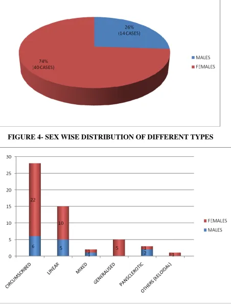
SEX RATIO
In our study 14 cases (26%) were males and 40 cases (74%) were females, the overall female: male ratio being 2.85:1. (Table 4, Figure 3,4)
TABLE 4- SEX RATIO
CLINICAL TYPES MALE FEMALE TOTAL CIRCUMSCRIBED TYPE 6 22 28 LINEAR TYPE 5 10 15 MIXED TYPE 1 1 2 GENERALISED TYPE 0 5 5 PANSCLEROTIC TYPE 2 1 3 OTHERS 0 1 1 TOTAL 14 40 54
TYPES OF LINEAR MORPHOEA NUMBER OF CASES
HEAD VARIANT a) En coup de sabre
56
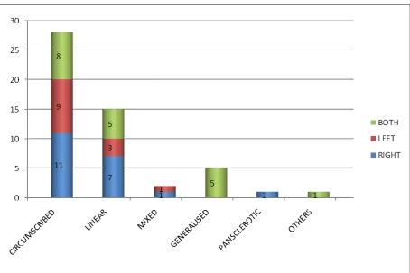
SIDE-WISE DISTRIBUTION
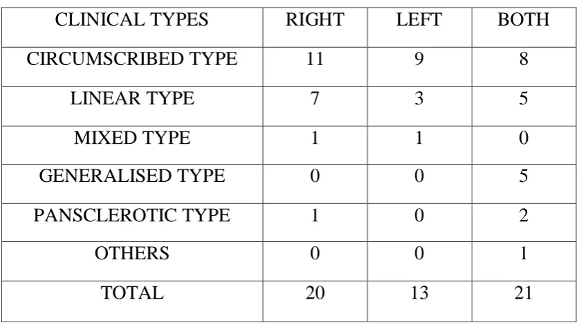
[image:56.595.111.530.327.562.2]Overall, both sides were involved in maximum number of cases with 39% of the patients followed by right side involvement with 37% of the cases and left side involvement constituting 24%. The right side involvement dominated in the individual types with the exception of pansclerotic and generalised morphoea where both sides were more commonly involved. (Table 5, Figure 5,6)
TABLE 5- SIDE WISE DISTRIBUTION
CLINICAL TYPES RIGHT LEFT BOTH
CIRCUMSCRIBED TYPE 11 9 8
LINEAR TYPE 7 3 5
MIXED TYPE 1 1 0
GENERALISED TYPE 0 0 5
PANSCLEROTIC TYPE 1 0 2
OTHERS 0 0 1
TOTAL 20 13 21
AGE OF ONSET
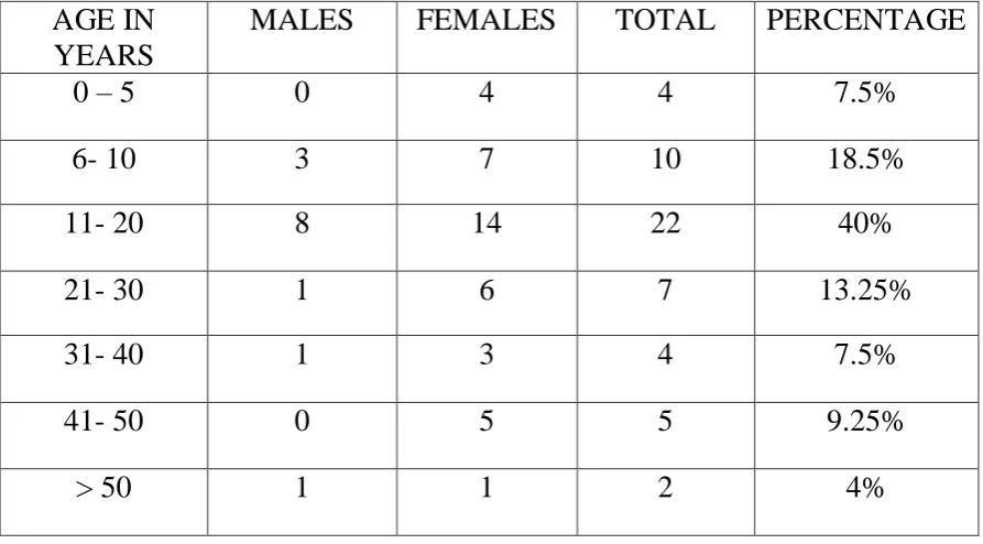
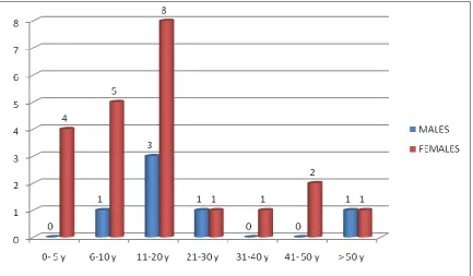
57
TABLE 6- AGE OF ONSET- OVERALL
AGE OF ONSET WISE DISTRIBUTION OF CIRCUMSCRIBED TYPE
[image:57.595.81.528.111.358.2]The commonest age group involved was 11-20 years with 11(39.5%) cases. (Table 7, Figure 8)
TABLE 7- AGE OF ONSET (CIRCUMSCRIBED TYPE)
AGE IN YEARS
MALES FEMALES TOTAL PERCENTAGE
0 – 5 0 4 4 7.5%
6- 10 3 7 10 18.5%
11- 20 8 14 22 40%
21- 30 1 6 7 13.25%
31- 40 1 3 4 7.5%
41- 50 0 5 5 9.25%
> 50 1 1 2 4%
AGE IN YEARS MALES FEMALES TOTAL
0 -5 0 4 4
6- 10 1 5 6
11- 20 3 8 11
21- 30 1 1 2
31- 40 0 1 1
41- 50 0 2 2
58
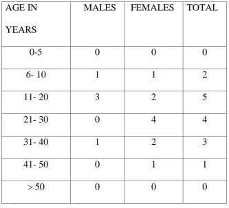
AGE OF ONSET WISE DISTRIBUTION OF LINEAR TYPE
[image:58.595.125.459.204.507.2]The commonest age group involved was 11-20 years with 5 (33.3%) cases. (Table 8, Figure 9)
TABLE 8- AGE OF ONSET (LINEAR TYPE)
AGE OF ONSET WISE DISTRIBUTION OF GENERALISED TYPE
The commonest age group involved was 41-50 years with 2 (40%) cases. (Table 9, Figure 10)
AGE IN YEARS
MALES FEMALES TOTAL
0-5 0 0 0
6- 10 1 1 2
11- 20 3 2 5
21- 30 0 4 4
31- 40 1 2 3
41- 50 0 1 1
59
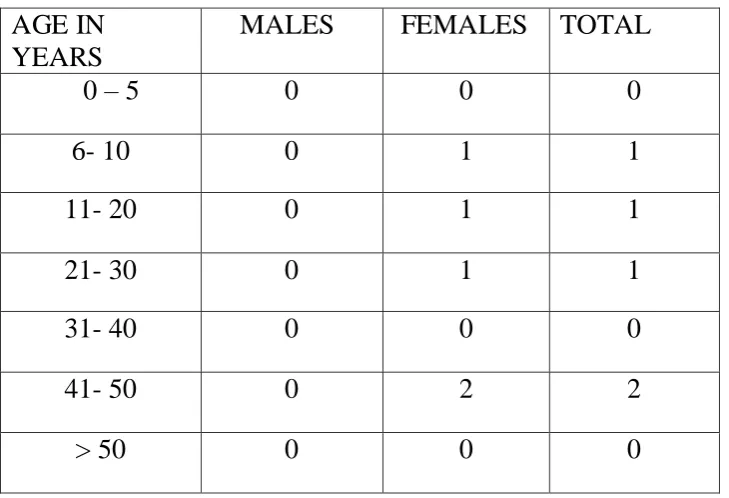
TABLE 9- AGE OF ONSET (GENERALISED TYPE)
AGE OF ONSET WISE DISTRIBUTION OF PANSCLEROTIC TYPE
All the cases occurred in the age group 11-20 years. (Table 10, Figure 11)
TABLE 10- AGE OF ONSET (PANSCLEROTIC TYPE)
AGE IN YEARS
MALES FEMALES TOTAL
0 – 5 0 0 0
6- 10 0 1 1
11- 20 0 1 1
21- 30 0 1 1
31- 40 0 0 0
41- 50 0 2 2
> 50 0 0 0
AGE IN YEARS
MALES FEMALES TOTAL
0 – 5 0 0 0
6- 10 0 0 0
11- 20 2 1 3
21- 30 0 0 0
31- 40 0 0 0
41- 50 0 0 0
60
AGE OF ONSET WISE DISTRIBUTION OF OTHER TYPES
Only one case of keloidal morphoea was seen in the age group 11-20 years.
AGE OF ONSET WISE DISTRIBUTION OF MIXED TYPE
One case was seen in 6-10 years age group and other in 11-20 years age group
DURATION OF LESIONS
The duration of lesions at the time of presentation was in the range of 6 months - 2 years most commonly with 22 (41%) cases. (Table 11, Figure 12)
TABLE 11- DURATION OF LESIONS
SITE WISE DISTRIBUTION- TOTAL
Overall, multiple sites involvement was seen in 25 cases (46%) and 29 cases (54%) showed single site involvement.
SINGLE SITE INVOLVEMENT- 29 CASES
In cases where only one site was involved, back and lower limbs were the most commonly involved sites with 28% cases each. (Table 12, Figure 13)
DURATION OF LESIONS
MALE FEMALE TOTAL PERCENTAGE
0-6 mo 7 14 21 39%
6mo- 2 years 3 19 22 41%
2-5 years 2 3 5 9%
61
TABLE 12- SITE WISE DISTRIBUTION-SINGLE SITE
SITE NUMBER OF
PATIENTS
PERCENTAGE HEAD AND NECK
a) FOREHEAD AND SCALP b) FACE c) BOTH 6 3 2 1 0 20%
CHEST 0 -
BACK 8 28%
ABDOMEN 1 4%
UPPER LIMBS 6 20%
LOWER LIMBS 8 28%
MULTIPLE SITES INVOLVEMENT- 25 CASES
In multiple sites involvement, upper limbs were most commonly involved with 72% of cases. (Table 13, Figure14)
TABLE 13- SITE WISE DISTRIBUTION- MULTIPLE SITES
SITE NUMBER OF
PATIENTS
PERCENTAGE HEAD AND NECK
a) FOREHEAD AND SCALP b) FACE 5 2 3 0 20%
CHEST 6 24%
BACK 10 40%
ABDOMEN 11 44%
[image:61.595.91.520.496.713.2]62
SITE WISE INVOLVEMENT OF CIRCUMSCRIBED TYPE
Circumscribed morphoea affected back most commonly in our study with 8 cases (28.5%). (Table 14, Figure 15)
TABLE 14- SITE WISE INVOLVEMENT (CIRCUMSCRIBED TYPE)
SITE NUMBER OF
PATIENTS
PERCENTAGE HEAD AND NECK
a) FOREHEAD AND SCALP b) FACE
2 1 1
7%
CHEST 0 -
BACK 8 28.5%
ABDOMEN 1 3.5%
UPPER LIMBS 4 14.5%
LOWER LIMBS 5 18%
MULTIPLE SITE INVOLVEMENT
8 28.5%
SITE WISE INVOLVEMENT OF LINEAR TYPE
63
TABLE 15- SITE WISE INVOLVEMENT (LINEAR TYPE)
SITE NUMBER OF
PATIENTS
PERCENTAGE HEAD AND NECK
a) FOREHEAD AND SCALP b) FACE
c) BOTH 4 2 1 1 27%
UPPER LIMBS 1 7%
LOWER LIMBS 2 13%
MULTIPLE SITE INVOLVEMENT
8 53%
SITE WISE DISTRIBUTION OF MIXED TYPE
Multiple sites were involved in both the cases.
SITE WISE DISTRIBUTION OF PANSCLEROTIC TYPE
Two cases involved lower limbs as well as upper limbs, out of which one involved all the 4 limbs. One female case showed involvement of just right lower limb.
SITE WISE DISTRIBUTION OF GENERALISED TYPE
Multiple sites were involved in all the 5 cases.
SITE WISE DISTRIBUTION OF OTHER (KELOIDAL) TYPE
64
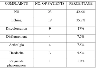
SYMPTOMS
[image:64.595.119.494.227.493.2]Although morphoea was mostly asymptomatic (42.6% of cases) but the commonest complaint reported was itching in 19 cases (35.2%). (Table 16, Figure 17)
TABLE 16- SYMPTOMS
COMPLAINTS NO. OF PATIENTS PERCENTAGE
Nil 23 42.6%
Itching 19 35.2%
Discolouration 9 17%
Disfigurement 4 7.5%
Arthralgia 4 7.5%
Headache 3 5.5%
Raynauds phenomenon
1 1.9%
EVOLUTION OF LESIONS
65
TABLE 17- EVOLUTION OF LESIONS
EVOLUTION OF LESIONS NO. OF PATIENTS PERCENTAGE
PROGRESSING 43 79.5%
STATIC 10 18.5%
REGRESSING 1 2%
PRECIPITATING FACTORS
Most of the cases in this study did not have any precipitating factors but 4 cases gave history of trauma at the site of morphoea whereas 1 patient developed the disease while she was pregnant. (Table 18, Figure 19)
TABLE 18- PRECIPITATING FACTORS
PRECIPITATING FACTORS NO. OF PATIENTS PERCENTAGE
Nil 49 90.7%
Pregnancy 1 1.9%
Trauma 4 7.4%
PAST AND FAMILY HISTORY
66
BIRTH HISTORY
In our study, 46 cases (85%) were born out of non consanguineous marriage followed by 3rd degree consanguineous marriage with 5 cases (9.5%). (Table 19, Figure 20)
TABLE 19- BIRTH HISTORY
CONSANGUINITY NUMBER OF PATIENTS
PERCENTAGE
NON CONSANGUINEOUS 46 85%
2ND DEGREE CONSANGUINEOUS
3 5.5%
3RD DEGREE CONSANGUINEOUS
5 9.5%
ASSOCIATED SYSTEMIC DISEASES
Almost 90% did not have any associated systemic diseases but Diabetes mellitus and Hypertension were seen in 3.7% of cases each. (Table 20, Figure 21)
TABLE 20- ASSOCIATED SYSTEMIC DISEASES
ASSOCIATED SYSTEMIC DISEASES
NUMBER OF PATIENTS
PERCENTAGE
BRONCHIAL ASTHMA 1 1.9%
DIABETES MELLITUS 2 3.7%
HYPERTENSION 2 3.7%
67
NUMBER OF LESIONS
Most of the cases in our study had multiple lesions, 67% to be exact. (Table 21, Figure 22)
TABLE 21- NUMBER OF LESIONS
NUMBER OF LESIONS NUMBER OF PATIENTS
PERCENTAGE
SINGLE 18 33.4%
MULTIPLE 36 66.6%
SENSATION
Out of the 54 patients, 3 patients had variable loss of sensation over the lesions. (Table 22)
TABLE 22- SENSATION
SENSATION NO. OF PATIENTS
INTACT 51
VARIABLE LOSS 3
68
CONTRACTURES
Out of the 54 patients, all 3 patients with pansclerotic morphoea had contractures. (Table 23)
TABLE 23- CONTRACTURES
HEMIATROPHY
No. of patients showing hemiatrophy of tongue- 2.
No. of patients showing hemiatrophy of face- 1. (Table 24, Figure 23)
TABLE 24- HEMIATROPHY
TOTAL NUMBER OF PATIENTS 54
PATIENTS WITH HEMIATROPHY 3
PERCENTAGE 5.55%
COMPLETE BLOOD COUNT
10 patients (18.5%) out of 54 showed eosinophilia and 6 patients (11%) had anaemia. (Table 25, Figure 24)
TOTAL NUMBER OF PATIENTS
54
PATIENTS WITH CONTRACTURES
3
69
TABLE 25- COMPLETE BLOOD COUNT
ABNORMALITY NO. OF PATIENTS
PERCENTAGE
NIL 42 77.8%
ANAEMIA 6 11%
EOSINOPHILIA 10 18.5%
LIVER FUNCTION TESTS
Only one patient with circumscribed morphoea showed abnormal liver function tests with raised liver enzymes.
HISTOPATHOLOGY
SITE OF BIOPSY
Biopsy was taken from the centre of the lesion for most of the cases (68.5%). (Table 26, Figure 25)
TABLE 26- SITE OF BIOPSY
SITE OF BIOPSY NUMBER OF PATIENTS PERCENTAGE
EDGE 7 13%
CENTRE 37 68.5%
BOTH 10 18.5%
Out of the 44 cases from which biopsy was taken from a single site,
54.5% of cases were classified as early morphoea and intermediate and late
stages accounted for 29.5% and 13.5% respectively. 2.5% of cases had
70
TABLE 27-HISTOPATHOLOGICAL STAGE OF MORPHOEA
STAGE OF MORPHOEA NUMBER OF PATIENTS
PERCENTAGE
EARLY STAGE 24 54.5%
INTERMEDIATE STAGE 13 29.5%
LATE STAGE 6 13.5%
OVERLAP OF TWO STAGES
1 2.5%
NAIL CHANGES
Leukonychia and nail dystrophy were most commonly seen in 3.7 % cases. (Table 28, Figure 27)
TABLE 28- ASSOCIATED NAIL CHANGES
NAIL CHANGES NO. OF PATIENTS PERCENTAGE
NIL 49 90.7%
LEUKONYCHIA 2 3.7%
MELANONYCHIA 1 1.9%
NAIL DYSTROPHY 2 3.7%
ASSOCIATED SKIN DISEASES
The commonest skin disease which was found to be associated was LSA in
71
TABLE 29- ASSOCIATED SKIN DISEASES
DISEASES NO. OF PATIENTS
NIL 43
HYPERTROPHIC SCAR 1
KELOID 1
PSORIASIS 1
LICHEN SCLEROSUS ET ATROPHICUS 4
SEBORRHEIC MELANOSIS 1
LICHEN STRIATUS 1
VITILIGO 2
COLLOID MILIUM 1
72
DISCUSSION
We encountered 54 cases of morphoea in this study which was done for a span of 2 years.
INCIDENCE
Out of a total 106368 cases visiting the dermatology outpatient department during the study period, the cases with morphoea were 54 in total. Thus the incidence rate of morphoea was 0.5 per 1000 dermatology cases. In a study done by Peterson et al12 in Olmsted county between 1960-1993 on 82 cases showed the incidence rate as 0.027 per 1000 population.
INCIDENCE OF VARIOUS CLINICAL TYPES
The incidence of various clinical types of morphoea in our study was
as follows
Circumscribed- 51.8%
Linear (Head and trunk/limb variant)- 27.8%
Generalised- 9.25%
Pansclerotic- 5.55%
Mixed- 3.7%
Other unclassified variants (Keloidal morphoea) - 1.8%
73
Our findings were in concurrence with the study conducted by Peterson et
al12 on 82 cases which reported that 56% of patients had plaque type morphoea, 20% linear, 13% generalized and 11% had deep morphoea. A study by Christianson et al11 of 235 cases showed an incidence of 35% of plaque type, and 46% of linear type and 19% of generalized morphoea.
SEX RATIO
The overall sex distribution in our study pointed towards a female predominance with female to male ratio being 2.85:1. This was found to be consistent with studies conducted by Christianson et al11 and Peterson et al12 with ratios being 3:1 and 2.6:1 respectively. The individual types of
morphoea also followed a similar pattern with exception of pansclerotic
morphoea where males were reported to be more commonly involved. Linear
morphoea in our study showed a female to male ratio as 2:1 whereas Peterson
et al12 reported an even sex distribution.
SIDE DISTRIBUTION
Bilateral involvement was seen in 39% of cases whereas 61% cases
showed unilateral involvement with right side being involved in 37% of cases