DISSERTATION ON
PATTERNS OF DIFFUSE PARENCHYMAL LUNG DISEASE
MANIFESTATIONS IN COLLAGEN VASCULAR DISEASE
AND IN RELATION TO DLCO
Submitted for
M.D., DEGREE EXAMINATION Branch-XVII
TUBERCULOSIS & RESPIRATORY DISEASES
INSTITUTE OF THORACIC MEDICINE Madras medical college & Govt. General Hospital
Chennai-600003
The Tamilnadu Dr. M.G.R.Medical University
Chennai 600032
CERTIFICATE
This is to certify that the dissertation on “PATTERNS OF DIFFUSE PARENCHYMAL LUNG DISEASE MANIFESTATIONS IN COLLAGEN VASCULAR DISEASE AND IN RELATION TO DLCO ” is a record of research work done by DR.G.SANGAMITHRA in partial fulfillment for M.D.BRANCH- XVII (T.B. AND RESPIRATORY DISEASES) EXAMINATION of
The Tamilnadu Dr. M.G.R.Medical University to be held in March 2009. The period of
study is from January 2008 to November 2008.
PROF .DR.D.Ranganathan, PROF.DR.N.Meenakshi, M.D, DTCD, DNB M.D, DTCD
Addl.Professor of Thoracic Medicine, Director,
Department of Thoracic Medicine, Institute of Thoracic Medicine,
Madras medical college & Govt. General Hospital, Chetpet,
Chennai. Chennai.
Dr. T. P. KALANITI, M.D,
Dean
Madras medical college & Govt. General Hospital
DECLARATION
I hereby declare that the dissertation entitled “ PATTERNS OF DIFFUSE PARENCHYMAL LUNG DISEASE MANIFESTATIONS IN COLLAGEN VASCULAR DISEASE AND IN RELATION TO DLCO ” submitted for the Degree of Doctor of Medicine in M.D., DEGREE EXAMINATION Branch-XVII TUBERCULOSIS & RESPIRATORY DISEASES is my original work and the
dissertation has not formed the basis for the award of any degree, diploma,
associate ship, fellowship or similar other titles. It had not been submitted to
any other university or Institution for the award of any degree or diploma.
Place: Chennai Signature of the Scholar
ACKNOWLEDGEMENT
I Would like to thank DR. T. P. Kalanithi MD Dean, Madras medical college & Govt. General Hospital for giving me permission to conduct the study in this
institution.
I am deeply indebted to Prof.DR.N.Meenakshi M.D,DTCD Director, Institute of Thoracic Medicine, Chennai for her guidance and constant inspiration throughout
my dissertation work. Words are few to express my gratitude to her for sparing her
precious time and energy in trying to bring out the best in me.
I would like to express my sincere thanks and heartfelt gratitude to Addl. Prof DR.D.Ranganathan M.D, DTCD, DNB. I shall always cherish in my heart for his constant encouragement, valuable guidance and relentless support throughout my
postgraduate course.
I would also like to thank Prof. DR. R.Porkodi M.D, DM, Director, Department of Rheumatology, Government General hospital, Chennai for her immense help. I
am very grateful to DR. Sridhar M.D, DTRD, DR. Sundar MD, DTCD, DR.Vinothkumar M.D, DNB, DR. Thiruppathi M.D, and all assistant professors at our Thoracic Department for providing valuable guidance and timely advice.
Last but not the least, I thank with deep gratitude to all the patients who had
participated in the study, without whose co operation this study would not have been
CONTENTS
Page No
I Introduction 1
II Aim of the study 3
III Review of Literature 4
IV Materials and Methods 32
V Results 37
VI Discussion 50
VII Conclusions 52
VIII Abbreviation
IX Bibliography
X Master chart
I. INTRODUCTION
Diffuse parenchymal lung diseases (DPLD) are a diverse group of pulmonary
disorders that are classified together because of similar clinical, roentgenographic,
physiologic, or pathologic manifestations1. They are also referred to as interstitial lung
diseases (ILD).DPLD alter mechanical and gas exchange properties of the lungs. In
general, the hall marks of DPLD are restrictive changes in pulmonary physiology i.e
decreased total lung capacity, reduced residual volume, diminished static compliance and
reduced vital capacity often with an increased FEV1/FVC ratio and a reduced diffusing
capacity with carbon monoxide.
Hence pulmonary function testing (PFT) aids in the evaluation and management
of patients with DPLD by provide an estimate of histologic severity, baseline estimation
of prognosis and be used to monitor disease progression or response to therapy. The
forced vital capacity and diffusion capacity are the most valuable serial measurements for
diagnosing DPLD.
The collagen vascular diseases are a heterogeneous group of immunologically
mediated inflammatory diseases. It is not surprising that, by virtue of their abundant
connective tissue and blood supply, the lungs are frequently involved in these disorders.
Consequently, collagen vascular diseases affect all areas of the lung (i.e. airways,
alveoli, vascular system, and pleura), and do so in various degrees and combinations2.
Diffuse parenchymal lung diseases in CVD patients are characterised by
deteriorating parenchymal fibrosis and gas exchange. It remains innocuous in the early
stage, as most of the rheumatic patients have restricted mobility manifesting with subtle
diagnosing the cases as early as possible. Because the pulmonary specialist often
involved in care of these patients, a comprehensive understanding of collagen vascular
disease and usual course of Diffuse parenchymal lung diseases are important.
The clinical assessment of a patient with DPLD requires a combination of history
and physical examination, laboratory investigation, lung function testing, chest imaging
studies, bronchoalveolar lavage, and histological examination.
Over the last few years, the abnormal patterns of DPLD on High Resolution
Computerised Tomography (HRCT) scans have been refined and are increasingly
recognised as diagnostic patterns. This has led to the increasing use of HRCT scans in
conjunction with thorough clinical assessment3.By pulmonary function tests (PFT) the
severity of physiologic disarrangements in DPLD, correlate well with the overall extent
of pathologic and HRCT abnormalities4. Abnormalities of pulmonary function tests also
verify the presence of disease particularly in those patients with normal chest
roentgenograms.
This study throws light on the non-invasive procedures rather than the invasive
procedures. Since most of the patients deny procedures like bronchoscopy or surgical
biopsy as well as they are in real respiratory compromise, it is better to opt an alternate
procedure like HRCT , Diffusing capacity for carbon monoxide (DLCO) and spirometry
II. AIM OF THE STUDY
To evaluate the Diffusing capacity for carbon monoxide among patients with
Diffuse parenchymal lung disease in proven Rheumatologic illness showing various
pattern of abnormalities in the High Resolution Computerised Tomography and correlate
the same with spirometry.
DESIGN OF THE STUDY:
Prospective study.
III. REVIEW OF LITERATURE
Diffuse parenchymal lung diseases (DPLD) commonly complicate the
management of collagen vascular diseases. Hence a comprehensive understanding of
collagen vascular disease and the usual cause of diffuse parenchymal lung disease is
important. Approximately 15% of the patients who present with diffuse parenchymal
lung diseases have an underlying collagen vascular disease5. Furthermore clinical
symptoms like cough, dyspnoea or Diffuse Parenchymal Lung Disease may be the first
manifestation of Rheumatic diseases. Finally it can result in significant morbidity and
mortality.
The autoimmune mediated inflammation and fibrosis that characterises these
diseases can easily and irreversibly disrupt the normal functioning of the lung. As a
group they can affect each portion of the lung :- The pleura, alveoli, interstitium,
vasculature, lymphatic tissue and of both small and larger airways. The lung diseases
associated with collagen vascular disease may precede the clinical presentation of
collagen vascular disease by five years or more5.
Full pulmonary function tests (PFT) should be performed in all cases of suspected
DPLD with collage vascular diseases. They aid in the diagnosis DPLD, assessment of
disease severity, response to treatment, and prognosis. DLCO is typically reduced in
DPLD to greater extent than the lung volumes at which it is measured. Co-existence of
DPLD with obstructive airway disease (i.e.emphysema), can confuse the results with a
mixed pattern of restriction observed as reduced lung volumes and obstruction
Baseline PFT may provide an estimate of prognosis. Exertional oxygen
desaturation to less than 88% and a decrease in FVC (greater than 10%) over a short
follow up period predict patients at increased risk of mortality. Severely decreased DLCO
(<30%) is also associated with increased mortality. Serial PFTs provide valuable
information in determining disease progression and response to medication. FVC and
DLCO are the most valuable serial measurements.
Many of these Collagen vascular diseases are characterised by the presence of the
specific type of autoantibody that may greatly assist in specific diagnosis.
AUTO ANTIBODY ASSOCIATED WITH SPECIFIC COLLAGEN VASCULAR DISEASES7
Diseases Associated Antibodies
Rheumatoid Arthritis
Systemic sclerosis
Mixed connective tissue diseases
Dermatomyositis/Polymyositis
Systemic lupus erythematosus
Sjogren’syndrome
Rheumatoid factor
Antiscl-70
Anticentromere Antibody
Anti ribonuclear protein
Anti-Jo-1
Anti-ds-DNA
Anti-sm
Anti ss-A(Ro)
The association of Collagen vascular disease and Diffuse parenchymal lung
disease (DPLD) is well established6. DPLD complicating collagen vascular diseases
account for high morbidity and mortality. Until recently, most of the studies of lung
involvement in the collagen vascular disease relied on clinical, physiological
and radiological data to define the presence of disease, determine the clinical
course, and assess the prognosis of the lung involvement.
RHEUMATOID ARTHRITIS (RA)
It is a sub acute or chronic inflammatory polyarthropathy of unknown
cause that particularly affects peripheral joints. Lung fibrosis associated with
Rheumatoid arthritis was first reported by Ellman & Ball 19488.
DPLD quickly appeared as the predominant pulmonary manifestation of
rheumatoid arthritis (RA) (after excluding drug-induced pulmonary disease).Systematic
PFTs detected a decreased diffusing capacity(DLCO) in 41% of patients, and among
them, 50% exhibited features of fibrosis associated with lymphoid infiltrates in lung
biopsy.Dawson and colleagues studied 29 patients with RA and found that DLCO < 54%
of predicted had an 80% sensitivity and a 93% specificity for predicting progressive
parenchymal lung disease over the next 2 years.
Prevalence of pleuropulmonary manifestations is clearly increased in males and in
smokers. Coexisting subcutaneous rheumatoid nodules, high titres of circulating
rheumatoid factor or antinuclear antibodies are also considered significant risk factors
Rheumatoid lung fibrosis is twice as common in men as in women with a mean
age of about fifty years. RA is characterised by the presence of symmetric arthritis,
morning stiffness and Rheumatoid factor in the blood.
Pleuropulmonary complications include interstitial pneumonitis with fibrosis,
Rheumatoid (necrobiotic) nodules, Bronchiolitis Obliterans Organising Pneumonia,
Bronchiectasis, Obliterative bronchiolitis, follicular bronchiolitis and pleural effusion or
pleural thickening 9,10.
Interstitial pneumonitis with fibrosis are the most common pulmonary
manifestations of Rheumatoid arthritis9.In fact pulmonary function abnormalities
consistent with interstitial fibrosis have been reported in as many as 40% of patients who
have Rheumatoid arthritis9.In more than half of these patients, however finding at chest
roentgenogram are normal. Evidence of interstitial fibrosis is seen at chest roentgenogram
in approximately 5%9 and at HRCT in 30% to 40%10 of patients with RA. The
complication is seen most frequently on men between 50 and 60 years of age.
TYPES OF INTERSTITIAL PNEUMONIA
Non-specific interstitial pneumonia(NSIP),Usual interstitial pneumonia(UIP)
Cryptogenic organising pneumonia(COP) and
Lymphocytic interstitial pneumonia(LIP).
The majority of the patients with interstitial fibrosis associated with RA have UIP,
lymphocytes may be prominent in both the parenchymal interstitium and in the interstitial
tissue in bronchiolar walls and interlobular septa(Follicular bronchiolitis).
RADIOLOGIC MANIFESTATIONS
Initial radiographic studies found a low incidence of 1.6–5% of DPLD in RA8. In
the early stage, the radiographic appearance consists of irregular linear hyper attenuating
areas in a fine reticular pattern. The abnormality usually involves mainly the lower lung
zones. With the progression of disease, the reticular pattern becomes coarser and diffuse
and later honeycombing may be seen11.
HRCT FINDINGS
HRCT findings consist of irregular linear hyper attenuating areas caused by a
combination of intralobular lines and irregular thickening of interlobular septa10.
Honeycombing is seen more markedly near the diaphragm. HRCT demonstrates
interstitial lung disease in patients with or without clinical evidence of the disease (69%–
80% and 20%–29% respectively) 10.
In a study by Akira et al,three major radiographic patterns of disease have been
identified in symptomatic patients who developed lung disease prior to or following the
diagnosis of RA12. These include reticulation with or without honeycombing (66%
patients), centrilobular branching lines with or without bronchial dilatation (17%), and
SPIROMETRY IN RA
The physiologic abnormalities of rheumatoid DPLD are identical to the other
fibrosing lung diseases13. There is a reduction in pulmonary compliance and lung
volumes. Physiologic testing also often shows evidence of obstruction to airflow, which
may reflect other pulmonary manifestations of RA including bronchiectasis, bronchiolitis
obliterans, chronic airway obstruction or cricoarytenoid arthritis 14,15.
DLCO IN RA
Abnormalities of gas transfer including a low DLCO is present in RA. Abnormal
pulmonary function may be found in individuals with normal chest roentgenograms14.
SYSTEMIC SCLEROSIS (SS)
It is a generalised collagen vascular disease characterised by the synthesis and
deposition of excessive extracellular matrix and vascular obliteration in various organs16.
Dr. Charlie Strange from the Medical University of South Carolina provided an
algorithm for the management of DPLD in scleroderma patients that includes chest
x-rays, spirometry, and DLCO. If the results are normal, these tests should be repeated on a
6-month basis. If any of the findings are abnormal, an HRCT should be obtained. All
patients with scleroderma undergo annual echocardiography and measurements of their
DLCO. A DLCO < 40% of predicted appears to be predictive for underlying Pulmonary
Arterial Hypertension(PAH), and an FVC/DLCO ratio >1.8 has good performance
characteristics in PAH.
SS is divided into two broad categories: Diffuse and limited forms based on the
Most common clinical manifestations are
Tightening, induration and thickening of the skin (SCLERODERMA),
Raynaud’s and other vascular anomalies,
Musculoskeletal manifestation and
Visceral involvement particularly Gastro Intestinal Tract, Lungs, Heart and
Kidney.
The diagnosis can be made with a high degree of certainty if the single major
criterion of proximal scleroderma is present or if there are 2 or more minor criteria
(sclerodactaly, pitting scars or loss of the substance of fingertips and bilateral basal
pulmonary fibrosis)8. Systemic Sclerosis has 3:1 female to male distribution and presents
more commonly in the third to fifth decade of life.
Lung is the fourth most commonly affected structure after skin, vessel and
oesophageal involvement7. Pulmonary involvement is more common and more severe in
systemic sclerosis than in other types of collagen vascular disease.Prognostic factors for
poor outcomes include male sex, the presence of lung involvement early in disease, low
DLCO, severe Raynaud’s phenomenon, and cigarette smoking.
RADIOLOGIC MANIFESTATIONS
Pulmonary fibrosis is the most common radiographic finding, present in
20% –65% of patients 16, 17. The fibrosis usually has a basilar predominance and appears
initially as a fine reticular pattern that progresses to coarse reticulation and
the disease but is less severe in the limited form. In Schurawitzki H, Stiglbauer R,
Graninger W, et al prospective study of 23 patients with systemic sclerosis, fibrosis was
identified at chest radiography in 39% of patients and at high-resolution CT in 91% of
patients 17.
HRCT FINDINGS
The predominant abnormalities at high-resolution CT consist of areas of
ground-glass attenuation, poorly defined sub pleural nodules, reticular pattern of attenuation,
honeycombing, and traction bronchiectasis 17.
In Kim et al18 longitudinal CT series of 40 patients with SS study , a variety of
radiological features were found including ground-glass opacity (100%), irregular linear
opacity (90%), small nodules (70%), honeycombing (33%), traction bronchiectasis
(68%), bilateral pleural thickening (45%), and enlarged mediastinal lymph nodes (15%).
After a mean follow-up of 40 months, regardless of the initial findings or presence of
treatment, the extent of parenchymal disease, including ground-glass opacity and
honeycombing, significantly increased with concomitant declines in forced expiratory
volume in 1 second (FEV1), and forced vital capacity (FVC) without significant change
in DLCO18.
SPIROMETRY IN SS
Physiological studies have shown a restrictive pattern with a decreased total lung
capacity (TLC), vital capacity (VC), forced vital capacity (FVC) and residual volume.
The greatest decline in lung function occurs within the first 4 years of disease and
predicted) restrictive lung disease is detectable in approximately 40% of patients with
scleroderma. Initially, there may be a reduction in DLCO, which may even precede
symptoms onset and/or loss of lung volume. Occult pulmonary impairment may be
present in patients with normal pulmonary function tests (PFT) which may be
demonstrated by cardio-respiratory exercise testing 19.
DLCO IN SS
In SS impairment of the transfer factor (or diffusing capacity) for carbon
monoxide (DLCO) is present. A reduced DLCO is the most common and most sensitive
PFT abnormality in patients with scleroderma and has been shown to correlate with the
presence of dyspnoea and with autopsy, evidence of DPLD. With progression of the
DPLD, the restrictive pattern is paralleled by the decrease in gas exchange. As
demonstrated by WELLS et al.20, DLCO is the best index of the extent of the DPLD
when compared with HRCT as the "gold standard". Interestingly, patients with
DPLD-scleroderma have a better survival rate (86% at 5 yrs) than patients with idiopathic
pulmonary fibrosis (IPF) (50% at 5 yrs) 21.
Reduced DLCO in SS
1. Related to alveolar septal thickening and vascular obliteration
2. May be the earliest PFT abnormality and the most sensitive indicator of SS 22
3. Degree of decline corresponds to the severity of dyspnoea
4. Severe isolated reduction in DLCO (<45% of the predicted value) is an early
A low DLCO is more common in limited form than in diffuse form. FVC and
DLCO are important prognostic factors in scleroderma24.
SYSTEMIC LUPUS ERYTHEMATOSUS (SLE)
The original description of SLE is credited to Kaposi in 1872.25. It is a
multisystem disorder that particularly affects vessel, serosa, kidneys, central nervous
system, skin , blood vessels, joints and lung. SLE is characterised by the presence
of anto antibodies against various nuclear antigens. It predominantly affects
women (F:M ratio 10:1) aged 20–50 years.
During the course of SLE, pleural disease develops in 50% of patients 26.
Pulmonary parenchymal abnormalities are also common. Pleuropulmonary involvement
occurs in approximately 50%–60% of patients. In one prospective study that included
1,000 patients, lung involvement was identified in only 3% at the onset of the disease; it
developed in an additional 7% of patients over the period of observation . Parenchymal
opacification may be caused by pneumonia, haemorrhage, acute lupus pneumonitis, or
pulmonary oedema, with pneumonia being the most common cause 26.
Pulmonary manifestations of systemic lupus erythematosus comprise both acute
and chronic lesions. Acute disease includes pulmonary haemorrhage, acute lupus
pneumonitis and pulmonary oedema. Chronic disease such as interstitial pneumonitis
with fibrosis is less common than in other connective tissue disorders 27.
Most frequent lung manifestation in SLE results from acute injury to
the alveolar-capillary unit, leading to acute lupus pneumonitis and/or alveolar
without infectious aetiology. The alveolar haemorrhage varies from mild to massive, and
BAL studies often reveal haemosiderin-laden macrophages. It presents with dyspnoea,
tachycardia, cough, fever, pleuritic chest pain and sometimes haemoptysis.
ACUTE LUPUS PNEUMONITIS
Acute lupus pneumonitis is characterized by the acute or sub acute onset of
tachycardia, tachypnea, dyspnoea, cyanosis, and cough. Fever is common, but
haemoptysis is infrequent, and clubbing is absent. The mean age in one series was 38
years with a mean duration of known SLE of 16 months (range 0 to 48 months). In 50%
of these patients, pneumonitis was the presenting manifestation of their SLE. Chest
examination may reveal fine or coarse rales, but signs of pleurisy are rare. In severe
cases, evidence of right ventricular overload may appear.
The chest radiograph plays a limited role in the differential diagnosis of
pulmonary complications of SLE. The most common findings in patients with SLE are
non-specific areas of air space consolidation. The consolidation may be unilateral or
bilateral and focal or diffuse, but it tends to involve mainly the lower lung zones. The
most common cause of consolidation is pneumonia
Chest roentgenograms in the acute lupus syndrome demonstrate diffuse or patchy
opacities that are predominantly basilar, although the middle and upper zones may be
affected. Usually bilateral, the opacities may be accompanied by pleural effusion and
cardiomegaly. A recent case report described acute lupus pneumonitis in a patient with a
normal chest radiograph and HRCT scan. Sub acute cases may demonstrate the
are often described in acute pneumonitis. Such deficits in particular cause an elevation of
the alveolar-arterial gradient; persist in the majority (of a small number) of patients
despite the apparent resolution of their lung disease.
RADIOLOGIC MANIFESTATIONS
The most common radiographic manifestation is unilateral or bilateral pleural
effusion that is frequently associated with pericardial effusion30 .Chest radiographs
typically show unilateral or bilateral patchy areas of consolidation, mostly predominant in
the lung bases, which may be associated with pleural effusion or atelectasis.Other chest
radiographic findings of SLE include loss of lung volume related to diaphragmatic
dysfunction, pulmonary oedema, musculoskeletal changes related to renal failure, and
bone changes related to corticosteroid therapy.
HRCT FINDINGS
Multiple patterns have been reported in SLE . The most common findings are
intralobular interstitial thickening and irregular thickening of the interlobular septa that
are predominantly found in the lower lobes31,32. Other common findings on HRCT
include traction bronchiectasis (architectural distortion and bronchial dilatation secondary
to the fibrosis), small foci of air space consolidation, and areas of ground-glass
attenuation31,32. Honeycombing changes may be present. Both pleural effusions and
cardiomegaly may be noted. Enlargement of the pulmonary artery accompanies cor
SPIROMETRY & DLCO IN SLE
Several studies indicate that physiologic abnormalities (e.g., DLCO, lung
volumes, or compliance) are more common than clinical or roentgenographic changes 33.
Most common physiologic deficit in patients with SLE is the reduction in DLCO with or
without decreased total lung capacity. Among patients with normal chest
roentgenograms, a reduction in VC was noted in 27% and in DLCO in 67% 34. In two
series of unselected patients, a reduction in DLCO occurred more frequently (72 to 80%)
than did volume restriction (43 to 49%) or roentgenographic abnormalities (30 to 50%)35.
POLYMYOSITIS/DERMATMYOSITIS (PM/DM)
Polymyositis is an autoimmune inflammatory myopathy characterised by
presence of symmetric weakness of the limb girdle and anterior neck muscles36.
Dermatomyositis is similar to polymyositis except for the presence of a characteristic
skin rash. PM/DM have an incidence of approximately 5–10 cases per million per year36
and occur twice as often in women as in men. The diagnostic criteria for PM/DM are
proximal muscle weakness (Symmetric, present for weeks or months) , Muscle biopsy
showing necrobiotic and inflammatory changes, Raised muscle enzymes ( Creatinine
phosphokinase), Characteristic electromyography and Characteristic skin rash. In
Dermatomyositis additional characteristic skin changes present: Heliotrope periorbital
rash and violaceous/red papular rash over bony prominence.
PM/DM is often associated with clinical, radiological or functional evidence of
pulmonary fibrosis. Mills and Mathews first described interstitial fibrosis in
and 9% of DM cases3. Lung involvement in PM/DM may precede muscle or skin
manifestations in 33% of cases. There is no correlation between the extent and severity of
muscle and skin involvement and the development of DPLD.
The clinical presentation may be arbitrarily divided into three forms.
1) DPLD may occasionally be rapidly progressive with acute fever, dyspnoea and
lung infiltrates similar to a Hamman-Rich-syndrome.
2) Patients may have a slowly progressive dyspnoea upon exertion with chest
radiographic abnormalities.
3) Some patients may have no pulmonary symptoms, but abnormal radiographs
and/or PFT.
RADIOLOGIC MANIFESTATIONS
Chest radiographs most often reveal bilateral basal infiltrates, but may be normal
in patients with biopsy proven DPLD. HRCT shows pleural irregularities, ground glass
attenuation and patchy consolidation38. The frequency of radiographic parenchymal
abnormalities is low (about 5%). The most common is a symmetric, predominantly basal
reticular pattern that may become diffuse over time and progress to honeycombing 36.
Bilateral areas of consolidation develop in some patients over a 2- to 3-week period.
HRCT FINDINGS
The HRCT findings are prominent interlobular septa, ground-glass attenuation,
patchy consolidation, parenchymal bands, irregular peribronchovascular thickening, and
abnormal chest radiographic findings or pulmonary function tests39.Patchy consolidation,
parenchymal bands, and irregular peribronchovascular thickening are seen to improve at
sequential CT, becoming pleural irregularities, prominent interlobular septa, ground-glass
attenuation, and sub pleural lines on follow-up CT scans. Therefore, consolidation with
patchy and sub pleural distribution, parenchymal bands, and irregular
peribronchovascular thickening are reversible. On occasion, areas of ground-glass
attenuation with parenchymal bands or sub pleural lines that represent a pathologic area
of usual interstitial pneumonia may progress to honeycombing.
SPIROMETRY & DLCO IN PM/DM
These patients usually have evidence of a restrictive ventilatory pattern with
reductions in VC and TLC. In addition, the DLCO may also be reduced. The profound
respiratory muscle weakness, which may often complicate these patient’s courses, may
be demonstrated by a decrease in maximal inspiratory pressure, maximal inspiratory flow
rate and maximal voluntary ventilation. Many patients have resting arterial hypoxemia,
which is made worse with exercise. Clinical exercise testing is of importance in PM/DM
patients to elucidate the cause of dyspnoea, which may not be limited to DPLD, but
might be due in part to pulmonary hypertension, cardiac dysfunction or muscle
weakness40.
SJOGREN’S SYNDROME
Sjogren’s syndrome, or the sicca complex, is an autoimmune exocrinopathy
characterized by lymphocytic infiltration of glandular and extra glandular organs. A
arthritis characterizes Sjogren’s syndrome. It is relatively common, affecting 0.1% of the
general population and 3% of older adults 41. It may be primary without features of other
collagen vascular disease, or secondary in association with other collagen vascular
disease most often rheumatoid arthritis.
Pulmonary manifestations occur frequently in Sjogren’s syndrome. Diffuse
parenchymal lung disease is probably the most common functional abnormality identified
in patients with primary Sjogren’s syndrome with lung involvement. The aetiology of
DPLD associated with Sjogren’s syndrome is unknown, and it appears that lymphocytic
pneumonitis represents only one pole of the spectrum of diseases, which includes pseudo
lymphomas and malignant lymphomas. DPLD occur more often in patients with extra
glandular manifestations than in those with glandular disease alone.
The most common thoracic complication, lymphocytic interstitial pneumonia is
followed in frequency by airway abnormalities such as follicular bronchitis,
bronchiectasis, and bronchiolitis. Less common complications include interstitial
pneumonitis and fibrosis, BOOP, lymphoma, pulmonary hypertension, and pleural
effusion or fibrosis. A large series of 343 patients reported by STRIMLAN 42 clearly
demonstrated that many patients do not manifest as a single type of pulmonary lesion.
RADIOLOGIC MANIFESTATIONS
Parenchymal abnormalities are evident at chest radiography in 10%–30% of
patients. The most common finding, a reticulonodular pattern that involves mainly the
lower lung zones, may reflect the presence of lymphocytic interstitial pneumonia or
restrictive pattern with a low diffusion capacity is commonly found on lung function
tests.
HRCT FINDINGS
In Franquet et al study of high-resolution CT findings among 50 patients
with Sjogren's syndrome showed the major abnormalities of bronchiectasis, bronchiolar
inflammation and increased parenchymal line43.There is an increased prevalence of
lymphocytic interstitial pneumonitis that is seen radiographically as a reticulonodular
pattern predominantly involving the lower lobes.
A recent article described the high-resolution CT findings of lymphocytic
interstitial pneumonia44, with the most common findings being areas of ground glass
attenuation, thickening of bronchovascular bundles with interlobular septa and cysts.
DPLD and bronchiolar inflammatory changes are common abnormal findings seen on
HRCT scans in primary Sjogren’s syndrome. For example, 20% of patients with primary
Sjogren’s syndrome undergoing HRCT had bronchial wall thickening with either
ground-glass opacification or a small nodular pattern in the majority of these cases.
SPIROMETRY & DLCO IN SJOGREN’S SYNDROME
Bariffi and colleagues studied 18 female non-smokers with Sjogren’s syndrome.
They found that 13 of the 18 had an FEV1/FVC ratio of less than 80% and 7 of 18 had
anFEV1 of less than 80%, whereas 9 of 14 had a maximum flow at 25% of VC (MEF25)
of less than 80%45 .In addition, these investigators found abnormalities in diffusing
capacity. Constantopoulos and colleagues, in a study of 61 patients with primary
whereas only 10% had a low FEV146. However, other investigators performed a
longitudinal study of 18 asymptomatic, non-smoking women with normal chest
radiographs. In this group, baseline FEV1/FVC, FVC, and TLC were normal in all
patients. Only 17% had a diffusion defect.
When compared with secondary Sjogren’s syndrome, there was a higher
incidence of restrictive pattern in primary Sjogren’s syndrome whereas an obstructive
ventilator defect was more common in secondary Sjogren’s syndrome or RA alone. The
significance of diffusion defects in patients with primary Sjogren’s syndrome remains of
uncertain significance; in a 10-year follow-up of pulmonary involvement in 30 patients, a
significant decline in diffusion was seen at 4 years followed by a significant improvement
at 10 years, in the absence of any intervention.
MIXED CONNECTIVE TISSUE DISEASE (MCTD)
The term mixed connective tissue disease refers to a condition in which patients
have mixed features of systemic lupus erythematosus, progressive systemic sclerosis, and
polymyositis. There is an 8:1female to male ratio and no racial predilection. Suggested
clinical criteria for the diagnosis are 1) Positive for anti RNP plus 2) Three or more
clinical features such as Hand oedema, Synovitis, Myositis, Raynaud’s phenomenon and
Acrosclerosis. After several years, MCTD may transform into one of the classic disease.
Pulmonary involvement has been described in 20–85% of patients with mixed
connective tissue disease (MCTD)47. Common pulmonary abnormalities include
interstitial pneumonitis with fibrosis, pulmonary hypertension and pleural effusion.
scleroderma and consist of Idiopathic Pulmonary Fibrosis with alveolar inflammatory
processes and progressive development of honeycomb formation; generally, the degree of
fibrosis appeared more severe in patients exhibiting scleroderma feature.
Radiologic manifestations
One-third of patients with mixed connective tissue disease had initial chest
roentgenograms that were abnormal demonstrating small irregular opacities involving the
lung base.
HRCT FINDINGS
In HRCT Pulmonary involvement in mixed connective tissue disease has been
characterized as the presence of ground-glass attenuation, nonseptal linear opacities, with
a peripheral and lower-lobe predominance. The frequency of pulmonary abnormalities
varies considerably in different series. In a review of the CT findings in 41 patients with
MCTD, Kozuka et al found ground glass, sub pleural micro nodules, non-septal linear
and reticular abnormality in more than 50% of cases.
In a retrospective study of 81 patients at the Mayo Clinic48, an interstitial pattern
was seen at chest radiography in 19%; on the other hand, careful prospective study of 34
patients in another investigation showed interstitial abnormalities in 85%. The
abnormalities consist of irregular linear hyper attenuating areas with a reticular pattern
and involving mainly the lung bases. With the progression of disease, the fibrosis
gradually extends superiorly; in the late stage, honeycombing may be identified. High
abnormalities include areas of parenchymal consolidation that may be related to
Bronchiolitis Obliterans Organising Pneumonia.
SPIROMETRY & DLCO IN MCTD
Pulmonary function abnormalities include a reduction of the DLCO, a decreased
VC, TLC, and FEV1.Abnormal PFT and chest radiographs are frequent. Impaired DLCO
has been reported in 67%, and restrictive lung volumes in 50% of MCTD. DLCO appears
to be the most sensitive single parameter in evaluating pulmonary dysfunction in MCTD.
RADIOLOGIC IMAGING IN DPLD
Patients with suspected DPLD will have a chest radiograph as the initial imaging
investigation. In most cases, this is abnormal and occasionally the radiographic
appearances are sufficiently characteristic to enable a specific diagnosis to be made when
taken in conjunction with the clinical and laboratory findings. Chest radiograph is an
essential test for the assessment of DPLD, however a normal Chest Radiograph cannot
exclude the diagnosis. If available, previous x-rays or reports of x-rays should be
obtained and compared with the recent x-rays. In appropriate clinical settings, HRCT
may be sufficiently characteristic to preclude the need for biopsy7.
PULMONARY HRCT
HRCT scanning is capable of imaging the lung with excellent spatial resolution
and providing anatomical detail similar to that seen by gross pathological examination.
Images are usually obtained in the supine and prone position. In normal patients,
dependent lung opacity is often seen in the posterior, subpleural regions of the lung. In
can be identical to those seen in normal patients. Prone images will differentiate between
these two possibilities, since normal dependent density in the posterior lung will
disappear on prone images, whereas true lung disease-related density will persist.
Dynamic expiratory images are also obtained to screen for air trapping.
1.Detection of DPLD
HRCT scanning is able to detect DPLD not visible on the chest radiograph. The
relative sensitivities of the two techniques for the detection of DPLD are 94% and 80%,
respectively.
2.Characterisation of disease and extent of disease
The diagnostic accuracy of HRCT scanning is further increased by concurrent
clinical evaluation49.Unlike chest radiography, HRCT scans provide cross sectional
images and the extent of disease is therefore much more readily appreciated than on the
chest radiograph. HRCT scanning may also elucidate patients with complex lung function
abnormalities—for example, co-existing fibrosing alveolitis and emphysema.
3.Impact on lung biopsy samples
HRCT scanning has a high degree of accuracy in many forms of DPLD. The
percentage of first choice diagnoses made with a high level of confidence in two studies
was 82% and 93%.Fibrosing alveolitis may be confidently distinguished from other
forms of DPLD with an accuracy of 88%. Using Bayesian analysis, Grenier et al
correct diagnosis with a high level of confidence in 61–80% of patients with DPLD.
Based on the findings it is evident that HRCT scanning can prevent the need for a
histological diagnosis. For patients in whom lung biopsy samples are required, HRCT
scanning is better able to differentiate between the need for transbronchial biopsy or open
lung biopsy samples. It is also able to determine the most appropriate areas from which
the biopsy samples should be taken.
4.Assessment of disease activity
There is evidence that a predominant ground glass pattern and is more likely to
represent active inflammatory disease and to respond to appropriate therapy Reticular
and honeycomb patterns on HRCT scans correlate well with histological evidence of
fibrosis.
5.Prediction of response to treatment
Because of its ability to differentiate between cellular and fibrotic disease with
reasonable accuracy, HRCT scanning can be used to predict response to treatment and is
significantly more accurate than chest radiography in this respect.
HRCT PATTERNS50
1. Reticular opacities: Thickening of the interstitial connective tissue network of
the lung will result in reticular opacities of varying morphology. This thickening can
2. Nodules: There are several ways to classify nodules: well defined vs. poorly
defined; upper vs. lower lobe distribution; and relationship to the secondary pulmonary
lobule. The last is the most useful characteristic, since it provides a focused differential
diagnosis and is reflective of the underlying disease pathophysiology. There are three
possible HRCT distributions of nodules: perilymphatic, random, and centrilobular.
3.Honeycombing: Honeycomb lung remodelling (honeycombing) reflects the end
stage of a number of diseases that cause parenchymal destruction. It presents a
characteristic HRCT pattern, with subpleural, thick-walled cysts that share walls and,
when advanced, are often stacked in multiple layers. Other signs of fibrosis (traction
bronchiectasis and reticulation) typically accompany it. Honeycombing is highly
suggestive of a pathologic diagnosis of usual interstitial pneumonia (UIP), although it can
be attributable to other diseases
4. Traction bronchiectasis: Bronchial dilatation occurring as a consequence of
interstitial fibrosis is referred to as traction bronchiectasis.The bronchi often appear
irregular (corkscrewed) and are not associated with radiologic evidence of bronchial
inflammation (gross bronchial wall thickening or mucous impaction). Other signs of lung
fibrosis (honeycombing or irregular reticulation) often accompany traction
bronchiectasis. While traction bronchiectasis is quite specific for fibrosis, the differential
diagnosis is broader than that of honeycombing. In patients with known collagen
vascular disease, bibasilar, peripheral, traction bronchiectasis accompanied by
5.Ground glass opacity:GGO is increased lung opacity that does not obscure the
associated vessels and represents abnormalities below the resolution of HRCT.GGO has
been associated with active or reversible lung disease. However, ground-glass opacity
can also be seen in cases in which fibrosis is the predominant abnormality. Ground-glass
attenuation can only be considered as reflecting the presence of potentially reversible
disease if there are no associated findings of fibrosis in the same area. The differential
diagnosis of ground glass opacity should be based upon the host immune status and
duration of symptoms. The presence of connective tissue diseases, environmental
inhalants, and drug use also should be considered when increased diffuse lung opacity is
present.
Pulmonary Function Tests
Full pulmonary function tests (PFT) should be performed in all cases of suspected
DPLD. They aid in the diagnosis, assessment of disease severity, response to treatment
and prognosis. PFT measurements should include forced expiratory volume in 1 second
(FEV1), forced vital capacity (FVC), FEV1/FVC ratio, vital capacity (VC) and diffusion
capacity of carbon monoxide (DLCO).
Lung function tests are usually restrictive with small lung volumes seen as
reduced VC in DPLD. The FEV1/FVC ratio is either maintained or often increased.
DLCO is typically reduced in DPLD to greater extent than the lung volumes. Coexistence
of DPLD with obstructive airway disease (e.g. emphysema), can confuse the results with
a mixed pattern of restriction observed as reduced lung volumes and obstruction
by restrictive lung function, by which is meant a reduction in lung volumes with
preserved ratio of forced expiratory volume in one second.
Diagnosis and assessment of diffuse parenchymal lung disease can be done with
FEV1, forced vital capacity (FVC), FEV1/FVC ratio, and VC together with a reduction
in carbon monoxide transfer factor (DLCO). However, in early disease lung volumes
and transfer factor may be within the normal range. In systemic sclerosis, an isolated
reduction in DLCO may point to pulmonary vascular disease. In one study, 19% of PSS
patients had such abnormal lung function, 11% of whom developed isolated pulmonary
hypertension and the risk increasing markedly with lower DLCO. Patients may often be
too ill to perform the tests and the investigations lack the appropriate sensitivity and
specificity to be of diagnostic value.
Lung function tests are conventionally used to give a global index of functional
impairment. Vital capacity (VC), total lung capacity (TLC), and DLCO are most
commonly used while in the UK, exercise testing is used relatively little. Several studies
have correlated lung function and exercise test parameters with the degree of pathological
abnormality on lung biopsy samples using the latter as the gold standard. Watters et al
developed a composite clinical radiographic physiological (CRP) score using seven
variables: dyspnoea, chest radiograph, FEV1 and FVC, TLC, DLCO/VA, resting A–a
ATS RECOMMENDATION FOR PERFORMING DLCO51
Equipment
1. Volume accuracy same as for spirometry(±3% over 8L range, all gases)
2. Documented analyzer linearity from 0 to full span ± 1%
3. Circuit resistance less than 1.5cm H2 O at 6 L/sec flow
4. Demand valve sensitivity less than 10 cm H2O to generate 6L/sec flow
5. Timing mechanism accurate to ±1% over 10 sec; checked quarterly
6. Documented instrument dead space(Inspiratory/Expiratory) less than 0.1L
7. Check for leaks and volume accuracy(3L calibration) daily
8. Validate system by testing healthy non smokers ( Biologic control ) quarterly
Technique
1. Subject should refrain from smoking for 24 hours before the test
2. Subject should be instructed carefully before the procedure
3. Subject should inspire rapidly;2.5 sec or less for healthy subjects, 4 sec or less in
obstruction
4. Subject should achieve an inspired volume greater than 90% of VC
5. Subject should achieve breath hold for 9-11 sec, relaxing against closed glottis or
6. VD wash out should be 0.75 L-1.0L(0.5 L if VC less than 2.0 L)
7. Alveolar sample volume should be 0.5-1.0 L collected in less than 4 sec
8. Visual inspection of VD washout and alveolar sampling should be used for
system that continuously analyzes expired gas
9. Test gas should contain 21% O2 at sea level; supplemented O2 should be
discontinued before testing if possible
10.Four minutes should elapse between repeat tests
Calculations
1. Average at least two acceptable tests; duplicate determinations should be within
10% or 3ml/min/mm of Hg
2. Use Jones method of timing of breath hold
3. Alveolar volume should be determined by single breath dilution of tracer gas
4. Adjust for VD volumes (Instrument and patient )
5. Determine inspired gas conditions( ATPS or ATPD )
6. Correct for CO2 and H2O absorption
7. Report DL/VA in ml CO ( STPD )/min/mm Hg per L( BTPS )
8. Correct for Haemoglobin concentration
10.Adjust for altitude
11.Use reference equations appropriate to the laboratory method and patient
population
Acceptable test criteria for diffusing capacity of the lung for carbon monoxide
Use of proper quality-controlled equipment,
VI (Inspired Volume) of >85% of largest VC in <4 sec,
A stable calculated breath hold for 10±2 s. There should be no evidence of leaks, or
Valsalva or Mueller maneuvers,
Expiration in <4 s (and sample collection time <3 s), with appropriate clearance of VD
(Dead space volume) and proper sampling/analysis of alveolar gas.
Pulmonary Function Tests in DPLD52
1. Severity and pattern of disease
Simple lung function testing using lung volumes and gas transfer factor gives a
reasonable measure of the extent of disease.
2. Monitoring the course of the disease
VC and DLCO are the most appropriate and simplest indicators of change in
DPLD. DLCO predict survival in some studies. In Wells et al20 study, increased
mortality is associated with reduced DLCO, FVC and TLC. The one index that
IV. MATERIALS AND METHODS
This prospective study was organized in the Institute of Thoracic medicine and
department of Thoracic medicine, Government General Hospital in association with
department of Rheumatology, Government General Hospital. Subjects were recruited
from the Pulmonary and Rheumatology outpatient clinic of our hospital. This study was
approved by the ethical committee of this institution.
The design of the work is a prospective study. The study extended from the period
of January 2008 to November 2008 and it was performed at the Institute of Thoracic
medicine with the same population referred from the department of Rheumatology,
Government General Hospital, Chennai.
INCLUSION CRITERIA
1. Clinically and radiologically confirmed cases of DPLD.
2. Age > 12 years.
3. Sex- Both genders.
4. Patients who are able to perform spirometry and diffusion capacity with a
breath holding period of at least 10 seconds.
5. Serologically positive collagen vascular disease patients.
EXCLUSION CRITERIA
1. Patient associated with history suggestive of Infection, Allergy and
2. Patient associated with any other respiratory disease, cardiovascular
disease, and malignancy.
3 Smokers are virtually eliminated from the study as a confounding factor
(For example respiratory bronchiolitis of smoking can be falsely attributed
to collagen vascular disease).
4. DPLD due to other causes or of unknown aetiology.
5. Breath-holding time <9 or >11 seconds, or with an inspiratory capacity
less than 85% of the largest previously measured vital capacity.
METHODOLOGY
In essence, without proper medical history all Diffuse parenchymal lung diseases
are of unknown cause. For an accurate diagnosis, there is no substitute for complete
clinical evaluation. This should be considered as the key diagnostic steps in the
evaluation of patient who has DPLD.
Thorough history elicitation with comprehensive evaluation of the chief
complaint and comprehensive review of multiple systems were done.
Medical history was taken with special reference to previous cardiopulmonary
disease, cough, exertional dyspnoea, sputum, chest pain and risk factors for pulmonary
disease, such as smoking.
Then followed by exhaustive review of past medical, social, family and
done. The clues that surface during this evaluation help to narrow the broad differential
diagnosis to few possible disorders as per ATS recommendation.
PULMONARY HRCT
The patients were then subjected to the High-resolution computed tomography
(HRCT) in the supine position, holding breath at deep inspiration, without contrast
medium. Prone sections were taken when posterior images obtained on supine sections
were suspected of having artefacts due to gravity dependent perfusion. The added value
of HRCT scanning in DPLD depends upon its ability to increase confidence of a specific
diagnosis by characterisation and profusion of lesion on representative anatomical level,
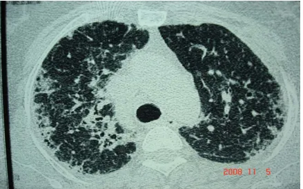
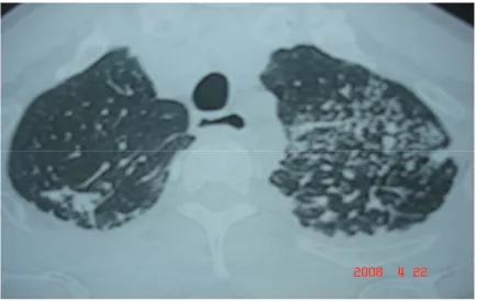
to alter patient management and if possible, to influence the outcome49. (Fig A, B, C
&D).
Pulmonary Function Tests
After completing the physical examination and HRCT of those who have fulfilled
criteria, were subjected to pulmonary function test. The various manoeuvres of PFT were
explained and practically demonstrated to them.
PFT was deferred to those persons who were suspected of having respiratory tract
infection. A course of antibiotics were given to them and asked to come for the next
session for the completion of PFT.
ATS RECOMMENDATION FOR PERFORMING SPIROMETRY51
Procedures for recording spirometry were done as per ATS recommendationthat
Preparation of the subject including asking about smoking, recent illness, medication use,
etc.and measurement of weight and height without shoes. The subject were then
instructed and demonstrated about the test including correct posture with head slightly
elevated, rapid and complete inhalation and exhalation with maximal force.
After assuming the correct posture nose clip was attached and mouthpiece was
placed in mouth with close lips around the mouthpiece. Patient was instructed to inhale
completely and rapidly with a pause of one second at Total Lung Capacity and then to
exhale maximally until no more air can be expelled while maintaining an upright posture.
Repeated instructions were given. Minimum of three manoeuvres were repeated; no more
than eight are usually required.
The variables recorded included FVC, FEV1 and FEV1 /FVC ratio. Three
technically satisfactory measurements were obtained in which FVC was reproducible
within 300 ml. The subject’s FVC was defined as the maximal FVC which was
determined before the DLCO test, as recommended by the ATS.
ATS RECOMMENDATION FOR PERFORMING DLCO53
DLCO measurements were performed in compliance with the American Thoracic
Society (ATS) guidelines. DLCO was measured using a single-breath technique. The
DLCO was routinely adjusted for haemoglobin if the value was outside the normal range.
Measurements of DLCO were made with a Collins automated system using a gas mixture
that contained 0.3% Methane tracer gas and 0.3% carbon monoxide. The breath holding
time was 10 seconds and the washout volume was 0.75 L. Each subject’s height and
The participants were seated, wearing nose clips, and performed at least two
DLCO manoeuvers separated by more than 4 minutes. The mean DLCO value from two
manoeuvers that matched within 3 ml/min/mm Hg was reported.
Before the test was performed, each subject was instructed about all of the
required manoeuvers. After the subject had adapted to the mouthpiece of the test
apparatus, four or five tidal volumes were recorded to determine a regular end expiratory
baseline. The subject was then asked to exhale as far as possible, to the point till maximal
exhalation had been reached (residual volume RV); making a rapid, maximal inhalation
within 2 to 2.5 seconds to VC continuing to hold the breath for 10 seconds while relaxing
against a closed glottis and exhaling rapidly. If after two attempts an acceptable
measurement could not be made, the procedure was then abandoned.
The values were interpreted as follows:
DLCO and DLCOHb : Normal(>80% predicted) Mild diffusion defect (65-80%),
Moderate diffusion defect(45-65%) and severe diffusion defect(<45%).
FVC:Normal(>80% predicted), Mild Reduction(60-80%), Moderate
Reduction(40-60%), Severe Reduction(<40%).
Statistical analysis
The following statistical analyses were performed to assess the strength of
association between the variables of the study by using Chi-Square Test . HRCT pattern
and DLCO in DPLD groups were compared by Mantel-Haenszel test for linear
V. RESULTS
In our study 55 patients were screened for Diffuse Parenchymal Lung Diseases
with collagen vascular diseases. Out of which 36 patients were taken up for the study
after satisfying eligible criteria. All were nonsmokers. Remaining patients were excluded
from the study group based on exclusion criteria.
AGE
In our study population age group ranges from 16-74 years.
Age Group ( Years ) No of Patients Percentage (%)
<35 36-45 46-55 >55
9
11
7
9
25
30.56
19.44
25
TOTAL 36 100
Maximum number of patients present presented between the ages of 36-45 years.
The mean age distribution found in our study is 44. The calculated standard deviation is
13.79.Hence 95% of confidence interval lies in the range of 44± 4.5 , indicating that most
SEX
The selected patients consisted of 66.67% Females and 33.33% males.
Male: Female sex ratio is 1:2 (Fig 2).
Disease classification
SEX No of Patients Percentage (%)
Male Female 12 24 33.33 66.67
Total 36 100
Disease No of patients Percentage (%)
Systemic Sclerosis
Rheumatoid Arthritis
Mixed Connective Tissue Disease
Systemic Lupus Erythematosus
Polymyositis/Dermatomyositis Sjogren’s syndrome 16 12 3 2 2 1 44.44 33.33 8.33 5.56 5.56 2.78
Clinical Features
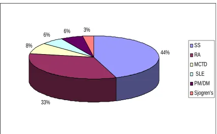
Rheumatoid arthritis and Systemic sclerosis form the bulk of the study population.
Both constitute more than 75% of the cases. Followed by three cases of MCTD and two
cases each from SLE and PM/DM and one case of Sjogren’s syndrome. (Fig 3).
The predominant symptom found in majority of the study cases are Exertional
dyspnoea(88.89%), followed by cough(50%) and chest pain(25%). Bibasilar crackles
heard in 16(55.56%) cases out of 36 cases.
Extra pulmonary symptoms are Dyspepsia, Dyphagia, Raynaud's phenomenon,
inflammatory arthritis, subcutaneous nodules, Sicca syndrome, Skin changes like rash,
discolouration and thickening, proximal muscle weakness, etc . These above-mentioned
symptoms present concomitantly in our cases and these symptoms have preceded or
accompany with respiratory illness.
No of Patients Clinical Features
Present(%) Absent(%)
Exertional Dyspnoea
Cough
Chest pain
Bibasilar crackles
32(88.89%)
18(50%)
9(25%)
16(44.44)
4(11.11%)
18(50%)
27(75%)
HRCT patterns
HRCT patterns No of patients Percentage (%)
RN GGO RGN RGHT RH RGH 14 7 6 5 3 1 38.9 19.5 16.6 13.9 8.3 2.8
Total 36 100
R-Reticular,G-Ground glass ,H-Honey combing,T-Traction bronchiectasis, N-Nodular
In our study population various combinations of Reticular, Ground glass opacity,
Honey combing, Traction bronchiectasis, and Nodular patterns were seen. Of which
ReticuloNodular pattern forms the most common presentation(38.9%).Followed by
Ground glass opacity(19.5%),RGN(16.6%)and RGHT(13.9%) . Less common being
RH(8.3%) and RGN(2.8%) (Fig 4).
Individual patterns in HRCT
HRCT pattern No of patients Percentage (%)
Reticular
Nodular
Ground glass opacity
Reticular pattern is the most frequent presentation (80.6%) followed by Nodular
pattern (58.3%) and GGO pattern(50%). Less common are Honeycombing and Traction
bronchiectasis(22.2% and 13.9% respectively) (Fig 5).
Spirometry
Forced Vital capacity(FVC) No patients Percentage(%)
Normal Mild restriction Moderate restriction Severe restriction 8 6 18 4 22.22 16.67 50 11.11
Total 36 100
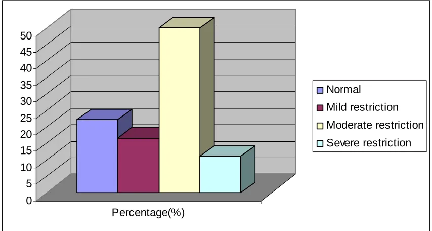
Out of 36 cases of study population 28 cases were presented with restriction
pattern (>75%) and remaining eight were presented with Normal FVC(22.22%). Forced
vital capacity suggestive of Moderate restriction constitutes 50% of study population
followed by Normal FVC(22.22%), Mild restriction(16.67%) and Severe restriction
(11.11%) in decreasing frequency(Fig 6).
Diffusing capacity
DLCO DLCO Hb
Diffusing capacity
No (%) No (%)
Normal
Mildly reduced diffusing capacity
Moderately reduced diffusing capacity
Severely reduced diffusing capacity
5 5 7 19 13.89 13.89 19.44 52.78 5 7 6 18 13.89 19.44 16.67 50
In DLCO Severely reduced diffusing capacity(52.78%) is the most common
presentation in study population followed by moderately reduced diffusing capacity and
then by equal presentations of Normal diffusing capacity and mildly reduced diffusing
capacity(13.89% each).
In DLCO for corrected Haemoglobin Severely reduced diffusing capacity(50%)
is the most common presentation in study population. Followed by mildly reduced
diffusing capacity(19.44%), moderately reduced diffusing capacity(16.67%) and Normal
diffusing capacity(13.89%). (Fig 7).
FVC Vs DLCO
Normal Mild Moderate Severe Total
FVC
No % No % No % No % No
Normal 3 37.5 3 37.5 2 25 - - 8
Mild 1 16.7 1 16.7 1 16.7 3 49.9 6
Moderate 1 5.6 1 5.6 4 22.1 12 66.7 18
Severe - - - - - - 4 100 4
FVC Vs DLCOHb
Normal Mild Moderate Severe Total
FVC
No % No % No % No % No
Normal 3 37.5 5 62.5 - - - - 8
Mild 1 16.7 1 16.7 1 16.7 3 49.9 6
Moderate 1 5.6 1 5.6 5 27.7 11 61.1 18
In both DLCO and DLCOHb Severly reducing diffusing capacity is associated
with severe reduction in FVC in all 4 cases(100%). Out of five normal DLCO and
DLCOHb patients two patients exhibit reduction in FVC (Fig 8).
HRCT Vs DLCO
Normal Mild Moderate Severe Total
HRCT patterns
No % No % No % No % No
RN
Reticulo Nodular
2 14.3 3 21.4 3 21.4 6 42.9 14
GGO
Ground Glass Opacity
2 28.6 2 28.6 3 42.8 - - 7
RNG
Reticulo Nodular with Ground Glass Opacity
1 16.7 - - 1 16.7 4 66.6 6
RGHT
Reticular, Ground glass opacity, Honeycombing &Traction bronchiectasis
- - - - - - 5 100 5
RH
Reticular & Honeycombing
- - - - - - 3 100 3
RGH
Reticular, Ground glass opacity& Honeycombing
- - - - - - 1 100 1
The various presentations of HRCT patterns in each individual were correlated
with Diffusing capacity(DLCO). Out of 14 cases of RN(ReticuloNodular) pattern 6
cases(42.9%) were present in severely reduced diffusing capacity followed by 3
cases(21.4%) each in mild and moderately reduced diffusing capacity, then 2
cases(14.3%) in normal diffusing capacity.
Out of 7 cases of GGO(Ground Glass Opacity) 3 cases(42.8%) were present in
moderately reduced diffusing capacity followed by 2 cases(28.6%) each in normal and
Out of 6 cases of RGN (Reticular, Ground glass opacity, Nodular) pattern 4
cases(66.6%) were present in severely reduced diffusing capacity followed by one case
(16.7%) each in Normal diffusing capacity and moderately reduced diffusing capacity.
All 5 cases(100%) of RGHT (Reticular, Ground glass opacity, Honeycombing
and Traction bronchiectasis) pattern present only in severely reduced diffusing capacity.
Out of 3 cases of RH (Reticular, Honeycombing) pattern all (100%) were present
in severely reduced diffusing capacity.
1 case(100%) of RGH (Reticular, Ground glass opacity, Honeycombing) pattern
was present in severely reduced diffusing capacity (Fig9).
HRCT Vs DLCOHb
Normal Mild Moderate Severe Total
HRCT patterns
No % No % No % No % No
RN
Reticulo Nodular
2 14.3 3 21.4 3 21.4 6 42.9 14
GGO
Ground Glass Opacity
2 28.6 4 57.1 1 16.7 - - 7
RNG
Reticulo Nodular with Ground Glass Opacity
1 16.7 - - 1 16.7 4 66.6 6
RGHT
Reticular, Ground glass opacity, Honeycombing &Traction bronchiectasis
- - - - - - 5 100 5
RH
Reticular & Honeycombing
- - - - 1 33.3 2 66.7 3
RGH
Reticular, Ground glass opacity& Honeycombing
The various presentations of HRCT pattern in each individual is correlated with
Diffusing capacity for corrected Haemoglobin(DLCOHb).
Out of 14 cases of RN(ReticuloNodular) pattern 6 cases(42.9%) were present in
severely reduced diffusing capacity followed by 3 cases(21.4%) each in Mild and
moderately reduced diffusing capacity, then 2 cases(14.3%) in Normal diffusing capacity.
Out of 7 cases of GGO(Ground Glass Opacity) 4 cases(57.1%) were present in
mildly reduced diffusing capacity followed by 2 cases(28.6%) in Normal and 1 case
(14.3%) in moderately reduced diffusing capacity.
Out of 6 cases of RGN (Reticular, Ground glass opacity, Nodular) pattern 4
cases(66.6%) were present in severely reduced diffusing capacity followed by one case
(16.7%) each in Normal diffusing capacity and moderately reduced diffusing capacity.
All 5 cases(100%) of RGHT (Reticular, Ground glass opacity, Honeycombing
and Traction bronchiectasis) pattern present only in severely reduced diffusing capacity.
Out of 3 cases of RH (Reticular, Honeycombing) pattern 2 cases (66.7%) were
present in severely reduced diffusing capacity followed by 1 case in(33.3%) moderately
reduced diffusing capacity .
1 case(100%) of RGH (Reticular, Ground glass opacity, Honeycombing) pattern
INDIVIDUAL HRCT PATTERN Vs DLCO
Normal Mild Moderate Severe
Individual HRCT
pattern
No % No % No % No % Total
Reticular 3 10.3 3 10.3 4 13.8 19 65.6 29
Nodular 3 14.3 3 14.3 4 19 11 52.4 21
GGO 3 16.7 2 11.1 3 16.7 10 55.6 18
Honeycombing - - - 8 100 8
Traction Bronchiectasis
- - - 5 100 5
Out of 29 cases of Reticular pattern 19 cases(65.6%) were present in Severely
reduced diffusing capacity followed by 4 cases(13.8%) in moderately reduced diffusing
capacity and 3 cases(10.3%) each in normal diffusing capacity and mildly reduced
diffusing capacity.
Out of 21 cases of Nodular pattern 11 cases(52.4%) were present in Severely
reduced diffusing capacity followed by 4 cases(19%) in moderately reduced diffusing
capacity and 3 cases(14.3%) each in normal diffusing capacity and mildly reduced
diffusing capacity.
diffusing capacity and normal diffusing capacity and 2 cases(11.1%) in mildly reduced
diffusing capacity.
All 8 cases(100%) of Honeycombing pattern 19 cases were present in Severely
reduced diffusing capacity.
All 5 cases(100%) of Traction Bronchiectasis pattern were present in Severely
reduced diffusing capacity (Fig 11).
INDIVIDUAL HRCT PATTERN Vs DLCOHb
Normal Mild Moderate Severe
Individual HRCT
pattern
No % No % No % No % Total
Reticular 3 10.3 3 10.3 5 17.2 18 62.2 29
Nodular 3 14.3 3 14.3 4 19 11 52.4 21
GGO 3 16.7 3 16.7 2 11.1 10 55.6 18
Honeycombing - - - - 1 12.5 7 87.5 8
Traction Bronchiectasis
- - - 5 100 5
Out of 29 cases of Reticular pattern 18 cases(62.2%) were present in Severely
capacity and 3 cases(10.3%) each in normal diffusing capacity and mildly reduced
diffusing capacity.
Out of 21 cases of Nodular pattern 11 cases(52.4%) were present in Severely
reduced diffusing capacity followed by 4 cases(19%) in moderately reduced diffusing
capacity and 3 cases(14.3%) each in normal diffusing capacity and mildly reduced
diffusing capacity.
Out of 18 cases of GGO pattern 10 cases(55.6%) were present in Severely
reduced diffusing capacity followed by 3 cases(16.7%) each in mildly reduced diffusing
capacity and normal diffusing capacity and 2 cases(11.1%) in moderately reduced
diffusing capacity.
Out of 8 cases of Honeycombing pattern 7 cases(87.5%) were present in Severely
reduced diffusing capacity and one case(12.5%) in moderately reduced diffusing
capacity.
All 5 cases(100%) of Traction Bronchiectasis pattern were present in Severely
reduced diffusing capacity (Fig 11).
Significant p value association occurs between the HRCT patterns and
DLCOHb at 5% level by using Chi- Square Mantel-Haenszel test for linear
association.
Significant p value association occurs between the HRCT patterns and FVC