This is a repository copy of
Imaging tests for the detection of osteomyelitis : a systematic
review
.
White Rose Research Online URL for this paper:
http://eprints.whiterose.ac.uk/153018/
Version: Published Version
Article:
Llewellyn, Alexis orcid.org/0000-0003-4569-5136, Jones-Diette, Julie
orcid.org/0000-0003-1769-8612, Kraft, Jeannette et al. (3 more authors) (2019) Imaging
tests for the detection of osteomyelitis : a systematic review. Health technology
assessment. pp. 1-128. ISSN 2046-4924
https://doi.org/10.3310/hta23610
[email protected] https://eprints.whiterose.ac.uk/
Reuse
Items deposited in White Rose Research Online are protected by copyright, with all rights reserved unless indicated otherwise. They may be downloaded and/or printed for private study, or other acts as permitted by national copyright laws. The publisher or other rights holders may allow further reproduction and re-use of the full text version. This is indicated by the licence information on the White Rose Research Online record for the item.
Takedown
If you consider content in White Rose Research Online to be in breach of UK law, please notify us by
Journals Library
DOI 10.3310/hta23610
Imaging tests for the detection of
osteomyelitis: a systematic review
Alexis Llewellyn, Julie Jones-Diette, Jeannette Kraft, Colin Holton,
Melissa Harden and Mark Simmonds
Health Technology Assessment
Volume 23 • Issue 61 • October 2019Imaging tests for the detection of
osteomyelitis: a systematic review
Alexis Llewellyn
o
,
1
Julie Jones-Diette
o
,
1
Jeannette Kraft
o
,
2
Colin Holton,
2
Melissa Harden
1
and Mark Simmonds
o
1
*
1
Centre for Reviews and Dissemination, University of York, York, UK
2
Leeds Teaching Hospitals NHS Trust, Leeds, UK
*Corresponding author
Declared competing interests of authors:none
Published October 2019
DOI: 10.3310/hta23610
This report should be referenced as follows:
Llewellyn A, Jones-Diette J, Kraft J, Holton C, Harden M, Simmonds M. Imaging tests for the detection of osteomyelitis: a systematic review.Health Technol Assess2019;23(61).
Health Technology Assessmentis indexed and abstracted inIndex Medicus/MEDLINE,Excerpta Medica/EMBASE,Science Citation Index Expanded(SciSearch®) andCurrent Contents®/
Health Technology Assessment
HTA/HTA TARISSN 1366-5278 (Print)
ISSN 2046-4924 (Online)
Impact factor: 3.819
Health Technology Assessmentis indexed in MEDLINE, CINAHL, EMBASE, The Cochrane Library and the Clarivate Analytics Science Citation Index.
This journal is a member of and subscribes to the principles of the Committee on Publication Ethics (COPE) (www.publicationethics.org/).
Editorial contact: [email protected]
The full HTA archive is freely available to view online at www.journalslibrary.nihr.ac.uk/hta. Print-on-demand copies can be purchased from the report pages of the NIHR Journals Library website: www.journalslibrary.nihr.ac.uk
Criteria for inclusion in theHealth Technology Assessmentjournal
Reports are published inHealth Technology Assessment(HTA) if (1) they have resulted from work for the HTA programme, and (2) they are of a sufficiently high scientific quality as assessed by the reviewers and editors.
Reviews inHealth Technology Assessmentare termed‘systematic’when the account of the search appraisal and synthesis methods (to minimise biases and random errors) would, in theory, permit the replication of the review by others.
HTA programme
Health Technology Assessment (HTA) research is undertaken where some evidence already exists to show that a technology can be effective and this needs to be compared to the current standard intervention to see which works best. Research can evaluate any intervention used in the treatment, prevention or diagnosis of disease, provided the study outcomes lead to findings that have the potential to be of direct benefit to NHS patients. Technologies in this context mean any method used to promote health; prevent and treat disease; and improve rehabilitation or long-term care. They are not confined to new drugs and include any intervention used in the treatment, prevention or diagnosis of disease.
The journal is indexed in NHS Evidence via its abstracts included in MEDLINE and its Technology Assessment Reports inform National Institute for Health and Care Excellence (NICE) guidance. HTA research is also an important source of evidence for National Screening Committee (NSC) policy decisions.
This report
The research reported in this issue of the journal was funded by the HTA programme as project number 16/103/03. The contractual start date was in July 2017. The draft report began editorial review in October 2018 and was accepted for publication in March 2019. The authors have been wholly responsible for all data collection, analysis and interpretation, and for writing up their work. The HTA editors and publisher have tried to ensure the accuracy of the authors’report and would like to thank the reviewers for their constructive comments on the draft document. However, they do not accept liability for damages or losses arising from material published in this report.
This report presents independent research funded by the National Institute for Health Research (NIHR). The views and opinions expressed by authors in this publication are those of the authors and do not necessarily reflect those of the NHS, the NIHR, NETSCC, the HTA programme or the Department of Health and Social Care. If there are verbatim quotations included in this publication the views and opinions expressed by the interviewees are those of the interviewees and do not necessarily reflect those of the authors, those of the NHS, the NIHR, NETSCC, the HTA programme or the Department of Health and Social Care.
© Queen’s Printer and Controller of HMSO 2019. This work was produced by Llewellynet al.under the terms of a commissioning
contract issued by the Secretary of State for Health and Social Care. This issue may be freely reproduced for the purposes of private research and study and extracts (or indeed, the full report) may be included in professional journals provided that suitable acknowledgement is made and the reproduction is not associated with any form of advertising. Applications for commercial reproduction should be addressed to: NIHR Journals Library, National Institute for Health Research, Evaluation, Trials and Studies Coordinating Centre, Alpha House, University of Southampton Science Park, Southampton SO16 7NS, UK.
NIHR Journals Library Editor-in-Chief
Professor Ken Stein Professor of Public Health, University of Exeter Medical School, UK
NIHR Journals Library Editors
Professor John Powell Chair of HTA and EME Editorial Board and Editor-in-Chief of HTA and EME journals.
Consultant Clinical Adviser, National Institute for Health and Care Excellence (NICE), UK, and Senior Clinical Researcher, Nuffield Department of Primary Care Health Sciences, University of Oxford, UK
Professor Andrée Le May Chair of NIHR Journals Library Editorial Group (HS&DR, PGfAR, PHR journals) and Editor-in-Chief of HS&DR, PGfAR, PHR journals
Professor Matthias Beck Professor of Management, Cork University Business School, Department of Management and Marketing, University College Cork, Ireland
Dr Tessa Crilly Director, Crystal Blue Consulting Ltd, UK
Dr Eugenia Cronin Senior Scientific Advisor, Wessex Institute, UK
Dr Peter Davidson Consultant Advisor, Wessex Institute, University of Southampton, UK
Ms Tara Lamont Director, NIHR Dissemination Centre, UK
Dr Catriona McDaid Senior Research Fellow, York Trials Unit, Department of Health Sciences, University of York, UK
Professor William McGuire Professor of Child Health, Hull York Medical School, University of York, UK
Professor Geoffrey Meads Professor of Wellbeing Research, University of Winchester, UK
Professor John Norrie Chair in Medical Statistics, University of Edinburgh, UK
Professor James Raftery Professor of Health Technology Assessment, Wessex Institute, Faculty of Medicine, University of Southampton, UK
Dr Rob Riemsma Reviews Manager, Kleijnen Systematic Reviews Ltd, UK
Professor Helen Roberts Professor of Child Health Research, UCL Great Ormond Street Institute of Child Health, UK
Professor Jonathan Ross Professor of Sexual Health and HIV, University Hospital Birmingham, UK
Professor Helen Snooks Professor of Health Services Research, Institute of Life Science, College of Medicine, Swansea University, UK
Professor Ken Stein Professor of Public Health, University of Exeter Medical School, UK
Professor Jim Thornton Professor of Obstetrics and Gynaecology, Faculty of Medicine and Health Sciences, University of Nottingham, UK
Professor Martin Underwood Warwick Clinical Trials Unit, Warwick Medical School, University of Warwick, UK
Please visit the website for a list of editors: www.journalslibrary.nihr.ac.uk/about/editors
Editorial contact: [email protected]
Abstract
Imaging tests for the detection of osteomyelitis:
a systematic review
Alexis Llewellyn
o
,
1Julie Jones-Diette
o
,
1Jeannette Kraft
o
,
2Colin Holton,
2Melissa Harden
1and Mark Simmonds
o
1*
1Centre for Reviews and Dissemination, University of York, York, UK
2Leeds Teaching Hospitals NHS Trust, Leeds, UK
*Corresponding author [email protected]
Background:Osteomyelitis is an infection of the bone. Medical imaging tests, such as radiography, ultrasound, magnetic resonance imaging (MRI), single-photon emission computed tomography (SPECT) and positron emission tomography (PET), are often used to diagnose osteomyelitis.
Objectives:To systematically review the evidence on the diagnostic accuracy, inter-rater reliability and implementation of imaging tests to diagnose osteomyelitis.
Data sources:We conducted a systematic review of imaging tests to diagnose osteomyelitis. We searched MEDLINE and other databases from inception to July 2018.
Review methods:Risk of bias was assessed with QUADAS-2 [quality assessment of diagnostic accuracy studies (version 2)]. Diagnostic accuracy was assessed using bivariate regression models. Imaging tests were compared. Subgroup analyses were performed based on the location and nature of the suspected osteomyelitis. Studies of children, inter-rater reliability and implementation outcomes were synthesised narratively.
Results:Eighty-one studies were included (diagnostic accuracy: 77 studies; inter-rater reliability: 11 studies; implementation: one study; some studies were included in two reviews). One-quarter of diagnostic accuracy studies were rated as being at a high risk of bias. In adults, MRI had high diagnostic accuracy [95.6% sensitivity, 95% confidence interval (CI) 92.4% to 97.5%; 80.7% specificity, 95% CI 70.8% to 87.8%]. PET also had high accuracy (85.1% sensitivity, 95% CI 71.5% to 92.9%; 92.8% specificity, 95% CI 83.0% to 97.1%), as did SPECT (95.1% sensitivity, 95% CI 87.8% to 98.1%; 82.0% specificity, 95% CI 61.5% to 92.8%). There was similar diagnostic performance with MRI, PET and SPECT. Scintigraphy (83.6% sensitivity, 95% CI 71.8% to 91.1%; 70.6% specificity, 57.7% to 80.8%), computed tomography (69.7% sensitivity, 95% CI 40.1% to 88.7%; 90.2% specificity, 95% CI 57.6% to 98.4%) and radiography (70.4% sensitivity, 95% CI 61.6% to 77.8%; 81.5% specificity, 95% CI 69.6% to 89.5%) all had generally inferior diagnostic accuracy. Technetium-99m hexamethylpropyleneamine oxime white blood cell scintigraphy (87.3%
sensitivity, 95% CI 75.1% to 94.0%; 94.7% specificity, 95% CI 84.9% to 98.3%) had higher diagnostic accuracy, similar to that of PET or MRI. There was no evidence that diagnostic accuracy varied by scan location or cause of osteomyelitis, although data on many scan locations were limited. Diagnostic accuracy in diabetic foot patients was similar to the overall results. Only three studies in children were identified; results were too limited to draw any conclusions. Eleven studies evaluated inter-rater reliability. MRI had acceptable inter-rater reliability. We found only one study on test implementation and no evidence on patient preferences or cost-effectiveness of imaging tests for osteomyelitis.
Limitations:Most studies included < 50 participants and were poorly reported. There was limited evidence for children, ultrasonography and on clinical factors other than diagnostic accuracy.
DOI: 10.3310/hta23610 HEALTH TECHNOLOGY ASSESSMENT 2019 VOL. 23 NO. 61
© Queen’s Printer and Controller of HMSO 2019. This work was produced by Llewellynet al.under the terms of a commissioning contract issued by the Secretary of State for Health and Social Care. This issue may be freely reproduced for the purposes of private research and study and extracts (or indeed, the full report) may be included in professional journals provided that suitable acknowledgement is made and the reproduction is not associated with any form of advertising. Applications for commercial reproduction should be addressed to: NIHR Journals Library, National Institute for Health Research, Evaluation, Trials and Studies Coordinating Centre, Alpha House, University of Southampton Science Park, Southampton SO16 7NS, UK.
Conclusions:Osteomyelitis is reliably diagnosed by MRI, PET and SPECT. No clear reason to prefer one test over the other in terms of diagnostic accuracy was identified. The wider availability of MRI machines, and the fact that MRI does not expose patients to harmful ionising radiation, may mean that MRI is preferable in most cases. Diagnostic accuracy does not appear to vary with the potential cause of osteomyelitis or with the body part scanned. Considerable uncertainty remains over the diagnostic accuracy of imaging tests in children. Studies of diagnostic accuracy in children, particularly using MRI and ultrasound, are needed.
Study registration:This study is registered as PROSPERO CRD42017068511.
Funding:This project was funded by the National Institute for Health Research Health Technology Assessment programme and will be published in full inHealth Technology Assessment; Vol. 23, No. 61. See the NIHR Journals Library website for further project information.
ABSTRACT
Contents
List of tables ix
List of figures xi
Glossary xiii
List of abbreviations xv
Plain English summary xvii
Scientific summary xix
Chapter 1Background 1
Osteomyelitis 1
Diagnostic imaging for osteomyelitis 1
Current diagnostic and treatment practice 2
Pathway to diagnosis in the NHS 2
Existing review evidence 3
Chapter 2Aims and objectives 5
Chapter 3Methods 7
Literature searches 7
Study selection 7
Participants 7
Index tests 7
Reference standards 8
Outcomes 8
Study designs 8
Data extraction 8
Quality assessment 9
Synthesis 9
Diagnostic meta-analysis 9
Inter-rater reliability and implementation review 11
Deviations from the protocol 11
Role of patient and clinical advisors 11
Chapter 4Results 13
Quantity and quality of research available 13
Included studies 13
Excluded studies 14
Assessment of diagnostic accuracy 14
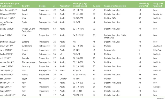
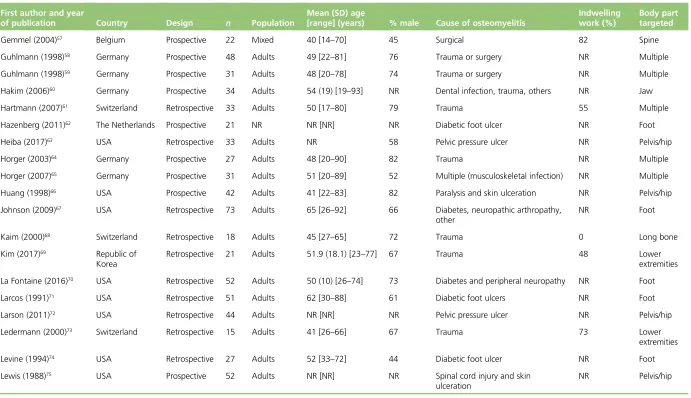
Characteristics of included studies 14
Critical appraisal 19
Synthesis of diagnostic accuracy in adults 22
Comparisons between tests 24
Synthesis of specific imaging tests 26
Other factors and subgroups 32
DOI: 10.3310/hta23610 HEALTH TECHNOLOGY ASSESSMENT 2019 VOL. 23 NO. 61
© Queen’s Printer and Controller of HMSO 2019. This work was produced by Llewellynet al.under the terms of a commissioning contract issued by the Secretary of State for Health and Social Care. This issue may be freely reproduced for the purposes of private research and study and extracts (or indeed, the full report) may be included in professional journals provided that suitable acknowledgement is made and the reproduction is not associated with any form of advertising. Applications for commercial reproduction should be addressed to: NIHR Journals Library, National Institute for Health Research, Evaluation, Trials and Studies Coordinating Centre, Alpha House, University of Southampton Science Park, Southampton SO16 7NS, UK.
Synthesis of diagnostic accuracy in people with diabetic foot ulcers 35
Comparisons of tests 36
Synthesis of diagnostic accuracy for osteomyelitis not related to diabetes 37
Comparisons of tests 39
Studies not included in the quantitative synthesis 39
Synthesis of studies in children 40
Review of inter-rater reliability 42
Characteristics of included studies 42
Critical appraisal 42
Results of inter-rater reliability studies 44
Review of implementation 44
Summary of previous systematic reviews 46
Clinical effectiveness summary and conclusions 46
Summary of included studies 46
General conclusions from synthesis 51
Diagnostic accuracy by cause or nature of osteomyelitis 51
Studies of children, inter-rater reliability and implementation 52
Chapter 5Discussion 53
Statement of principal findings 53
Diagnostic accuracy in adults 53
Diagnostic tests in children 54
Inter-rater reliability and implementation 54
Strengths and limitations of the assessment 54
Strengths 54
Limitations 55
Uncertainties 56
Considerations from patient representatives 56
Chapter 6Conclusions 57
Implications for health care 57
Suggested research priorities 58
Acknowledgements 59
References 61
Appendix 1Literature searches 73
Appendix 2Results of critical appraisal of diagnostic accuracy studies
(QUADAS-2) 91
Appendix 3Participant selection criteria in diagnostic accuracy studies 105
Appendix 4Results of diagnostic meta-analyses 111
CONTENTS
List of tables
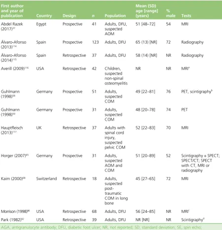
TABLE 1 Study design and participant characteristics 15
TABLE 2 Diagnostic accuracy studies according to test and body part targeted 19
TABLE 3 Summary of critical appraisal of diagnostic accuracy studies 19
TABLE 4 Results of univariate meta-analyses 26
TABLE 5 Summary of regression model to compare imaging tests 27
TABLE 6 Summary of diagnostic accuracy of ultrasound studies 30
TABLE 7 Comparison of tests in people with diabetic foot ulcers 36
TABLE 8 Comparison of tests in people without diabetes 39
TABLE 9 Studies not included in the quantitative synthesis 40
TABLE 10 Diagnostic accuracy results of studies in children 41
TABLE 11 Characteristics of inter-rater reliability studies 43
TABLE 12 Critical appraisal of inter-rater reliability studies 44
TABLE 13 Results of inter-rater reliability studies 45
TABLE 14 Summary of previous systematic reviews 47
TABLE 15 The QUADAS-2 signalling questions for risk-of-bias assessment 91
TABLE 16 The QUADAS-2 risk-of-bias decisions 92
TABLE 17 The QUADAS-2 applicability decisions 102
TABLE 18 Participant selection criteria in diagnostic accuracy studies 105
TABLE 19 Summary of diagnostic accuracy studies 111
TABLE 20 Results from logistic regression model to compare tests 115
TABLE 21 Summary of studies of scintigraphy 115
TABLE 22 Results from logistic regression model to compare tests 118
TABLE 23 Univariate meta-analyses of studies of people with diabetic foot ulcers 122
TABLE 24 Results of logistic regression to compare tests–diabetic foot ulcers 123
TABLE 25 Univariate meta-analysis–patients without diabetes 126
TABLE 26 Results of logistic regression to compare tests–patients without diabetes 127
DOI: 10.3310/hta23610 HEALTH TECHNOLOGY ASSESSMENT 2019 VOL. 23 NO. 61
© Queen’s Printer and Controller of HMSO 2019. This work was produced by Llewellynet al.under the terms of a commissioning contract issued by the Secretary of State for Health and Social Care. This issue may be freely reproduced for the purposes of private research and study and extracts (or indeed, the full report) may be included in professional journals provided that suitable acknowledgement is made and the reproduction is not associated with any form of advertising. Applications for commercial reproduction should be addressed to: NIHR Journals Library, National Institute for Health Research, Evaluation, Trials and Studies Coordinating Centre, Alpha House, University of Southampton Science Park, Southampton SO16 7NS, UK.
List of figures
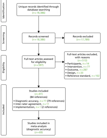
FIGURE 1 A PRISMA flow diagram showing study selection 13
FIGURE 2 Summary ROC plot of diagnostic accuracy in adults 22
FIGURE 3 Bivariate meta-analysis of sensitivity and specificity: all adult studies 23
FIGURE 4 Summary HSROC curves: all adult studies 23
FIGURE 5 Forest plots of sensitivity and specificity for MRI 25
FIGURE 6 Comparing DORs within studies 27
FIGURE 7 Summary ROC plot for scintigraphy studies 28
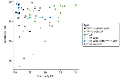
FIGURE 8 Diagnostic accuracy of scintigraphy according to type of test performed 29
FIGURE 9 Diagnostic accuracy of SPECT studies according to type of SPECT used 30
FIGURE 10 Diagnostic accuracy results for combination tests 31
FIGURE 11 Diagnostic odds ratios for combination tests 31
FIGURE 12 Sensitivity and specificity according to whether or not any imaging
test was used before the main test 33
FIGURE 13 Sensitivity and specificity according to whether the study was of AOM
or COM 34
FIGURE 14 Bivariate meta-analysis according to acute or chronic status 34
FIGURE 15 Sensitivity and specificity in studies of patients with diabetic foot ulcers 35
FIGURE 16 Bivariate analysis of sensitivity and specificity in people with diabetic
foot ulcers 36
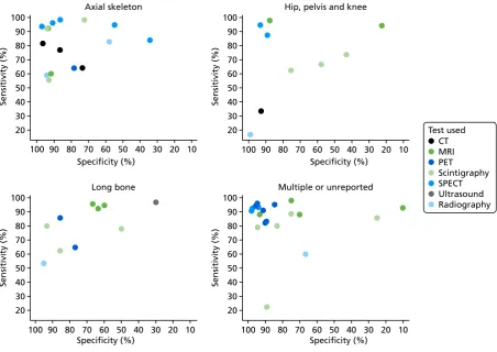
FIGURE 17 Sensitivity and specificity according to scan location 37
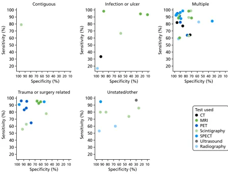
FIGURE 18 Sensitivity and specificity according to likely cause of osteomyelitis 38
FIGURE 19 Bivariate meta-analysis in studies of people without diabetes 39
FIGURE 20 Summary of risk of bias and applicability assessment 91
FIGURE 21 Summary of PPV and NPV in adults 113
FIGURE 22 Summary of PPV and NPV from bivariate analysis 113
FIGURE 23 Comparisons of DORs within studies 114
DOI: 10.3310/hta23610 HEALTH TECHNOLOGY ASSESSMENT 2019 VOL. 23 NO. 61
© Queen’s Printer and Controller of HMSO 2019. This work was produced by Llewellynet al.under the terms of a commissioning contract issued by the Secretary of State for Health and Social Care. This issue may be freely reproduced for the purposes of private research and study and extracts (or indeed, the full report) may be included in professional journals provided that suitable acknowledgement is made and the reproduction is not associated with any form of advertising. Applications for commercial reproduction should be addressed to: NIHR Journals Library, National Institute for Health Research, Evaluation, Trials and Studies Coordinating Centre, Alpha House, University of Southampton Science Park, Southampton SO16 7NS, UK.
FIGURE 24 Association between specificity and incidence of osteomyelitis in MRI
studies 117
FIGURE 25 Sensitivity and specificity for PET and PET-CT studies 117
FIGURE 26 Sensitivity and specificity according to test and reference standard used 118
FIGURE 27 Sensitivity and specificity according to risk of bias owing to patient
selection 119
FIGURE 28 Sensitivity and specificity according to risk of bias owing to index test 119
FIGURE 29 Sensitivity and specificity according to presence of indwelling metalwork 120
FIGURE 30 The PPV and NPV in studies of people with diabetic foot ulcers 120
FIGURE 31 The HSROC curves in studies of people with diabetic foot ulcers 121
FIGURE 32 Summary PPV and NPV in studies of people with diabetic foot ulcers 121
FIGURE 33 Meta-analysis of scintigraphy studies in people with diabetic foot ulcers 122
FIGURE 34 Difference in DOR between studies–patients with diabetic foot ulcers 123
FIGURE 35 The PPV and NPV by test location 124
FIGURE 36 The PPV and NPV by cause of osteomyelitis 124
FIGURE 37 Summary sensitivity and specificity by test location 125
FIGURE 38 Summary sensitivity and specificity by cause of osteomyelitis 125
FIGURE 39 Comparisons of DORs across tests–patients without diabetes 127
LIST OF FIGURES
Glossary
Diagnostic odds ratio Ratio of odds of sensitivity to odds of specificity (summarises diagnostic accuracy).
Diphosphono-1,2-propanodicarboxylic acid A substance used in scintigraphy.
False negative Incorrect negative test result–an affected individual with a negative test result.
False positive Incorrect positive test result–an unaffected individual with a positive test result.
Fludeoxyglucose A radiopharmaceutical used in positron emission tomography.
Hexamethylpropyleneamine oxime A substance used in scintigraphy.
Histopathology The microscopic study of tissue samples to enable the diagnosis of osteomyelitis.
(Hydroxy)methylene diphosphonate A substance used in scintigraphy.
Index test The test for which diagnostic accuracy is being evaluated.
Indium-111 A radioisotope used in scintigraphy.
Meta-analysis Statistical (or quantitative) techniques used to combine the results of two or more studies and obtain a combined estimate of effect.
Microbiology A laboratory method used to diagnose osteomyelitis; a microbiological culture involves multiplying microbial organisms by letting them reproduce under controlled laboratory conditions.
Negative predictive value Proportion of people who test negative (for osteomyelitis) who do not have the condition.
Positive predictive value Proportion of people who test positive who have the condition.
Positive rate Proportion of people with a positive index test result (i.e. who might be diagnosed with osteomyelitis).
Sensitivity Proportion of people with the condition (osteomyelitis) who are correctly diagnosed.
Specificity Proportion of people without the condition who are correctly diagnosed.
Technetium-99m A radioisotope used in scintigraphy and single-photon emission computed tomography.
True negative A correct negative test result–an unaffected individual with a negative test result.
True positive A correct positive test result–an affected individual with a positive test result.
DOI: 10.3310/hta23610 HEALTH TECHNOLOGY ASSESSMENT 2019 VOL. 23 NO. 61
© Queen’s Printer and Controller of HMSO 2019. This work was produced by Llewellynet al.under the terms of a commissioning contract issued by the Secretary of State for Health and Social Care. This issue may be freely reproduced for the purposes of private research and study and extracts (or indeed, the full report) may be included in professional journals provided that suitable acknowledgement is made and the reproduction is not associated with any form of advertising. Applications for commercial reproduction should be addressed to: NIHR Journals Library, National Institute for Health Research, Evaluation, Trials and Studies Coordinating Centre, Alpha House, University of Southampton Science Park, Southampton SO16 7NS, UK.
List of abbreviations
18F-FDG fludeoxyglucose
3D three-dimensional
99mTc technetium-99m
AGA antigranulocyte antibody AOM acute osteomyelitis
CDSR Cochrane Database of Systematic Reviews
CENTRAL Cochrane Central Register of Controlled Trials
CI confidence interval
CINAHL Cumulative Index to Nursing and Allied Health
COM chronic osteomyelitis CRD Centre for Reviews and
Dissemination
CT computed tomography
DARE Database of Abstracts of Reviews of Effects
DOR diagnostic odds ratio
DPD diphosphono-1,
2-propanodicarboxylic acid GP general practitioner
(H)MDP (hydroxy)methylene diphosphonate
HMPAO hexamethylpropyleneamine oxime HSROC hierarchical summary receiver
operating characteristic HTA Health Technology Assessment MDP methylene diphosphonate MeSH medical subject heading MRI magnetic resonance imaging NICE National Institute for Health and
Care Excellence
NPV negative predictive value PET positron emission tomography PPV positive predictive value
PR positive rate
PRISMA Preferred Reporting Items for Systematic Reviews and Meta-Analyses
QUADAS-2 quality assessment of diagnostic accuracy studies (version 2) ROC receiver operating characteristic SPECT single-photon emission computed
tomography WBC white blood cell
DOI: 10.3310/hta23610 HEALTH TECHNOLOGY ASSESSMENT 2019 VOL. 23 NO. 61
© Queen’s Printer and Controller of HMSO 2019. This work was produced by Llewellynet al.under the terms of a commissioning contract issued by the Secretary of State for Health and Social Care. This issue may be freely reproduced for the purposes of private research and study and extracts (or indeed, the full report) may be included in professional journals provided that suitable acknowledgement is made and the reproduction is not associated with any form of advertising. Applications for commercial reproduction should be addressed to: NIHR Journals Library, National Institute for Health Research, Evaluation, Trials and Studies Coordinating Centre, Alpha House, University of Southampton Science Park, Southampton SO16 7NS, UK.
Plain English summary
O
steomyelitis is an infection of the bone and is treated with antibiotics. Left untreated, it can cause permanent damage and can lead to amputation.The best method to diagnose osteomyelitis is to take a bone sample (bone biopsy) but this is invasive and painful. Imaging may help target the best locations for biopsies or remove the need for a biopsy entirely. Several methods are available, including radiography, ultrasound, magnetic resonance imaging (MRI), single-photon emission computed tomography (SPECT) and positron emission tomography (PET).
This project systematically reviewed the relevant literature to determine which tests are the most accurate and relevant for clinical practice. All types of patients and all types of osteomyelitis were reviewed. Studies were pooled using statistical methods (meta-analyses) to estimate the overall accuracy of the imaging tests.
The review identified 81 studies and concluded that MRI, PET and SPECT all had similar accuracy, correctly identifying over 85% of people who did have osteomyelitis and over 80% of people who did not have osteomyelitis. Radiography and computed tomography were less accurate. Modern forms of scintigraphy have accuracy similar to PET or MRI.
There was no evidence that the accuracy of the imaging tests was different depending on the cause of osteomyelitis or which body part was affected. In particular, diagnostic accuracy in people with diabetic foot ulcers was similar to other types of osteomyelitis in adults. There was not enough evidence about which tests are most accurate in children, so further studies in children are needed.
DOI: 10.3310/hta23610 HEALTH TECHNOLOGY ASSESSMENT 2019 VOL. 23 NO. 61
© Queen’s Printer and Controller of HMSO 2019. This work was produced by Llewellynet al.under the terms of a commissioning contract issued by the Secretary of State for Health and Social Care. This issue may be freely reproduced for the purposes of private research and study and extracts (or indeed, the full report) may be included in professional journals provided that suitable acknowledgement is made and the reproduction is not associated with any form of advertising. Applications for commercial reproduction should be addressed to: NIHR Journals Library, National Institute for Health Research, Evaluation, Trials and Studies Coordinating Centre, Alpha House, University of Southampton Science Park, Southampton SO16 7NS, UK.
Scientific summary
Background
Osteomyelitis is an infection of the bone and bone marrow that may result in bone infarction and loss of limb or joint function and, in extreme cases, may necessitate amputation of the affected limb. In children, osteomyelitis may also inhibit limb growth. Osteomyelitis is common in people with vascular deficiency, such as adults with diabetes mellitus.
Patients usually present with a range of symptoms including swelling, joint pain and fever. These symptoms are often not specific to osteomyelitis, leading to delays in correct diagnosis. Blood tests are used initially to assess inflammatory markers; when these tests show evidence of possible infection, patients are referred for further diagnostic testing. The most accurate diagnostic tool is a bone biopsy or aspiration of a pus collection from the bone or tissue surrounding the bone, with a histological and/or microbiological assessment of the sample to identify the organism causing the infection. The primary treatment for osteomyelitis is a course of antibiotics, but surgery may also be used.
Diagnostic imaging for osteomyelitis
A range of diagnostic imaging methods are available, including radiography, magnetic resonance imaging (MRI) scans, computed tomography (CT) scans, scintigraphy, positron emission tomography (PET) scans, single-photon emission computed tomography (SPECT) and ultrasound.
Little formal guidance [such as guidelines produced by the National Institute for Health and Care Excellence (NICE)] exists for which imaging techniques to use to diagnose osteomyelitis. The only current NICE
guidance is for the treatment of diabetic foot ulcers. In those patients, radiography is recommended, followed by MRI if osteomyelitis is suspected, but not confirmed, by radiography.
Objectives
The key objectives were to:
l perform a systematic review of all studies reporting the diagnostic accuracy of any relevant imaging test, or combination of tests used to detect osteomyelitis
l perform diagnostic meta-analyses of identified studies to formally assess their diagnostic accuracy l investigate diagnostic accuracy across the range of different types of osteomyelitis and types of patient l compare the diagnostic accuracy of diagnostic tests both statistically and pragmatically by systematically
reviewing inter-rater reliability and also the broader issues around implementation of imaging tests, such as availability of machinery, radiation exposure and acceptability to patients
l provide useful guidance as to which imaging tests should be preferred, according to type of disease and patient, in the UK.
Methods
A systematic review of the clinical effectiveness was performed following the general principles
recommended in the University of York Centre for Reviews and Dissemination’s (CRD’s) guidance and the PRISMA (Preferred Reporting Items for Systematic Reviews and Meta-Analyses) statement. The protocol details have been registered on PROSPERO (number CRD42017068511).
DOI: 10.3310/hta23610 HEALTH TECHNOLOGY ASSESSMENT 2019 VOL. 23 NO. 61
© Queen’s Printer and Controller of HMSO 2019. This work was produced by Llewellynet al.under the terms of a commissioning contract issued by the Secretary of State for Health and Social Care. This issue may be freely reproduced for the purposes of private research and study and extracts (or indeed, the full report) may be included in professional journals provided that suitable acknowledgement is made and the reproduction is not associated with any form of advertising. Applications for commercial reproduction should be addressed to: NIHR Journals Library, National Institute for Health Research, Evaluation, Trials and Studies Coordinating Centre, Alpha House, University of Southampton Science Park, Southampton SO16 7NS, UK.
Literature searches
Comprehensive searches for published and unpublished literature were carried out during August 2017 and updated in July 2018. Databases searched included MEDLINE and EMBASE.
Study selection
Titles and abstracts and the full texts of studies were independently assessed for inclusion by two reviewers using the inclusion criteria outlined in the following sections.
Participants
Participants included any patients with suspected osteomyelitis (based on symptoms, surgical samples or blood tests). No restrictions were made for age or disease aetiology.
Index tests
Index texts included any diagnostic imaging technique that could potentially identify osteomyelitis, including radiography, MRI, CT, PET, scintigraphy, SPECT and ultrasound.
Reference standards
Histopathology or microbiology based on bone biopsy or pus aspiration, and surgery, were the reference standards. As biopsies are invasive, clinical follow-up of at least 6 months with no signs or symptoms of osteomyelitis was also accepted as confirmation of the absence of osteomyelitis.
Outcomes
Studies reporting diagnostic accuracy of imaging tests compared with a reference standard expressed in terms of sensitivity (percentage with osteomyelitis with a positive diagnostic test result) and specificity (percentage without osteomyelitis with a negative test result) were included.
Studies reporting inter-rater reliability data or other data on test interpretation were included. General implementation outcomes considered were cost-effectiveness (of relevance to the UK), availability of tests, radiation exposure and experience of patients and clinicians.
Study designs
Any study that considered an imaging test or tests for osteomyelitis and which reported data on any of the specified outcomes was included. Only studies explicitly considering testing for osteomyelitis were included.
Data extraction
Study and patient characteristics were extracted by at least one reviewer and checked by a second reviewer. The numbers of true-positive, true-negative, false-positive and false-negative test results were extracted where possible.
Inter-rater reliability estimates were extracted from the papers and tabulated. For implementation studies, relevant results (e.g. from surveys of clinicians) were extracted and summarised narratively.
Quality assessment
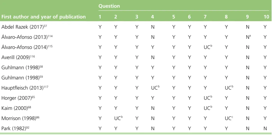
The quality of the included diagnostic accuracy studies was assessed using the QUADAS-2 [quality assessment of diagnostic accuracy studies (version 2)] tool. Critical appraisal was performed by one reviewer and independently checked by another.
Synthesis
Data were synthesised in meta-analyses across studies using logistic regression modelling. Random-effects models were used to account for potential heterogeneity in diagnostic accuracy across studies. Results were presented as summary sensitivity and specificity estimates, with 95% confidence intervals (CIs), and as summary HSROC (hierarchical summary receiver operating characteristic) curves. Analyses were performed separately for adults and children.
SCIENTIFIC SUMMARY
When the studies were deemed too diverse for meta-analysis to be suitable, or where only one or two studies were available, the reported diagnostic accuracy from each available study was presented in tables and on ROC (receiver operating characteristic) plots.
Separate meta-analyses were conducted for each diagnostic imaging test and, when sufficient data were available, in subcategories of patients including:
l patients with diabetic foot ulcers l cause of osteomyelitis
l anatomical site.
When studies report diagnostic accuracy data for two or more imaging tests, these tests were compared by extending the bivariate logistic regression models to include all imaging tests in one model.
Inter-rater reliability results and qualitative data on implementation were reported narratively and tabulated. Areas where few or no data have been published were also identified.
Results
Diagnostic accuracy
The review of diagnostic accuracy included 77 studies. The sample size of the studies ranged from 7 to 339, but most (80%) included fewer than 50 participants. Nearly one-quarter of the studies were considered as being at a high risk of bias, although poor reporting meant that there was significant uncertainty about the quality of most studies. Most of the evidence focused on the diagnostic accuracy of MRI, scintigraphy and radiography for the diagnosis of diabetic foot osteomyelitis. Few studies specifically focused on the axial skeleton, the pelvis/hip/knee and long bones.
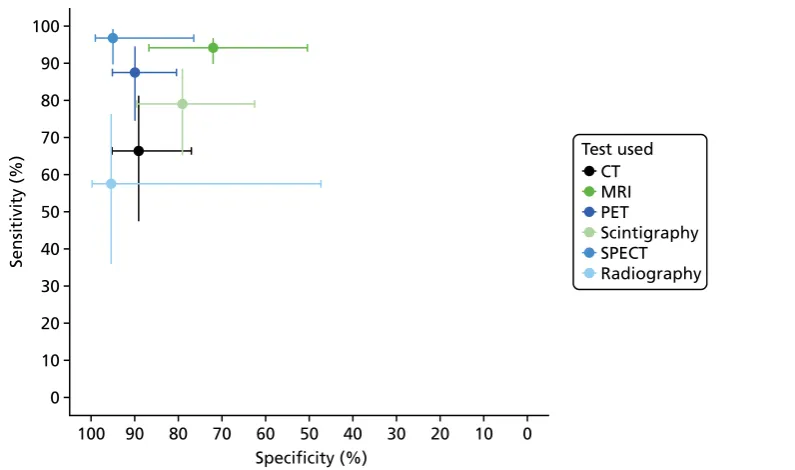
The overall meta-analysis of diagnostic accuracy in adults found that MRI can detect osteomyelitis with high accuracy (95.6% sensitivity, 95% CI 92.4% to 97.5%; 80.7% specificity, 95% CI 70.8% to 87.8%). PET also had high diagnostic accuracy (85.1% sensitivity, 95% CI 71.5% to 92.9%; 92.8% specificity, 95% CI 83.0% to 97.1%), as did SPECT (95.1% sensitivity, 95% CI 87.8% to 98.1%; 82.0% specificity, 95% CI 61.5% to 92.8%). There were similar diagnostic odds ratios and summary HSROC curves for MRI, PET and SPECT, suggesting that the three imaging tests have similar diagnostic performance.
Scintigraphy (83.6% sensitivity, 95% CI 71.8% to 91.1%; 70.6% specificity, 95% CI 57.7% to 80.8%), CT (69.7% sensitivity, 95% CI 40.1% to 88.7%; 90.2% specificity, 95% CI 57.6 to 98.4) and radiography (70.4% sensitivity, 95% CI 61.6% to 77.8%; 81.5% specificity, 95% CI 69.6% to 89.5%) all had generally inferior diagnostic accuracy when compared with MRI, PET or SPECT. The most up-to-date forms of
scintigraphy, such as99mTc HMPAO WBC (technetium-99m hexamethylpropyleneamine oxime white blood
cell) scintigraphy (87.3% sensitivity, 95% CI 75.1% to 94.0%; 94.7% specificity 95% CI 84.9% to 98.3%), had high diagnostic accuracy, similar to that of PET or MRI. There were insufficient studies of ultrasound to assess its diagnostic accuracy.
Key participant subgroups
The main patient subgroup was patients with diabetic foot ulcers, representing nearly half of all studies. The results of the meta-analyses for these patients were similar to those from the main meta-analysis, although there were too few studies of SPECT or CT to reliably assess diagnostic accuracy.
Studies of patients without diabetes were divided according to scan location and by potential cause of osteomyelitis. Data within each category were generally limited, but there was no evidence that diagnostic accuracy varied by scan location or cause, or that results differed substantially from the main analysis.
DOI: 10.3310/hta23610 HEALTH TECHNOLOGY ASSESSMENT 2019 VOL. 23 NO. 61
© Queen’s Printer and Controller of HMSO 2019. This work was produced by Llewellynet al.under the terms of a commissioning contract issued by the Secretary of State for Health and Social Care. This issue may be freely reproduced for the purposes of private research and study and extracts (or indeed, the full report) may be included in professional journals provided that suitable acknowledgement is made and the reproduction is not associated with any form of advertising. Applications for commercial reproduction should be addressed to: NIHR Journals Library, National Institute for Health Research, Evaluation, Trials and Studies Coordinating Centre, Alpha House, University of Southampton Science Park, Southampton SO16 7NS, UK.
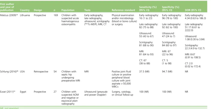
Diagnostic tests in children
The evidence for the accuracy of ultrasound and MRI in children was mixed and limited overall. Ultrasonography had moderate sensitivity and specificity in children with suspected acute haematogenous osteomyelitis but had perfect sensitivity and specificity in a small study of children with negative or equivocal initial radiographic findings. MRI had good sensitivity and specificity in children with suspected acute haematogenous
osteomyelitis in one study, but preoperative MRI had poor sensitivity and near perfect specificity in another study of patients with septic hip.
Inter-rater reliability and implementation
Eleven studies evaluated the inter-rater reliability of at least one imaging test, and one study provided data on clinician opinions on imaging tests for osteomyelitis.
Magnetic resonance imaging appeared to have acceptable inter-rater reliability. There was some evidence suggesting that PET and scintigraphy showed near perfect inter-rater reliability, although this is limited to two small studies. We found no evidence on patient preferences and cost-effectiveness of imaging tests for osteomyelitis.
Only one study on the implementation of diagnostic test imaging for osteomyelitis was included. A Dutch survey of clinicians found that preferred imaging strategies for diagnosing post-traumatic osteomyelitis depended on specialty and availability of machinery. Most responders were not aware of local hospital protocols for diagnosing osteomyelitis.
Discussion
Strengths and limitations of the analyses
This systematic review was the largest and most comprehensive review of the diagnosis of osteomyelitis to date, and the first to comprehensively compare all relevant imaging tests across all types of patient.
There were few studies identified that included children. This may be because studies of children do not discuss osteomyelitis directly, but instead mostly in the context of other conditions, such as septic arthritis. Hence, it is possible that some possibly relevant studies were missed.
Some imaging tests were reported, particularly ultrasound and CT scans. Some aspects of studies were inconsistently reported, such as varying descriptions of the cause of osteomyelitis, non-reporting of whether osteomyelitis was acute or chronic and lack of clarity on whether or not radiography (or other tests) had been used prior to the main test. This made assessment in these subgroups difficult.
We identified very few data beyond those on diagnostic accuracy, with few studies discussing broader implementation issues such as access to machinery, costs or radiation exposure.
Uncertainties
The main uncertainties remaining following this review arise largely because of limitations in the identified studies.
The diagnostic accuracy of imaging tests in children remains highly uncertain because of the very limited nature of the evidence. We could reach no firm conclusions on the diagnostic accuracy of any imaging test in children.
The diagnostic accuracy of ultrasound is currently unknown as the only two studies in adults had conflicting results.
SCIENTIFIC SUMMARY
Although we found no evidence that diagnostic accuracy varied across subgroups of patients, limited or inconsistent reporting of some characteristics, such as acute versus chronic osteomyelitis, or the cause of osteomyelitis, means than differences between tests or between subgroups cannot be ruled out.
Generalisability of the findings
The apparent consistency of diagnostic accuracy across the various types of patient and causes of
osteomyelitis suggests that the diagnostic accuracy findings are likely to be generalisable to any population being tested for osteomyelitis. The similarity in diagnostic accuracy across MRI, PET and SPECT scans suggests that these tests should have similar accuracy in most clinical circumstances.
The review found considerable variation in the specificity of MRI across studies. This may mean that the observed specificity of MRI in any given setting may differ from the summary estimates calculated in this review, depending on how MRI is implemented.
The limited evidence on diagnosis of osteomyelitis in children means that results may not be generalisable beyond the populations in the included studies.
Conclusions
Implications for service provision
Magnetic resonance imaging, PET and SPECT all have broadly similar and high accuracy when diagnosing osteomyelitis. All are likely to be suitable imaging tests for diagnosing osteomyelitis. No clear reason to prefer one test over the others in terms of diagnostic accuracy was identified. The wider availability of MRI machines, and the fact that MRI does not expose patients to harmful ionising radiation, may mean that MRI is preferable in most cases, unless it is unsuitable for a particular patient. A PET or SPECT scan may be required if a MRI scan is inconclusive.
Positron emission tomography had poorer sensitivity but higher specificity than MRI, with more consistent results across studies. This may make PET better suited to situations where avoiding false-positive diagnoses is important, for example when the test would be followed by surgery or other invasive procedures.
There is no evidence to suggest that the diagnostic accuracy varies with the potential cause of osteomyelitis or with the body part scanned, although data on patients other than those with diabetic foot ulcers were limited. The review identified very limited data on diagnosing osteomyelitis in children, so considerable uncertainty remains over the diagnostic accuracy of imaging tests in children. Clinicians should be aware of this limitation in the evidence base.
Suggested research priorities
The most urgent research priority is to perform diagnostic accuracy studies of imaging tests in children. Large diagnostic accuracy studies are needed, which must be of high quality, with proper blinding of test assessors and consecutive recruitment of patients. The priority tests should be MRI and ultrasound, ideally comparing the two tests in the same children.
Ultrasound has not been widely assessed in adults. Current results suggest that ultrasound on its own may not be sufficiently accurate to diagnose osteomyelitis, but further accuracy studies are needed to resolve the uncertainty. It may be more appropriate to investigate the diagnostic accuracy of ultrasound as a precursor to MRI or other tests (e.g. as a replacement for radiography).
Given the similarities in diagnostic accuracy of MRI, PET and SPECT, suitable investigation of patient and clinician experience and opinion of these tests, through surveys or focus groups, would be useful to identify practical reasons for the choice of test. Similarly, a formal economic evaluation of these tests, accounting for test cost, availability and risk of radiation exposure, would help to clarify the choice between these tests.
DOI: 10.3310/hta23610 HEALTH TECHNOLOGY ASSESSMENT 2019 VOL. 23 NO. 61
© Queen’s Printer and Controller of HMSO 2019. This work was produced by Llewellynet al.under the terms of a commissioning contract issued by the Secretary of State for Health and Social Care. This issue may be freely reproduced for the purposes of private research and study and extracts (or indeed, the full report) may be included in professional journals provided that suitable acknowledgement is made and the reproduction is not associated with any form of advertising. Applications for commercial reproduction should be addressed to: NIHR Journals Library, National Institute for Health Research, Evaluation, Trials and Studies Coordinating Centre, Alpha House, University of Southampton Science Park, Southampton SO16 7NS, UK.
Study registration
This study is registered as PROSPERO CRD42017068511.
Funding
Funding for this study was provided by the Health Technology Assessment programme of the National Institute for Health Research.
SCIENTIFIC SUMMARY
Chapter 1
Background
Osteomyelitis
Osteomyelitis is an infection of the bone and bone marrow.1,2Left untreated, it may result in bone infarction
and loss of limb or joint function and, in extreme cases may require amputation of the affected limb. If the infection spreads, it may lead to potentially fatal septicaemia.3In children, osteomyelitis may also inhibit limb
growth, requiring extensive orthopaedic intervention in later childhood.Staphylococcus aureusis the most common organism causing osteomyelitis, but other common organisms such asStreptococcusspp. or Escherichia colimay also be responsible in some cases. Bone infections occur most commonly in people aged < 20 years or > 50 years. It accounts for around 1% of all childhood hospital admissions. The incidence of osteomyelitis has increased over recent decades, notably in children and in patients > 60 years of age. This growing incidence has been associated with increased prevalence of meticillin-resistantS. aureus (MRSA) in children and an increase in diabetes mellitus-related infections in adults.4
Osteomyelitis may be acute, subacute or chronic and is divided between haematogenous osteomyelitis, in which infection transfers from a remote location in the body via the bloodstream, and contiguous osteomyelitis, in which infected material comes into direct contact with the bone.5The haematogenous
type is more common in children whereas the contiguous type is more common in adults, usually as a result of trauma or surgery.6Osteomyelitis is also common in people with vascular deficiency, such as
adults with diabetes, as a complication of diabetic foot ulcers.7Osteomyelitis may lead to infection of the
adjacent joint (septic arthritis) or occur secondary to septic arthritis by contiguous spread.
Patients usually present with a range of symptoms including swelling, joint pain and fever. These symptoms are often not specific to osteomyelitis, leading to delays in correct diagnosis. Blood tests are used initially to assess inflammatory markers indicative of infection in the body, including white blood cell (WBC) count, C-reactive protein (CRP) levels and erythrocyte sedimentation rate (ESR).8When these tests show evidence
of possible infection, patients are referred for further diagnostic testing. The most accurate diagnostic tool is a bone biopsy or aspiration of a pus collection from the bone or tissue surrounding the bone, with a microbiological assessment of the sample to identify the organism causing the infection. Biopsies are invasive and generally require a local or a general anaesthetic. The analysis of the results may take several days. Alternative diagnostic tools include blood or tissue cultures, which may be less accurate but are useful in identifying the organism causing an infection in the body, which enables the selection of the appropriate antibiotic for treatment. The primary treatment for osteomyelitis is a course of antibiotics, but surgery may also be used.9
Diagnostic imaging for osteomyelitis
Diagnostic imaging of the affected area before performing a biopsy may help improve diagnosis and avoid unnecessary biopsies in people who may have an infection but are unlikely to have osteomyelitis. It can also be useful to identify pus collections, assess the need for drainage procedures and establish the best way for surgical access.
A range of diagnostic imaging methods are available, including radiography, magnetic resonance imaging (MRI), computed tomography (CT), scintigraphy, positron emission tomography (PET), single-photon emission computed tomography (SPECT) and ultrasound.9–12These imaging methods each have their
advantages and disadvantages.
DOI: 10.3310/hta23610 HEALTH TECHNOLOGY ASSESSMENT 2019 VOL. 23 NO. 61
© Queen’s Printer and Controller of HMSO 2019. This work was produced by Llewellynet al.under the terms of a commissioning contract issued by the Secretary of State for Health and Social Care. This issue may be freely reproduced for the purposes of private research and study and extracts (or indeed, the full report) may be included in professional journals provided that suitable acknowledgement is made and the reproduction is not associated with any form of advertising. Applications for commercial reproduction should be addressed to: NIHR Journals Library, National Institute for Health Research, Evaluation, Trials and Studies Coordinating Centre, Alpha House, University of Southampton Science Park, Southampton SO16 7NS, UK.
Radiography are easily available and cheap to perform, but are poor at detecting osteomyelitis in its early stages. Radiography may be most useful in identifying and ruling out other causes of the patient’s symptoms, such as bone fractures.11MRI is probably the most widely recommended and used technique.
It is more accurate than radiography and able to detect osteomyelitis in its early stages, but is more expensive to perform12and when used in children necessitates the use of sedation or general anaesthesia.
PET and bone scintigraphy are more expensive and less widely available than MRI or radiography.11,13These
methods expose patients to ionising radiation. Ultrasound avoids the radiation exposure and is readily available, but its diagnostic accuracy is currently uncertain.12There is also a distinction between methods
that provide two-dimensional images (radiography, scintigraphy) and those producing three-dimensional images (PET, MRI, CT, SPECT). Some tests (e.g. MRI) may be less suited to patients with hip replacements or other indwelling metalwork because the metalwork can alter the reliability of the imaging.
Current diagnostic and treatment practice
Once osteomyelitis is suspected on the basis of physical examination and blood tests, MRI is currently generally recommended as the imaging test of choice because it can detect osteomyelitis early and it can identify pus collections within bone that might require surgical drainage. Radiography is not usually recommended in isolation, because of their failure to detect early osteomyelitis, but are generally used as a first-line investigation to rule out or confirm bone fractures or other causes of symptoms.12CT,
scintigraphy and PET are less widely recommended, but are an alternative for patients in whom MRI is not possible.
Ultrasonography is suggested as an alternative to radiological tests7,9,11,12,14and is widely used in paediatric
practice to exclude joint effusions and pus collection next to bone.14This is especially helpful in young
children (aged < 6 years), who would require a general anaesthetic for MRI. Ultrasound is also used to guide aspiration and biopsy.
Little formal guidance [such as guidelines produced by the National Institute for Health and Care Excellence (NICE)] exists about which imaging techniques to use to diagnose osteomyelitis. The only current NICE guidance is for the treatment of diabetic foot ulcers.1In those patients, radiography is recommended to either
confirm advanced osteomyelitis or confirm that the symptoms are due to other causes (e.g. broken bones). Radiography is followed by MRI if osteomyelitis is suspected but not confirmed by radiography. In children, ultrasonography is sometimes used in place of MRI. Antigranulocyte Fab fragment antibody scintigraphy should not be used in patients with diabetic foot ulcers.15Recommendations about its use have also been
published in the USA.16,17
Osteomyelitis is treated with a 4- to 6-week course of antibiotics.18,19Treatment is initially intravenous,
switching to oral antibiotics after around 2 weeks. The choice of antibiotics will depend on the infecting organism, as determined by tests such as microbiological culture and the patient’s medical history. Surgery may also be used for debridement of necrotic tissue and affected bone, to drain pus and to reduce bacterial load.
Pathway to diagnosis in the NHS
There are a number of ways in which a patient might be referred for imaging to diagnose osteomyelitis. Patients may present with fever and be admitted as inpatients, or may be referred directly by their general practitioner (GP) to an orthopaedic clinic. This pathway to clinic is slower than presenting directly to accident and emergency and such patients often have less virulent infection or subacute osteomyelitis. Patients may be referred from other hospitals, particularly those that lack the facilities to treat children (e.g. if the hospital does not offer MRI under general anaesthesia). Patients presenting with acute symptoms may have a musculoskeletal issue (often limping or joint pains) or non-specific systemic
BACKGROUND
symptoms and sepsis (e.g. immunodeficient patients as a result of underlying chronic condition). Generally unwell patients with sepsis are more difficult to diagnose because they might be in intensive care and joint symptoms could initially be missed while the focus is on treating severe symptoms.
The range of symptoms and possible causes of these symptoms mean that osteomyelitis may not be suspected at first. Patients may undergo one or more radiographic assessments (and ultrasound scans in children) and repeated blood tests prior to final diagnosis. Patients may also have received a course of antibiotics before diagnosis, with osteomyelitis suspected only because that treatment course was not successful. This complicates the diagnostic process, and the practical pathway to most diagnoses of osteomyelitis in many patients will differ from that used in formal diagnostic accuracy studies.
Existing review evidence
Several systematic reviews or meta-analyses have been performed to assess diagnostic imaging techniques for osteomyelitis.20–30
Four of these reviews considered primarily, or only, people with diabetic foot ulcers.20,22,23,26Their conclusions
varied, depending on the tests included, but MRI, PET and WBC scintigraphy were all suggested as suitable imaging tests. Three reviews of osteomyelitis in the general population mostly recommended PET and SPECT as having the best diagnostic accuracy.25,27,28One review focused on patients with peripheral post-traumatic
osteomyelitis and concluded that WBC scintigraphy with SPECT/CT or18F-FDG (fludeoxyglucose)-PET/CT had
the best diagnostic accuracy in this population.21Another review of MRI in patients with pressure ulcers was
inconclusive as a result of insufficient evidence.29Two reviews were conducted in children. One focused on
calcaneal osteomyelitis and was inconclusive because of the limited evidence.24The other children’s review
focused on haematogenous acute and subacute paediatric osteomyelitis, and found that MRI had the highest sensitivity and specificity compared with radiography, scintigraphy, CT and ultrasound.30
DOI: 10.3310/hta23610 HEALTH TECHNOLOGY ASSESSMENT 2019 VOL. 23 NO. 61
© Queen’s Printer and Controller of HMSO 2019. This work was produced by Llewellynet al.under the terms of a commissioning contract issued by the Secretary of State for Health and Social Care. This issue may be freely reproduced for the purposes of private research and study and extracts (or indeed, the full report) may be included in professional journals provided that suitable acknowledgement is made and the reproduction is not associated with any form of advertising. Applications for commercial reproduction should be addressed to: NIHR Journals Library, National Institute for Health Research, Evaluation, Trials and Studies Coordinating Centre, Alpha House, University of Southampton Science Park, Southampton SO16 7NS, UK.
Chapter 2
Aims and objectives
T
he overall aim of this project was to systematically review the literature on diagnostic imaging for osteomyelitis in order to identify the techniques with the best diagnostic accuracy and the greatest clinical utility, across the range of types of disease and patients. The key objectives were to:l perform a systematic review of all studies reporting the diagnostic accuracy of any relevant imaging test, or combination of tests, used to detect osteomyelitis
l perform diagnostic meta-analyses of identified studies to formally assess their diagnostic accuracy l investigate diagnostic accuracy across the range of different types of osteomyelitis and types of patient l compare the diagnostic accuracy of diagnostic tests both statistically and pragmatically, by
systematically reviewing inter-rater reliability, and the broader implementation of imaging tests, accounting for key factors such as availability of machinery, radiation exposure and acceptability to patients
l provide useful guidance as to which imaging tests should be preferred, according to type of disease and patient, in the UK.
DOI: 10.3310/hta23610 HEALTH TECHNOLOGY ASSESSMENT 2019 VOL. 23 NO. 61
© Queen’s Printer and Controller of HMSO 2019. This work was produced by Llewellynet al.under the terms of a commissioning contract issued by the Secretary of State for Health and Social Care. This issue may be freely reproduced for the purposes of private research and study and extracts (or indeed, the full report) may be included in professional journals provided that suitable acknowledgement is made and the reproduction is not associated with any form of advertising. Applications for commercial reproduction should be addressed to: NIHR Journals Library, National Institute for Health Research, Evaluation, Trials and Studies Coordinating Centre, Alpha House, University of Southampton Science Park, Southampton SO16 7NS, UK.
Chapter 3
Methods
A
systematic review of the clinical effectiveness was performed following the general principles recommended in the Centre for Reviews and Dissemination’s (CRD’s) guidance and the Preferred Reporting Items for Systematic Reviews and Meta-Analyses (PRISMA) statement. The protocol details have been registered on PROSPERO (number CRD42017068511).Literature searches
The search strategy was developed by an information specialist with input from the review team and clinical advisors. The strategy was developed in MEDLINE (via Ovid) and included search terms for osteomyelitis and relevant diagnostic imaging techniques. No language, date, geographical or study design limits were applied. The MEDLINE strategy was adapted for use in the other resources searched.
The searches were carried out during August 2017 and updated in July 2018 to capture more recent studies. The following databases were searched: MEDLINE (including Epub Ahead of Print, In-Process & Other Non-Indexed Citations, Ovid MEDLINE Daily and Ovid MEDLINE), Cochrane Central Register of Controlled Trials (CENTRAL), Cochrane Database of Systematic Reviews (CDSR), Cumulative Index to Nursing and Allied Health (CINAHL) Plus, Database of Abstracts of Reviews of Effects (DARE), EMBASE, Health Technology Assessment (HTA) database and PubMed.
In addition, ClinicalTrials.gov and PROSPERO were searched for ongoing and unpublished studies. Relevant guidelines were identified through searches of the National Guidelines Clearing House, NHS Evidence, the NICE website and the Trip database. The reference lists of relevant systematic reviews were manually checked to ensure that all relevant studies from previous reviews were identified.
The search results were imported into EndNote X8 [Clarivate Analytics (formerly Thomson Reuters), Philadelphia, PA, USA] and de-duplicated. The complete search strategies can be found inAppendix 1.
Study selection
Titles and abstracts and the full texts of studies were independently assessed for inclusion by two reviewers using the inclusion criteria outlined in this section. Disagreements were resolved through discussion and, where necessary, consultation with a third reviewer. Study selection was performed using EPPI-Reviewer 4 software (Evidence for Policy and Practice Information and Co-ordinating Centre, University of London, London, UK).
Participants
Participants were any patients with suspected osteomyelitis (based on symptoms, surgical samples or blood tests). No restrictions were made for age or disease aetiology.
Index tests
Index tests considered included any diagnostic imaging technique that could potentially identify
osteomyelitis, either alone or in combination with other relevant tests, such as radiography, MRI, CT, PET, SPECT and ultrasound. Variations on these tests were included, such as variations in the radioisotopes used and differences in protocols or contrast agent use.
Scintigraphy was not a protocol-specified imaging test for this review as it was not expected to be widely used in the UK, particularly because three-dimensional (3D) SPECT imaging may be preferred to planar scintigraphy. However, as the protocol specified that any relevant imaging test would be considered,
DOI: 10.3310/hta23610 HEALTH TECHNOLOGY ASSESSMENT 2019 VOL. 23 NO. 61
© Queen’s Printer and Controller of HMSO 2019. This work was produced by Llewellynet al.under the terms of a commissioning contract issued by the Secretary of State for Health and Social Care. This issue may be freely reproduced for the purposes of private research and study and extracts (or indeed, the full report) may be included in professional journals provided that suitable acknowledgement is made and the reproduction is not associated with any form of advertising. Applications for commercial reproduction should be addressed to: NIHR Journals Library, National Institute for Health Research, Evaluation, Trials and Studies Coordinating Centre, Alpha House, University of Southampton Science Park, Southampton SO16 7NS, UK.