RESEARCH
PIWIL2 promotes progression
of non-small cell lung cancer by inducing CDK2
and Cyclin A expression
Xiaohan Qu
1*, Jinlu Liu
2, Xinwen Zhong
1, Xi Li
1and Qigang Zhang
1Abstract
Background: PIWI proteins have important roles in tumorigenesis due to their interaction with piRNAs. Recent stud-ies suggest that PIWI proteins affect prognosis of various cancers.
Methods: In the present study, PIWI genes expression was assayed in non-small cell lung cancer (NSCLC). To deter-mine the effects of PIWIL2 on NSCLC cells, overexpression and interference assays were performed using the A549 and H460 cell lines. The tumor formation model was performed to demonstrate the effects of PIWIL2 on tumor forma-tion in vivo.
Results: PIWIL2 was increased both at the RNA and protein level in malignant cancer tissues compared with adja-cent normal tissue. Moreover, increased PIWIL2 gene expression was negatively correlated with prognosis in NSCLC patients. Overexpression and interference of PIWIL2 promoted and depressed cell proliferation, respectively. Mean-while, PIWIL2 interference arrested cells at the G2/M stage. In addition, we found that CDK2 and Cyclin A expression were correlated with PIWIL2 expression. Moreover, transfection of PIWIL2 promoted tumor growth in nude mice. Conclusion: Our findings shed light on the function of PIWIL2 in NSCLC and suggest potential prognostic and thera-peutic value.
Keywords: PIWIL2, Non-small cell lung cancer, Cyclin A, CDK2, Prognosis
© 2015 Qu et al. This article is distributed under the terms of the Creative Commons Attribution 4.0 International License (http:// creativecommons.org/licenses/by/4.0/), which permits unrestricted use, distribution, and reproduction in any medium, provided you give appropriate credit to the original author(s) and the source, provide a link to the Creative Commons license, and indicate if changes were made. The Creative Commons Public Domain Dedication waiver (http://creativecommons.org/publicdomain/ zero/1.0/) applies to the data made available in this article, unless otherwise stated.
Background
Non-small cell lung cancer (NSCLC) is one of the most common cancers worldwide and is associated with high mortality rates [1]. Several biomarkers have been used to detect progression have been used in diagnosis of NSCLC mortality. However, both NSCLC-specific anti-gens and mRNA-based biomarkers are associated with overdiagnosis and overtreatment [2]: mRNA-based bio-markers are seldom specific to NSCLC tissue and are commonly expressed by epithelial cells, including nor-mal, hyperplastic and cancerous cells [2]; meanwhile, NSCLC-specific antigen serum levels could reflect cancer progression or NSCLC hyperplasia, infection or inflam-mation [3]. Thus, there is a need for improved NSCLC
biomarkers. Meanwhile, the most common therapeutic strategy for NSCLC, including exairesis, chemotherapy and radiotherapy, are associated with poor prognosis and low long-term survival rates [4]. New strategies such as gene therapy and stem cell therapy have been used in clinical treatment [5, 6]. In addition, epigenetic research presents another potential cancer therapeutic avenue; understanding epigenetic changes during NSCLC pro-gression could contribute to diagnosis, predicting prog-nosis and exploring new treatment strategies [7, 8].
Chromosomal instability is a common feature of can-cer cells that often displays deletions, rearrangements and duplications of different DNA fragments in the genome [9]. Specific alterations in miRNA expression, methylation levels and histone modifications are all regarded as causative factors in the etiology of cancer [10, 11]. Indeed, aberrant DNA methylation has been found in transposable elements (TEs)–regions that may
Open Access
*Correspondence: [email protected]
be associated with cancer progression [12]; erratic TEs could lead to genomic instability and affect expression levels of both oncogenes and tumor suppressor genes [13,
14]. Hyperactive TEs therefore appear to play an integral role in cancer development. PIWI proteins are key fac-tors in controlling TE stability [15, 16]. By interacting with PIWI-interacting RNAs (piRNAs), PIWI protects the integrity and stability of the genome from silencing TEs [17]. Much progress has been made in understand-ing the molecular function of PIWI in several cancers; however, the role of PIWI in NSCLC remains unknown.
Although the PIWI-piRNA pathway was first identi-fied in the testis, PIWI has subsequently been found to be highly expressed in many different types of cancer [18]. PIWI is expressed in breast cancer [19] and gastrointes-tinal cancer [20, 21] but not in the corresponding normal tissues. PIWI homologs are common in vertebrates; for example, in mice, three different homologs of PIWI were found [22] while in humans, PIWIL1, PIWIL2, PIWIL3 and PIWIL4 were identified [23, 24]. These homologs may have different functions depending on the type of cancer. PIWIL1 promotes cancer growth and is associ-ated with increased mortality [20, 25]; increased lev-els of PIWIL2 have been reported in breast [26, 27] and cervical cancer [28]; and increased levels of PIWIL3 and PIWIL4 have been reported in colon cancer [29]. To date, PIWI gene expression has not been investigated in NSCLC; the expression patterns and function of PIWI in NSCLC therefore warrants investigation.
In the present study, we focus on the expression pattern of PIWI homologs in the context of NSCLC progression and prognosis. We investigated the function of PIWI in a NSCLC cell line, using cell transfection. The biologi-cal activities of the NSCLC cell line were confirmed after PIWI over-expression and silencing. Finally, Cyclins and Cyclin-dependent kinases (CDKs) were detected after PIWI over-expression and silencing to investigate the mechanism of PIWI on NSCLC cell progression both in vivo and in vitro. This study provided novel insights into the role of PIWI in NSCLC that suggest potential diagnostic, prognostic and therapeutic value for PIWI in NSCLC.
Methods
Materials
NSCLC samples were provided by the China Medical University (Shenyang, China) in accordance with the guidelines of the China Medical University of Medi-cine Research Ethics Committee. All patients provided informed consent. NSCLC tissues and adjacent tissues were collected from patients who underwent resection. Tumor tissues were confirmed as such by haematoxylin and eosin staining. Only those samples with more than
70 % tumor content were used for further study as tumor tissues.
Cell culture and transfection
The human NSCLC cell line, A549 and H460 were obtained from the Shanghai Cell Bank at the Chinese Academy of Sciences. The cells were cultured in Dulbec-co’s modified Eagle’s medium (DMEM) (Gibco, Gaith-ersburg, Maryland, USA) containing 10 % fetal bovine serum (Gibco) at 37 °C in a incubator at 5 % CO2.
The pcDNA3.1+ vector containing c-Myc-tagged PIWIL2 was constructed in our laboratory as previously described [30]. The c-Myc promoter was synthesized by Sangon Biotech (Shanghai, China). The PIWI fragment was amplified from human iPS cell lines with the primer set: 5′-TTC TCG AGA TGG ATC CTT TCC GAC CAT C-3′ and 5′-TTC CAT GGT CAC AGG AAG AAC AGG TTC TC-3′. The primer sets were designed to contain the full length of coding sequence. Amplified PIWI cDNA was digested with NcoI and XhoI (New England BioLabs [NEB], Hertfordshire, UK). The digested fragment was then subcloned between the NcoI site and the XhoI site of the pcDNA3.1+ vector. The construction of shRNA for PIWIL2 was previously reported [30] (PIWIL2 shRNA1: 5′-ACC GGC CUG GGU UGA ACU AAA-3′, PIWIL2 shRNA2: 5′-ACA GAA UCA AAC ACU GUG A-3′). Transfection was performed in 6-well plates with Lipofectamine 2000 (Invitrogen, Carlsbad, CA, USA) according to the manufacturers’ protocol. Cells were then replated at a density of 2 × 10⁵ cells per well and
cultured for 48 h after which they were assayed by real-time PCR, western blot and immunofluorescence stain-ing. For rescue groups, after 24 h post interference or over-expression transfection, the second over-expression transfection or interference were performed, respectively. Then the cells were analyzed at 48 h post the second transfection.
MTT assays
To assay the cell proliferation rate, 24-well plates at a den-sity of 2 × 10⁴ cells/well were seeded after transfection.
Subsequently, the cells were analyzed by methyle thiazol tetrazolium assay (MTT assay, Sigma-Aldrich, St. Louis, MN, USA). The amount of MTT formazon product was determined by using a microplate reader and the absorb-ance was measured at 570 nm (Berthold, Tokyo, Japan).
Real‑time PCR
1 μg total RNA was transcribed for each sample to
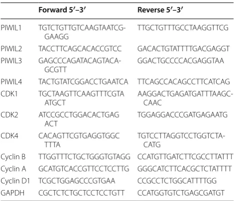
com-plementary DNA (cDNA) using M-MLV reverse tran-scriptase (Invitrogen) according to the manufacturer’s protocol. The primers for real-time PCR are shown in Table 1. The PCR conditions were as follows: 95 °C for 3 min, 40 cycles at 95 °C for 12 s and 55 °C for 40 s. GAPDH was used as an internal PCR control. Mean-while, a no-template control and melt curve analysis were used to monitor contamination. The relative expression levels were calculated using the 2−ΔΔCt method.
Western blot
The levels of PIWI proteins, CDK proteins, Cyclin pro-teins and GAPDH were determined by western blot. Primary rabbit polyclonal antibodies against PIWIs (PIWIL1, SAB1300682; PIWIL2, SAB2105190; PIWIL3, SAB4200150; PIWIL4, SAB2105885), CDKs (CDK1, SAB4500050; CDK2, SAB4503706; CDK4, SAB4300695), Cyclins (Cyclin B, SAB4503501; Cyclin A, SAB4503499; Cyclin D1, SAB4502603) and GAPDH (SAB2100894) were purchased from Sigma-Aldrich (St. Louis, MO, USA). Sec-ondary antibodies were HRP-conjugated anti-rabbit IgG (Sigma-Aldrich). Both tissues and cells were homogenized using RIPA buffer (50 mM Tris pH 8, 150 mM NaCl, 1 % NP-40, 0.5 % DOC, 0.1 % SDS, 1 mM DTT, protease and phosphatase inhibitors). Extracted proteins were quanti-fied using the BCA protein assay kit from Sangon Bio-tech (Shanghai, China). For each sample, 15 μg protein
were electrophoresed on a 12 % sodium dodecyl sulfate polyacrylamide gel. The sample was next transferred onto polyvinylidene fluoride membranes (Millipore, Billerica, MA, USA). Proteins were blocked at room temperature for 1 h using 4 % skim milk and incubated with each antibody
(for PIWIs, 1:1000; CDKs, 1:1000; Cyclins, 1:100; GAPDH, 1:2000) at 4 °C overnight. After three washes with TBST buffer (pH 7.6, 20 mM Tris–HCl, 137 mM NaCl, 0.01 % Tween-20), membranes were incubated with HRP-con-jugated anti-rabbit IgG and visualized using enhanced chemiluminescence (ECL, Millipore, Billerica, MA, USA).
Immunohistochemical staining (IHC)
Tissue samples were fixed with 10 % buffered formalin and 6-μm-thick tissue sections were cut. The sections
were then incubated with monoclonal antibody (for PIWIs, 1:100; CDKs, 1:200; Cyclins, 1:200) and the same secondary antibody that was used for western blotting. Horseradish peroxidase streptavidin complex (Beyotime, Wuhan, China) was used to visualize the signals. Sections were color-developed with diaminobenzidine and stained with hematoxylin (Beyotime).
Immunofluorescence microscopy
Protein expression was measured by immunofluores-cence, using monoclonal antibodies (for CDKs, 1:100; Cyclins, 1:100) that were detected by FITC-conjugated goat anti-rabbit IgG (Sigma-Aldrich) at a dilution of 1:200. DAPI (1 μg/ml) was used to stain nuclei.
Flow cytometry
Cell activities and apoptosis were measured by flow cytometry after over-expression and inhibition of PIWIL2 in NSCLC cells. Annexin V-FITC and propidium iodide (PI) (BioVision, Milpitas, CA, USA) staining was used according to the manufacturer’s instructions. For each sample, at least 30,000 cells were analyzed. Experi-ments were performed in triplicate.
Tumor formation in a nude mouse model
Five-week-old nude mice were used to confirm the effect of PIWIL2 on tumor formation. The experiment was approved by the ethics committee of the China Medi-cal University (Shenyang, China). Mice were randomly divided into two groups of ten (n = 5). One group was treated with A549 cells while other was treated with A549 cells containing PIWIL2 expression vectors; 30,000 A549 cells (in 1 ml DMEM) were injected every 3 days for 21 days on the dorsal side of the mouse. Tumor volumes were calculated every 3 days according to the following the formula: Volume = length × width × height × 3.14/6. At 21 days, tumor tissues were assayed by real-time PCR, western blot and IHC.
Statistical analysis
Real-time PCR, MTT assays and apoptosis rates are pre-sented as mean ± standard deviation (SD). One-way-ANOVA analysis was used to analyze differences among
Table 1 Primers used in the present study Forward 5′–3′ Reverse 5′–3′
PIWIL1
TGTCTGTTGTCAAGTAATCG-GAAGG TTGCTGTTTGCCTAAGGTTCG PIWIL2 TACCTTCAGCACACCGTCC GACACTGTATTTTGACGAGGT PIWIL3
GAGCCCAGATACAGTACA-GCGTT GGACTGCCCCACGAGGTAA PIWIL4 TACTGTATCGGACCTGAATCA TTCAGCCACAGCCTTCATCAG CDK1 TGCTAAGTTCAAGTTTCGTA
ATGCT AAGGACTGAGATGATTTAAGC-CAAC CDK2 ATCCGCCTGGACACTGAG
ACT TGGAGGACCCGATGAGAATG CDK4 CACAGTTCGTGAGGTGGC
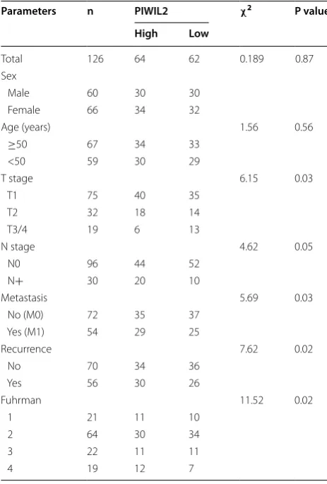
[image:3.595.56.291.523.725.2]groups and results where P < 0.05 were considered sig-nificant. Survival curves were plotted according to the Kaplan–Meier method. Of the 126 patients involved in this study, 62 expressed relatively low levels of PIWIL2 (fold-change relative expression to adjacent tissue was between 2 and 4) while 64 expressed high levels of PIWIL2 (fold-change relative expression to adjacent tissue was between 5 and 7) in cancer tissues, as judged by real-time PCR (Table 2). Multivariate analysis was conducted using Cox’s proportional hazards regression model. Patients’ survival information was collected telephoni-cally. Tumor stage was classified based on the 2011 Union for International Cancer Control (UICC) TNM classifica-tion of malignant tumors. The nuclear grading was per-formed according to Fuhrman’s system [31]. All data were analyzed using SPSS 17.0 (SPSS Inc., Chicago, IL, USA).
Results
PIWIL2 expression is elevated in NSCLC tissues
To demonstrate the roles of PIWI in NSCLC, real-time PCR, western blot and IHC were employed to assay the
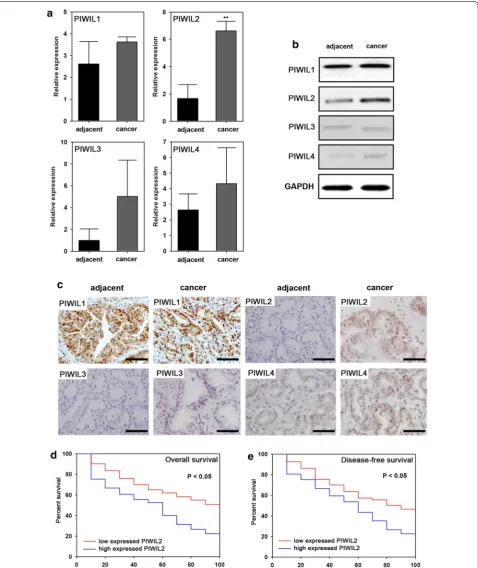
expression levels of the four PIWI homologs (PIWIL1, PIWIL2, PIWIL3 and PIWIL4) in NSCLC tissues and adjacent tissues. PIWIL2 expression was significantly higher, both at the mRNA (P < 0.05) (Fig. 1a) and pro-tein levels (Fig. 1b, c), in malignant cancer tissues com-pared to adjacent tissues. No significant difference was observed in other PIWI genes (PIWIL1, PIWIL3 or PIWIL4) between NSCLC tissues and adjacent tissues (Fig. 1).
PIWIL2 is associated with poor prognosis in NSCLC
We observed 126 patients for a period of 100 months to determine the relationship between PIWIL2 expression and NSCLC prognosis. Patients were divided into two groups: those that had high levels of PIWIL2 expression (mRNA expression levels higher than 5), and those with low levels of PIWIL2 expression (mRNA expression lev-els lower than 4). Overall survival and disease-free sur-vival were estimated. We found a significant negative correlation between PIWIL2 and overall survival as well as disease-free survival (Fig. 1d, e).
PIWIL2 regulates NSCLC cell progression
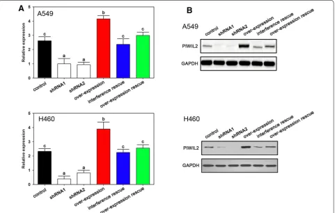
To determine the role of PIWIL2 on NSCLC cells, we used RNA interference and overexpression vectors to control PIWIL2 expression. As expected, the overex-pression vectors increased, while the interference assay decreased PIWIL2 levels, as confirmed by real-time PCR (Fig. 2A) and western blot (Fig. 2B). Both shRNA1 and shRNA2 depressed expression of PIWIL2 which con-firmed no off target effects in the present experiment (Fig. 2A, B).
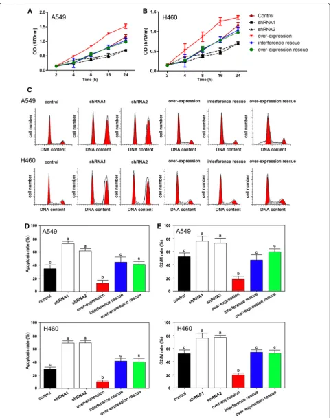
The MTT assays indicated that cell proliferation could be depressed by both shRNA1 and shRNA2 interfer-ence while overexpression of PIWIL2 induced cell prolif-eration both in A549 and H460 (Fig. 3A, B). Using flow cytometry, we found that the interference group had higher levels of apoptosis compared with the overexpres-sion group (Fig. 3C, D). Furthermore, in the interference group, more cells arrested at the G2/M stage compared with the other groups (Fig. 3C, E).
PIWIL2 regulates CDK2 and Cyclin A expression in NSCLC cells
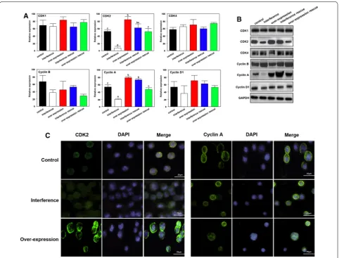
We next investigated the mechanism whereby PIWIL2 causes G2/M cell cycle arrest by measuring the expres-sion of Cyclins and CDKs. The interference of PIWIL2 was used shRNA1 which has been proved with no off target effects. The A549 cell line was used to deter-mined the gene expression. The present result indicated that no significant differences were observed for CDK1, CDK4, Cyclin B or Cyclin D at mRNA or at the protein level (Fig. 4A–C). On the contrary, CDK2 and Cyclin A
Table 2 Clinical characteristics of patients with liver can-cer in the present study
Parameters n PIWIL2 χ2 P value
High Low
Total 126 64 62 0.189 0.87 Sex
Male 60 30 30
Female 66 34 32
Age (years) 1.56 0.56
≥50 67 34 33
<50 59 30 29
T stage 6.15 0.03
T1 75 40 35
T2 32 18 14
T3/4 19 6 13
N stage 4.62 0.05
N0 96 44 52
N+ 30 20 10
Metastasis 5.69 0.03
No (M0) 72 35 37 Yes (M1) 54 29 25
Recurrence 7.62 0.02
No 70 34 36
Yes 56 30 26
Fuhrman 11.52 0.02
1 21 11 10
2 64 30 34
3 22 11 11
[image:4.595.56.288.385.728.2]Fig. 1 Expression of PIWIL2 in NSCLC and multivariate Cox’s regression analyses of primary NSCLC patients (n = 126). Real-time PCR (a), western blot (b) and IHC (c) analysis of adjacent and NSCLC tissues. d Overall survival rates and its correlation with PIWIL2 expression. The cumulative overall survival rate for patients in the PIWIL2-high group was significantly lower than that for patients in the PIWIL2-low group (χ2= 7.54; P = 0.03). e
[image:5.595.60.539.89.658.2]expression were significantly decreased or increased fol-lowing PIWIL2 interference, or overexpression, respec-tively (Fig. 4A–C).
PIWIL2 promotes tumor growth in vivo
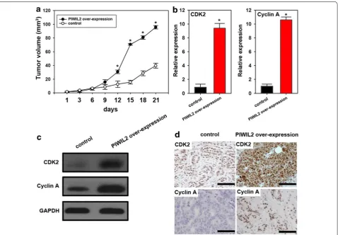
We found that mice treated with PIWIL2-transfected A549 cells had significantly increased tumor volumes compared to controls (Fig. 5a). Moreover, PIWIL2 expression promoted expression of CDK2 and Cyclin A both at mRNA and protein level (Fig. 5b–d), which indi-cates that PIWIL2 induces CDK2 and Cyclin A expres-sion and promotes tumorigenesis in nude mice.
Discussion
In this study, we explored PIWI expression in NSCLC as well as its role in tumor progression. By comparing the expression of the four PIWI homologs (PIWIL1, PIWIL2, PIWIL3 and PIWIL4), we showed that only PIWIL2 was highly expressed in malignant NSCLC tissues com-pared with adjacent tissues. These results suggested that PIWIL2 may play a crucial role in the progression of NSCLC. Moreover, high levels of PIWIL2 expres-sion were associated with decreased overall survival
and disease-free survival rates. Similar results have been demonstrated in breast [27] and endometrial cancer [28]. Thus, in cancers, the overexpression of PIWIL2 is key evidence in the malignant and pernicious [30]. How-ever, PIWI expression patterns vary by cancer type. For instance, all PIWI proteins are overexpressed in colon cancer [29], while only PIWIL2 is expressed in breast cancer [26]. Although PIWI genes are potentially useful diagnostic and prognostic biomarkers, the heterogene-ous expression patterns between different cancer types is still not well understood. We showed that PIWIL2 is highly expressed in NSCLC and is a potential indicator of NSCLC prognosis.
Once we confirmed the aberrant expression of PIWIL2 in NSCLC, we investigated the potential role of PIWIL2 in NSCLC progression in NSCLC cells using overex-pression and interference of PIWIL2. High PIWIL2 expression significantly promoted cell proliferation and activities. Conversely, inhibition of PIWIL2 triggered apoptosis and G2/M cell cycle arrest. It is widely rec-ognized that PIWIL2, as a small RNA binding protein, mediates genome stability and regulates genes expression [32, 33]. The PIWI-piRNA pathway was first illustrated
[image:6.595.59.541.90.396.2]in 2006 [22, 34], however, little is known regarding the PIWI-piRNA pathway in cancer. Although abnormal PIWI expression resulting in poor prognosis has previ-ously been reported, the specific role of PIWI in cancer cells remains unknown. Additionally, PIWIL2 interfer-ence triggers apoptosis, which implies a potential thera-peutic strategy for NSCLC.
Because PIWIL2 regulates progression of NSCLC cells by controlling the cell cycle, we quantified cell-cycle-related proteins affected by PIWIL2. CDK2 and Cyclin A are key factors that control DNA synthesis and the cell cycle [35]; lack of CDK2 and Cyclin A leads to apopto-sis [36]. We showed that PIWIL2 promotes expression of CDK2 and Cyclin A both in vitro and in vivo. In a pre-vious study, PIWIL2 could inhibit TGF-β signaling via
Hsp90 and by promoting TβR degradation [37]. Further,
PIWIL2 and TGF-β expression are negatively correlated
[37]. These findings suggest that PIWIL2 may affect cell proliferation through various means. Our results support previous findings that PIWIL2 promotes cell proliferation.
Over the last decade, evidence has emerged that an epigenetic switch occurs during NSCLC transforma-tion where non-coding RNAs, including miRNAs, piR-NAs, and long non-coding RpiR-NAs, mediate self-renewal of cancer initiating cells [38]. Lim and colleagues showed that piRNA pathway genes are overexpressed in ovarian cancer [39] and they proposed that PIWIL1 and MAEL inhibit cell invasion. A subsequent study indicated that the RASSF1C-dependent promotion of lung cancer cell proliferation is dependent on IGFBP-5 and PIWIL1, implicating the PIWI-piRNA pathway in tumorigenesis [40]. In vertebrates, abnormal expression of PIWI genes was related to tumorigenesis, sterile and hypogenesis
Fig. 4 The effects of PIWIL2 on CDK2 and Cyclin A in NSCLC cells. Real-time PCR (A) and western blot (B) analysis indicated that only CDK2 and Cyclin A is affected by changes in PIWIL2 expression. The characters on the bars represent significant differences between groups (P < 0.05). C
[image:8.595.58.541.88.455.2][41–46]. However, until now, the PIWI-piRNA pathway remains poorly understood. In addition to its role in sta-bilizing the genome, the PIWI-piRNA pathway is likely involved in tumorigenesis. So far, detailed evidence for the role of PIWI proteins in tumorigenesis remains lim-ited. We showed that PIWIL2 interference inhibited NSCLC progression, both in vitro and in vivo, which sug-gests potential therapeutic value for PIWIL2 in NSCLC.
Conclusions
In sum, we showed that increased expression of PIWIL2 is associated with worse prognosis of NSCLC and that PIWIL2 interference could inhibit cell proliferation and activities. Furthermore, CDK2 and Cyclin A were regu-lated by PIWIL2 both in vitro and in vivo. These findings suggest that PIWIL2 participates in the progression of NSCLC via CDK2 and Cyclin A.
Authors’ contributions
XQ and JL designed the experiment, performed most of the experiments and drafted the manuscript. XZ, XL and QZ carried out the flow cytometric
analysis, participated in the design of the study and helped in writing the manuscript. All authors read and approved the final manuscript.
Author details
1 The First Affiliated Hospital, China Medical University, NO. 155, Nanjing North Street, Heping District, Shenyang 110001, Liaoning, China. 2 The Forth Affili-ated Hospital, China Medical University, Shenyang, China.
Acknowledgements
A special thanks to Zhang Zhang for his invaluable assistance in statistical analysis.
Compliance with ethical guidelines
Competing interests
The authors declare that they have no competing interests.
Received: 28 April 2015 Accepted: 9 September 2015
References
1. Siegel R, Ma J, Zou Z, Jemal A. Cancer statistics, 2014. CA Cancer J Clin. 2014;64:9–29.
[image:9.595.61.540.88.420.2]2. Chang A. Chemotherapy, chemoresistance and the changing treatment landscape for NSCLC. Lung Cancer. 2011;71:3–10.
3. Wroblewski JM, Bixby DL, Borowski C, Yannelli JR. Characterization of human non-small cell lung cancer (NSCLC) cell lines for expression of MHC, co-stimulatory molecules and tumor-associated antigens. Lung Cancer. 2001;33:181–94.
4. Rivera MP. Multimodality therapy in the treatment of lung cancer. Semin Respir Crit Care Med. 2004;25 Suppl 1:3–10.
5. Weinstein IB, Joe AK. Mechanisms of disease: oncogene addiction—a rationale for molecular targeting in cancer therapy. Nat Clin Pract Oncol. 2006;3:448–57.
6. Bertolini G, Roz L, Perego P, Tortoreto M, Fontanella E, Gatti L, Pratesi G, Fabbri A, Andriani F, Tinelli S. Highly tumorigenic lung cancer CD133 + cells display stem-like features and are spared by cisplatin treat-ment. Proc Natl Acad Sci USA. 2009;106:16281–6.
7. Risch A, Plass C. Lung cancer epigenetics and genetics. Int J Cancer. 2008;123:1–7.
8. Juergens RA, Wrangle J, Vendetti FP, Murphy SC, Zhao M, Coleman B, Sebree R, Rodgers K, Hooker CM, Franco N. Combination epigenetic therapy has efficacy in patients with refractory advanced non-small cell lung cancer. Cancer Discov. 2011;1:598–607.
9. Stephens PJ, Greenman CD, Fu B, Yang F, Bignell GR, Mudie LJ, Pleasance ED, Lau KW, Beare D, Stebbings LA. Massive genomic rearrangement acquired in a single catastrophic event during cancer development. Cell. 2011;144:27–40.
10. Sharma S, Kelly TK, Jones PA. Epigenetics in cancer. Carcinogenesis. 2010;31:27–36.
11. Kanwal R, Gupta S. Epigenetic modifications in cancer. Clin Genet. 2012;81:303–11.
12. Chénais B. Transposable elements and human cancer: a causal relation-ship? Biochim Biophys Acta. 2013;1835:28–35.
13. Laird PW, Jaenisch R. The role of DNA methylation in cancer genetics and epigenetics. Annu Rev Genet. 1996;30:441–64.
14. Esteller M, Herman JG. Cancer as an epigenetic disease: DNA methylation and chromatin alterations in human tumours. J Pathol. 2002;196:1–7. 15. Aravin AA, Hannon GJ, Brennecke J. The Piwi-piRNA pathway provides an
adaptive defense in the transposon arms race. Science. 2007;318:761–4. 16. Slotkin RK, Martienssen R. Transposable elements and the epigenetic
regulation of the genome. Nat Rev Genet. 2007;8:272–85.
17. Siomi MC, Sato K, Pezic D, Aravin AA. PIWI-interacting small RNAs: the vanguard of genome defence. Nat Rev Mol Cell Biol. 2011;12:246–58. 18. Suzuki R, Honda S, Kirino Y. PIWI expression and function in cancer. Front
Genet 2012;3:204.
19. Wang D-W, Wang Z-H, Wang L-L, Song Y, Zhang G-Z. Overexpression of hiwi promotes growth of human breast cancer cells. Asian Pac J Cancer Prev. 2013;15:7553–8.
20. Wang Y, Liu Y, Shen X, Zhang X, Chen X, Yang C, Gao H. The PIWI protein acts as a predictive marker for human gastric cancer. Int J Clin Exp Pathol. 2012;5:315.
21. Cui L, Lou Y, Zhang X, Zhou H, Deng H, Song H, Yu X, Xiao B, Wang W, Guo J. Detection of circulating tumor cells in peripheral blood from patients with gastric cancer using piRNAs as markers. Clin Biochem. 2011;44:1050–7.
22. Girard A, Sachidanandam R, Hannon GJ, Carmell MA. A germline-specific class of small RNAs binds mammalian Piwi proteins. Nature. 2006;442:199–202.
23. Qiao D, Zeeman A-M, Deng W, Looijenga L, Lin H. Molecular characteriza-tion of hiwi, a human member of the piwi gene family whose overexpres-sion is correlated to seminomas. Oncogene. 2002;21:3988–99.
24. Sasaki T, Shiohama A, Minoshima S, Shimizu N. Identification of eight members of the Argonaute family in the human genome. Genomics. 2003;82:323–30.
25. Liu W, Jiang X, Zhang Z. Expression of PSCA, PIWIL1, and TBX2 in endome-trial adenocarcinoma. Onkologie. 2010;33:241–5.
26. Lee JH, Jung C, Javadian-Elyaderani P, Schweyer S, Schütte D, Shoukier M, Karimi-Busheri F, Weinfeld M, Rasouli-Nia A, Hengstler JG. Pathways of proliferation and antiapoptosis driven in breast cancer stem cells by stem cell protein piwil2. Cancer Res. 2010;70:4569–79.
27. Liu JJ, Shen R, Chen L, Ye Y, He G, Hua K, Jarjoura D, Nakano T, Ramesh GK, Shapiro CL. Piwil2 is expressed in various stages of breast cancers and has the potential to be used as a novel biomarker. Int J Clin Exp Pathol. 2010;3:328.
28. He G, Chen L, Ye Y, Xiao Y, Hua K, Jarjoura D, Nakano T, Barsky SH, Shen R, Gao J-X. Piwil2 expressed in various stages of cervical neoplasia is a potential complementary marker for p16INK4a. Am J Transl Res. 2010;2:156.
29. Li L, Yu C, Gao H, Li Y. Argonaute proteins: potential biomarkers for human colon cancer. BMC Cancer. 2010;10:38.
30. Yao Y, Li C, Zhou X, Zhang Y, Lu Y, Chen J, Zheng X, Tao D, Liu Y, Ma Y. PIWIL2 induces c-Myc expression by interacting with NME2 and regulates c-Myc-mediated tumor cell proliferation. Oncotarget. 2014;5:8466. 31. Fuhrman SA, Lasky LC, Limas C. Prognostic significance of morphologic
parameters in renal cell carcinoma. Am J Surg Pathol. 1982;6:655–64. 32. Chapman EJ, Carrington JC. Specialization and evolution of endogenous
small RNA pathways. Nat Rev Genet. 2007;8:884–96.
33. Peters L, Meister G. Argonaute proteins: mediators of RNA silencing. Mol Cell. 2007;26:611–23.
34. Aravin A, Gaidatzis D, Pfeffer S, Lagos-Quintana M, Landgraf P, Iovino N, Morris P, Brownstein MJ, Kuramochi-Miyagawa S, Nakano T. A novel class of small RNAs bind to MILI protein in mouse testes. Nature. 2006;442:203–7.
35. Cardoso MC, Leonhardt H, Nadal-Ginard B. Reversal of terminal differ-entiation and control of DNA replication: cyclin A and Cdk2 specifically localize at subnuclear sites of DNA replication. Cell. 1993;74:979–92. 36. Katayose Y, Kim M, Rakkar AN, Li Z, Cowan KH, Seth P. Promoting
apopto-sis: a novel activity associated with the cyclin-dependent kinase inhibitor p27. Cancer Res. 1997;57:5441–5.
37. Zhang K, Lu Y, Yang P, Li C, Sun H, Tao D, Liu Y, Zhang S, Ma Y. HILI inhibits TGF-β signaling by interacting with Hsp90 and promoting TβR degrada-tion. PLoS One. 2012;7:e41973.
38. Costa FF. Non-coding RNAs: new players in eukaryotic biology. Gene. 2005;357:83–94.
39. Lim SL, Ricciardelli C, Oehler MK, Tan IMDA, Russell D, Grützner F. Overex-pression of piRNA pathway genes in epithelial ovarian cancer. PLoS One. 2014;9:e99687.
40. Reeves ME, Firek M, Chen S-T, Amaar YG. Evidence that RASSF1C stimula-tion of lung cancer cell proliferastimula-tion depends on IGFBP-5 and PIWIL1 Expression levels. PLoS One. 2014;9:e101679.
41. Pan Y, Hu M, Liang H. Wang J-j, Tang L-j: The expression of the PIWI family members miwi and mili in mice testis is negatively affected by estrogen. Cell Tissue Res. 2012;350:177–81.
42. Houwing S, Kamminga LM, Berezikov E, Cronembold D, Girard A, van den Elst H, Filippov DV, Blaser H, Raz E, Moens CB, et al. A role for Piwi and piRNAs in germ cell maintenance and transposon silencing in Zebrafish. Cell. 2007;129:69–82.
43. Houwing S, Kamminga L, Berezikov E, Cronembold D, Girard A, Elst H, Filippov D, Blaser H, Raz E, Moens C, et al. A role for Piwi and piRNAs in germ cell maintenance and transposon silencing in Zebrafish. Cell. 2007;129:69–82.
44. Kuramochi-Miyagawa S, Kimura T, Ijiri TW, Isobe T, Asada N, Fujita Y, Ikawa M, Iwai N, Okabe M, Deng W. Mili, a mammalian member of piwi family gene, is essential for spermatogenesis. Development. 2004;131:839–49. 45. Zhou Y, Zhong H, Liu S, Yu F, Hu J, Zhang C, Tao M, Liu Y. Elevated
expres-sion of Piwi and piRNAs in ovaries of triploid crucian carp. Mol Cell Endocrinol. 2014;383:1–9.