METHODS FOR THE INVESTIGATION OF STRUCTURE
IN POLYPEPTIDES AND PROTEINS
A Thesis Submitted for the
Degree of Doctor of Philosophy
by
Malcolm David Fenn
B.Sc. (Qld), M.Sc. (A.N.U.) A.R.A.C.I.
PREFACE
This thesis is submitted for the Degree of Doctor of Philosophy, having been submitted for no other degree. The work reported in this thesis was carried out in the Department of Chemistry of the Australian National University under the supervision of
Dr. J.R.
Bradbury. The work is wholly original except where due reference i s made in the text.(M.D.
FENN)I
ACKNOWLEDGMENTS
I would like to thank in general the staff of the Department of Chemistry and in particular my supervisor Dr. J.H. Bradbury for their encouragement and assistance during the course of the project. My thanks also go to Imperial Chemical Industries of Australia and New Zealand for their financial assistance in the form of an ICIANZ Research Fellowship.
Lastly, I express my thanks to my typist, Mrs. C. Grant, for persevering with a difficult task.
I i
1
SYNOPSIS
This thesis is concerned with two aspects of structural peptide chemistry.
The first project reported in this thesis is an nmr study of the effect of strongly protic solvents on simpl e amides and a polypeptide, poly-y-benzyl-L-glutamat e (PBG). The results for simple amides are consistent with protonation on the addition of dichloroacetic acid (DCA)
and relatively slow exchange of the amide nitrogen proton with solvent
(T
>
10-2 sec)oSpin decoupling experiments conducted on solutions of PBG in protic and non protic solvents have shown that the greatest contributor to the width of the aCH resonance is due to coupling with the ~CH2 protons of the side chain o The
exchange of the amide nitrogen proton with solvent was observed to be slow. A kinetic scheme for the helix to coil transition in PBG is proposed and a possible mechanism for the transition described.
I
I
complex to the amino acid complex and pept ide less one residue determined. Both reaction rates were found to be dependent on the nature of the N terminal residue.
i
INDEX
GENERAL INTRODUCTION 1
NMR THEORY 3
SECTION 1: NMR STUDY OF THE HELIX TO COIL
TRANSITION IN PBG
INT RODUCT I ON
1. Model compounds
2. Poly-amino acids
11 17
18
(a) Infra red spectroscopy and
Conductivity 19
(b) Viscosity 19
(c) Circular dichroism and optical
rotatory dispersion 23
(d) Electro optical properties 24
(e) nmr spectroscopy 26
EXPERIMENTAL
Poly-amino acids
Solvents
Reagents
NMR methods
28
29
3031
RESULTS AND DISCUSSION
Model amides in protic solvents 32 Model amides in basic solvents 43
Other protons 46
PBG in mixed solvents of DCA andCDC1
3 51
Spin decoupling experiments 52
Kinetic scheme for PBG 60
Mechanism of the helix to coil transition 66
SECTION II: A NEW METHOD FOR DETERMINING THE SEQUENCE OF AMINO ACIDS IN PEPTIDES INTRODUCTION
(a) Fragmentation of proteins (b) Separation of fragments (e) Determination of Sequence
1. Mass Spectrometry 2. NMR Spectroscopy 3. Enzymic degradation 4. Chemical Methods
(i) C terminal (ii) N terminal
68 69
70
71
74
75
i
u
EXPERIMENTAL Materials Experimental
Identification of complexes (a) Chromatography
(b) Visible and near ultra violet spectroscopy
(c) NMR spectra
RESULTS AND DISCUSSION Polymer supports
Reaction of carboxyls
Protection of free carboxyls Hydrolysis of peptide complexes Reaction of C-terminal protected peptides
CONCLUSION
APPENDIX A
APPENDIX B
BIBLIOGRAPHY
80 83
113
117
117
119
127
129
136
139
145
151
152
GENERAL INTRODUCTION
Very few problems in natural science have been
subjected to such intensive investigation as the overall
structure of proteins. One of the main features of protein
investigations is the vast variety of experimental methods which have been used, from sophisticated techniques such as X-ray crystallography, nuclear magnetic resonance and
electro optical methods to viscometry, osmotic pressure, electrical conductance, titration and simple organic
chemistry. Such a multitude of techniques serves to
illustrate the enormous complexity of not only the simplest living cell but also a native protein molecule. Proteins are considered to consist of three distinct structural groupings, aptly named the primary, secondary and tertiary structures
(Linderstrom-Lang 1952). The primary structure can be defined as the actual chemical structure of the basic protein molecule. Secondary structure refers to stable configurations induced by hydrogen bonding of the primary
structureo Tertiary structure describes the folding of this
secondary structure in a compact molecular unit ioe. the
2.
first sight appear to be completely unrelated. However, in
a broad sense, this is not quite correct. Both projects are concerned with a particular aspect of structural protein
chemistry. The first
is
a continuation of project startedpreviously (Fenn, 1967) and is concerned with an aspect of
secondary structure, the transition between one secondary structure and another. The second project is concerned with the development of an inorganic technique for the determination
of the primary structure in peptides and proteins •
f
NUCLEAR MAGNETIC RESONANCE THEORY
If an external magnetic field is applied to a system of nuclei with magnetic moments, these nuclear
magnets will experience torques and will tend to be lined up parallel to the field. Although direct observation of such an orientation is difficult to observe, i t is possible, under appropriate conditions, for these magnets to absorb energy from a magnetic field oscillating with a frequency in the radio frequency· region o Such absorption gives rise to what are called nuclear magnetic resonance (nmr) spectrao Spectra are not observed for nuclei with no magnetic moment o
Essentially, nmr spectra are used as a probe to investigate local magnetic effects inside a molecular system o The local magnetic field near a particular nucleus will depend on its chemical environment and is determined by several factors including the polarisation of remote parts of the molecule, magnetic moments (nuclear and electronic) of neighbouring molecules, and intra molecular effects due to other nuclei
and electrons in the same molecule.
If a nucleus has a magnetic moment and a magnetic field is applied then the nucleus will precess in order to
orientate its magnetic moment with the field o The energy
[
Ir,
If
4.
which is given
by:-Y
Hov
=
2 rr
where
v
is the frequency in HzHo the applied field
and y the gyromagnetic ratio which 1S constant for each
type of nucleus.
The second environmental effect _is that of the
surrounding electrons. If any atom or molecule is placed in
a magnetic field it acquires by virtue of the induced orbital
motion of its electrons a diamagnetic moment. These moving
electrons constitute effective currents within the molecule
and thereby produce secondary' magnetic fields which also act
on the nuclei present. Since the induced currents are
proportional to the applied field, the magnitude of the
induced field will also be proportional to the applied field,
the local field at the nucleus being given by:
-H = Ho (1-
0
)
local (2)
where 0 is a constant known as the screening constant
dependent on the chemical environment of the nucleuso The
effect of the screening constant can be seen from equation (1).
For a finite positive value of 0 the local field is lower
the nmr spectra of a given species of nucleus in various
chemical environments, there will be a corresponding set
of different values of screening constants and resonance
will occur at different frequencies for each chemically
distinct environment 0 This displacement of signal due to
the creation of screening constants is known as a chemical
shift. When two or more nuclei have identical screening
constants, identical chem~cal shifts are obtained and the
nuclei are said to be equivalent.
In view of equations (1) and (2) the number of
nuclei giving rise to each resonance is directly proportional
to the intensity of the signal.
The most important single parameter to be derived
from the nmr spectrum is the chemical shift. Since the
absorption frequency of nuclei of different elements occur
at very different parts of the spectra, normally only one
type of nuclei is considered, in this case hydrogeno Since
the absorption frequency is dependent on the applied magnetic
field i t is most convenient to express the chemical shift in
terms of a non-dimensional unit defined by:
-6
=H-H
~, I
1
6.
where H is the resonant frequency of the signal being
measured and Hs lS the resonant frequency of a second proton
signal chosen as a reference signalo
The nmr spectrum may be modified if the molecules
being measured are taking part in various rate processes,
an example of which is the phenomenon of proton exchange between different chemical positions. If this exchange is
rapid enough, the signals due to the protons at the various
exchange sites will coalesce. Actually, this is merely the
operation of the uncertainty principle, which in this case
is given by equation
(4):-1 ( 4)
T =
2 TTO
where T is the smallest lifetime for which the resonance of
the two states can be distinguished and
0
is the separation(in Hz) of the two resonances for large lifetimes. For
lifetimes above this critical value, distinct resonances are obtained for protons at the exchanging sites and the intensity
of the resonance is directly'proportional to the number of
protons at that exchange site. Furthermore, the width of e
the resonance is inversly proportional to the population.
f.
1
7.
Where Wa and Wb are the widths of the resonances
A and B respectively and P
a and Pb are their populationso
Because of equation (5) when the population at one site is
greatly in excess of the population at the other site, the
resonance due to the latter site is broadened considerably and
may become so broad as to be invisible in the spectrum
(exchange broadening).
When the lifetimes are much smaller than the critical
value, a single sharp resonance is observed. The observed
chemical shift,
0
b of the proton resonance is related too s
the chemical shifts of the various states
by:-w'here
=
P.1
O
1.
(6)o
is the chemical shift of the protons at the ith iexchange site and P . is the mole fraction of exchanging protons
1
at this site. (stewart et al, 1967).
When the lifetime of the exchanging protons is of
the same order as the critical value, the resonances collapse
to a single broad peak.
When all the magnetic nuclei in the molecule are
equivalent then only one resonance signal is obtained.
However, when the nuclei have two different sets of equivalent
8.
addition to the two distinguishable signals, each of these
may be split into further components by spin-spin interactiono
Due to interaction of the bonding electrons, two adjacent
nuclei give rise to two different spin states (in the case of
two adjacent
protons):-( a) where the nuclear spins are parallel;
( b) where the nuclear spins are anti paralleL
This gives rise to two slightly different magnetic
states for each nuclei and hence two resonance frequencies
for each nuclei. Since the difference in magnetic states is
a function of the lmolecule i t is independent of the strength
of the applied field.
Spin-spin coupling may be effectively removed by
several mechanisms which cause the spin multiplet to collapse
to a less complex resonance.
The collapse of spin multiplets may be caused by
the rapid exchange of nuclei between identical molecules.
During exchange the exchanging nucleus has an equal probability
of attaching itself to a molecule with anyone of the spin
orientations possible. If the exchange is rapid enough then
9.
of the exchanging nucleus) but an average and the spin
multiplet collapses. The critical value for this process
is again given by the uncertainty' principle and the same
observations for lifetimes greater than, smaller than or of
the same order as the critical value applyo
Spin multiplets may also be collapsed by rapid
disturbance or relaxation of the second nucleus causing the
splittingo Such occurs with the resonance due to protons
attached to a nitrogen nucleus. Under normal circumstances
due to the three spin states of the nitrogen nucleus the
resonance attributed to the proton is a triplet. When, as
in certain molecules rapid transition of the nitrogen nucleus
between its three spin states occurs, the proton sees only
an average spin state and a single resonance ensues o
A third method is the application of a strong rf
magnetic field, the frequency being adjusted to the resonance
frequency of the nucleus causing the splitting. This causes
frequent transitions between the various spin states of the
nucleus, and just as with the preceding mechanism, an average
spin state is seen and the spin-spin coupling is removed.
A final method of decoupling is to replace one of
the coupling nuclei with a nucleus where the interaction of
bonding electrons(hence splitting)is very small or negligible
10.
Hence by careful examination of the chemical shift,
spin-spin splitting and width of the resonances, one may not
only obtain information concerning the molecu~( structure of
molecules but also the rates of any exchange processes which
SECTION 1:
NMR STUDY OF THE HELIX TO COIL
11 •
INTRODUCTION
Proteins are naturally occurring high molecular
wei ht compounds consisting of one or more chains of amino
acid. residues which are linked together by peptide bonds.
The order in which the amino acid residues are arranged in
the chain is known as the sequence (primary structure) and
is unique for a particular protein. The chains mayor may
not contain inter or intra molecular crosslinks, and in many
cases are associated with other compounds such as long chain
fatty acids or sugars. Their molecular shapes vary from rigid rod like structures to the almost spherical symmetry of the
globular proteins. Because of the great variation in the
above factors the study and explanation of the
physico-chemical properties of proteins has frequently proved to be difficult.
Understanding of these properties is often
clarified by the study of simple lprotein like! model
compounds. Although many small, low molecular weight
peptides have been prepared by the well known step wise
methods of synthesis and have proved useful models, the
synthesis of reasonable quantities of high molecular weight polypeptides of a predetermined sequence is not yet
technologically feasible. Thus there remains a large gap
between the low molecular weight peptide models and the high
1
I
I
ii
120
molecular weight proteins. This gap is bridged to some
extent by the poly-~amino acids.
Poly-«-amino acids are synthetic polymers of
~-amino acids linked by peptide bonds, being prepared by the
polymerisation of the corresponding N-carboxy acid anhydride
monomer {Blout and Karlson, 1956, Katchalski et al 1964)0
Like other synthetic polymers a sample of poly-~-amino acid
suffers from the disadvantage of being polydisperse i.eo
the chain lengths of the molecules in the sample are of
varying lengths.
The presence of the peptide bond and the relative
ease with which poly-~amino acids of a great variety may be
synthesised make poly-~-amino acids very useful models for
studying the physico-chemical properties of high molecular
Iveight proteins in the solid state and in solution o
Physico-chemical properties of proteins which
contain a large percentage of one amino acid may be
approximated by a polymer of the amino acid concerned. For
example poly-L-alanine has proved a useful model in the
elucidation of the structure of tussah silk which contains
a large amount of alanine. Similarly, polyglycine has been
used as a model for silk fibroin and copolymers containing
kno,vn sequences of glycine and proline for collageno
13.
Information pertinent to the primor\ al synthesis of
"
proteins is obtained from the thermal polymerisation of
mixtures of free amino acids, while kinetic studies of the
polymerisation of N-carboxy acid anhydrides of amino acids
may be useful in determining the probability of certain
sequences occurring in proteinso
Multichain poly~-amino acids exhibit the same
physico-chemical properties as glob! ular proteins and may
prove useful models as such.
The information on poly-~-amino acids in the solid
state has attracted the attention of a great number of
investigators. Initially, the main purpose of this work
was to arrive at a clearer interpretation of data obtained
with fibrous and globular proteins and i t soon became clear
that poly-~-amino acids provide excellent model compounds
for this purpose. X-ray analysis of poly~-amino acids
provided the first evidence for the existence of the d-helix
conformation in proteins proposed by Pauling and Corey (1951).
The work of Perutz (1951) and of Bamford et al (1952, 1956)
showed that in the solid state poly-~-amino acids may attain
oIrhelical conformations. It was, therefore, of interest to
see whether such conformations existed in solution. The
investigations carried out on the optical and hydrodynamic
1,
If
14.
properties, in particular those of Doty and coworkers (1954,
1956) on poly-¥-benzyl-L-glutamate have yielded evidence
that poly-~amino acids may exist in solution both in the
~helical form and random coil conformations, depending on the
conditions chosen (e.g. solvent, temperature). It was found
that in weakly interacting solvents such as chloroform,
ethylene dichloride and dimethyl formamide poly-"'-amino acids
attain the helical conformation while in strongly interacting
solvents (DCA, TFA) they exist in the form of random coils.
Thus it is possible to induce a helix to coil transition by
the gradual addition of a strongly interacting solvent to a
solution of a poly-~-amino acid in a weakly interacting
solvent. Studies on the helix-coil transition may shed
some light on the process of protein denaturation which i t
appears to resemble in certain aspects. Although whether
the transition is a good model for denaturation is doubtful
since the rates for the two processes are vastly different
(Bradbury and King, 1971) 0
The mechanism by which this helix to coil transition
is achieved in such
phy~ical
properties as viscosity (Dotyet al, 1954, 1956; Teramoto et al, 1967; Bradbury and Fenn,
1967, Fenn, 1967; Bradbury and Fenn, 1968), specific volume
I
I
I !
15.
(Bradburyet aI, 1965; Noguchi, 1966), heat capacitance
(Ackermann and Ruterjans, 1964; Karasz et aI, 1964; Karasz
and OYReilly, 1966; Ackermann and Neumann, 1967), optical
rotatory dispersion (Doty and Yang, 1956; Yang and Doty,
1957; Perlmann and Katchalski, 1962; Moffitt and Yang,
1956; Fasman, 1962; Balusbramanian, 1967; Urnes and Doty,
1961; Quadrifoglio and Urry, 1967; Bovey, 1968; Marlborough
et aI, 1965) electrical birefringence (Watanabe et aI, 1964;
Milstein and Charney, 1970) and conductance (Stake and
Klotz, 1965) has been a controversial subject for several
years.
The effectiveness of these acids in producing
transitions in the above physical properties has been
attributed to their ability to disrupt the hydrogen bonds
between the C=O and N-H groups which stabilise the helix, by
forming their own competing hydrogen bonds with the C=O and
N-H groups (Singer, 1962). However, i t must be realised
that DCA and TFA are strong acids and there exists the
possibility of protonation of the amide group with subsequent
collapse of the helix to the random coil, being brought about
by electrostatic repulsion between charged amide groups.
I
Evidence purporting to show the validity of either
one or the other of the above theories has been presented by
many authors using a variety of techniques. Because of this
apparent conflict of opinion i t is proposed to summarise the
17.
1. Model compounds
Specific volume, conductance, infra red and nmr
studies of simple amides in concentrated aqueous solutions
of mineral acids or in strongly protic solvents such as DCA
or TFA have led to the conclusion that simple amides are
indeed protonated in such solvents. (Berger et aI, 1959;
Herbison-Evans and Richards, 1962; Becker and Davidson,
1963; Klotz et aI, 1964; Stewart et aI, 1967; Nawrot and
Veiss, 1970 (a))o Furthermore, it has been demonstrated that
simple oligopeptides are protonated in formic acid, a much
less protic solvent than either DCA or TFA (Nawrot and Veiss,
1970 (b)). Nmr studies of amides dissolved in a variety of
protic and non protic solvents have demonstrated that when
dissolved in DCA the resonance attributed to protons on the
carbon adjacent to the amide nitrogen was approximately 0024
ppm downfield from the position of the resonance in CDCI 3•
This has been interpreted as being indicative of protonation
(Fenn, 1967; Bradbury and Fenn, 1969 (a)). There is also
evidence that protonation is preceded by a hydrogen bonded
intermediate (Stewart et aI, 1967; Nawrot and Veiss, 1970 (a)).
In brief, amides are protonated in DCA or TFA via
a hydrogen bonded intermediate. On protonation a downfield
shift of the resonance of the protons adjacent to the amide
18.
2. Poly-~amino acids
Further direct evidence in favour of charging
stems from the effect of the addition of amines, water
(Steigman et aI,
1969)
and in particular the dichloroacetateion (Zezine et aI,
1968)
to the solvento Protonation of thepolY-o<-amino acid can be represented by the following
equationo
H H
\
I
-N-C- +CHC1
2COOH < '" -N-C + CHC12COO- (7)
1\
-
;
,,
\
°
OHWhereas, the addition of amines or water results as shown
by equations
R-NH2
( g) and
(q)
+ + CHC1
2COOH
~ R-NH3
..--+
+ CHC1
2COOH ~ H3 0
+ CHC1
2COO (8)
(9)
The addition of amines or water to the solvent
will, as shown by equations (8) and
(9),
~ increase thedichloroacetate ion. Hence, the equilibrium in equation (7)
will be moved to the left. This results in the coil to helix
19.
(a) Infra red spectroscopy and conductivity
The possibility of protonation of the amide group of poly-d-amino acids was first proposed from infra red
studies of several poly-~amino acids in mixtures of non protic (EDC, CHC1
3) and protic (TFA, DCA) solvents. (Hanlon et al,
1963;
Hanlon and Klotz,1965;
Hanlon,1966).
Thisproposal was substantiated by conductivity studies in the
above solvents (stake and Klotz,
1966;
Watanabe andYoshioka,
1966).
Hanlon
(1966)
estimated that the extent of chargingof the amide groups of PBG in EDC-DCA mixtures in the region
of 2-70% DCA was about 20% rising to 60%0 This figure is
greatly in excess of the amount estimated from other methods
(Fenn,
1967;
Bradbury and Fenn,1968;
this work) 0(b) Viscosity
It is a reasonable assumption that if protonation
were to occur when poly-~-amino acids are dissolved in
organic acid solvents, then, presumably, one would find
evidence of a polyelectrolyte effect in the viscometry.
, of a macromolecule The intrinsic viscosity,
TJSP/
c
is generally defined as Lt being obtained from the
C=o
well known Huggins! equation
=
c
by extrapolation of a graph of
~SP/c
against~zeroconcentration. For uncharged macromolecules whether rigid
or flexible the above relationship has been found to be
applicableo However, in the case of a charged macromolecule
~SP/
in the absence of a simple salt C will increase
with increasing dilution.
When a macromolecule is charged, due to repulsion between charges, there will be a relatively high free energy associated with a compact configuration and a relatively low
free energy" associated with an expanded one. For a high
ionic strength the electrostatic free energy becomes
considerably reduced by moderate expansion and the average configuration may be expected to continue to have spherical
symmetry but with slightly increased radi.us of gyration.
~
At low ionic strength, however, considerably more expansion
is required. For a poly"electrolyte in solution in the absence
of a simple salt, the only mobile ions present are the polymer
counterions. On dilution with pure solvent the mobile counter
ions will distribute themselves at a greater distance from
the polyion. The concentration of these counter ions, and
hence the ionic strength, will decrease in the vicinity of
the polyion and the macromolecule will expand 0 Thus for
21.
a plot of
YJ
SP vs C having a characteristic negativeslope.
t
Such an effect has been observed for poly-~
-benzyl-L-aspa~ate, poly-E-carbobenzoxy-L-Iysine and PBG in DCA
(Fenn, 1967; Bradbury and Fenn, 1968) poly-L-methionine
in TFA (Bradbury and Chapman, 1970) and poly-L-Iysine in
concentrated H2S04 (Peggion et aI, 1970). It is also
observed in 20% DCA/80% EDC for PBG samples of low degree
of polymerisation (DPw < 155). A similar polyelectrolyte
effect has been noted for polyamides in formic acid
(Schaefgen and Trivosonno, 1951, 1952; Saunders, 1962,
1964). Harrap and Woods (1961) observed a polyelectrolyte
effect with proteins dissolved in formic acid. The effect
was presumably due to protonation of side chain amines of the
protein in this case.
An
increase in flexibility of the helix on theinitial addition of DCA to non protic solutions of high
molecular weight polY-oV-amino acids as shown by the decrease
in the intrinsic viscosity and the decrease of the exponent
in the Mark-Houwink equation is noted for PBG (Fenn, 1967;
Teramoto et aI, 1967; Bradbury and Fenn, 1968) and
poly-~-methyl-L-glutamate (Yoshida et aI, 1962). It is
worth noting that for high DP molecules where end effects
220
the intrinsic viscosity by approximately 10% (see appendix
A). As this is the order of magnitude of the observed
decrease in
['1 ]
one must assume that in the region 0-50%DCA/100-50% EDC only a very small number of internal breaks
occuro The polyelectrolyte effect observed for small DP
PBG in the solvent region associated with helix supporting
properties has been interpreted as indicative of the presence
of short, charged random coil breaks internally and at both
ends of the helix. (Fenn, 1967; Bradbury and Fenn, 1968)0
The amount of terminal disorder has been postulated
to be independent of DP and small (10-20 residues at low
acid concentrations). Hence, the effect from it will
predominate at low Dpy s • On the other hand, the number of
interruptions in the centre of the helix are proportiondl to
the length of the chain but are still very few at high DPo
(Fenn, 1967; Bradbury and Fenn, 1968)0 Thus the number of
random residues will be virtually independent of DP, and due
any effect/to terminal disorder will only be observed for
poly-~amino acids of low DP.
The viscometry is considered to offer unequivocal
proof of the presence of protonation of poly~amino acids
in strongly protic solvents, since there is no other mechanism
for the expansion of a macromolecule on dilution except if it
(c) Circular Dichroism and optical rotatory dispersion
Circular dichroism (Quadrifoglio and Urry, 1967)
and optical rotatory dispersion studies (Balusbramanian,
1967; Perlmann and Katchalski 1962) on high DP poly- -amino
acids in non-interacting solvents to which either TFA or DCA
had been added showed no change in the various ORD or CD
parameters until the normal helix to coil transition point
was reached. Since viscometry (Fenn, 1967; Bradbury and
Fenn, 1968) indicatesthat at low acid concentrations some
10-20 residues are in the random coil form, and hence
protonated, at low molecular weights with, most probably,
a slow increase with increase in molecular weight it is sample
probable that in a high molecular weight/these random coil residues will amount to a few per cent and any deviation in
the CD and ORD parameters be within experimental erroro
If this explanation is correct, then examination
of a sample of PBG of low molecular weight should produce an
appreciable effect. Such an effect has been observed by
Fraser et al (1965) for a low molecular weight sample of
poly-y-ethyl-L-glutamate but not by Bovey (1968) for low
molecular weight PBG.
D oxolupanine, a model amide, thought to be
24.
as indicating the absence of protonation of the amide groups
of D oxolupanine and hence the amide groups of poly~~amino
acids in the above solvents. However, Bovey assumed that the
pKats of the amide groups in D oxolupanine would be similar
to those in poly-~amino acids; an assumption which might
well be incorrect o Neither did he offer any additional
evidence ioe. conductivities to support his postulate of the absence of protonation.
Recent observations by Balusbramanian (1970) have
indicated that the CD spectra of several poly-~amino acids
with ionisable side chains dissolved in a variety of non-protic helix disrupting media (fluoroketones and fluoroglycols) are similar to spectra obtained in concentrated sulphuric acid and methane sulphuric acid and again similar to spectra of poly-L-lysine and poly-L-glutamic acid in salt free aqueous
media. In view of these observations there appears to be some
doubt In the ability of CD and ORD spectra to distinguish uniquely between charged and uncharged species.
(d) Electro optical properties
Studi es involving the measurement of electric dichroism,
electric birefrngence and dielectric constant of non-protic
solutions of high molecular weight PBG to which DCA had been
added have been made by several authors. (Watanabe and
25.
It was observed that on the initial addition of
DCA or TFA the electric birefringence and the dielectric
constant drop to an observable plateau followed by an abrupt
change accompanying the helix to coil transition in the
vicinity of 75% DCA. Since electric birefringence can be
related to the intrinsic viscosity (Tanford, 1961)and
considering the high molecular weight of the PBG samples
used, i t is not surprising that the change in electric
bire-fringence reflects the changes in intrinsic viscosity of a
sample of PBG of similar molecular weight (Fenn, 1967;
Bradbury and Fenn, 1968). However, the authors have
interpreted the decrease in electric birefringence and
di-electric constant as being due to a decrease in effective
dipole momento Watanabe has suggested the cause as being due
to protonation of the terminal amide groups.
On the other hand, the addition of small amounts of
TFA caused complete disappearance of electric dichroism.
The authors (Milstein and Charney, 1970) interpreted this as
being consistent with strong interaction of TFA with the
benzyl ester carboxyls by hydrogen bondingo A similar effect
has been observed on the addition of small amounts of DCA to
CDCl
3 solutions of PBG (this work).
It can be said that the above studies have given some
26.
(e) Nuclear magnetic resonance spectroscopy
In recent years a great number of nmr investigations
of the helix to coil transition of poly-~amino acids in non
aqueous solvents have been reported (Goodman and Masuda,
1964;
stewart et al1967;
Markley et aI,1967;
Liu et aI,1967;
Fenn,1967;
Bradbury et aI,1967, 1968;
Bradburyand Fenn,
1969,
(a), (b); Feretti and Paolillo,1969;
Feretti and Ninham,
1970;
Liu and Lignowski,1970).
Although i t is not proposed to discuss the experimental
results of these investigations in detail, several basic
observations can be made.
For poly-~amino acids of high DP ( )
100)
the nmrresonance of the o(.CH proton is generally too broad to be observed in CDC13, but during the transition from the helix
to the random coil occurs as a broad single peak which moves
downfield. The presence of a single resonance has been interpreted as indicative of a magnetic environment for the
dv CH protons which is the average of that due to helical and
random coil residues which are in rapid equilibrium. On the
other hand with polypeptides of low DP (<
100)
there are tw·oresonances which can be attributed to the ~CH protonso Such
double resonances are a clear indication of two magnetic
27.
Although these two peaks have been assigned to protonated
and unprotonated residues (Fenn 1967; Bradbury and Fenn,
1969, (a), (b)) other authors (Feretti and Ninham, 1970;
Bradbury et aI, 1970; Ullman, 1970) have interpreted them
differently.
It is the aim of this thesis to attempt to solve
the apparent inconsistency between the spectra of high DP and
low DP and the controversy over protonation of poly-~-amino
28.
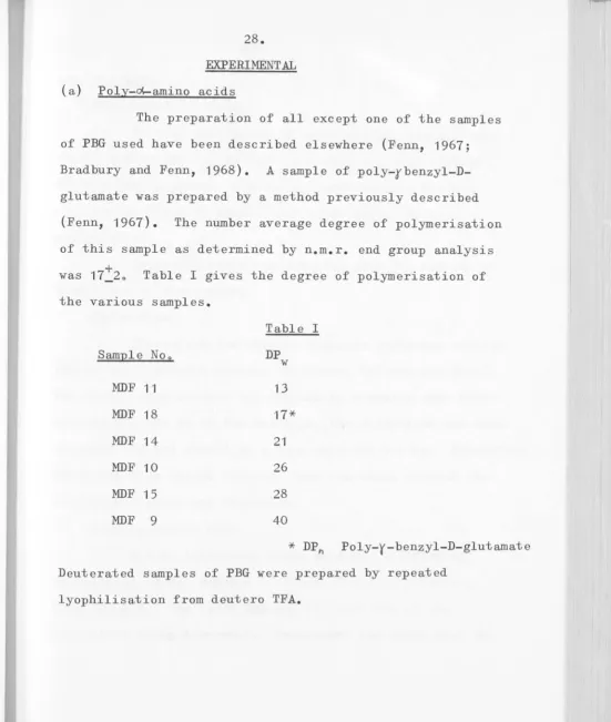
EXPERIMENTAL
(a) Poly-~amino acids
The preparation of all except one of the samples
of PBG used have been described elsewhere (Fenn,
1967;
Bradbury and Fenn,
1968).
A sample of poly-rbenzyl-D-glutamate was prepared by a method previously described(Fenn,
1967).
The number average degree of polymerisation of this sample as determined by n.m.r. end group analysiswas 17:20 Table I gives the degree of polymerisation of
the various samples.
Sample Noo
MDF
11
MDF
18
MDF
14
MDF
10
MDF
15
MDF
9
Table I
DP w
13
17*
21
26
28
40
*
DP
h Poly-y-benzyl-D-glutamateDeuterated samples of PBG were prepared by repeated
[image:37.607.47.598.16.666.2]29.
(b) Solvents
Deuterated Solvents
Deutero chloroform of spectroscopic quality was
obtained from Stohler Isotope chemicals and used without
further purification. Similarly, spectroscopic deutero
trifluoroacetic acid (Merck) was used without further
purification.
Deuterium oxide was obtained from the Australian
Atomic Energy Commission o
Chloroform
Chloroform for nuclear magnetic resonance studies
was of spectroscopic quality (Matheson, Coleman and Bell)o
The stabilising alcohol was removed by standing over Linde
molecular sieve No 4A for tw·o days, the chloroform was then
decanted off and stored in a dark coloured bottle. Chloroform
which had been stored for more than two weeks without the
stabilising agent was discarded o
Dichloroacetic acid
B.DoH. laboratory grade acid 'vas purified by
distillation under reduced pressure at 1-2 mm pressure,
B.P.
50-60oC., the first 20% and the last 20% of the30.
middle fraction used contains between 0.1% and 0.3% water (Fenn, 1967) 0
Deuterated DCA was prepared by mixing
quantities of DCA anhydride (B.D.H.) and D
20 in a dry boxo
Trifluoroacetic acid
Light laboratory grade acid was dried over
phosphorus pentoxide for several hours then distilled under
anhydrous conditions (Klotz et aI, 1964).
Other Solvents
The solvents, piperidine (M&B), pyrrolidine
(Light), triethylamine (BDH) and benzene (M&B) were of
laboratory grade quality and were used after drying over molecular sieve No 4A.
(c) Reagents
The following reagents were used without further
purification.
Laboratory grade reagents
N methyl formamide (BDH) N methyl acetamide (BDH)
diethyl acetamido malonate (Light)
&
NN dimethylacetamide (K
&
K)Analytical grade reagents
31.
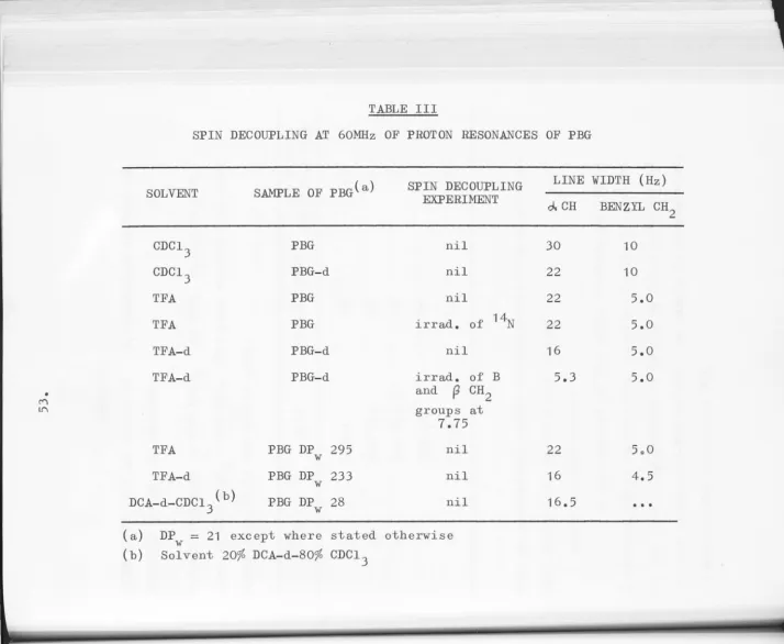
(d) Nuclear Magnetic Resonance Methods
Spectra were obtained at 100mHz on a Varian HA100 instrument and at 60MHz on a Perkin Elmer R10 machine.
Double resonance experiments were performed at 60MHz on a suitably modified Varian HA60 instrument by Dro A.G. Moritz of the Defence Standards Laboratories, Melbourne (Long and Moritz, 1968). All spectra were obtained at 350C using an
r.f. input power of 1mV and a scanning speed of 1.6 ppm per minute. All solutions except where specifically stated were
10% w/v concentration. Mixed solvents were prepared by mixing accurately known volumes of the two components and
32.
N-methyl acetamide CH
3-CO-NH-CH3
Spectra were obtained in mixed solvents of CDCl 3 and DCA at 60MHz. The spectrum in CDCl
3 consists of a
doublet at 7028 tau and 7.20 tau (amide methyl) and a sharp
singlet at 8.30 tau (aldehydic methyl). The resonance due to the NH proton was obscured by solvent. On deuteration of the amide by lyophilisation from D
20, the doublet
collapses to a singlet. Hence the splitting is due to spin-spin coupling with the amide hydrogen and not cis
trans isomerisationo The addition of small amounts of DCA
(9%) results in the collapse of the doublet to a broad
singlet at 7.09 tau (Fig I). Additional acid results in
little change in the spectrum. No additional broadening of the amide methyl is observed. The collapse of the doublet to a broad singlet indicates an increase in the rate of
exchange of the amide proton with solvent protonso Clearly,
this is an intermediate situation in which the lifetime
each state is of the same order as 2 1
2-rrS
_2
~ 3 x 10 sec
of
w"here
S
is the chemical shift between the two states and inthis case is given by the coupling constant (J
=
4.8 Hz). This lifetime is of the same order as that obtained by Stewart et al (1967) for N-methyl acetamide in 10% TFA -90% CDCI3• It is interesting to note that Stewart et al
2.7
2.8 FIG 2
N methyl acetamide
2.1
2.2
o
330
reappearance of the methyl doublet for TFA concentrations
)16%. No such effect is observed in the DCA _
system.
In addition to the collapse of the amide methyl
doublet, both this resonance and that due to the aldehydic
methyl shift downfield by 0.23 ppm and 0033 ppm respectively
with a large proportion of the shift occurring with the
initial addition of acid (Fig 2).
In agreement with stewart et al (1967) it is
believed that this initial downfield shift and increase in
rate of exchange of the amide proton is indicative of
protonation of the amide. Although exchange of the amide
nitrogen proton is of the intermediate case, equilibrium
between the unprotonated species and the protonated species
must be fast. If this equilibrium were slow then for low
concentrations of DCA
«
9%) a third resonance at 70 09 dueto the protonated species would be visible, increasing in area
as the acid content is increased. As no such resonance
is visible one must conclude that this equilibrium is
o
H\\
/
C-N
/
\
CH
3
CH
3
34.
"
fast
reI. slow ,
o
H~
T
C-N
+ CHCl
2
COOH
/
\
CH3 CH
35.
N-methyl formamide H-CO-NH-CH 3
Spectra were obtained at 60MHz in mixed solvents of CDC1 3 and DCA. Representative spectra are shown in Fig 3. Throughout the entire solvent range the methyl group appears as a doublet
(J
= 4Hz). Deuteration of the amide by lyophilisation from D20 causes this doublet to
collapse to a singlet. Thus the splitting is due to spin-spin coupling with the amide nitrogen proton and not cis trans isomerisation. The presence of the doublet also indicates that exchange between the amide proton and the
1 -2
4 x 10 sec.
solvent is slow i. e.
'i'
~2 -;r
S
As theacid content of the solvent is increased both the methyl doublet
and the broad singlet due to the aldehydic proton move downfield by 0.20 ppm and 0.15 ppm respectively (Fig 4)0 No broadening of these peaks outside of experimental error
is observed o
The presence of one doublet throughout the solvent range indicates that the equilibrium between protonated and unprotonated species lS fast.
On increasing the acid content of the solvent, the peak which can be attributed to the amide proton on area
consideration, sharpens, moves downfield and increases in area
[image:46.606.42.597.15.681.2]a:o
9
o
9 e
Solvent (O:A)
L
---JL
L
ppm from 1":3
:=IG 3
N methyl fo:rma.ciJe
:lI!ll
FIG 4
N methyl formamide
8.1
8.2
rICO
8.) v
en
~ 2.8
6 0
~ ...
8 n. n.
2.9
a.
3
.0
o
3.1
20 40 60 80 100
[image:48.606.77.592.14.673.2]3
4
FIG5(a)
N met~l acetamide
5
shift of acid proton
6
7
8
9
10
11
20
40
60 80100
[image:49.606.65.591.16.678.2]%
DCA100
33
20
5
3
1
o
8
+ NH
FIG
5(b)
7
[image:50.606.50.595.12.705.2]hydrogen of the uncharged species and the acid prot on. A
peak which appears at approx. 3 tau and increases in area
as the acid content is increased appears to be due to the
amide proton of the protonated species (Fig 5b)o
As only one resonance is observed for the acid
proton and the amide proton one must assume that exchange
between the two protons is rapid o Also the appearance of
a peak corresponding to the protonated species indicates
that the equilibrium between protonated and unprotonated
species must be slow. Yet interpretation of the spectra
of the methyl protons is exactly the opposite. Because of
the discrepancy between these sets of observations the
37.
Diethyl acetamide malonate
Sample No o 1
60MHz spectra for this sample were obtained in
solvent mixtures of DCA andCDCl
3• The resonance of the
proton on the carbon atom adjacent to the amide nitrogen
was found to be a doublet both in 100% DCA and 100% CDCl 3
(J = 3.5 Hz), the resonance in DCA being 0023 ppm further
downfield. In intermediate solvents two doublets which
can be attributed to the '~CHY are visible (Fig 6).
The downfield doublet observed moves rapidly downfield and
increases in area with increasing acid content of the
solvent. The upfield doublet shows only a slight chemical
shift but decreases in area finally disappearing in the
region 75-80% DCA (Fig 7). The variation in chemical
shifts with increasing acid content of the solvent are
shown in Fig 7.
Sample No.2
Although this sample was obtained from the same
source (Lo Light and Co., but different batch number) the
NMR spectra obtained in solvent mixtures of DCA and CDCl 3
are significantly different. Only one '~CHf doublet
(J = 3.5 Hz) is visible throughout the entire solvent range
(Fig 8)0 This doublet behaves in a manner similar to the
lowfield doublet observed in sample number one, the final
o
~,.,
FI'} 7
6.5
+
100 MHzo 60 l.:Hz
7·5
,ill
en 8.0
~ 6 0 H
'--<
6
0-5.2
5·3 CH
5.4 o
20 40 60 80 100
~ f; N
.
' " 0 ~ 0 'i 0..
." <-~ ' " ~,
1
.
,
'"g
~ ~.
!
c: C F. e. 0..
X ~ . ~ : rJ~
1
38.
The gradual downfield shift of the lowfield
I ~CH' in sample one clearly indicates that two or more
forms are in rapid equilibrium, these forms being in slow
equilibrium with yet another form, represented by the
upfield i ~CHi. The shift observed for sample two
indicates two or more forms in rapid equilibrium only.
Such difference in behaviour can be rationalised
by the assumption that sample one is a mixture of the cis
and trans isomers of the amide while sample two contains
only one of the isomers. If one assumes that only one
protonated form is possible which is in rapid equilibrium
with one of the isomers then the different behaviours of
samples one and two can be explained by the following kinetic
scheme.
I
OH R
\,~-
/
C-N
/
\
I
II
H
slow
o
H
\
\
/
C-N
/ 'R
39.
Since I and III are in rapid equilibrium with
each other the observed chemical shift of the Y ~CHt
resonance will represent an average of the relative
amounts of the two. As the acid content of the solvent is
increased the relative amount of III is increasedo Since
the t ~CHt resonance of the protonated amide III can be
expected to be further downfield than in II (Bradbury and
Fenn 1969, (a)), the average resonance will move downfield.
As II is not in rapid equilibrium with I two resonances will
be seen. With an increase in the relative amount of III
more of I i s removed and the equilibrium between I and II
shifts to the left thus decreasing the relative amount and
hence the area of II. Sample one containing both isomers
exhibits the behaviour outlined above. However, sample two
containing only one isomer, which must be I, shows only one
doublet for the Y~ CHY which moves downfield with increasing
acid content of the solvent due to increasing amounts of
1110
An
alternative explanation can be that both samplesone and two are pure, but different isomers. The isomer in
sample one requires an intermediate with which it is in slow
equilibrium, the intermediate form being in fast equilibrium
~----~ -~---"""""""""""--II--~
o
H\\
/
C-N
/
\
R
40.
slow intermediate
"
fast
+H+
-H+
"
OH
H\\.t
I
C-N
/
\
R
While the isomer in sample two either requires no intermediate
or has an intermediate which is in rapid equilibrium with both
the isomer and the final protonated formo
o
R~ I
C-N
/
\
H
__ -=f~a=s~t __ ~~ intermediate
<-
"
fast
+H+
-H+
oH
H\
/
C-N
/ \
R
Although the equilibrium between protonated and unprotonated
species appears to be rapid, the rate of exchange of the
amide proton with solvent appears to be slow as shown by
the presence of a doublet for the fo('CHf resonance
(J
=
3.5Hz)o
Also a separate doublet for the amide proton(J
=
3.5Hz)
distinct from the acid proton is observed(Fig 8). This resonance moves downfield as the acid content
of the solvent is increased again signifying fast equilibrium
between the protonated and unprotonated forms. (It is noted
that the interpretation of the chemical shifts of all
resonances lead to the same conclusions, a fact not observed
---
-
- -.. ---...
""11~
410 NN dimethyl acetamide
Spectra of NN diemethyl acetamide in mixed solvents of DCA and CDC1
3 were obtained at
60MHz
(Fig 9). In CDC13the spectrum consists of a doublet at
6.98
and7.07
tau(amide nitrogen methyls) and a singlet at 7093 tau (aldehydic
methyl). The doublet is attributed to the two methyl groups
on the amide nitrogen having different magnetic environments which are due to restricted rotation about the amide bond
(Phillips
1955;
Stewart et al1967).
With the addition ofDCA to the solvent
(9%),
the doublet collapses to a singletindicating an increase in the rate of rotation about the amide bond to such an extent that the different magnetic environments of the methyl groups are averaged out and a single resonance
is observedo Similar behaviour was observed by Stewart et
al in a TFA/CDC1
3 system. Although in the TFA/CDC13 system
the doublet collapsed at a slightly higher concentration of
acid
(12%)
than in the DCA/CDC13 system used hereo In an
agreement with other authors (Stewart et al) i t is felt that the collapse of the doublet is due to protonation of the amide group with consequent loss of double bond character of the
CO-N bond. Such acid catalysed lowering of the rotational
barrier has also been observed in aqueous hydrochloric acid
FI
G
9
i
I DCA9
5
3
o
4
3
2 [image:60.648.14.638.15.707.2]FI::; 10
Dim8th'yl acetamlde
2.0
2.2
Ul COCH
3
f~
E! 2·4 0
0
1-4 0
( . ,
E 0. p,
2.8
3.0
3
.
2
o
--
-42"
aldehydic methyl group and the amide methyl resonances move downfield by 0.30 ppm and 0.24 ppm respectively
(Fig 10).
It is also noted from Fig 10 that the majority of the downfield chemical shift observed occurs during
protonationo This seems to indicate that the chemical
shift observed on dissolution of amides in acidic solvents
is due to protonationo
Since only one doublet is observed in the spectrum during protonation the equilibrium between the protonated
species and unprotonated species must be fast.
o
OR\\ / fast \\
+/
C-N - - - - -" d""::N
+
CRC12
COO-/
\
"/
\
43.
NMR study of simple amides in basic solvents
Previous work (Fenn,
1967;
Bradbury and Fenn,1969,
(a)) had shown that for a simple amide the n.m.roresonance of the protons on the carbon atom adjacent to the
nitrogen moved downfield by a value of 00 24 ppm from its
position in CDC1
3 when the amide was dissolved in DCA o
It therefore seemed reasonable to assume that if this shift
were due to protonation then on deprotonation ioeo placing,
a negative charge on the amide group, the resonance of the
proton in question should have a shift equal in magnitude
but opposite in sense to the shift observed in acid
solvents o Accordingly, the nmr spectra of N methyl formamide,
N methyl acetamide and N isopropyl benzamide have been
obtained in several basic solvents. The results are shown
in table II.
TABLE II
Chemical shifts of protons of amides in CDC1
3 and basic
[image:63.644.55.632.18.666.2]44.
CDC1
3 solvent (ppm) in
Amide
Et3N Piperidine Na/C 6H6
t
Bu OK/DMSo-d 6
HCONH-CH
3 0 a -0003 -0.22
CH
3CONH-CH3 0 a -0.05 -0.23
PhCONH-C!!(CH
3)2 0 -0.03 -0007 -0.24
a obscu red by solvent
Triethylamine and piperidine are not strong enough
bases to cause a chemical shift due to deprotonation,
however, sodium in benzene has a small effect. Apart from
this the spectra are essentially unchanged in these solvents
and the NH resonance is visible. When equimolar amounts of
the amide and potassium tertiary butoxide are dissolved in
dimethyl sulphoxide-d
6 the following spectral changes are
observed:-(a) a decrease in the amount of splitting of the
proton resonance adjacent to the amide group
and a chemical shift of 0.23 ppm which is
equal in magnitude but opposite in sense to
the average shift of 00 24 ppm observed on
dissolution in DCA (Fenn 1967, Bradbury and
-45.
(b) the NH resonance disappears;
(c) the occurrence of a new resonance at 8.72
due to the methyl groups of tertiary butanol
which is produced by deprotonation of the
amide group.
Fig. 11 illustrates the above being the spectra of
t
N isopropyl benzarnide in CDC1
3 and Bu OK/DMSo-d6"
It is also observed from the table that since
N isopropyl benzamide is the most difficult to protonate
[image:65.645.30.632.13.687.2]Q) '" ....
i
..
c Q) ~ " rl H ~ . <. 0 ... Po 0'" ....
Z +' C g: rl o I n :c u rl'" U ..., "
to
..
"' Ul ~ S 0 ...
..
.
S46.
OTHER PROTONS
By examining the chemical shifts of the protons
in DCA one may obtain information concerning changes in the
various chemical states of the acid, in particular the
formation of the acid anion and intermolecular hydrogen
bondso Nevertheless, such information could only be obtained
at low concentrations of acid, since under normal conditions
the concentration of DCA unassociated with solute would be
greatly in excess of that associated with solute. Hence by
equation (6) there would be no appreciable difference in
chemical shift of the respective DCA protons in pure solvent
and solvent with solute.
Stewart et al (1967) have found that the addition
of TFA to a solution of N methyl acetamide inCDCl
3 causes not
only a downfield chemical shift of the amide nitrogen methyl
but also a large initial downfield chemical shift of the acid
proton of TFA. This large initial downfield shift of the
acid proton was interpreted as indicative of protonation of
the amide. However, the addition of TFA to a solution of
poly-L-alanine in CDCl
3 produced only a relatively small
downfield shift of the acid proton of TFA. From this they
concluded that protonation of poly-L-alanine did not occur
Since the molar concentration of the N methyl
acetamide was about six times greater than that of the
peptide residues the two experiments cannot be compared.
In order to clarify this point, spectra were
obtained in mixed solvents of DCA and CDCl
3 at 60 MHz
where the molar concentrations of N methyl acetamide and
PBG (DP
=
21) were 0044M and 0.46 moles peptide residuesw
per litre.
In solvents containing no solute, a single sharp
resonance is obtained for the acid proton of DCA. This
resonance is displaced downfield by nearly 3 ppm in the
range 1-30% DCA, but little change occurs from 30-100% DCA
(Fig 12)0 This behaviour is similar to that observed for
other carboxylic acids and has been interpreted in terms
of the formation of intermolecular hydrogen bonded structures
(Reeves, 1961).
With the addition of either solute, the acid
proton of DCA could not be observed in solutions containing
5% or less of DCA.
An interesting observation is that when small
amounts of DCA (5% or less) are added to CDCl
3 solutions
PBG:-:'1:; 12
• N methyl acetamide
o
:
0 s
olute+
PEG9
(f)
i2
10E
0
H
(,...-E
a. a.
11
--~~~-+---~~---~---20 to 60 80
100
FIG 13
acid ,:Jroton
DCA
proton
- 200 Hz
9 e 7 6
ppm from TItS
benzyl
::H2
5
PSG
aCH
4
% DCA
20
9
5
[image:70.644.37.632.16.689.2]