CORRELATION OF RETINAL NERVE
FIBRE LAYER THICKNESS IN OPTICAL
COHERENCE TOMOGRAPHY WITH
VISUAL OUTCOME IN OPTIC NEURITIS
Submitted in partial fulfillment of requirements of
M.S. OPHTHALMOLOGY
BRANCH - III
REGIONAL INSTITUTE OF OPHTHALMOLOGY
MADRAS MEDICAL COLLEGE
CHENNAI - 600 003
THE TAMIL NADU
DR. M.G.R. MEDICAL UNIVERSITY
CHENNAI
CERTIFICATE
This is to certify that this dissertation entitled "CORRELATION
OF RETINAL NERVE FIBRE LAYER THICKNESS IN OPTICAL
COHERENCE TOMOGRAPHY WITH VISUAL OUTCOME IN
OPTIC NEURITIS" is a bonafide record of the research work done by
Dr. C.USHA, post graduate in Regional Institute of Ophthalmology and
Government Ophthalmic Hospital, Madras Medical College and
Government General Hospital, Chennai-03, in partial fulfillment of the
regulations laid down by The Tamil Nadu Dr. M.G.R. Medical University
for the award of M.S. Ophthalmology Branch III, under my guidance and
supervision during the academic years 2013 - 2015.
Prof.Dr.K.NAMITHA BHUVANESWARI MS, DO.
CHIEF - DEPT OF CORNEA RIO - GOH
EGMORE
CHENNAI - 600 008
Prof.Dr.K.NAMITHA BHUVANESWARI MS. DO
DIRECTOR AND SUPERINTENDENT RIO - GOH
EGMORE
CHENNAI - 600 008.
Prof. Dr. R. VIMALA, MD
DEAN
MADRAS MEDICAL COLLEGE AND GOVERNMENT GENERAL HOSPITAL
DECLARATION BY THE CANDIDATE
I hereby declare that this dissertation entitled, "STUDY ON
CORRELATION OF RETINAL NERVE FIBRE LAYER
THICKNESS IN OPTICAL COHERENCE TOMOGRAPHY WITH
VISUAL OUTCOME IN OPTIC NEURITIS" is a bonafide and
genuine research work conducted by me under the guidance of
Prof. Dr. K. NAMITHA BHUWANESWARI, M.S., Professor
Department of Cornea, Regional Institute of Ophthalmology,
Government Ophthalmic Hospital, Chennai - 600 008.
Date :
I express my sincere thanks and gratitude to
Prof. Dr. R. VIMALA, M.D., Dean, Madras Medical College and
Government General Hospital for permitting me to conduct this study.
I have great pleasure in thanking Prof. Dr. K. NAMITHA
BHUVANESWARI, M.S., Director and Superintendent, RIO - GOH,
Madras Medical College, who has also been my guide and mentor for her
valuable guidance in preparing this dissertation.
I am very grateful to my unit Chief Prof. Dr. M.R.CHITRA,
M.S., for valuable advice and guidance for the study.
I express my profound gratitude to my co-guide
Dr. B. PRAMILA, M.S., for her valuable guidance and constant support
at every stage throughout the period of this study.
I am very grateful to my unit assistant professor,
Dr. R. MUTHIAH, M.S., Dr. T.G. UMA MAHESWARI, MS., and
Dr. K.S.T. LATHA, M.S. for rendering their valuable advice and
professors and all my colleagues who had helped me in bringing out this
study.
Finally, I am indebted to all the patients for this sincere
ONTT - Optic Neuritis treatment trial
MRI - Magnetic resonance imaging
RB Neuritis - Retrobulbar Neuritis
CSF - Cerebrospinal fluid
OCT - Optical coherence tomography
RNFL - Retinal Nerve Fibre Layer
OCT, SLO - OCT-scanning laser ophthalmoscopy
ICM - Internal Limiting membrane
RAPD - Relative afferent Pupillary defect
SA SPACE - Sub Arachnoid Space
VEP - Visually evoked potential
HIV - Human Immuno Defeciency virus
VDRL - Venereal Disease Research Laboratory
MMR - Mumps, Measles, Rubella
SLE - Systemic Lupus Erythematosus
PAN - Poly Arteritis Nodosa
ONH - Optic Nerve Head
PL - Perception of Light
FB - Foreign Body
VZ - Varicella Zoster
TB - Tuberculosis
DM - Diabetes Mellitus
FFA - Fundus Fluorescein Angiography
RPE - Retinal Pigment Epithelium
Sl.No. Title Page No.
PART - I
1. Introduction 1
2. Anatomy of Optic Nerve 5
3. Physiology of Optic Nerve 14
4. Optic Neuritis 17
5. OCT 33
6. Review of Literature 45
PART - II
1. Aims and Objectives 58
2. Materials and Methods 59
3. Results 66
4. Discussion 89
5. Conclusion 96
6. Summary 99
7. Proforma 101
ANNEXURE
1. Key to Master Chart
2. Master Chart
INTRODUCTION
Optic neuritis is an inflammation of optic nerve. The
annual incidence is 1-5/1,00,000. It can occur as unilateral or
bilateral disease, usually bilateral in pediatric population.
Optic neuritis affects females more commonly than males.
Majority will present in 20-50 years group, mean age is 30-50
years, but it can occur in any age group.
Patients present with painful visual loss of acute onset
associated with field defect. Visual acuity ranges from 20/20 to
no perception of light.
On examination there will be relative afferent pupillary
defect in almost all cases, the exception being bilateral disease or
the fellow eye with optic atrophy, and defective colour and
contrast sensitivity .The contrast sensitivity will at times be
Examination of fundus will reveal disc edema with
blurring of margin and hyperemia, but the disc will appear
normal in retrobulbar neuritis .
Unilateral or bilateral optic neuritis and myelitis in Devic’s
syndrome.Common field defect in optic neuritis is central or
centrocecal scotoma, but other defects like arcuate and
altitudional (ONTT study) can occur. The common causes for
optic neuritis are idiopathic and demyelination.
Investigations like MRI (to look for demyelination), blood
investigation and x-ray sinuses (to rule out septic foci), and x-ray
chest(to exclude sarcoidosis), MRI spine and NMO-IgG
(Neuromyelitis optica), CSF analysis(Multiple sclerosis and
Neuromyelitis optica) are done in cases of optic neuritis.
Patients of optic neuritis are treated with intravenous
followed by oral prednisolone 1mg per kg body weight for 11
days(according to ONTT study), this speeds up the recovery and
prevents relapse of disease.
Optical coherence tomography is a new tool in optic
neuritis to quantify axonal loss after optic neuritis, hence can be
used to assess visual outcome in these patients. OCT utilizes
infrared light of 800-820nm light.
It utilizes Michelson interferometry to produce cross
sectional images based on optical scattering of light. 3.4mm
circular scan centered around the disc measures the nerve fiber
layer thickness(RNFL scan) and compares the thickness with age
adjusted normative data. Other protocols available for evaluation
of retina are macular scan and optic disc analysis.
RNFL scans are useful to assess the progressive changes
There is thinning of RNFL in optic neuritis after an attack.
With the thinning , visual recovery and the chances of going for
optic atrophy can be predicted. In our study spectral domain
OCT-SLO is used to quantify RNFL thickness. Hence OCT can
ANATOMY OF OPTIC NERVE
It is second cranial nerve extends from optic disc upto
optic chiasma where the two nerves meet each other. It is nothing
but the continuation of nerve fiber layer of retina. It is
comparable to a sensory tract of the brain morphologically and
embryologically. Because it is an outgrowth of the brain it is
surrounded by meninges and like other nerves it is not covered
by neurilemma.
Optic nerve is divided into 4 parts they are intraocular
(1mm) intraorbital (30mm) intracanaliculer (6–9 mm)
intracranial (10m).
Intraocular part appears in the eyes as optic disc and has an
average diameter of 1.5mm which expands to 3mm just behind
I. Intraocular Part:
The optic nerve head is divided into 4 portions. Surface
nerve fiber layer : It is essentially composed of axonal bundles
and covered by astrocytes, ILM of elschnig which separates it
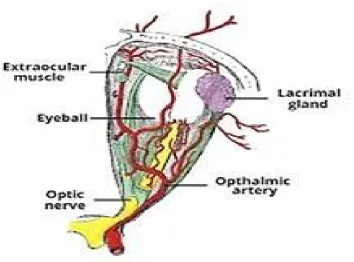
from vitreous. Figure -1 describes the relations of optic nerve to
ocular structures.
Pre laminar region : composed of neurons and astroglial
tissue separated from choroid by a cuff of astrocytes.
Lamina cribrosa: It is made up of fenestrated sheets of
scleral tissue lined by glial
tissue. Bundles of optic nerve leave the eye through this.
The lamina cribrosa gets its blood supply from circle of zinn.
Retro laminar region: Here the nerve gets its myelin sheath
II. Intra orbital Part:
This parts extends from back of the eyeball to the optic
foramina and it is sinous to play for the eye movements. It is
covered by dura, arachnoid and pia. The central retinal artery
with vein crosses in SA space to enter the nerve on its infero
medial aspect. Posteriorly the nerve is closely surrounded by
annulus of Zinn and origin of 4 recti muscles. Some of superior
rectus and medial rectus are adherent to its sheath.
The long and short ciliary nerves and arteries surround the
nerve. Ophthalmic .A, superior Ophthalmic.V, and nasociliary
nerve cross superiorly from lateral to medial side of the nerve.
Ciliary ganglion, Branches of oculomotor nerve,
nasociliary nerve, sympathetic and Abducent nerve, are situated
III. Intra canalicular Part:
Ophthalmic A is closely related and crosses the nerve from
medial to lateral side inferiorly sphenoid and posterior ethmoidal
sinuses lie medial to it and separated only by a thin bone. Hence
there are chances of RB neuritis (Retrobulbar neuritis) following
sinusitis.
IV. Intracranial part:
This lie above cavernous sinus and converge to form the
optic chiasma. The internal carotid.A runs below and the lateral
Arrangements of nerve fibers in optic nerve:
In nerve head it is same as in retina, behind the eye the
upper and lower temporal half are separated by papillomacular
bundle situated on temporal side. The upper and lower nasal
fibers are situated on nasal side, near chiasma the macular fibers
are centrally placed.
Blood supply of optic nerve:
I. The intraocular parts
The surface nerve fibre layer is supplied by capillaries of
retinal vessels. The prelaminar region by vessels of ciliary
region. The Laminacribrosa region by the short posterior ciliary
arteries and circle of Zinn –Haller. The retrolaminar region by
ciliary (from recurrent pial vessels) and retinalcirculation. The
central retinal. A provides centripetal branches from pial plexus
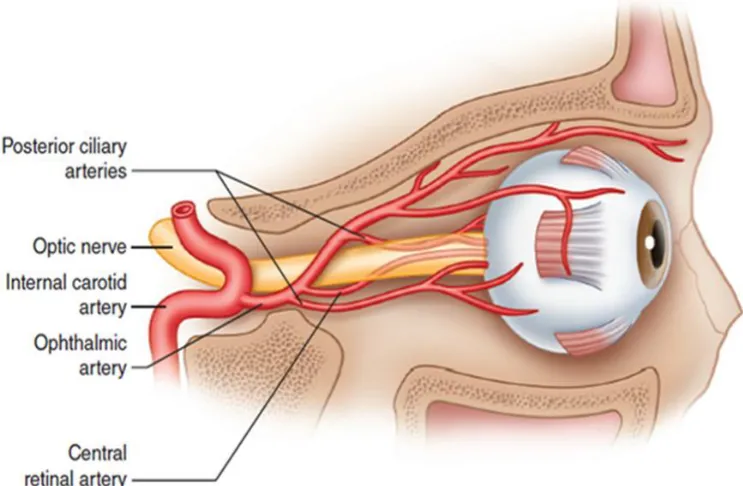
and also centrifugal branches. Figure-2 depicts the blood supply
II. Intra Orbital Part- Supplied by 2 system
A periaxial system is derived from 6 branches of ICA
namely ophthalmic. A, long and short posterior citiary. A,
Lacrimal.A, Central retinal A, Circle of Zinn.
The axial system is derived from the intraneural branches
of central retinal.A, central collateral arteries from CRA, and
central artery of optic nerve.
III. Intracanalicular part is supplied by periaxial system of
vessels.
Venous drainage
· From optic nerve head venous drainage is by the central
retinal.V .
· The orbital part in its proximal end drained by pial plexus
and distal end by central retinal.V.
· The intracranial part is drained by the pial plexus which
OPTIC NERVE PHYSIOLOGY
There are two system of retinal ganglion cells- P cell
system and M cell system.
P cell system - 80-90% of these cells are of small to
moderate size and they are concentrated in macula and projects
into the parvocellular layer of lateral geniculate ganglion. They
thought to subserve high contrast high spatial frequency
resolution.
M cell (Large cells) system is fast conducting and comprise
about 10-20% of ganglion cell population. M cells project into
magnocellular layer of lateral geniculate ganglion. They are
TOPOGRAPHIC ANATOMY OF OPTIC NERVE
Peripheral ganglion cells occupy the periphery of optic disc
and the central portion of the disc is occupied by ganglion cells
located closer to optic disc.
The arrangement of fibers in the disc and distal portion of
nerve corresponds to topographic distribution in retina. The
papillomacular bundle occupies temporal optic disc and these
fibers moves centrally in the distal portion of optic nerve. All
retinal fibers retain their position throughout the visual pathway.
Because some axons decussate in optic chiasma the axons lose
their retinotopic order to some extent.
Hence retinal disease is divided into 1.those affecting the
detection apparatus and 2.those affecting the conducting
Retinal diseases have to be differentiated from macular
diseases by the following ways:
Features Optic nerve
disease
Macular disease
Metamorphopsia Absent Present
Pain on eye movements Present Absent
Relative afferent
pupillary defect
Present Absent
Colour vision Markedly
reduced
Slightly reduced
Contrast sensitivity Markedly
reduced
Variable
OPTIC NEURITIS
INTRODUCTION :
Optic neuritis is an inflammation of one or both optic
nerves resulting in temporary visual loss. Affects young to
middle aged adults between 20 to 50 years of age, with a mean
age of 30-35. But it can affect individuals of any age. Children
are affected bilaterally whereas in adults it is usually unilateral.
Butin adults bilateral disease can occur in patients of multiple
sclerosis. The female to male ratio is 3:1 and the incidence is
1-5/1,00,000.
Apart from idiopathic variety two variants of optic neuritis
are optic perineuritis and neuroretinitis. In optic perineuritis
there will be involvement of optic nerve sheath without
involvement of optic nerve. Neuroretinitis is involvement of
SYMPTOMS :
Patient of optic neuritis will present with:
Loss of central vision (in more than 90%).
Loss of peripheral vision in superior or inferior fields
associated with orbital pain(90%) above or behind the eye .
Visual acuity decreases over next several days from 20/20
to no perception of light .
There will be loss of colour vision and perception of
phosphenes (less common symptom)(30%).
There will be reduced contrast sensitivity in all cases.
After an attack of optic neuritis even after complete
recovery of vision some patient may complain of transient loss of
SIGNS :
1. Visual acuity ranges from 20/20 to no PL .
2. Visual field defects – central scotoma , centrocecal
scotoma, arcuate scotoma, superior or inferior altitudinal
scotoma , Bi temporal or left or right hemianopia . Any
type of field defect can occur but in ONTT study with
automated perimetry of central 30°, the field revealed
diffuse loss in 48% and focal loss is 52%.
3. Reduced contrast sensitivity and colour vision . The loss of
contrast sensitivity is proportionate to or sometimes worse
than loss of visual activity. The colour dysfunction is more
severe than visual activity level. In ONTT, no particular
type of colour vision defect was consistently associated
with optic neuritis .
4. Relative afferent pupillary defect is always present in optic
atrophy. It is diagnosed by swinging lash light test . By
swinging the flash light the amplitude and thevelocity of
pupillary constriction are compared with other eye and it
should be equal when there is no RAPD.
5. Fundus findings – Disc edema with minimal blood vessel
enlargement and rarely peripapillary hemorrage in
papillitis . Normal appearing fundus is retrobulbar neuritis .
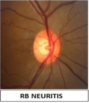
Figure 3-Showing disc changes in optic neuritis
Figure-3 shows the fundus changes in optic neuritis.
Papillitis is shown with disc edema and in retro bulbar neuritis
[image:29.595.356.509.219.397.2]DIFFERENTIAL DIAGNOSIS OF OPTIC NEURITIS
1. Acute ischemic optic neuropathy – Painless visual loss ,
associated with usually altitudional field defects . In fundus
more papillary hemorrhage with sectoral disc edema of
pallid type will be seen. It is the disease of elderly above
50 years . It can be anterior or posterior depending upon
the location of ischemic damage, and it can be arteritic or
non arteritic depending on etiology.
Arteritic AION is usually associated with giant cell
arteritis and other connective tissue disorder. Non arteritic
ischemic optic neuropathy in majority is due to idiopathic
and ischemia due to causes other than arteritis.
2. Leber’s hereditary optic neuropathy – Patients are usually
males of 15 to 35 years and present with painless loss of
vision in one eye followed by other eye. Fundus findings
are circum papillary telangiectatic microangiopathy,
testing shows DNA(mitochondrial) mutations.
Corticosteroids is of no use here in leber’s optic
neuropathy.
3. Vogt koyonagi harada disease – hyperaemia of optic disc
and optic neuritis in addition to uveitis, choroiditis and
exudative retinal detachments are seen .
INVESTIGATIONS
Diagnostics studies are performed to rule out some other
lesions like compressive lesion for optic neuropathy, to
determine other than demyelination causes responsible for
inflammation of nerve and to determine the visual and
neurological prognosis of optic neuritis .
MRI brain is not for the diagnosis of optic neuropathy but
best in recurrent opticneuritis and is assessing the progression of
MS. 40-70% of patients with isolated optic neuritis will have
images. Diffuse enlargement of optic nerve will be seen on fat
suppressed scan with or without contrast enhancement.
Gadolinium enhancement will show inflammatory infiltrate and
expansion of extra cellular space. MRI brain and optic nerve is
done usually. MRI is indicated in diagnosing optic neuritis in
patients above 45 years, in bilateral cases, in cases of optic
neuritis which shows progression beyond 2wks and in patients of
optic neuritis with atypical fields like hemianopic defects.
MRI is contra indicated in patients with ferromagnetic
implants, FB, cardiac pace makers, metallic cardiac valves and in
claustrophobia.
VEP (Visually evoked potential) -In visually asymptomatic
or suspected or known MS patients VEP seems to bea sensitive
indicator .It is helpful in predicting the progression of MS. VEP
is the measurement of signal in occipital cortex in response to
light stimulus, damage anywhere along the afferent pathway will
P1(positive peak) followed by second negative(N2) and
positive(P2). Positive peak is usually at 100 msec.
Latency and amplitude are analysed and compared with the
other eye Demyelination will produce increased P-100 latency
without affecting the amplitude. Multifocal VEP can detect
small abnormalities of transmission in optic neuritis. In toxic,
ischemic and compressive lesions the amplitude will get affected
first.
Chest x ray , HIV,VDRL, X-ray of sinuses , blood routine
are important to exclude other causes of optic neuritis .
CSF analysis is important in patients with abnormal MRI
suggestive of MS to look for oligoclonal bands, anti MBP and
PROGNOSIS
Complete visual , field , colour vision, contrast sensitivity
recovery by 5 weeks after onset . Recurrence rate is 35%
according to ONTT . The risk of recurrence increases(41%) in
those treated by only oral steroids.Optic neuritis occurs in 50%
as initial presentation in patients with MS and the risk of
developing MS after optic neuritis in 5 years is 30% .
RESIDUAL DEFICITS AFTER OPTIC NEURITIS
Disc pallor, RAPD,Colour vision defects and prolonged
latency in VEP.ONTT showed 30% of contrast sensitivity,55%
of colour vision was normal after recovery and 58% showed no
TREATMENT
As per ONTT guidelines patients after acute optic neuritis
are treated with 250 mg methyl prednisolone four times daily for
3 days and followed by oral prednisolone of 1 mg/kg/day for 11
days. Long term treatment with interferon beta- 1a should be
considered in high risk patients .
OTHER DEMYELINATING CONDITIONS ASSOCIATED WITH OPTIC NEURITIS
Devic’s disease
Devics disease occurs in children and young adults of both
sexes represents <1% of demyelinating diseases . Here the brain ,
spinal cord and optic nerves are affected . Patient presents with
bilateral visual loss and paraplegia. In devic’s disease cerebellum
will never get affected, excavation leading to cavity formation
will be there and there will be no gliosis are all in contrast to
disease. There is disc edema in fundus examination and most of
them develop disc pallor. The mortality is 50% in thesepatients.
Diagnosis is by CSF analysis ,MRI , NMO-IgG(50%)in
serum and it is treated with corticosteroids .
Schilder’s Disease
Patients will present with visual loss due to damage of post
chiasmal damage to visual pathway. There will be homonymous
hemianopic and quadrantic defects or cerebral blindness. MRI
shows demyelination with CSF changes similar to MS. It is
progressive and lethal to life, death can occur within months.
Causes of optic neuritis are mainly demyelinating and idiopathic .
Other causes are:
1. Optic neuritis in other primary demyelinating diseases like
2. Infectious end para infectious optic neuritis – following
viral infection in children due to immune medicated
demyelination – adenovirus, coxsakie virus, CMV, HIV,
measles, mumps,VZ,rubella and Hepatitis A
viruses.Bacterial causes include syphilis , lymes disease,
catscratch disease, anthrax,B haemolytic streptococcal
infection , brucellosis, meningococcal infection , TB , and
typhoid disease . Aspergillus and mucor infection of
sinuses in DM patients can involve optic nerve .
3. Post vaccination optic neuritis – Both retrobulbar and
papillitis can occur. Usually occur 1-3 weeks after
vaccination. It can occur with BCG, Hepatitis-B, Rabies
vaccine, Tetanous toxoid, and MMR (Mumps,measles and
rubella)
4. Inflammatory optic neuritis – in sarcoidosis, SLE, PAN,
neuritis or anterior optic neuritis. Disc is lumpy white in
sarcoidosis with inflammatory reaction in vitreous.
5. Auto immune optic neuropathy . Patients usually will
present with recurrent episode of visual loss with good
response to steroids.
OPTIC PERINEURITIS
It is due to inflammation of nerve sheath without
involvement of the nerve proper. There is bilateral disc edema
similar to papilledema. This is not associated with MS. In most
cases there is an associated infectious process.
NEURORETINITIS
Patients will present with unilateral visual loss without
pain and usually occurs in third or fourth decade. RAPD will be
present. Common field defects here are centerocecal,central
scotoma but arcuate and altitudinal defects can occur. There will
like exudates. Small white discrete chorioretinal lesions may be
seen associated with inflammation of vitreous. It is a self-
limited condition with good visual prognosis. It is due to
infectious or para infectious diseases.FFA(Fundus fluorescein
angiography) will show swelling of disc and leakage from
vessels on the disc. MS is not associated with this and it can
occur in some systemic inflammatory disorders like sarcoidosis
and in infectious diseases like cat-scratch disease. It can be
caused by infections like syphilis, lyme’s disease and
OPTICAL COHERENCE TOMOGRAPHY (OCT)
It allows non invasive cross sectional imaging of living
tissue. It works through low coherence interferometry. It utilizes
800-820nm infra red light from super luminescent diode and is
projected on the retina.It has an axial resolution of 8µm to 3 µm .
The exiting light is split into 2 , with one beam entering the eye
and the other to a reference mirror, if the path length of the two
arms is closely matched to within the coherence length, an
interference signal is detected and translated into two
dimensional colour coded representation of retinal layers . The
operator view in real time an infrared video image of fundus
displayed as the computer to ensure proper alignment . The
patient view a fixation light . By scanning the mirror in
reference arm reflectivity profile is obtained and it is called
Ascan which has information on spatial dimension and location
of structures. Cross sectional tomographic B scan is obtained by
A sequence of axial scans at various transverse positions is
obtained to form two dimensional representation of retina . The
ability to image the morphology and any abnormalities is based
upon the fact that biologic tissues of differing densities have
different back scattering properties .
OCT representation of normal retina:
· OCT allows retina to be studied in terms of cross sections
of vitreous,from ILM to choriocapillaries and superficial
choroidal layers.
· Due to the contrast between the nonreflective vitreous and
reflective retina the ILM is clearly defined.
· RNFL is more visible in areas of papillomacular bundle
because of its density.
· Reflectivity is greater in plexiform than that of nuclear
layer.
· Photo receptors because of its vertical orientation, forms a
poorly reflective band in front of RPE.
· Retinal pigment layer is highly reflective.
In normal retinal scans the interface between the posterior
hyaloid and the internal limiting membrane can only be
visualized if hyaloid is elevated off the surface of the retina , the
fovea is displayed with its normal thinning due to the absence of
inner retinal layers . A false colour coding system is used , those
with high reflectivity like RNFL and RPE are represented in red
colour and medium reflectivity are with yellow or green and low
reflectivity are represented by black colour . A colour coded map
along with a legend is displayed in order to facilitate rapid
interpretation of numerical values.
Greater thickness is represented by hot colours like red and
white, average thickness by green and thin areas by cooler
colours like blue or black .The standard deviation of centre point
the centre in the scanning protocol and is recorded with the
centre point .
Image processing algorithms allow automated
measurements of retinal thickness. This is accomplished by
determining the anterior and posterior surfaces of retina . The
anterior surface is defined by transition from the low reflectivity
of vitreous cavity to the high reflectivity of nerve fibre layer. The
posterior surface is defined by high reflectivity of the junction
between retinal tissue and RPE/chorio capillaries complex .
RNFL scanning involves a circular 3.4mm diameters
centred in optic nerve heads which then displayed as cross
sectional image.Characteristic changes in reflectivity at inner and
outer retinal boundaries are converted into RNFL thickness data
by computer algorithm . The data is then divided into and
displayed in superior, inferior, nasal and temporal quadrants as
The boundary of ONH is defined by the termination of
RPE , thus minimising the need for the user to identify the disc
margin . The machine is able to scan the macula rising 6 equally
spaced linear scan .
The OCT scan of retina allows cross sectional study of
macular ,peripapillary region including RNFL and optic nerve
head region .
Figure -5 shows the principle of OCT.
Types of OCT:
1. Time domain OCT- Here the interference is achieved
when the pathdifference is within the coherence length of
light source.It has a resolution of 8-10 microns. 400 A sans
per second can be obtained.
2. Frequency domain OCT- For interference spectrally
· Spectral domain OCT- Here by distributing different
optical frequency on detector stripe a spectral
information is extracted. The resolution is 5-2
microns in tissues. 20,000-52000 A scans can be
obtained per seconds.
· Time domain OCT-Here the spectral component is
In SD-OCT the moving part which is present in
conventional instrument is replaced with spectrometer and this
captures scans quikly before the patient their gaze. Faster speed,
high resolution and improved signal to noise ratio improve the
detection of true boundary of outer retina and enable the
detection and quantification of clinically relevant disease
features. In 3-D OCT C-sans are produced from B-scans to
produce three dimensional images. Various types of 3-D
OCT available are:1.TOPCON 3-D OCT 1000, 2.OTI-OCT
SLO, 3.OPTOVUE- RTVUE 100, 4.OPTOPOL-SOCT
The following table compares the features of different OCTs
FEATURES CONVENTIONAL
OCT
UHR OCT 3 D- OCT
Measurement
principle
Time domain Timedomain Spectral
domain
Light source Super luminescent
diode laser Femtosecond sapphire laser Super luminescent diode laser Measurement capabilities A scan B scan A scan B scan A scan B scan C scan
Wave length 820 nm 815 nm 840nm
Band width 25 125 50
Axial
resolution
10 microns 3 microns 5microns
Transverse
resolution
20 microns 15-20
microns
<20 microns
Dilatation of
pupil
Required Required Not required
Scanning
speed
1.3 seconds 4 seconds 0-0.5
seconds
OCT-SLO
Optical coherence tomography – scanning laser
ophthalmoscope is a new imaging modality, that combines the
abilities of SLO&OCT .With this OCT B-scan images and
simultaneous SLO pixel images is created for precise OCT image
localization. B scan images are created by x-y scanning at
increasing depths and also permits accumulation of information
from entire planes of tissue at varying depths thus creating
c-scans. It uses beam splitter to create two channels, one uses SLO
to create red free images the o. It uses beam splitter to create two
channels, one uses SLO to create red free images and the other
is used to create OCT images. With this instrument precise
anatomic localization of OCT images is possible.
RNFL thickness is super imposed against a normative
database graph with green is considered within normal limits and
healthy peripapillary RNFL thickness range between 89.8 and
113.38 microns.
Several studies showed progressive thinning of RNFL with
increasing age by OCT. A decline of 2µm per decade in the
mean RNFL thickness.
RNFL thickness decreased by 2.2µm for every 1mm
increases in axial length and it is decreased by 0.9µm for every 1
diopter change in spherical equivalent power towards high
myopia .
Benefits of OCT are :
1. Creates live sub surface images at near microscopic
resolution.
2. Obtains instant and direct imaging of tissues.
3. With this OCT no preparation of sample or subject is
required.
Despite normal visual recovery there is loss of nerve fibre
in the form of thinning of RNFL in cases of fully recovered optic
neuritis in OCT . In case of MS, OCT has found thinning of
nerve fibre layer even without an episode of optic neuritis.
deseze et al found that RNFL thinning was more pronounced in
neuromyelitis optical and controls and this corresponds with the
REVIEW OF LITERATURE
1. A Review on optic neuritis by the National Hospital for
Neurology & Neurosurgery, London, and Moorfields Eye
Hospital, London,and St Thomas Hospital, London. They
found that low contrast acuity and contrast sensitivity have
high correlation with structural changes like RNFL
thickness measured by optical coherence tomography,
linking visual function with structural derangements.
2. Andrew P. D. Henderson et al had done a serial study on
retinal changes following optic neuritis for neuroprotection
trial. Found in Brain(2010) 133(9):2592-2602.All cases of
clinically isolated unilateral optic neuritis were made to
undergo optical coherence tomography, visual assessments
and visual evoked potentials at presentation (median 16
days from onset of visual loss) and after 3, 6, 12 and 18
month. The retinal nerve fibre layer thickness of the
significantly reduced at all later time points when
compared with the unaffected eye. They found that
thinning appears at around 1.6 months from the onset of
symptom and further loss thereafter.
3. Fiona Costello et al studied about sex differences in RNFL
thinning after acute optic neuritis. In this study men were
older than women and 62% of men had relapsing MS.
They found that after 6 months mean retinal nerve fibre
layer values were lower(74 μm) in men than women(91
μm)
4. R. T. Naismith, MD et al in Neurology March 24, 2009
vol. 72, 1077-1082 done study on OCT findings and found
that they differ in neuromyelitis optica from MS.
Neuromyelitis optica (NMO) is associated with destructive
inflammation, resulting in necrosis and axonal injury.
Disability from multiple sclerosis (MS) is due to a
involvement. Optical coherence tomography (OCT), by
measuring retinal nerve fiber layer (RNFL) as a surrogate
of axonal injury, has potential to discriminate between
these two conditions.
5. Brain (2012) 135 (2): 521-533. Optical coherence
tomography segmentation reveals ganglion cell layer
pathology after optic neuritis. Thinning was observed in
the ganglion cell layer of eyes of acute optic neuritis 3 and
6 months after onset (P < 0.001). Peripapillary RNFL
oedema was observed in affected eyes (P = 0.008) and
subsequently thinned over the course of this study.
Ganglion cell layer thickness was lower in both
participants with multiple sclerosis and participants with
neuromyelitis optica, with and without a history of optic
neuritis, when compared with healthy controls (P < 0.001)
6. Costello F et al done a study on quantification of axonal
loss with OCT in optic neuritis, I t is found in Ann neurol
2006 jun ;59(6):963-9.In this study they correlated visual
acuity with degree of changes in retinal nerve fibre layer
thickness. Here 54 patients after optic neuritis underwent
repeated evaluation of RNFL with OCT and Regression
analyses were used to determine the relationship between
RNFL thickness and visual function. They found thinning
of RNFL in 74% and they also found that it occur within 3
to 6 months of ON. The average RNFL value was
thinnerwas (p<0.0001) in the affected (78 micron)
compared with the unaffected eye (100 micron).
Regression analyses demonstrated a threshold of 75
micron, below which there is persistent visual dysfunction.
More thinning noted in patients with impaired visual
7. Dr. M. Kolappan, A. P. D.et al in journal of neurology
march 2009,vol 256,issue 3,305-319 said that there is
retinal nerve fibre layer thinning in patients after an attack
of optic neuritis. Confocal scanning laser ophthalmoscope,
scanning laser polarimetry, optical coherence tomography
can be used to quantify RNFL thickness. MRI is of value
in atypical cases but not in routine cases.
8. Ahn HC et al done a study on quantitative analysis of
retinal nerve fibre layer in normal children and adolescents
by OCT. It is there in Korean j ophthalmol, 2005
sep;19(3): 195-200. In this study 144 eyes of 72 normal
children and adolescents were analysed with OCT-3 and
compared with Korean adults. They found the mean RNFL
thickness of the 72 normal children and adolescents was
105.53 +/- 10.33 micron. The mean values for left and
right eyes were 104.28 +/- 7.68 micron and 106.79 +/-
12.98 micron, respectively. They also found there was no
4 quadrants of the left and right eyes (p=0.926). There was
no significant gender difference in the mean RNFL
thickness and no significant difference between children
and adults in RNFL thickness (p=0.822 and p=0.08).
9. Fisher JB et al studied the relation of visual function to
RNFL in MS. The study was published in ophthalmology
2006 Feb; 113(2);324-32. It is a cross sectional study. The
purpose of the study is to examine the relation of visual
function to retinal nerve fiber layer (RNFL) thickness as a
structuralbiomarker for axonal loss in multiple sclerosis
(MS), and to compare RNFL thickness among MS eyes
with a history of acute optic neuritis (MS ON eyes), MS
eyes without an optic neuritis history (MS non-ON eyes),
and disease-free control eyes.it was done on 180 eyes of 90
patients of MS and 72 eyes of36 patients of disease free
controls. RNFL was measured using optical coherence
tomography with fast RNFL thickness software protocol
binocularly before oct using low-contrast letter acuity
(sloan charts, 2.5% and 1.25% contrast levels at 2 m) and
contrast sensitivity (pelli-robson chart at 1 m). Visual
acuity (retroilluminated early treatment diabetic
retinopathy charts at 3.2 m) was also measured.Even
though the snellen acuity equivalents were better than
20/20 in both groups, RNFL thickness was reduced
significantly in MS patients (92 mum) versus controls (105
mum) (p<0.001) and more reduced in MS patients after
optic neuritis (85 mum; p<0.001); lower visual function
scores were associated with reduced average overall RNFL
thickness in MS eyes; for every 1-line decrease in
low-contrast letter acuity or low-contrast sensitivity score, the mean
RNFL thickness decreased by 4 micron.They concluded
that Scores for low-contrast letter acuity and contrast
sensitivity correlate well with OCT-RNFL as a structural
biomarker, supporting that these visual function tests as
results also suggest a role for ocular imaging techniques
such as OCT in trials that examine neuroprotective and
other disease-modifying therapies. They also concluded
that axonal loss is seen in MS patients even without optic
neuritis.
10. Pro MJ et al in J Neurol Sci.2006 Dec
1;250(1-2);114-9.The purpose of the study is to demonstrate whether
OCT-3 and HRT-2 can be used to measure changes of the optic
disc and peripapillary retinal nerve fiber layer (RNFL) in
eyes with acute retrobulbar optic neuritis that have no
clinically apparent optic disc swelling. To correlate these
with MRI of the affected optic nerve. 8 patients of acute
retrobulbar optic neuritis with no prior optic neuritis were
made to undergo OCT-3, HRT-2 and MRI at presentation,
1 and 3 months after the disease. They found the affected
eyes without clinically seen optic disc swelling, there was a
non-significant trend to increased thickness in the total
affected eyes showed smaller mean cup to disc ratio
(p=0.003) and a smaller cup area (p=0.002) compared with
the unaffected eye. The MRI did not correlate with OCT
and HRT. Follow-up imaging of the affected eyes showed
normalization of HRT cup size parameters and OCT RNFL
thickness (p<0.04). At follow-up,the temporal RNFL had
thinning in 7/8 affected eyes (46.8 mum, p=0.021)
compared with fellow unaffected eyes (57.8 mum), which
did not change. Hence RNFL imaging is useful in
assessing residual injury after optic neuritis.
11. Sony P et al in Indian journal of ophthalmology 2004 Dec;
52(4); 303-9,studied the retinal nerve fibre layer thickness
in normal Indian eyes with OCT-3. It is a cross sectional
study. In 146 (84 males and 62 females) normal
subjectsPeripapillary retinal nerve fibre layer was imaged
by OCT-3. The RNFL thickness was measured in four
quadrants and the data wasanalysed using SAS commercial
variance (ANOVA) and Pearson's correlation coefficient
influence of age and gender was evaluated. They found
that the average RNFL thickness in the population under
study was 104.27 +/- 8.51 (95% CI 87.25-121).Age had a
significant negative correlation with average RNFL
thickness (r = -0.321, P = 0.000) and with average superior
(r = -0.233, P = 0.005) and average inferior RNFL
thickness (r = -0.234, P = 0.004). There was no effect of
gender on various RNFL thickness parameters.
12. Trip SA et al in Ann neurol 2005 sep;589(3);383-91.They
studied retinal nerve fibre layer axonal loss and visual
outcome in optic neuritis. In optic neuritis to quantify
axonal loss of the retinal nerve fiber layer (RNFL) and
secondary retinal ganglion cell loss in the macula with
optical coherence tomography. 25 patients were enrolled
who had a previous single episode of optic neuritis and 15
control subjects. Optical coherence tomography
quantitative visual testing, and electrophysiological
examination were performed. There were highly significant
reductions (p < 0.001) of RNFL thickness and macular
volume in affected patient eyes compared with control eyes
and clinically unaffected fellow eyes. There were
significant relationships among RNFL thickness and visual
acuity, visual field, color vision, and visual-evoked
potential amplitude. This study has demonstrated
functionally relevant changes indicative of axonal loss and
retinal ganglion cell loss in the RNFL and macula,
respectively, after optic neuritis.
13. Keltner JL et al studied the visual field profile of optic
neuritis in Arch ophthalmol2010Mar;128(3):330-7.They
evaluated visual field abnormalities after an episode of
optic neuritis among participants in the Optic Neuritis
Treatment Trial. They evaluated 10 443 visual fields from
454 patients and classified visual field abnormalities into
types of visual loss: diffuse, localized, and artifactual.
Classification frequency was determined and reader
agreement was evaluated. The association of visual field
abnormality classifications with mean deviation, pattern
standard deviation, visual acuity, and foveal threshold was
assessed. They found that diffuse loss of 66.2% in the
affected eyes but only 6.2%in the fellow eyes. During
years 1 through 15,both the groups exhibited
predominantly localized loss in the nerve fiber bundle
region (partial arcuate, paracentral, and arcuate defects). At
year 1, 35.7% in the affected eyes and 34.4% in the fellow
eyes consisted of localized defects. At year 15, 39.5% in
the affected eyes and 26.3% in the fellow eyes consisted of
localized defects. Foveal threshold was highly correlated
with visual acuity and contrast sensitivity in the affected
14. Costello et al 2008 showed that earliest significant
difference between the two eyes were seen in temporal
region 2 months after optic neuritis and thinning
progressed upto 6 months and there after stabilized.
15. Cedric Lamirel MD et al studied OCT in MS and optic
neuritis and they concluded that RNFL thickness in OCT
may become part of routine tests to measure disease
activity, follow up MS patients and effects of treatment in
AIMS AND OBJECTIVES
1. To correlate the changes in OCT and visual outcome in
patients with opticneuritis .
2. To evaluate the patients of optic neuritis.
3. To treat all patients with intravenous methyl prednisolone
250 mg 4 times a day for 3 days followed by oral
prednisolone 1mg per kg body weight for 11 days.
4. To include OCT as a tool in the management of patients
with optic neuritis.
5. To identify the patients who are all at risk for optic atrophy
MATERIALS AND METHODS
SUBJECT SELECTION
All patients attending to outpatient department with
symptoms and signs of optic neuritis for the period of one year.
SAMPLE SIZE
Sample size is 25 patients.
DURATION OF STUDY
All patients are followed up for one year
PLACE OF STUDY
INCLUSION CRITERIA
All patients with acute visual loss with colour
desaturation and pain on ocular movements suggestive of optic
neuritis.
Patients of optic neuritis with RAPD.(Relative afferent
pupillary defect).
EXCLUSION CRITERIA
Patients with dense media opacity and macular disease.
Patients of traumatic optic neuropathy.
Patients with toxic optic neuropathy.
Patients who are not willing for the study.
Patients with glaucomatous cupping.
Patients with bilateral disc edema suggestive of
METHODOLOGY:
All patients presenting to OPD with history of acute onset
visual loss with pain on ocular movements and colour
desaturation are registered. Complete ophthalmic examination
was done to look for the presence of relative afferent pupillary
defect , Field charting with help of automated perimetry, testing
colour vision with the help of ishihara chart, testing contrast
sensitivity with pelli-robson chart, fundus examination to rule out
retro bulbar optic neuritis or optic neuritis with disc edema .
History of previous similar episodes in the same or other
eye was enquired in the history. Patients who are chronic
alcoholic and smoker are exempted from the study because of
the risk of toxic amblyopia. History of any drug intake or
nutritional diseases are asked for to exclude toxic amblyopia. In
children with optic neuritis history of vaccination and any recent
viral infection are asked. Known cases any intra cranial tumors
of diabetes mellitus, hypertension and ischemic arterial disease
are enquired. On general examination other systems examined,
pulse, blood pressure were recorded.
On ocular examination
· visual acuity with the help of snellan’s chart was recorded.
· Extra ocular movements were noted.
· Pupillary examination including size, shape,direct and
consensual light reaction were noted. Relative afferent
pupillary defect is elicited by swinging flash light test.
Torch is shined on one eye pupil for 2-3 seconds and then
rapidly shifted to other eye for 2-3 seconds. This is
repeated for 4-5 times and only the direct response to light
is observed. To say normal the velocity and amplitude
should be symmetrical whereas light stimulation will
produce sluggish response in affected eye then the pupil
will redilate then the light is moved to other eye and that
due increase in pupillo motor inputs. It is graded 1-4+ in
ascending order and can be quantified with neural density
filters.
· Anterior segment examination was done with the help of
slit lamp.
· A dilated and detailed fundus examination with direct
ophthalmoscope and slit lamp examination with 90 D lens.
· Refraction was done.
· Intra ocular pressure is measured with goldmann
applanation tonometer.
· Colour vision tested with ishihara pseudo isochromatic
plates,
· Fields recorded with octopus perimeter and
· Contrast sensitivity with pelli-robson chart.
All patients were advised to undergo blood investigation,
MRI brain to rule out demyelination, MRI spine in Devic’s
disease, visually evoked potential, chest and para nasal sinus
laser ophthalmoscope optical coherence tomography to quantify
the axonal loss after an attack of optic neuritis. Cerebrospinal
fluid assay and NMO IgG assay in cases of multiple sclerosis and
transverse myelitis.
Blood investigations done are total count, differential
count, erythrocyte sedimentation rate, blood sugar, HIV, VDRL.
Neurologist opinion sought in all cases and septic foci are
eliminated after concerned specialist opinion.
All patients were treated with intravenous methyl
prednisolone 250 mg four times daily for three days and
followed by oral prednisolone 1mg per kilogram body weight for
eleven days (according to ONTT).
Visual acuity, colour vision, fields, contrast sensitivity
were tested and fundus examination, pupillary assessment done
after treatment and second weekly for one month and then at
SD OCT-SLO was done on all patients at the end of
4weeks or after the resolution of disc edema. Retinal nerve fiber
layer thickness assessment made and correlated with visual
outcome.
All patients were followed up for the study period of one
year. Visual acuity, Colour vision, Fields, Contrast sensitivity,
Pupillary assessment, Fundus examination, Refraction were done
RESULTS
[image:74.595.103.509.269.581.2]AGE DISTRIBUTION
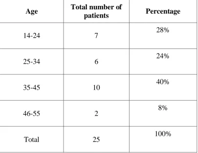
TABLE-1:AGE WISE DISTRIBUTION
Age Total number of
patients Percentage
14-24 7 28%
25-34 6 24%
35-45 10 40%
46-55 2 8%
Total 25 100%
In our study 40% of them were in 35-45 years of age, 28%
were in 14-24 years of Age, among them only one patient was
below 15 years all others were above 18 years. 24% were in
GRAPH-1(Age distribution)
In our study majority of patients were in the age group of
18 to 35 years, only one Patient was below 15 years, the diseases
was only 8% in those Individual above 45 years of age. It is 40%
in 35-45 years group. So we found that the disease is more
SEX DISTRIBUTION
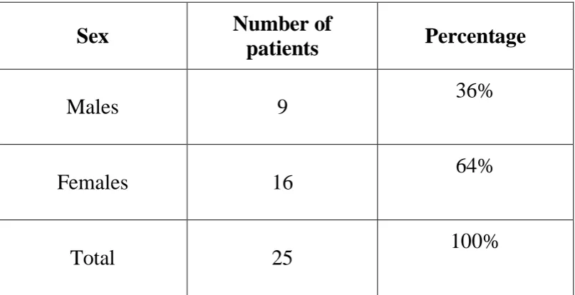
TABLE-2 (SEX DISTRIBUTION)
Sex Number of
patients Percentage
Males 9 36%
Females 16 64%
Total 25 100%
In our study, females out -numbered males by 28%, that is
64% of patients in our study are females whereas males are only
36%. From this table above we conclude that the disease is more
[image:76.595.104.508.262.469.2]GRAPH 2: SEX DISTRIBUTION
LATERALITY
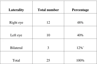
TABLE-3 (LATERALITY)
Laterality Total number Percentage
Right eye 12 48%
Left eye 10 40%
Bilateral 3 12%`
Total 25 100%
In this study, right eye was affected in the majority of
patients. 12 patients out of 25 study group (48%)presented with
right sided optic neuritis. 10 out of 25 patients presented with left
sided optic neuritis (40%). Three patients developed optic
[image:78.595.102.508.209.477.2]GRAPH-3 (LATERALITY)
In our study 48% of individual had disease in right eye,
40%had disease in left eye and in 12% of individual both the
FIELD DEFECTS
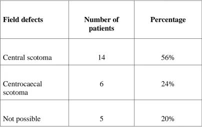
TABLE-4 (FIELD DEFECTS)
Field defects Number of patients
Percentage
Central scotoma 14 56%
Centrocaecal scotoma
6 24%
Not possible 5 20%
14 Patients (56%) out of 25 were presented with central
scotoma whereas 6 were (24%)Presented with centrocaecal
scotoma, and in 5 patients assessment of fields was not possible
because of poor vision (worse than 1/60). We found that most
[image:80.595.102.507.211.466.2]GRAPH -4 (FIELD DEFECT)
So according to our study the most common field defect
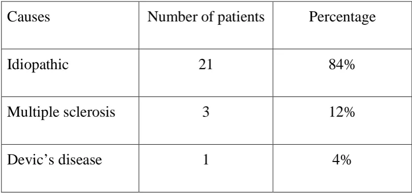
CAUSES OF OPTIC NEURITIS
In our study the most common cause being idiopathic, only
3 patients out of 25 showed demyelination plaques (12%)
suggestive of multiple sclerosis in MRI brain. Only one patient
(4%) was positive for MRI brain and MRI spinal cord with
NMO-IgG positivity suggestive of devic’s disease.
TABLE-5 (CAUSES OF OPTIC NEURITIS)
Causes Number of patients Percentage
Idiopathic 21 84%
Multiple sclerosis 3 12%
[image:82.595.101.507.400.590.2]GRAPH-5(CAUSES)
In our study we found that the most common cause for
optic neuritis was idiopathic in nature.
IDIOPATHIC 84% DEMYELINATION
12%
DEVIC`S DISEASE 4%
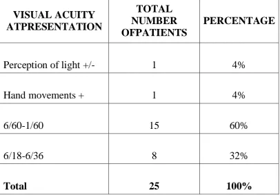
VISUAL ACUITY AT PRESENTATION
TABLE-6(VISUAL ACUITY AT PRESENTATION)
VISUAL ACUITY ATPRESENTATION
TOTAL NUMBER OFPATIENTS
PERCENTAGE
Perception of light +/- 1 4%
Hand movements + 1 4%
6/60-1/60 15 60%
6/18-6/36 8 32%
Total 25 100%
In our study majority of patient’s (15) visual acuity at the
time of presentation was between 6/60-1/60 by snellen’s chart, 8
patients had visual acuity of 6/18-6/36. Only one patient
presented with hand movement and one patient with no
perception of light. From this table we found that the visual loss
[image:84.595.103.510.197.480.2]GRAPH-6(VISUAL ACUITY AT PRESENTATION)
In our study 60% of patients were presented to our
department with visual acuity of 6/60-1/60, 32% were presented
with the visual acuity of 6/18-6/36 and 4% of them had visual
acuity of HM+ or NO PL at presentation. So we found that the
visual acuity in optic neuritis can range from 6/18 to No PL.
0 2 4 6 8 10 12 14 16
RECURRENCE RATE
In our study 4 patients had recurrence during study period.
Out of the 4, one is a case of devic’s disease, one showed
demyelination plaque in MRI brain suggestive of multiple
sclerosis and the other two with no associated risk factor.During
our study period 3 patients developed optic neuritis in felloweye,
all had no associated risk factor.
TABLE-7(RECURRENCE OF OPTIC NEURITIS)
Factors associated with recurrence
of disease
Number of
patients
Percentage
Multiple sclerosis 1 25%
Devic’s disease 1 25%
No other associated factors 2 50%
[image:86.595.104.510.471.677.2]GRAPH-7 (RECURRENCE RATE)
In our study and from the above charts we found that there
was no associated risk factors involved with recurrence in the
same eye or the involvement of fellow eye during the study
period.
0 0.5 1 1.5 2 2.5 3 3.5 4
RECURRENCE
FELLOW EYE INVOLVEMENT 4
FUNDUS FEATURES
In our study only three patients were presented with
retrobulbar neuritis whereas all other patients had disc edema and
blurring of disc margin. Six patients developed disc pallor during
recovery after treatment.
GRAPH 8- FUNDUS FEATURES
12%
88%
0% 0%
FUNDUS FEATURES
RETINAL NERVE FIBER LAYER THICKNESS IN OPTICAL COHERENCE TOMOGRAPHY
ANALYSIS – UNPAIRED T TEST
Descriptive Statistics, Table-8(RNFL thickness in OCT)
RNFL
THICKNESS
Number of
individual
Minimum Maximum Mean Std.
Deviation
RNFL –
FELLOW
EYE
25 81 156 122.32 20.012
Descriptive Statistics, Table-9(RNFL thickness in OCT)
RNFL
THICKNESS
Number of
individual Minimum Maximum Mean
Std.
Deviation
RNFL –
AFFECTED
EYE
RNFL
THICKNESS
Number of
individual
Mean Std.
Deviation
SEM
RNFL –
FELLOW
EYE
25 122.3 20.01 4.002
RNFL –
AFFECTED
EYE
25 97.48 19.82 3.964
Difference 24.84 5.633
95% confidence interval for difference: 13.51 to 36.17.t =
4.410 (VALUE) with 48 degrees of freedom; P = 0.0001 –
HIGHLY SIGNIFICANT . NULL HYPOTHESIS IS REJECTED AT P VALUE 0.0001
RNFL – THINNING OCCURS IN OPTIC NEURITIS OF
TREATMENT GIVEN
All patients were treated with intravenous methyl
prednisolone 250 mg four times daily after assessment of general
condition of them.
It was followed by oral prednisolone 1 mg per kilogram
VISUAL ACUITY AFTER TREATMENT
TABLE- 10(Visual acuity after treatment)
Visual acuity Number of patients
Percentage
Less than 6/36 2 8%
6/36-6/18 9 36%
Better than 6/18 14 56%
Total 25 100%
All patients in our study had good visual acuity after
treatment. Out of 11 patients Who showed less improvement
after treatment, 6 developed optic atrophy during the study
period and 5 had lens changes but through which fundus
examination was possible. We found that patient who had more
[image:92.595.102.505.210.432.2]GRAPH 9 ( VISUAL ACUITY AFTER TREATMENT)
After treatment with ONTT regimen 56% had visual acuity
better than 6/18,that is they all improved upto 6/9 and 6/6. Six
patients developed disc pallor after optic neuritis.
0 5 10 15 20 25
Less than 6/18
6/36-6/18 better than 6/18
total
CORRELATION OF RNFL THICKNESS WITH VISUAL OUTCOME
In our study if the difference in retinal nerve fibre layer
thickness between The two eyes is more than 25 microns, it is
found that the recovery of vision is poor after treatment. We also
found that the thinning is more marked in patients with optic
atrophy. In patients who recovered well after treatment, showed
minimal thinning of retinal nerve fibre layer in optical coherence
tomography. The thinning of retinal nerve fibre layer was more
marked in the temporal quadrant in OCT in all 25 patients who
underwent the study. Those who had optic atrophy showed
TABLE-11 (RNFL THICKNESS WITH VISUAL OUTCOME)
Mean thickness of
RNFL in microns
Visual acuity
after treatment
Number of
patients
Percentage
80 Worse than
6/36
3 12%
91 6/36-6/18 8 32%
104.92 Better than
6/18
[image:95.595.103.503.246.475.2]GRAPH 10: CORRELATION OF RNFL THICKNESS WITH VISUAL OUTCOME
In our study the mean retinal nerve fibre layer thickness is
more than 100 microns in patients who are all improved to 6/18
or better after treatment, whereas in patients in whom the visual
acuity was worse than 6/36 after treatment the RNFL thickness
was 80 microns. Hence from our study those with marked
thinning of RNFL show less improvement in vision after
treatment.
0 20 40 60 80 100 120
Fundus Picture of
Patient 6
Fundus Picture of
Patient 10
Colour Plate 3
Fundus Picture of
Patient 15
Colour Plate 4
Fundus Picture of
Colour Plate 5
Colour Plate 6
Colour Plate 7
Colour Plate 8
Colour Plate 9
Colour Plate 10
Colour Plate 11
Colour Plate 12
Colour Plate 13
Colour Plate 14
Colour Plate 15
Colour Plate 16
DISCUSSION
We conducted a study on the correlation of retinal fibre
layer thickness in optical coherence tomography with visual
outcome in optic neuritis. It is a prospective clinical study done
on 25 patients in the regional institute of ophthalmology,
Chennai. All patients were followed up for one year. All patients
were made to undergo optical coherence tomography after the
resolution of disc edema.
In our study we found that the disease was more common
among (40%) 35-45 years of age and least common among
46-55(8%) years of age. Only one patient presented at the age of
14 years. 12 patients were in the age group of 15-34 years. So
from our study we found that optic neuritis is more common in
18-45 years of age and we also found that optic neuritis is least
likely in patients above 45 years. J Terrence et al39 in 1991, Arch
ophthalmol on clinical profile of optic neuritis found that the
In this study we found that 64% of patients are females and
36% are males, out of 25 patients 16 of them are females and 9
of them are males. So the disease optic neuritis affects females
commonly than males. J Terrence et al39 found in his study on
clinical profile of optic neuritis that 77.2% are females and
females out-numbered males.
In our study we found that right eye is affected more than
left eye, the disease is bilateral in 12% of individual. Out of 25
subjects in this study 12 patients had disease in right eye (48%)
and 10 patients had disease in left eye (40%). It is not a
significant difference. 3 patients out of 25 developed disease in
the fellow eye during the study(12%).
This study revealed that the common field defect
associated with optic neuritis is central and centrocaecal
scotoma. Among the 25 patients 14 (56%) had central scotoma
and 6patients(24%) developed centrocaecal scotoma. Keltner JL
optic neuritis and they found diffuse loss in 66.2% in affected
eye and 6.2% in fellow eye.
Regarding the causes for the inflammation in optic neuritis,
we found in our study that 21 patients out of 25 cases no obvious
cause was found and it is said to be idiopathic. 3 patients (12%)
in this study showed demyelination plaque in MRI brain
suggestive of multiple sclerosis. And in our study there was
only one had positive lesion in both brain and spine MRI and the
patient was also positive for NMO-IgG antibody suggestive of
devic’s disease. We also found that the disease is bilateral in this
patient and ended with optic atrophy. H Wang JM et al40 that
optic neuritis is bilateral in pediatric population. Pirko.I et al
found that patients with NMO positivity has worse visual
outcome. Wang JC et al41 in his study found that in 83.9% there
In our study majority of patients were presented with the
visual acuity of 6/60-1/60. In this study 60% (15 patients)