JOURNALOFVIROLOGY, June 1984, p.929-938 Vol. 50,No. 3 0022-538X/84/060929-10$02.00/0
Location
of DNA-Binding Proteins and Disulfide-Linked
Proteins
in
Vaccinia Virus
Structural
Elements
YASUO ICHIHASHI,1* MASAYASU OIE,1 ANDTAKASHI TSURUHARA2
Department ofVirology, Faculty of Medicine, Niigata University, Asahimachi, Niigata
951,1
andDepartment ofMicrobiology, Niigata College
of Pharmacy,
Kamishineicho 5827,Niigata
950-21,2
JapanReceived6April 1983/Accepted28November 1983
Treatmentwith sodium dodecyl sulfate(SDS)converted the vaccinia virus strain IHD-J into
particles
of twotypes: (i) ghosts which possesseda thin-membrane vesicle derived from basement part of the virusmembrane with attached lateral bodies and a membranous structure derived from the core wall and (ii)
aggregates ofa DNA-nucleoprotein eluted from the core. These particles lacked lipids, and all the viral
phospholipids
weredetected inthe SDS-soluble fraction. The viral membrane was composed of an SDS-soluble coat layer and the basement membrane, and the basement membrane was maintained by amechanism other than the lipid bilayer. By comparisons of protein species in morphologically distinct subviral particles
prepared
by severalsolubilizing
methods, protein compositions of viral structural elements weresuggested
asfollows: 25,000-molecular-weight viral protein-17,000-molecular-weight viralprotein
(VP25K-VP17K), viral
basement membrane; VP13.8K, major component of the lateral body;VP70K, VP69K, VP66K, andVP64K, minorcomponents of the lateralbody; VP61K, outerlayer ofcore wall; VP57K-VP22K, inner layer of core
wall;
andVP27K-VP13K,
nucleoprotein. These structural elements found in the SDS-insoluble particles dissolved in the same SDS solution under reducing conditions, indicating that the disulfide linkages seem to have a principal role in maintaining theirmorphological integrity.
VP57K, VP27K, VP13.8K, and VP13KwererevealedtopossessaffinityforDNA.Denaturedcalf thymusDNAand viral DNA in
double-
orsingle-stranded formassociatedequallywellwiththese
proteins,
but RNAdid not bind. Therefore, itwas strongly suggested that disulfide-linked VP27K-VP13Krepresented
thenucleoproteins
of vaccinia virus. A structural model of vaccinia virus isproposed anddiscussed.The morphology of vaccinia virus has been well
docu-mented (5, 18, 19,23, 30), andoverallclassification of viral outer-regionproteins and corecomponents has been
estab-lished basedon differential solubilization of viral membrane
with Nonidet P-40(NP-40) plus 2-mercaptoethanol (2-ME) or
digestion
with enzymes (2, 7, 8, 17, 22, 25). However,exceptforthesurface tubule, whichhasbeenreportedtobe composed of 58,000-molecular-weight (58K) protein (29),
componentsof vaccinia virus structural elements, eventhe
nucleoprotein, have notbeenidentified.
Werecentlyfoundthat someviralproteins, whicharenot
soluble in sodium dodecyl sulfate (SDS) solution but are
soluble inthe sameSDSsolutionunderreducingconditions,
areprobably linked with disulfide bonds (10, 22). The high
molecular weightsofthedisulfide-linked proteins suggested aroleforthedisulfide bonds in assembly ormaintenance of
viral substructural elements. To identify which of these
disulfide-linked complexes constructs which structural ele-mentsof the virus,theviruswasdegradedunderconditions
thatproduce particles constituted of different subviral struc-tures, followedby electrophoretic analyses of the proteins.
Astaining method for detection of DNA-binding activitywas used to identifynucleoproteins of vaccinia virus.
MATERIALSANDMETHODS
Cells andvirus. KBcells were grown in Dulbeccomodified
Eagle mediumsupplementedwith
10%
calf serum. Vaccinia virusstrain IHD-J waspropagatedin KB cells as described*Correspondingauthor.
previously(11, 12) and was purified by the surcrose gradient
centrifugationmethod (14) withouttreatment with proteases. Thevirus titer wasassayed in Vero cells.
Treatment of virus withdetergents. Purified vaccinia virus
(200 ,ugof protein per ml, 108 PFU/ml) was mixed with an
equal volume of SDS solution
(1%
SDS, 50 mMTris-hydrochloride buffer,pH 7.4) orNP-40 + 2-ME solution (1% NP-40, 2%2-ME, 50 mMTris-hydrochloridebuffer, pH 7.4) andincubatedat37°Cfor 30 min. Samples were layered onto
36% sucrose andcentrifuged at 25,000 rpm for 30 min. The
supernatant above the sucrose layer was passed through a
membrane filter (Millipore Corp.; 0.22 ,um) and designated asthesolublefraction.The pellet beneath the sucrose layer was resuspended in Tris-hydrochloride buffer (50 mM, pH
7.4) by gentle pipettingand was pelleted again by
centrifuga-tion. Particles that were pelleted by centrifugation after treatmentwithNP-40 + 2-ME (core fraction;
60-tLg
portions) were treated with trypsin (Worthington Diagnostics;tolyl-sulfonyl
phenolalanyl
chloromethyl ketone-trypsin, 20 ,ug/ml, 37°C,15 min), followed byadjustmentof pH to2.0 with 0.1 N
HCl,
andwere centrifugedat 25,000 rpm for 30 min. Thepelletwassubjectedtoelectrophoresis.TheNP-40 + 2-ME-treated core fraction was further treated with 0.2%aceticacid solution (37°C, 15 min)andcentrifugedat 25,000 rpmfor30min.Thesupernatantand pellet were lyophilized and subjected to electrophoresis. Samples for electron
mi-croscopicanalysis were prepared by standard methods.
Electrophoresis. SDS-polyacrylamide gel electrophoresis (PAGE)wasperformed as described by O'Farrell (21) for the second dimension oftwo-dimensional electrophoresis with
12.5% gels. Samples were dissociated in lysis buffer (2.3% 929
on November 10, 2019 by guest
http://jvi.asm.org/
930 ICHIHASHI1, OIE, AND TSURUHARA
'-Xx*t V 4 h * V C~~~~~~~~~~~~~~~~~~~~~-
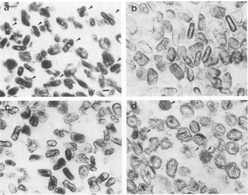
-~-FIG. 1. Thin-section electronmicrographs of virus treated with detergents. (a) Virus treated withNP-40 + 2-ME.Arrowsindicate cores withattached lateralbodies. Bar, 100 nm. (b) NP-40+2-MEcorefraction treated withtrypsin. (c)NP-40+2-MEcorefraction treated with acetic acid. (d) Virus treated with SDS.Arrowsindicate filamentous aggregates.
SDS, 5% 2-ME, 10% glycerol, 60 mM Tris-hydrochloride
buffer, pH 6.8)and heated at100°Cfor 1min.
Detection ofDNA-binding proteins. DNA-binding activity of vaccinia virus structural proteins was detected by a
staining methodbasically similartothatreported by Bowen etal.(3).Viralpolypeptideswereelectrophoresedina12.5%
polyacrylamide gel slab, and the proteins were transferred
ontoanitrocellulose sheet (ToyoRoshi; TM-2, 0.45,um) as
describedpreviously (13).Theblotsweresoakedfor5minin
lx SSC solution (SSC is 0.15 NaCl plus 0.015 M sodium
citrate, pH 6.5) containing0.1% bovine serum albuminand
then incubated in denatured DNA solution (50 ,ug of calf thymus DNA perml, 0.1% bovine serum albumin, 10 mM
EDTA, 0.02% sodium azide, 1x SSC) for 30 min at room
temperature. Calf thymus DNA(Sigma Chemical Co.; type
I)wasdissolvedat 100 ,ug/mlin 1x SSCcontaining 10 mM
EDTA and denatured by 20 min of incubation at 100°C, followedbyquenchinginice. Insomeexperiments vaccinia
virus DNA, yeast RNA (Sigma; 100 ,ug/ml), or synthetic
polyadenylate-polyuridylate in helicalform(Sigma; 100 ,ug/ ml) was used instead ofcalf thymus DNA. Vaccinia virus
DNAwasisolated from
32P-labeled
virus bytreatment withproteinaseK(Merck &Co., Inc.; 20,ug/ml) in the presence
of 0.5% SDS, followed by extraction with
phenol-chloro-form, and was used with or withoutheat denaturationat aconcentration ofabout 5
,ug/ml
(105
dpm/ml). Aftersoaking
in either ofthe above DNA solutions, the blot was rinsed
briefly with 1x SSC, fixed with 10% trichloroacetic acid for
10 min, rinsed, and then stained for 5 min with ethidium
bromide
(0.02%
in lx SSC). The blot wasphotographed
under short-wave UV light through a
gelatin
filter(Kodak;
no.22),orautoradiographedbyusingX-ray film(Kodak; X-OmatAR). KB cell nuclei,
processed
inparallel,
servedas control samples.Thin-layer
chromatography.
Lipids
in bothdetergent-treated virus and control viruswereexaminedby thin-layer chromatography (28) with
precoated
plates(Merck;
silicaplate60).
RESULTS
Structural elements ofdetergent-treated virus. Studies on the solubility characteristics ofvaccinia virus have shown J. VIROL.
ARM
on November 10, 2019 by guest
http://jvi.asm.org/
[image:2.612.64.560.74.465.2]STRUCTURE OF VACCINIA VIRUS 931 that the pellet fraction obtained by treatment with SDS
under nonreducing condition contains particlescomposedof 10 proteins and DNA, and theparticles were thought to be "'nucleoids" (9, 26, 27). However, in our repeated experi-ments, virus particles pelleted after treatment with SDS appeared to be of two types when negatively stained;
aggregates of filamentous material and empty bags with
partially bulging walls. Thin-sectioned profiles revealed the
former structure to consist of an aggregate of 2-nm-wide electron-dense filaments without a limitingmembrane. Simi-lar-sized filaments were observed in some of the treated viruses (Fig. ld and Fig. 2f and g). The latter structure was a virus ghost particle which possessed athin-membrane vesi-cle together with attached lateral bodies and part of the core wall, and a large part of its DNA-nucleoprotein filament was eluted from the core
chamber.
The attached lateral bodiesindicated that the membrane of the ghost particle was derived from a basement part of the virus membrane. The thickness of the membrane was reduced from 20 nm (intact virus membrane) to 5 to 7 nm. The viral membrane was composed of a SDS-resistant membrane (virus basement membrane) seen in the ghost particles and coat layer soluble in the SDS solution under nonreducing conditions. Treat-ment with higher SDS concentrations (up to 2%) produced similar particles.
When the SDS-insoluble fractionwastreated with DNase, empty bags composed of viral basement membrane and lateral bodies remained, but filamentous aggregates disap-peared (Fig. 3). The filaments eluted from the viral core chamber were identified as DNA,probably complexed with nucleoproteins. Residual core wall whichwas seen in some of SDS-treated ghostparticlesdisappeared, which suggested association between the DNA filament and the core wall components.
Viral proteins soluble inNP-40 + 2-ME havebeen known to constitute the virus membrane (7, 22, 25); therefore, the component proteins ofthe ghost membrane could be deter-mined bycomparisons of proteins soluble in SDS and those soluble in NP-40 + 2-ME.ThefactsreportedbyEasterbrook (7) were confirmed again. Treatment ofvaccinia virus with NP-40 + 2-ME solubilizes its membrane; particles pelleted after such treatment consist of the viral core and two attached lateral bodies (core fraction), and additional treat-ment of the core fraction with trypsin removes lateral bodies. Furthermore, the core fraction was treated with acetic acid to obtain particles of different structural ele-ments. The treatment liberated the lateral bodies, and the pellet contained particles which had a thin membrane de-rived from the core wall and debris probably derived from lateralbodies (Fig. 1 and2). Treatmentwith DNase reduced
electron density ofthe core chamber (Fig. 2d).
Figure 4 is aschematicdrawing ofvacciniavirus degrada-tion by treatment with detergents, and Table 1 presents a summary ofmorphological findings.
Protein composition of viral structural elements. The pro-teins present in the various viral substructures described above were analyzed by SDS-PAGE. Figure 5 shows pro-files ofsupernatant and pelletfractions of virus dissociated
in SDS or NP-40 + 2-ME solution. Proteins existing in the soluble fraction of the treatments have been analyzed (22, 25), and presentresults coincidentally showedthat the NP-40 + 2-ME-soluble fraction contained the 54K viralprotein
(VP54K), VP37K, VP34K, VP32K, VP29K, VP25K,
VP21K, VP18K, VP17K, and VP16K. However, VP11OK,
VP88K, and VP37K were moreprominentin the NP-40 + 2-ME precipitate than in the soluble fraction. These proteins
a
b
.: to
c 'St
g_a.s = ^
,jlCe s >
''
itE44F;
£Wt}
4
>,
..
t,
A,.
R
,;. i,.^.. }'
t
as, v .z.;
_S
_w_;.bt->E
w.
K K.-I.
fat w
'5 s t: ..1
.4-- d.e -F N. Wf- 1,.1M
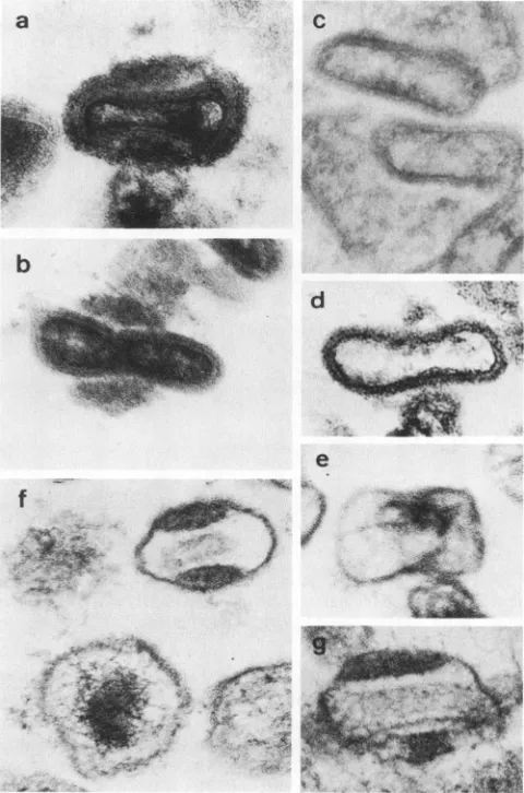
FIG. 2. Highmagnification electronmicrographs of virus treated withdetergents. (a) Control virus. (b) NP-40+ 2-ME corefraction. (c) NP-40 + 2-ME core fraction treated with trypsin. (d)NP-40 + 2-ME core fraction incubated with DNase. (e) NP-40 + 2-ME core fraction treated with acetic acid. (f and g) Virus treated with SDS. Bar, 100 nm.
may possibly be core components partially soluble in the NP-40 + 2-ME solution. But otherexplanations cannot be excluded; they may distribute both core and viral mem-brane, or differentproteins of similar size may composeviral core and membrane structures. Todetermine which ofthese possibilitiesisthe case,a newanalysis method isrequired to overcomethislimitation ofthe present method basedonthe solubility of proteins. As VP54K migrated with VP57K in 12.5% gel, they were distinguished by electrophoresis in a 8% gel and also by a staining method for DNA-binding
protein which is described in the following section. These NP-40 + 2-ME-soluble proteins were components of the viral coat layer and basement membrane. Compared with
SDS-soluble proteins, VP25K-VP17K was suggested to be the component of the viral basement membrane.
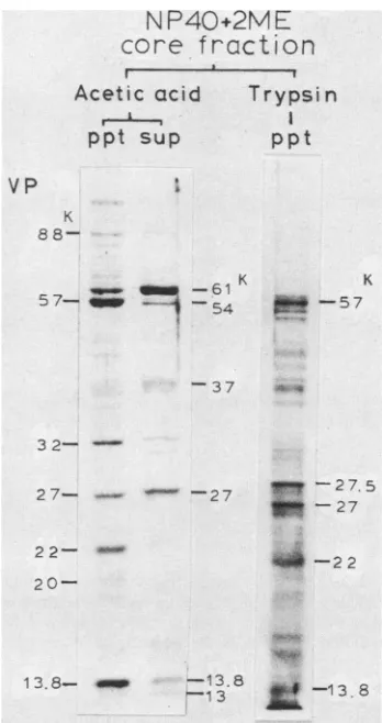
Protein composition of the core fraction was analyzed further by comparing the SDS-PAGE profile of the core fraction with those obtained after additional treatment with trypsin oracetic acid (Fig. 6). The major components in the core fraction (VP61K, VP57K, VP27K, and VP22K) corre-sponded to VP4a, VP4b, VP8, and VP9a ofprevious reports (22, 25). Core particles pelleted after treatment withtrypsin
VOL. 50,1984
I
jh..-:-y:k-'..
.1 .i .-..
Ik
it`
'..II
m .9 V
!101
11
.4 % -.
on November 10, 2019 by guest
http://jvi.asm.org/
[image:3.612.315.555.78.441.2]932 ICHIHASHI, OIE, AND TSURUHARA
... s
_ ,. §' :!
g l,-, E 2
s. r . .. 5
... >- s25 sh_ r
.,;_ -.sst, :
'j
o:-.X?:...k
S.>
.RzaFt)K9t
S -.:'s: w,
s ...
:.
::S ...
..s,,,,.,,,,,.r,#-s.
-e s0>r¢' p9'
:. Ni,
. :i.
e:tS..<kit.Sse:fIE.,$|es.
gFe.egsx;Z 5.sEsa ..
7,,,e.>...,.wr...:...
:.:....
..:'
'
.:,
:' .'
j'.
,:, ., ,§.+.t. ,:
__.i$t;.p."@S>S'
qr ....
VI
,.wc t, F
X
ji.
ily
pa
;T:4k
"WW,
ia
FIG. 3. Effect of DNase on SDS-treated virus. Virus (1 mg) suspended in a 1% SDS solution was centrifuged, and the precipitate was incubated at room temperature for 30 min with DNase (Worthington; DNase I, 100 U in 1 ml of 50 mM Tris-hydrochloride buffer [pH7.41-10 mMCaC12). Thin-sectioned electron micrograph. Bar, 100 nm.
contained VP57K, VP27K, VP22K, and a trace amount of
VP13.8K, but other distinct proteins, such as that which appeared at27.5K, were cleavage products. Disappearance ofVP61Kaftertreatmentwithtrypsin shows that it probably
occupies the surface of cores. Since VP13K exists as VP27K-VP13K complex (10), VP57K, VP27K-VP13K, and VP22K weresuggested to be majorcomponentsof theinner
layer ofcore walls andof the nucleoprotein.
Treatment of the core fraction with trypsin removed
lateral bodies. Theproteinsthatclearly werediminishedor
reduced in amountaftertreatment with trypsinwere
deter-mined by comparison with the profile of the core fraction; these reduced proteins were VP11OK, VP88K, VP79K, VP70K, VP69K, VP66K, VP64K, VP61K, VP37K,VP32K, and VP13.8K. Some of these proteins (VP11OK, VP88K,
VP79K, VP74K,VP37K, and VP32K) wereconsideredto be
components ofthe coatlayerasmentionedabove.Thus,the other trypsin-sensitive proteins (VP7OK, VP69K, VP66K, VP64K, VP61K and VP13.8K) weresuggestedto be compo-nents of lateral bodiesor theouterlayerofthe core wall.
¢@
~~~Tryps
in,Acet ic acid
FIG. 4. Diagram ofthecontrolleddegradation of vaccinia virusbytreatmentwithdetergents.
J. VIROL.
..1
.,r..j., :,;:w, ;,".., ..:,
...41': .7-`:'i
~"i *'.:,;
,:A
.-*,OR
:-.w:,
.1
4L
on November 10, 2019 by guest
http://jvi.asm.org/
[image:4.612.131.494.75.333.2]STRUCTURE OF VACCINIA VIRUS 933
TABLE 1. Summary of morphologicalstudies of the effect of treatmentwith detergent onviral substructures
Presence (+) or absence (-)in pellet aftertreatmentwithb:
Virus sub- NP-40 + 2-ME SDS +
structurea NP4++Aei DS DNs
+Trypsin acid
CL - _ _ _
BM - - - + +
L + - ± + +
OCw + + _ _
-ICw + + + + +
NP + + + +
-aAbbreviations: CL, coat layer; BM, basement membrane; L,
lateralbodies; OCW,outerlayer ofcorewall;ICW, inner layer of corewall; NP,DNA-nucleoprotein.
b +, Degradedresidual forms of thestructure.
Treatmentofthe corefraction with 2% acetic acid liberat-ed three proteins (VP61K, VP54K, andVP37K)andpartof VP32K, VP27K, VP16K, VP13.8K,and VP13K. Thepellet contained VP57K, VP32K, VP22K, VP13.8K and reduced amounts of VP61K and VP27K. Thus, the treatment with
acetic acid did not clearly distinguish the location ofthese
S
D
S
I
HD-J
IN
P40+
2M E
r-- I I
ppt
supppt
sup
VP
-K
110-
....-Ace
PP
NP40+2ME
core
fraction
tic acid Trypsin
t sup
ppt
VP
:K 8
8-5 7-
---
-54-1 KK
-57
-37 A *
3 2-
-2 7- - 2 7
22-
20--27.5
a-27
4-22
13.8- -13.8 1 8
13 -13.
6
1
~
57
54-4 5
--37-
m-3
4
-32-
--27- am
25-
21-2 l
--*.,,3b _
_~
i.
1
7-1 6- 14-13.8
13
FIG. 5. Proteins in the supernatant and pi
with SDS or NP-40 + 2-ME. Virus was susp
concentration of1%)orNP-40+ 2-ME (0.5% mM Tris-hydrochloride buffer, pH 7.4) and c
rpm for 30 min. Samples were electrophore:
VP54Kin the supernatant solutionwas
disting
by SDS-PAGE in a 8% gel and also by stai methodtodetect DNA-binding protein.ppt,Pre
supernatant.
-88 FIG. 6. SDS-PAGE profilesof viral corefractions treated with trypsinoracetic acid.Theviralcorefractionprepared bytreatment with NP-40 +2-ME was suspended inacetic acid (final concentra-tion, 2%) or treated with trypsin (final concentration, 20 ptg/ml; 5 4 37°C, 15 min) and centrifuged at 25,000 rpm for 30 min. The precipitated pellets (ppt) of the trypsin-treatedcoresand the ppt and supernatants (sup) of the lyophilized acetic acid-treated samples weresubjectedtoSDS-PAGE in a12.5%gel.
m
-37
___w
-34
proteins; however, VP57K and VP22K were exceptional3 2
components; they were predominantly detectable in theacetic acid pelletfraction. As these two proteins appeared
together in pelleted samples prepared by treatment of the
viruswith SDS, NP-40 + 2-ME, NP-40 + 2-ME + DNase, -25 NP-40 + 2-ME +
trypsin,
orNP-40 + 2-ME + aceticacid,
they probably form a substructure which is commonly detectable in these pellets, namely, the inner layer of the
-21 corewall
(VP57K-VP22K complex).
Disulfide-linked viral proteins. The virus that was pelleted _ l
7
after treatment with SDScontained no detectable amountsoflipids when examined by thin-layer chromatography; all
viral lipids were found in the SDS-soluble fraction. The
_w
absence oflipids in the viral ghost particlesand the existence of membrane structures in them indicated that themem-braneswere not maintained bya lipid bilayerbut ratherby
,ellet after treatment linked proteins. The fact that the SDS-insoluble fraction can )ended in SDS
(final
be dissolved inSDS +2-MEsolutionstrongly suggestedthat NP-40, 2% 2-ME, 50 disulfide linkages in the proteins participate in maintainingsentrifugen
at25,00
g the viralstructures.guished
from VP57KFigure
7 shows the effect of 2-ME concentration on theining VP57K by the solubility of viral proteins which are suspended in SDS -cipitated pellet; sup, solution. SDS-soluble proteins, oligomers, and unit
com-plexes appeared in the profile of the lane with SDS alone. VOL.50,1984
*slow~
-Z,--l I
a
4..
499mop-54
on November 10, 2019 by guest
http://jvi.asm.org/
[image:5.612.58.299.95.203.2] [image:5.612.56.301.324.647.2]934 ICHIHASHI, OIE, AND TSURUHARA
SDS 1°/.
2 ME
0.01o0.01 0.1 1 50°fo
88-6 1
-VP
-110K
_ e -88
54 t&D - _
36.
16-37*34-4- _
27, 131
25-- w
29-2
1- 16-
14-1
3--61
40-57
-4 7
-43
4-g_~~~~~J0- f37
____ _ -34
- -32-29
X -25
-2 20
-1 7
16
14
Jm~1 3 8
-0 -it3
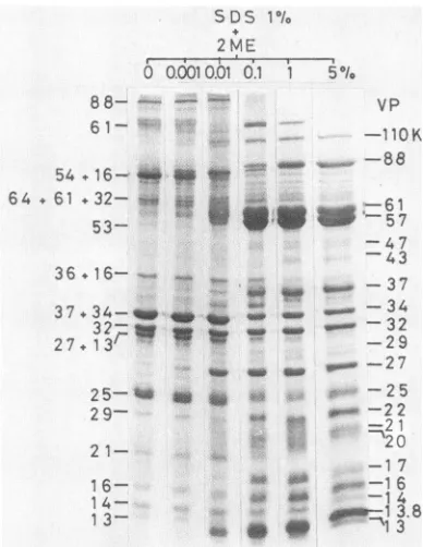
FIG. 7. Effect of2-ME concentrationonthe solubility of virus
proteins. 2-ME wasadded at various final concentrations tovirus suspended in1%SDS.Thesampleswereloaded inwidely separated lanestoavoid effects of 2-ME in the samplesof adjacent lanes.
The subunit proteins of the unit complexes have been identified by the two-dimensional SDS-PAGE method, where thefirst dimensionwasperformed under nonreducing
conditions and thesecond dimensionunderreducing condi-tions. VP25K has been identified as the dimer form of
VP17K (VP25K-VP17K), VP27K-VP13K exists as a
com-plex, and some proteins (VP21Kand VP29K) migratewith
different speedswhenthey aresolubilized inSDSorinSDS
+2-MEsolutions (10). Results obtained by such analysisare
presented at the left side ofFig. 7. Inthis experiment, the virus samples were loadedon thegel without separation of
SDS-resistant particles to reveal all SDS-soluble
compo-nents. If the particles were eliminated from the sample, VP25K,VP27K-VP13K,VP88Koligomer,VP61K oligomer, and VP70KtoVP63K minor componentswereallreducedto trace amounts. The parallel lanes show that lower-molecu-lar-weight proteins, suchasVP14K, VP13.8K, andVP13K, migrated faster when virus was solubilized in 1% SDS containing 0.01 to1% 2-ME than those thatweresolubilized in 1% SDS containing5% 2-ME. Trace amounts of VP27K and VP13K were detectable in the lane with SDS alone;
however,significantdissociation of theVP27K-VP13K
com-plexinto the subunits tookplaceataconcentration of 0.01%
2-ME in 1%SDS solution, andcomplete separationof these subunitproteins occurredatahigherconcentration of 2-ME (1%). VP13.8Kwas detectable in the lane where thesample was treated with 1% SDS + 0.01%2-ME and seemedtobe solubilized completelyin1% SDS +0.1% 2-ME. Twomajor
core components (VP61K and VP57K) appeared in the profile when the virus was treated with 1% SDScontaining
higherthan0.1% 2-ME. TheVP54K-VP16Kcomplex disso-ciated into subunits at 0.01% 2-ME, the VP37K-VP34K complexes dissociated into subunits at 0.1% 2-ME, and complete dissociation ofVP133K required 5% 2-ME. Thus,
each structural protein was affected differentially by the reducing conditions in SDS solution.
VP57K-VP22K, VP13.8K, and a large partof VP61K were barely soluble in SDS and did not migrate in the 12.5% gel. Since proteins smaller than about 2 x
105
daltons could be electrophoresed in the 12.5% polyacrylamide gel, all these proteins apparently existed in the virus particles in highly multipliedform,exceeding2 x105
daltons.Therefore,it was stronglysuggested thatthelateral bodies, core outerlayer,and core inner layer, which remained after treatment with 1% SDS, were constructed by a bond highly sensitive to a reducing condition such as disulfide linkage. On the other hand, VP25K-VP17K and VP27K-VP13Kcomplexes migrat-ed in the gel. These two complexes also associated with SDS-insolubleparticles; however,theassociation seemedto be weak and breakableby electrophoretic drive.
The results of SDS-PAGE analysis are summarized in Table 2.
Viral DNA-binding proteins. To identify vaccinia virus
nucleoprotein, we employed the method for detecting pro-teins withDNA-binding capacity. This method is basically
similar to that reported by Bowen et al. (3). Purified virus was electrophoresed in a slab gel by SDS-PAGE, and the viral proteins were electrophoretically transferred onto a
nitrocellulose sheet. The nitrocellulose sheet was then
soaked in a solution containing DNA, and the adsorbed
DNA was revealed by ethidium bromide staining or by autoradiography if the DNA wasradioactively labeled. The
adsorption ofDNAtookplace inthepH rangeof5 to 9.2in ix SSC as well as in 10 mMTris-hydrochloride buffer, but adsorbed amountswere low in 4x SSC.
Figure 8, lane 2, is an autoradiogram by 32P-labeled vaccinia DNA, and lane 3 is stained by the procedure
described above. Both lanes show that VP57K, the 33K
protein, the 32K protein, VP27K, VP13.8K, and VP13K bound vaccinia DNA and denatured calf thymus DNA.
Double-stranded 32P-labeled vaccinia DNA and its dena-tured form adsorbed to the same
proteins
as did denatured calfthymus DNA;however,
double-strandedsynthetic
RNAand yeast RNA did not. Thus the
DNA-protein
associationdid not depend on strandedness or on a
specific
nucleotide sequence. The association of DNA withVP57K, VP27K,
and VP13.8K was
stable,
and the 33Kprotein,
the 32Kprotein, and VP13Kwere
low-affinity
proteins;
theadsorbed DNAcould be releasedby repeatedlywashing
theblotwith ix SSC.The DNA-binding canacity ofunit
complexes
wasexam-inedto test whethertheactivity iscarried by unit complexes
containing vaccinia DNA-binding proteins. The VP27K-VP13K complex showed DNA-binding activity, and VP57K
andVP13.8K appeared at the upper endofthe stackinggel (Fig. 9). TheVP27K-VP13K complex had reactivity similar
to thatof VP27K and showed nohigher-order specificity.
Since histones sometimes adsorbed to virus samples, histones in KB cell nuclei were used as a control (Fig. 10). When the virus and KB cell nuclei were processed in
parallel,the 33K and 32K bands ofvirus sampleappearedin
positions identicalto thoseof the histone
Hi
bands,suggest-ing slightcontamination by
Hi
of the virus sample, whereasother viral DNA-binding proteins migrated to
positions
different from those occupied byhistones.
Model for vaccinia virus structure. The locations of viral
proteins in vaccinia virus particles were determined by comparing the results presented in Table 1 and 2, and the
locations are presented in the last column of Table 2. NP-40 + 2-ME-soluble proteins have been classified as
J. VIROL.
64 - 61 -3 2
-53-
Is's
on November 10, 2019 by guest
http://jvi.asm.org/
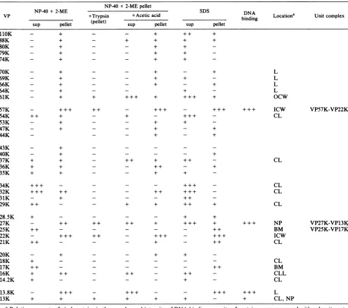
[image:6.612.82.276.73.324.2]STRUCTURE OF VACCINIA VIRUS 935 TABLE 2. Proteinconstituents of virus fractions treated withdetergents'
NP-40+2-MEpellet
NNP-40+ 2-ME N SDS DNA
VP +Trypsin +Acetic acid binding Locationb Unitcomplex
sup pellet (pellet) sup pellet sup pellet
110K - + - - + + + +
+
_ +
+
_ +
+ +
+ + + +
_+ +_
_ + +_
_+ +_
+ +
_ +
_ +
+++ +
+ _
_ +
+ _
+++ +
L L L L OCw
+
- +++ ++ -+ ++ - +++ +++ ICw
++ + - + - +++ - CL
_+ +_
VP57K-VP22K
_ + -+
_+
++ +
_+
+ + _
_ ++ - +
_ + +_
_ ++ ++
-_- ++ _
+ + ++ +
CL
CL CL CL
+ +
_ ++ ++ ++ + +++ + +++ NP
++ - - - ++ BM
+++ ++ - +++ - +++ ICW
++ _ _ _ + _ ++ CL
_+
+ + _
+
+
_ ++
+ + _
VP27K-VP13K VP25K-VP17K
CL BM CLL
+ - CL
+++ - - +++ +++ L
+ + + - + CL, NP
aRelativeamountsof viralproteinsin the samplesandintensityofDNA-binding capacityofproteinsweremeasured withadensitometer
andexpressedasfourgrades(-, +, ++, + ++). sup, Supernatant.
bAbbreviationsarethesameas those in Table1,footnotea.
components of the virus membrane, which is composed of
coat layerand virus basement membrane. The virus base-ment membrane seems to be composed of the VP25K-VP17K, and the other proteinsintheNP-40+ 2-ME-soluble fractionaregroupedtocoatlayercomponents.The NP-40 +
2-ME-solublefraction formedabandatadensity of1.22gIml when it was centrifuged in 10 to 25% tartrate density gradient. The density was similarto thatofsurface tubule (29); however, the artificially aggregated material in the tartrategradient hadnotubularfigureandcontainedVP54K,
VP20K, and VP16K. Apparently, further studiesare neces-sarytoelucidate theorganizationofthesecoatlayer
compo-nentsontheviralmembraneortoidentify thesurface tubule components. By comparisonof thecorefraction before and
aftertreatmentwithtrypsin,thelateral bodieswere
suggest-ed to be composed of VP13.8K, VP70K, VP69K, VP66K, and VP64K.Because of theappearanceofVP57K-VP22Kin
allthe examined pellet fractions which containedthe inner layer of the core wall, VP61K was concluded to be a
componentoftheouter layerof thecorewall, and VP57K-VP22Kwasconcludedtobeacomponent ofthe innerlayer
ofthe corewall.
Among detected vaccinia DNA-binding proteins, the amounts of33K and 32Kproteins are too small tobe viral nucleoprotein and are probably derived from host histone Hi. VP13.8K isascribed to be thelateralbody component; thus, either VP27K-VP13KorVP57K-VP22Kisthe nucleo-protein of vaccinia virus.When thecorefractionwastreated
88K 80K 79K 74K 70K 69K 66K 64K 61K 57K 54K 53K 47K 44K 43K 40K 37K 36K 35K 34K 32K 31K 29K
_ +
_ +
_ +
+ +
+ +
+ +
+++ ++
_ +
+ + _
+_
28.5K 27K 25K 22K 21K 20K 18K 17K 16K 14.2K 13.8K 13K
+_
+-_
+ ++ _
+-_
+ +
VOL. 50, 1984
on November 10, 2019 by guest
http://jvi.asm.org/
936 ICHIHASHI, OIE, AND TSURUHARA
SDS+
2ME
VP K
110-88-o
37-4 34-32-4
-Upper end of stacking gel
-::
_ 32
2 7-2 5- be 22- -f 2
1--1 7- s&q
1 6- *i. 1 4-
13.8-_
:
..:. ;:: *:: .:
...
s,. & s;.
_|:
_ ....
-2 7
-1.3.8
[image:8.612.92.270.71.371.2]13
FIG. 8. DNA-bindingproteins of vaccinia virus. Nitrocellulose sheets blotted with viral proteins were immersed for 30 min in denatured 32P-labeled vaccinia virus DNA solution (105 dpm/ml, 0.01% bovine serum albumin, 0.02% sodiumazide, lx SSC)orin denaturedcalf thymus DNA solution (50 ,ug ofDNAperml,0.01% bovine serum albumin, 0.02% sodium azide, 10 mM EDTA, lx SSC).Bound DNAwasdetectedbystainingwithethidium bromide orautoradiographically by using Kodak X-OmatARfilm. Lane1, SDS-PAGE profile of control virus stained with Coomassie brilliant blue.Lane2, autoradiogramofanitrocellulose sheetprocessedas
described in thetextandsoaked in32P-labeledvacciniaDNA. Lane 3, photograph of a nitrocellulose sheet processed as above and stained with denatured calf thymus DNA and ethidium bromide solutions. The bright band at the upper end of the stacking gel represents viral DNAin theelectrophoresedsample.
of biological unit membranes can be dissociated easily by treatment with SDS. The absence of lipids in SDS-treated
virus particles and the solubility of the viral ghost particles in SDSunder reducing conditions indicate that disulfide bonds in the polymer of VP25K-VP17K complexes are the major bondsholding the viral membranestructuretogether. Based onthesefindings,we hypothesize that the morphogenesis of vaccinia virus membrane in the viroplasm starts by self-assembly of the basement membrane via interaction between VP25K-VP17K unit complexes. Spicules which may be composed of coat layer proteins then associate with the basementmembrane, andlipids are incorporated into specif-ic receptors of the membrane or coat proteins, orboth. This hypothesis is consistent with the comment of Blough and Tiffany (1) that theamountof viral lipids istoosmallandthat thelipids are ofinadequate composition to form a continu-ous biological lipid bilayer membrane around the virus particle. Transfer of membrane phospholipids to purified virus (28) also supports this hypothesis. It is an interesting characteristic of the viral membrane that, despite differences inhow it isconstructed andwhat its componentsare, it can fuse with the cellular membrane; this has been shown by electronmicroscopic study of virus penetration (4).
Inaddition to the VP25K-VP17K complex located in the virus basement membrane, VP57K-VP22K (inner layer of the core wall), VP27K-VP13K (nucleoprotein), and VP13.8K(majorcomponentof the lateral body)werefound
SDS
1
2
VP K 61
--__ Upper end of stacking gel
57,
13.854 + 16r' '
with aceticacid,DNAand VP27K-VP13Kwerefound inthe supernatant, and VP57K-VP22K were recovered in the
pellet. Based on the simultaneous solubilization of VP27K-VP13Kand DNA, andontheexistence of VP57K-VP22Kin
the core fraction treated with
DNase,
the VP27K-VP13Kcomplex is concluded to be the
nucleoprotein
of vaccinia virus.DISCUSSION
The viral membrane, which is
composed
ofproteins
andlipids, isassembled in theviroplasm
according
to aprocess that is quite different from that observed in cellular unit membrane formation.Morphologically,
immatureviruspar-ticles possess a membrane
resembling
alipid bilayer
unitmembrane coatedexternallywithauniform
spicule layer (6).
Thepresent results showthat theunitmembrane defined
by
electron microscopy corresponds to the membrane of the
SDS-treated ghost particle, but it is not a
lipid
bilayer
membrane. The virusmembrane isstable in SDSeven aftercompleteremovalof
phospholipids,
whereas thelipid
bilayer
37
+34-27 +
132
'm
~2 7+ 1325- m
FIG. 9. DNA-binding activity ofthe VP27K-VP13K unit com-plex. Vaccinia virus suspended in 1% SDS solution (30 ,ugof protein in20,ul of SDSsolution)waselectrophoresedin a12.5%gel.Lane 1,profile by Coomassie bluestaining. Lane2, anautoradiograph. Theproteins in the gelweretransferredto anitrocellulosesheet,and the sheet was incubated with 32P-labeled vaccinia virus DNA followingtheproceduredescribed in thetext.
J.VIROL.
on November 10, 2019 by guest
http://jvi.asm.org/
[image:8.612.354.515.368.659.2]STRUCTURE OF VACCINIA VIRUS 937 toexistashigh-molecular-weight multiplicated proteins.The
disulfide bonds may participate in formation of virus sub-structuresbyintramolecular or intermolecular bindings. The abundanceof such proteins suggests that not only the virus membrane but also coremorphogenesis is ascribable to self-assembly, which is largelydependent onsulfhydryl-disulfide
exchange reactions. Polymerization coupled with cleavage
processing ofprecursor protein is suggested in the finding that VP63K, which exists in the cytoplasm of the virus-infected cellsin dissolved form, has a peptide composition similartothatofVP57K(13, 16). The enzymescatalizingthe
association of sulfhydryl proteins and dissociation of disul-fide-bonded proteins may have important roles in
morpho-genesis ofthe virusparticleandprobablyalso in the
uncoat-ingprocess when virusinfects cells.
We found that vaccinia basic proteins, namely, VP57K, VP27K, VP13.8K, and VP13K, possess affinity for DNA, buttheother viral basicproteins, such as VP61K, VP32K,
and VP25K-VP17K, did not show affinity for DNA. The
binding occurs in solutions ofwide pH and ionic-strength
ranges, with no apparent specificity for strandedness or DNA sequence. However, since viral proteins had been solubilized in SDS + 2-ME (100°C, 1 min) and then trans-ferred onto a nitrocellulose sheet in aelectrophoretic buffer
containing 20% methanol, such drastic conditions may de-stroy higher-order specificity. Italsosuggests that detected
DNA-binding capacity itself could possibly be artifactual, but this
possibility
is thought to be less plausible becauseKB IHD J KB IHD:J
DNAbindingcapacity of histone and adenovirus
nucleopro-tein is specificallydetected by this staining method.
Vaccinia virus DNA-binding proteins have been studied
bycolumn chromatography byusing affinityto DNA-bound substrate. By comparison to the reported SDS-PAGE pro-filesofDNA-bindingproteins, themolecular size of VP27K seems to correspond to FP14 of Nowakowski et al. (20).
VP13K sharesfeatures such as DNA-binding capacity,basic
charge, phosphorylation, and solubility in NP-40 with the previouslyreportedarginine-richbasic proteinof Pogo et al. (24) and theDNA-bindingprotein ofKao etal. (15). Howev-er, because the experimental conditions and procedures
usedbythoseworkers werequitedifferent fromours, direct comparison between their
proteins
andVP13Kisnotpossi-ble. And there seem to be severalviralproteins of about 13K that have different isoelectricpoints (22). Nevertheless, we propose thattheirproteinsareourVP13K, because theother DNA-bindingproteinofsimilar molecularsize (VP13.8K) is differentin twofeatures: itis notsoluble inNP-40andlacks phosphate.
One can speculate on the function of the DNA-binding proteins by their location in the virion; the function of
VP57K-VP22K core wall proteins may be to package the DNAnucleoprotein complex; the function ofVP27K-VP13K maybethatof
nucleoprotein,
tostabilizeDNAand modulategene function. Considering that the lateral bodies are re-leased into the cytoplasm just after virus
penetration,
VP13.8K may play a role in the shut-downof hostcellDNA synthesis. Apparently further studies arenecessary to con-firm thesepoints.
ACKNOWLEDGMENTS
We thank W. K. Joklik and D. J. Pickup for critically reviewing this manuscript.Y.I.appreciates the continuousencouragementand support of the late S. Matsumoto.
Histone
Mg
.-1X W
fx..
H*-Im,=
VP
57 K
33
32
-27
H 3 <
H2A-w
H 2Br H 4
--FIG. 10. Comparison of histones and vaccinia DNA-binding
proteins. KB cell nuclei were prepared by treating KB cells with
0.02% NP-40 in lx SSC. KB cell nuclei and vaccinia viruswere
electrophoresed in parallel, blotted, and stainedasdescribedinthe
text.KBcell histoneswereidentified by comparison with standard
histone samples.
LITERATURECITED
1. Blough, H. A., and I. M.Tiffany. 1973. Lipidin viruses. Adv. LipidRes.11:267-339.
2. Boisvert, J., and T. Yamamoto. 1977. Effect ofdetergents on
purified vaccinia virus: analysis by SDS polyacrylamide gel electrophoresis and electron microscopy. Can. J. Microbiol. 23:240-252.
3. Bowen, B., J. Steinberg, U. K. Laemmli, and H. Weintraub. 1980. The detection ofDNA-binding proteins byprotein blot-ting. Nucleic AcidsRes.8:1-20.
4. Chang,A., and D. H. Metz. 1976.Furtherinvestigationsonthe modeofentryof vacciniavirus into cells. J. Gen. Virol. 32:275-282.
5. Dales, S. 1963.Theuptake anddevelopmentof vacciniavirusin strainLcellsfollowed with labeled viraldeoxyribonucleicacid. J. Cell Biol. 18:51-72.
6. Dales, S., and E. H. Mosbach. 1968. Vacciniaas a model for membranebiogenesis. Virology 35:564-583.
7. Easterbrook, K. B. 1966. Controlled degradation of vaccinia virionsin vitro: anelectron microscopic study. J. Ultrastruct. Res. 14:484-496.
8. Holowczak, J. A., and W. K. Joklik. 1967. Studies of the structural proteinsof virions and cores. Virology33:717-725. 9. Holowczak, J. A., V. L. Thomas, andL. Flores. 1975.Isolation
and characterization of vaccinia virus "nucleoids." Virology 67:506-519.
10. Ichihashi,Y.1981.Unitcomplex of vacciniapolypeptideslinked by disulfide bridges. Virology113:277-284.
11. Ichihashi, Y., andM.Oie. 1980.Adsorptionandpenetration of thetrypsinized vacciniavirion. Virology101:50-60.
12. Ichihashi, Y., and M. Oie. 1982. Proteolytic activation of vaccinia virusfor thepenetration phase of infection. Virology
VOL. 50, 1984
on November 10, 2019 by guest
http://jvi.asm.org/
[image:9.612.80.262.385.671.2]938 ICHIHASHI, OIE, AND TSURUHARA
116:297-305.
13. Ichihashi, Y., T.Tsuruhara, and M. Oie. 1982. The effect of proteolytic enzymes on the infectivity ofvaccinia virus. Virolo-gy122:279-289.
14. Joklik, W. K. 1962. Thepurification of four strains of poxvirus-es.Virology 18:9-18.
15. Kao, S.-Y., E. Ressner, J. Rates, and W. R. Bauer. 1981. Purification and characterization ofasuperhelix bindingprotein fromvaccinia virus. Virology 111:500-508.
16. Kats, E., and B. Moss. 1970. Formation of a vaccinia virus structuralpolypeptide form ahighmolecularweightprecursor: inhibition by rifampicin. Proc. Natl. Acad. Sci. U.S.A. 66:677-684.
17. McCrae, M. A., and J. F. Szilagyi. 1975. Preparation and characterization ofasubviralparticle of vaccinia virus contain-ing the DNA-dependent RNA polymerase activity. Virology 68:234-244.
18. Medzon, E. L., and H. Bauer. 1970. Structural features of vaccinia virus revealed by negative staining, sectioning and freeze-etching. Virology 40:860-867.
19. Morgan, C. 1976. Vaceinia virus reexamined:development and release.Virology 73:43-58.
20. Nowakowski, M., J. Kates,and W. Bauer.1978.Isolation of two DNA-binding proteins from the intracellular replication com-plex of vacciniavirus. Virology 84:260-267.
21. O'Farrell,P.H. 1975.High resolution two-dimensional electro-phoresis of proteins. J. Biol. Chem. 250:4007-4021.
22. Oie, M., and Y. Ichihashi. 1981. Characterization of vaccinia
polypeptides. Virology 113:263-276.
23. Peters, D., and G. Muller. 1963. Thefinestructureof the DNA-containingcoreof vaccinia virus.Virology21:266-269. 24. Pogo, B. G. T., J. R. Katz, and S. Dales. 1975. Biogenesis of
poxviruses: synthesis and phosphorylation ofa basic protein associated with the DNA.Virology 64:531-543.
25. Sarov, I., and W. K. Joklik. 1972. Studies on the nature and location of the capsid polypeptides of vaccinia virion. Virology 50:579-592.
26. Soloski, M. J., C. V. Cabrera, M. Esteban, and J. A. Holowczak. 1979. Studies concerning the structure and organization of the vaccinia virus nucleoid. I. Isolation and characterization of subviral particles prepared by treating virions with guanidine-HCl, Nonidet P-40, and 2-mercaptoethanol. Virology 99:209-217.
27. Soloski, M. J., and J. A. Holowczak. 1981. Characterization of supercoiled nucleoprotein complexes released from detergent-treatedvaccinia virions. J. Virol. 37:770-783.
28. Stern, W., and S. Dales. 1974. Biogenesis of vaccinia: concern-ingthe origin of the envelope phospholipids. Virology 62:293-306.
29. Stern, W., and S. Dales. Biogenesis of vaccinia: isolation and characterization of a surface component that elicit antibody suppressing infectivity and cell-cell fusion. Virology 75:232-241.
30. Westwood, J. C. N., W.J. Harris, H. T. Zwartouw, D. H. J. Titmuss, and G. Appleyard. 1964. Studies on the structure of vaccinia virus. J. Gen. Microbiol. 34:67-78.
J.VIROL.