Introduction
Breast cancer is a heterogeneous and complex disease that encompasses diff erent entities with distinct bio-logical features and clinical outcomes [1-3]. Adjuvant systemic therapies are employed to eradicate potential micrometastatic disease after surgery for early-stage cancers but their management remains challenging in clinical practice. Treatment decisions still are based largely on clinicopathological criteria, including age, tumor size, histological grade, lymph node metastasis, lympho-vascular invasion, and estrogen receptor (ER), proges-terone receptor (PR), and human epidermal growth factor receptor 2 (HER2) status. Th ese parameters have
been incorporated into guidelines such as those of St. Gallen [4] and the National Institutes of Health consensus or integrated in internet-based decision tools (like Adjuvant! Online [5,6]) to aid clinicians evaluating the risk of distant recurrence and the need for adjuvant
chemo therapy (Figure 1). While this approach has
improved survival for the average population, it has progressively widened the indications of adjuvant chemotherapy [7]. Currently, approximately 60% of all patients with early breast cancer receive some form of chemotherapy; although all patients will be exposed to the toxicity of these agents, only a minority will benefi t from it [7,8]. Reliable prognostic and predictive markers are needed to guide the selection of the most appropriate adjuvant therapies for individual patients with breast cancer. In fact, a shift from defi ning the cancer patients who should receive chemotherapy on the basis of their prognostic characteristics to defi ning the patients who are likely to benefi t most from this modality of adjuvant treatment is currently taking place.
In the past decade, the development of gene expres sion
profi ling using high-throughput microarray-based
methods has allowed the concurrent analysis of the
expres sion level for thousands of genes in a tumor
sample. Th ese technologies were hailed as a new dawn in cancer biology and oncology practice; however, after the initial wave of enthusiasm, a wave of (over)skepticism followed [9,10]. Fortunately, with the signifi cant number of studies based on gene expression profi ling in the last decade and the availability of datasets for reanalyses and meta-analyses, the fi eld of gene expression profi ling has matured.
Microarray-based gene expression profi ling studies undoubtedly have contributed to our understanding of the heterogeneity and complexity of breast cancer behavior. It was through a series of seminal studies by the Stanford group [11-13] that the breast cancer research community has come to terms with the idea that breast cancer is by no means a single disease and that distinct
Abstract
Breast cancer comprises a collection of diseases with distinctive clinical, histopathological, and molecular features. Importantly, tumors with similar histological features may display disparate clinical behaviors. Gene expression profi ling using microarray technologies has improved our understanding of breast cancer biology and has led to the development of a breast cancer molecular taxonomy and of multigene ‘signatures’ to predict outcome and response to systemic therapies. The use of these prognostic and predictive signatures in routine clinical decision-making remains controversial. Here, we review the clinical relevance of microarray-based profi ling of breast cancer and discuss its impact on patient management.
© 2010 BioMed Central Ltd
Microarrays in the 2010s: the contribution of
microarray-based gene expression profi ling to
breast cancer classifi cation, prognostication and
prediction
Pierre-Emmanuel Colombo
1, Fernanda Milanezi
1, Britta Weigelt*
2and Jorge S Reis-Filho*
1R E V I E W
*Correspondence: [email protected]; jorge.reis-fi [email protected]
1Molecular Pathology Team, Breakthrough Breast Cancer Research Centre, Institute
of Cancer Research, 237 Fulham Road, London, SW3 6JB, UK
2Signal Transduction Laboratory, Cancer Research UK London Research Institute,
44 Lincoln’s Inn Fields, London, WC2A 3LY, UK
molecular subtypes, often with identical histopathological features, do exist [11]. Moreover, numerous multigene signatures associated with prognosis and response to systemic therapies have emerged [1-3]. Some of these signatures are commercially available (Table 1) and two
of them (MammaPrint, Agendia BV, Amsterdam, Th e
Netherlands, and Oncotype DX, Genomic Health, Redwood City, CA, USA) are currently being tested in randomized pros pective clinical trials [14,15]. Here, we discuss the potential clinical relevance of gene profi ling in breast cancer and its potential impact on patients’ clinical care.
Molecular classifi cation of breast cancer
Th at breast cancer comprises a heterogeneous and
complex group of tumors has been known for decades, and attempts to develop standardized classifi cation systems to account for the diversity of this disease were initiated in the late ’60s [16]. Nevertheless, clinical and translational investigators had historically considered breast cancer to be a single group of tumors in the
context of clinical trials. Th e observation that tumors that had similar histopathological characteristics behaved in distinct manners was often used to disregard the histo-logical heterogeneity of breast cancer.
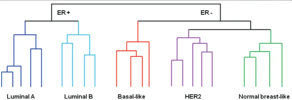
Th e whole landscape of breast cancer research changed with the publication of seminal, class discovery, microarray-based gene expression profi ling studies [11-13], in which the heterogeneity and complexity of breast cancers were rediscovered at the molecular level (Figure 2). To the average ‘microarrayer’ and bioinfor ma-tician, the experiments performed by Perou and colleagues [11] may now sound almost quaint, but in 2000 they had a major impact on how breast cancer was perceived given that they demonstrated that (a) positive and ER-negative breast cancers were funda mentally distinct at the transcriptomic level and (b) breast cancer could be divided into at least fi ve molecular subtypes: luminal A, luminal B, normal breast-like, HER2, and basal-like [12,17] (Figure 2).
[image:2.612.67.545.90.418.2]Several groups have now demonstrated that ER-positive and ER-negative breast cancers have their prog-nosis governed by distinct biological processes [18,19]
Figure 1. Clinical decision-making for adjuvant chemotherapy. Criteria included in the St. Gallen guidelines (green font) and in Adjuvant! Online (underlined) are shown. ER, estrogen receptor; HER2, human epidermal growth factor receptor 2; PR, progesterone receptor; uPA/PAI-1, urokinase-type plasminogen activator and plasminogen activator inhibitor-1.
Estimation of the risk of recurrence (prognostic
factors) and of the benefit from systemic treatments
(predictive factors)
Clinico-pathological features
Additional prognostic
and predictive factors
Ki67
Age
Tumor size
Histological grade
Multigene signatures
Histological grade
Mitotic index
Lymph node involvement
ER
uPA/PAI-1
ER
PR
HER2
Lympho-vascular invasion
CHEMOTHERAPY BENEFIT
Lympho vascular invasion
General status and co-morbidities
CHEMOTHERAPY BENEFIT
Improvement of 10-year disease-free
survival
<5%
>5%
and that at least some of these subtypes (for example, basal-like) have distinct risk factors, clinical presentation, histological features, response to therapy, and outcome [2,3,20]. Th ese data have led some experts in the fi eld to suggest that traditional clinicopathological features and immunohistochemical markers be replaced by this molecular taxonomy [21].
Th e initial approach employed for the identifi cation of the molecular subtypes was based on hierarchical clustering analysis. It should be noted, however, that this approach requires large datasets, is to some extent subjective, and cannot be employed for the classifi cation of individual samples prospectively [22-25]. Th erefore, ‘single sample predictors’ (SSPs) were developed on the basis of the correlation between the expression profi le of a given sample with the centroids for each molecular sub-type (that is, average expression profi le of each molecular subtype) [13,17,26]. Over the last decade, three distinct
SSPs were developed [13,17,26]. Further more, on the
basis of this approach, Parker and colleagues [17] developed a quantitative reverse transcriptase-poly merase chain reaction (qRT-PCR)-based or NanoString-based method (PAM50) that can be used to classify formalin-fi xed
paraffi n-embedded (FFPE) samples into the molecular
subtypes. Our group [27] and others [28,29] have demonstrated that subtle variations in data normalization and centering, as well as in the proportion of samples from each of the subtypes, may lead to changes in the classi fi cation of samples using SSPs. Moreover, indepen-dent groups have demonstrated that the classifi cation of tumors into the molecular subtypes, except for the basal-like subtype, is dependent on the SSP used [27,28]. Th is is
best exemplifi ed by the modest agreement in the
classifi cation of samples (agreement of 64%, kappa score of 0.527, and 95% confi dence interval of 0.456 to 0.597) when a cohort of 295 breast cancers was classifi ed into the molecular subtypes by the authors of the original studies on the molecular classifi cation using SSPs by Sorlie’s [13,30] and Perou’s [26,31] groups.
[image:3.612.69.545.90.254.2]Despite the enthusiasm for the use of this molecular taxonomy for the design of clinical trials and routine oncology practice, there are several issues that ought to be considered. First, the subdivision of luminal tumors into A and B is strongly dependent on the SSP used [27] and principally depends on the expression of prolifera tion-related genes [17,26,32]; there is burgeoning evidence to
demonstrate that the expression of proliferation-related genes in luminal cancers forms a continuum [3,19,33] and that the division of these tumors into two subgroups on the basis of the currently available SSPs [13,17,26] may be artifi cial. Th e subclassifi cation of ER-positive breast cancers into subtypes is not only a challenge for the ‘intrinsic’ subtype classifi cation. In fact, given that proli-fera tion is a continuum in ER-positive cancers and that proliferation is a strong determinant of outcome in this group of tumors, the allocation of ER-positive breast cancer patients into good or poor prognosis by using other microarray-based methods (for example, MammaPrint and genomic grade index) or into low, intermediate, or high histological grade should be considered arbitrary to
some extent (see ‘Multigene prognostic signa tures’
section). Second, normal breast-like cancers are now considered by some to be an invalid molecular subtype given that these tumors are likely to constitute an artefact of frozen tissue procurement and representation (that is, samples with a disproportionately high content of normal breast and stromal cells) [3,17,26,27,34,35]. Th ird, the HER2 (or HER2-enriched) subtype as defi ned by micro-arrays does not encompass all cases classifi ed as HER2-positive by clinically validated methods (that is, immuno-histochemistry and in situ hybridization with methods approved by the US Food and Drug Adminis tration), and not all HER2-positive cancers by clinical methods are classi fied as HER2 subtype by microarrays [3,17,21,36,37].
Th e above discrepancies do not invalidate the existence of the ‘intrinsic’ subtypes. As recently pointed out by Perou and colleagues [38], this is an evolving classifi cation system and PAM50 [17], rather than the SSPs described by Sorlie and colleagues [13] or Hu and colleagues [26], should be employed. With the development of the PAM50 assay, prospective testing of this classifi cation by independent groups will determine its prognostic and predictive power and clinical utility above and beyond the clinicopathological parameters currently available.
Th e putative histogenetic implications of the molecular subtypes (that is, luminal cancers would originate from luminal cells and basal-like cancers would stem from basal/ progenitor cells) [12,13,39-42] have proven incorrect. Although this observation does not have a direct impact on the clinical utility of the ‘intrinsic’ molecular subtypes, it has led to the assumption that diff erent subtypes of breast cancer would originate from diff erent cell types [13,39-42]. Importantly, there is independent direct evidence to demonstrate that the likeliest cell of origin of basal-like breast cancers lies in the luminal progenitor population rather than the ‘basal’ population of the normal breast [43,44].
Additional evidence to support the contention that the ‘intrinsic’ molecular taxonomy remains a working model in development stems from the recent identifi cation of at
least three additional molecular subtypes of ER-negative cancers: the ‘interferon-rich’ subtype [26,45], the ‘mole-cu lar apocrine’ subtype [46-48], and the ‘claudin-low’ subgroup [35,49] (Figure 2). Th e ‘interferon-rich’ subtype, fi rst described by Hu and colleagues [26], is characterized by high expression of interferon-regulated genes, such as
STAT1 [26,45]; the ‘molecular apocrine’ subtype, which is characterized by activation of androgen receptor signal-ing, frequently displays HER2 gene amplifi cation and may
be associated with PTEN germline mutations [46-48];
and the ‘claudin-low’ subgroup, which comprises tumors that express low levels or lack of expression of E-cadherin and claudin mRNA, displays an enrichment for the expres sion of genes often expressed in the process of epithelial-to-mesenchymal transition and immune res-ponse genes and allegedly harbors features suggestive of a ‘cancer stem cell-like’ phenotype [35,49]. Intriguingly, greater than 40% of these cancers do express E-cadherin and claudins at the protein level, despite the low expression levels of these genes by microarray analysis [35]. Importantly, a substantial proportion of tumors classifi ed as of claudin-low subtype by using the cell line-derived SSP described by Prat and colleagues [35] were previously classifi ed as normal breast-like by using other SSPs; these samples may have a disproportionately high content of stromal and normal breast cells. Hence, it remains to be determined whether breast cancers that do express E-cadherin and claudins at the protein level and that were classifi ed as claudin-low by the SSP predictor
were not classifi ed as such due to stromal cell
contamination. Another point for considera tion is the overlap between the transcriptomic charac teristics of the claudin-low subtype and those of spindle cell metaplastic breast carcinomas [49,50].
Given the above observations, but despite recent claims that PAM50 models derived from archival formalin-fi xed RNA are ‘a potential replacement for grade-, hormone receptor-, Ki67-, and HER2-based prognostic models’ [21], we argue that the microarraybased gene classifi -cation for breast cancer is not yet ready for clinical use in prognostic models or otherwise [1,3,8,27]. In fact, stan-dardi zation of the defi nitions and the methodologies for the identifi cation of the molecular subtypes and pros pec-tive clinical trials to validate the contribution of the ‘intrinsic’ subtypes in addition to the current clinico-pathological parameters for the management of breast cancer patients are still required [1,3,8,27]. Robust, independently validated methods for the identifi cation of these subtypes are yet to be published.
Multigene prognostic signatures First-generation signatures
been pursued by many groups in the last decade [51-58] with the aim of defi ning which patients would have such a good prognosis that they could forgo chemotherapy. Th e fi rst prognostic gene signature [51] consisted of 70 genes and was shown to identify a group of good-prognosis patients with minimal risk of development of distant metastasis within 5 years in patients who were systemic therapy-naïve. In a subsequent study, van de Vijver and colleagues [59] demonstrated that the 70-gene signature was a predictor of outcome independently of the current clinicopathological prognostic markers in a dataset comprising 295 cases (64 cases from the analysis that led to the development of the 70-gene signature and 231 new cases). Importantly, in that [59] and subsequent [60,61] studies, it has been repeatedly demonstrated that the 70-gene signature classifi es greater than 95% of ER-negative cancers as poor prognosis and that there is a strong correlation between 70-gene signature-defi ned poor prognosis and high histological grade. Furthermore, the studies demonstrated that the 70-gene signature would outperform the current methods based on clinico-pathological parameters for chemotherapy use [51,59].
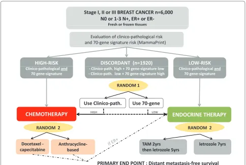
Th is has led to the development of MammaPrint, a
commercially available version of the 70-gene signature. Subsequent studies have led to the development of several other prognostic signatures, including the 76-gene signature [54,62] and genomic grade index [55,63-65], which were also shown to be independent predictors of outcome. MammaPrint is currently being tested in the MINDACT (Microarray In Node-negative and 1-3
positive lymph-node Disease may Avoid ChemoTh erapy)
trial [15] (Figure 3), which will deter mine whether this signature can actually replace clinico pathological para-meters for the identifi cation of patients who could be spared from the use of chemotherapy. Table 1 summar-izes the prognostic signatures more extensively studied to date. For comprehensive reviews on microarray-based prognostic gene signatures, readers are referred to Sotiriou and Pusztai [2], Weigelt and colleagues [3], and Kim and Paik [66].
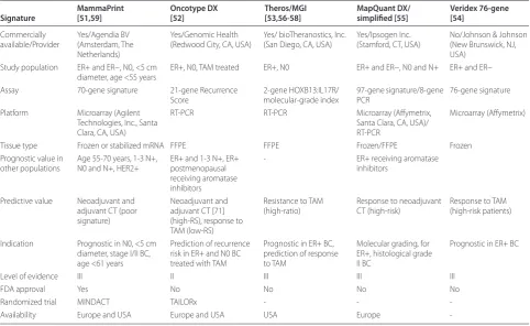
In parallel with the development of microarray-based prognostic signatures, Paik and colleagues [52] developed Oncotype DX, a qRT-PCR-based analysis of 21 genes (16 cancer-related and 5 reference genes), which can be used for risk stratifi cation of ER-positive, node-negative breast cancers from patients treated with adjuvant tamoxifen. In contrast to microarray-based predictors, Oncotype DX can be applied to FFPE samples, and this test was developed and validated on the basis of a retrospective analysis of the existing material from two randomized
clinical trials (NSABP-B-20 and NSABP-B-14). Th e
signature is based on the expression of genes that are associated with proliferation, ER signaling, HER2, and invasion [52]. Th e expression of these genes is presented
as a recurrence score (RS) that ranges from 0 to 100.
Th ese scores provide an estimate of 10-year distant
recurrence risk. For clinical use, patients are separated into three categories: low-risk (RS <18), inter mediate-risk (RS ≥18 and <31), and high-mediate-risk (RS ≥31) [52]. Oncotype DX has been shown to be an independent prognostic factor for patients with ER-positive, node-negative breast cancers treated with tamoxifen and to outperform standard clinicopathological parameters for the prediction of 10-year distant recurrence risk [52]. Oncotype DX has been subsequently evaluated in other populations of breast cancer [67] and shown to be an independent prognostic parameter in patients with ER-positive tumors with up to three ER-positive nodes receiving adjuvant chemotherapy [68] and in postmenopausal patients with ER-positive tumors treated with aromatase inhibitors (that is, anastrozole) [69].
Oncotype DX RSs have also been shown to be corre lated with the benefi t patients derive from adjuvant chemo-therapy in samples from clinical trials [70-72]. In fact, patients with tumors displaying high RSs despite the poor prognosis derive signifi cantly more benefi t from chemo-therapy than those with low-RS tumors. In addition, patients with low-RS cancers appear to derive negligible benefi t from the addition of chemotherapy to tamoxifen [70,71]. Th erefore, Oncotype DX has also been considered a predictive marker of benefi t from chemotherapy.
Despite the numerous publications on fi rst-generation signatures, level II evidence to support the prognostic role was achieved only for Oncotype DX; for the remaining signatures, only level III evidence has been obtained so far. Given the level of evidence that has been accrued, Oncotype DX has received approval from the American Society of Clinical Oncology [73] and was included in the National Comprehensive Cancer Network guidelines (Breast Cancer version 1.2011 [74]) as an option to evaluate prognosis and as a complement to clinicopathological features to predict response to chemotherapy for patients with ER-positive, node-negative breast cancer. None of the microarray-based prognostic signatures has been endorsed by these professional bodies.
Are the fi rst-generation prognostic gene signatures ready for use in clinical practice?
for Treatment Rx) [14] (Figures 3 and 4), which evaluate the genomic signatures MammaPrint and Oncotype DX, respectively.
First-generation signatures have been shown not to be stable in terms of the list of genes they are composed of [75,76]; however, comparative studies and meta-analyses have demonstrated that, despite having a negligible over-lap in their constituent genes, the fi rst-generation signa-tures tend to have similar performance and show a relatively good concordance in their prognostic classifi -cation, identifying similar but not identical subgroups of patients with poor prognosis [31,33,77].
Th e ability of these signatures to determine prognosis seems to be directly correlated to the assessment of proliferation-/cell cycle-related genes [18,33]. Th e fact that these fi rst-generation signatures arguably are mere surrogates of proliferation poses some important
[image:6.612.64.549.88.413.2]problems for their use. First, given that proliferation has been shown to be prognostic in ER-positive disease and not in ER-negative cancers, fi rst-generation signatures are applicable only for the prognostication of patients with ER-positive and HER2-negative breast cancers [18,54, 60,61]. As the expression level of proliferation-related genes in ER-positive cancers has been demon-strated to follow a continuum rather than a bimodal distribution, the subdivision of ER-positive cancers into good-prognosis (that is, luminal A) and poor-prognosis (that is, luminal B) groups is artifi cial [18,33]. In fact, the continuous nature of the Oncotype DX RS is more representative of the ranges of prognosis of patients with ER-positive disease. It should be noted, however, that this approach for clinical decision-making may be proble-matic. For instance, the prognostication and management of patients with an intermediate RS remain unclear, and
Figure 3. MINDACT (Microarray In Node-negative and 1-3 positive lymph-node Disease may Avoid ChemoTherapy) randomized trial design. The clinical impact of MammaPrint is being evaluated in MINDACT, a prospective multicenter randomized trial conducted by the European Organization for Research and Treatment of Cancer. The trial compares the recurrence-risk assessment of the 70-gene signature with that provided by Adjuvant! Online in selecting patients for adjuvant chemotherapy. Patients with concordant results are being treated accordingly (high-risk: chemotherapy with or without endocrine therapy, depending on estrogen receptor (ER) status; low-risk: hormonal therapy if ER-positive without chemotherapy). Discordant cases are being randomly assigned to receive adjuvant therapy on the basis of either clinicopathological or 70-gene signature risk assessment. Launched in 2006, the trial intends to confi rm the validity of the signature and demonstrate that its clinical use would reduce the number of patients receiving unnecessary treatments, but the results will probably take years to be revealed. Clinico-path, clinicopathological; N, lymph node; N0, lymph node-negative; RANDOM, randomization; TAM, tamoxifen; yrs, years.
Stage I, II or III BREAST CANCER n=6,000 N0 or 1-3 N+, ER+ or
ER-Fresh or frozenƟƐƐƵĞƐ Fresh or frozen ƟƐƐƵĞƐ
EvaluaƟŽŶŽĨ cůŝŶŝcŽ-patŚŽůŽgical risk ĂŶĚ 70-geŶe sigŶature risk (MammaPriŶt)
HIGH-RISK DISCORDANT (n=1920) LOW-RISK
Clinico-pathological and 70 gene-signatƵre
Clinico-pathological and 70 gene-signatƵre - Clinico-path. high + 70 gene-signatƵre low
- Clinico-path. low + 70 gene-signatƵre high
RANDOM 1 RANDOM 1
Use Clinico-path. Use 70-gene
CHEMOTHERAPY
ENDOCRINE THERAPY
RANDOM 2
W O L H
G I H
RANDOM 2 RANDOM 2
TAM 2yrs letrozole 7yrs
AnthracyclineDocetaxel
-RANDOM 2
then letrozole 5yrs based
capecitabine
up to 40% to 60% of clinically intermediate-risk patients (that is, breast cancers combining ER-positive, HER2-negative, and grade II status) are allocated to the intermediate-risk RS group [78]. Th erefore, the actual contribution of Oncotype DX to the management of this particular group of patients remains to be elucidated [78]. Th e lack of prognostic power of fi rst-generation prognostic signatures in ER-negative breast cancer and their association with proliferation in ER-positive breast cancer have brought to the forefront of cancer research the limitations of histological grading. In a way akin to fi rst-generation prognostic gene signatures, histological grade is not prognostic in ER-negative disease and is strongly associated with proliferation [18,79]. It should be noted, however, that the levels of intra- and inter-observer agreement of histological grade remain sub-optimal, despite the numerous eff orts to implement a standardized histological grading system [79]. It could be argued, on the basis of the above obser vations, that the major contribution of fi rst-generation prognostic gene signatures is to provide a standardized proliferation assay for breast cancer.
A second limitation of the fi rst-generation prognostic signatures stems from the fact that most of them were
developed to predict short-term distant recurrence (<5 years) and were shown to have a strong ‘time dependence’ and a reduced prognostic value after 5 to 10 years of follow-up [61,80]. Hence, these signatures may represent merely early distant recurrence surrogates and are unable to predict late relapses with the same accuracy. Th us, there is still a need to develop signatures that could identify patients who have a higher risk of late relapse and who may benefi t from prolonged therapy.
[image:7.612.65.548.100.396.2]Another important consideration in relation to the currently available fi rst-generation prognostic signatures is that they were derived on the basis of the analysis of tissue samples with varying contents of neoplastic cells, stromal cells, infl ammatory infi ltrate, and normal breast tissue. Th ere is evidence to suggest that the percentage of non-neoplastic cells has a substantial impact on the fi nal expression profi le of a tumor and on the ability to derive biologically meaningful prognostic signatures [81]. It should be noted that, although stromal cells and infl am-matory infi ltrate may be integral parts of the expression profi le of a tumor and provide important prognostic and predictive information, most studies employed samples with percentages of stromal cells, infl ammatory infi ltrate, and normal breast tissue ranging from 0% to 50%.
Table 1. Prognostic multigene signatures in breast cancer commercially available or in commercial development
MammaPrint Oncotype DX Theros/MGI MapQuant DX/ Veridex 76-gene Signature [51,59] [52] [53,56-58] simplifi ed [55] [54]
Commercially available/Provider
Yes/Agendia BV (Amsterdam, The Netherlands)
Yes/Genomic Health (Redwood City, CA, USA)
Yes/ bioTheranostics, Inc. (San Diego, CA, USA)
Yes/Ipsogen Inc. (Stamford, CT, USA)
No/Johnson & Johnson (New Brunswick, NJ, USA)
Study population ER+ and ER−, N0, <5 cm diameter, age <55 years
ER+, N0, TAM treated ER+, N0 ER+ and ER−, N0 and N+ ER+ and ER−
Assay 70-gene signature 21-gene Recurrence Score
2-gene HOXB13:IL17R/ molecular-grade index
97-gene signature/8-gene PCR
76-gene signature
Platform Microarray (Agilent Technologies, Inc., Santa Clara, CA, USA)
RT-PCR RT-PCR Microarray (Aff ymetrix,
Santa Clara, CA, USA)/ RT-PCR
Microarray (Aff ymetrix)
Tissue type Frozen or stabilized mRNA FFPE FFPE Frozen/FFPE Frozen
Prognostic value in other populations
Age 55-70 years, 1-3 N+, N0 and N+, HER2+
ER+ and 1-3 N+, ER+ postmenopausal receiving aromatase inhibitors
- ER+ receiving aromatase
inhibitors
Predictive value Neoadjuvant and adjuvant CT (poor signature)
Neoadjuvant and adjuvant CT [71] (high-RS), response to TAM (low-RS)
Resistance to TAM (high-ratio)
Response to neoadjuvant CT (high-risk)
Response to TAM (high-risk patients)
Indication Prognostic in N0, <5 cm diameter, stage I/II BC, age <61 years
Prediction of recurrence risk in ER+ and N0 BC treated with TAM
Prognostic in ER+ BC, prediction of response to TAM
Molecular grading, for ER+, histological grade II BC
Prognostic in ER+ BC
Level of evidence III II III III III
FDA approval Yes No No No No
Randomized trial MINDACT TAILORx - -
-Availability Europe and USA Europe and USA USA Europe
It remains to be determined whether repeated samples of the same tumor with drastically diff erent percentages of neoplastic cells (for example, 50% versus 100%) would be allocated to the same prognostic subgroup consistently. Th erefore, methods to estimate the non-neoplastic cell content of samples or tissue microdissection to standard-ize the proportion of neoplastic/non-neoplastic cells would be desirable in the development of new micro-array-based classifi ers and implementation of currently available gene expression signatures.
Despite the initial claims that these signatures would replace current clinicopathological parameters for the management of patients with breast cancer, clinicopatho-logical variables have been shown to add prognostic infor-mation independent from that off ered by fi rst-genera tion signatures [1-3]. Th erefore, these gene signa tures should be perceived as ancillary tools that complement current
methods based on the clinicopatho logical features of the
tumors rather than as a replace ment for them [1-3].
Importantly, the additional prog nostic information pro-vided by fi rst-generation signa tures appears to be limited when clinicopathological parameters are analyzed in a centralized fashion with standardized methods (that is, centralized reassessment of histological grade and standard ized assessment of ER, PR, HER2, and prolifera-tion rate as defi ned by Ki67 immunohisto chemical analy-sis) [82]. Th erefore, the true contribution of the commer-cially available fi rst-generation signatures beyond tumor morphology and immunohistochemistry remains to be determined [8].
[image:8.612.67.555.88.406.2]Recently, ‘second-generation’ signatures specifi c for the distinct subtypes of breast cancers have been reported by studying breast cancer microenvironment or host immune response [1,83-87]. Immune response-related signatures
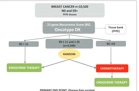
Figure 4. TAILORx (Trial Assigning IndividuaLized Options for Treatment Rx) randomized trial design. Oncotype DX is being tested in TAILORx, a prospective randomized phase III trial coordinated by the Breast Cancer Intergroup. The purposes of this trial are to confi rm the prognostic value of the 21-gene recurrence score (RS), to determine the optimal management of patients with intermediate-RS, and to refi ne the utility of the assay in clinical practice. The accrual was recently completed and the fi rst results will be disclosed in 2013. Patients with estrogen receptor (ER)-positive, node-negative breast cancers were eligible and were separated in three study groups according to their RS. High-RS patients (RS >25) received chemotherapy plus endocrine therapy, whereas low-RS patients (RS <11) were assigned to endocrine therapy alone. Patients with intermediate-RS (RS = 11 to 25) were randomly assigned to receive either hormonal therapy alone or hormonal therapy plus chemotherapy. To minimize potential under-treatment in both the high-risk and the randomly assigned groups, the RS ranges for TAILORx were diff erent from those originally defi ned (11 to 25 instead of 18 to 31). FFPE, formalin-fi xed paraffi n-embedded; N0, lymph node-negative; RANDOM, randomization.
BREAST CANCER n=10,500
N0 and ER+
FFPEƟ
FFPE Ɵssues
21-gene Recurrence Score (RS)
21 gene Recurrence Score (RS)
Oncotype DX
Tissue bank
(FFPE)
RS < 11
RS
ш
(n=4,390)
11 and
ч
25
RS >25
RANDOM
CHEMOTHERAPY
ENDOCRINE THERAPY
ENDOCRINE THERAPY
OC
have been shown to be potential prog nosticators in ER-negative or triple-ER-negative breast cancers [83-85]. Although these signatures are promising, additional evidence in support of the use of these signatures as potential predictors of outcome is still required.
Multigene predictive signatures
Beyond prognostic classifi ers, an important challenge is to provide physicians with biomarkers that could predict the response or lack of response to treatments and determine the most eff ective regimen for a specifi c patient or subgroup of patients. In clinical practice, only ER and HER2 are currently used as predictive markers for the selection of patients likely to respond to endocrine therapy and trastuzumab, respectively.
In addition to Oncotype DX, whose RSs have been shown to be associated with benefi t from the addition of chemotherapy to tamoxifen, other prognostic signatures were also shown to have predictive value for the incre-mental benefi t of chemotherapy [1-3,65,88,89]. However, unlike Oncotype DX, the predictive power of MammaPrint [88,89] and genomic grade index [65] have only been tested in retrospective datasets from patients treated with multidrug chemotherapy regimens.
Gene expression signatures and response to chemotherapy
With the clinical need for predictive markers for specifi c chemotherapy agents and multidrug regimens, several groups have developed multigene signatures specifi cally designed to predict response in patients receiving either chemotherapy or endocrine therapy. Using supervised approaches, several studies have attempted to identify multigene signatures of response to chemotherapy by
comparing gene expression profi les between
high-sensitivity and low-responsiveness tumors [90-93]. Th e majority of the studies focused on neoadjuvant chemo-therapy and, by means of microarrays or RT-PCR, analyzed tumor samples obtained from biopsies taken at diagnosis before initiation of chemotherapy (Table 2). Chemotherapy sensitivity usually was estimated with rate of pathological complete response to neoadjuvant therapy (pCR) as a surrogate of long-term benefi t from the treatment. For example, the MD Anderson Cancer Center group developed a 30-gene signature in 82 breast cancer patients receiving T/FAC chemotherapy
(pacli-taxel, fl uorouracil, doxorubicin, cyclophosphamide)
[90,92]. Th is DLDA-30 predictor was then validated in 51 independent patients and predicted pCR probability with higher sensitivity and negative predictive value than clinical variables based on age, grade, and ER status [92].
Th e accuracy of this predictor was confi rmed in an
indepen dent study [94]. Despite these interesting pre-liminary results, the accuracy of the 30-gene predictor was not found in a recent study in which it was not an
independent predictor of pCR after multivariate analysis and did not perform better than clinical variables, questioning its potential utility in the clinical setting [95].
An alternative attempt to predict chemosensitivity to specifi c chemotherapy regimens was developed with the use of in vitro models [96]. Th e combination of in vitro
signatures associated with drug sensitivity in cell lines was thought to provide composite signatures that could predict response to multidrug regimens and be translated to patients receiving multidrug chemotherapy [96]. Th ese ‘regimen-specifi c’ signatures tested in patients who, as participants in the European Organization for Research and Treatment of Cancer (EORTC) BIG00-01 clinical trial, received TET (docetaxel, epirubicin-docetaxel) or FEC (fl uorouracil, epirubicin, and cyclo phos phamide) chemotherapy resulted in a validation study published in 2007 [97]. Importantly, problems with the methodology of these studies have been identifi ed [98-100] and serious concerns about the validity of the published results were raised [101,102]. Subsequently, after a series of investi-gations, the fi ndings derived from in vitro studies were considered invalid, and this led to the discontinuation of the clinical trials based on these prediction models.
Furthermore, several high-profi le publi cations have
recently been retracted.
Another method to develop multigene classifi ers of chemosensitivity is based on the use of metagenes (that is, groups of coexpressed genes associated with a small number of biological processes). A retrospective micro-array analysis of prospectively collected ER-negative breast cancer samples demonstrated that increased stromal gene expression predicted resistance to FEC chemotherapy [103]. Th is ‘stromal’ multigene classifi er was subsequently validated in two independent cohorts [103]. Further validation of this metagene is awaited.
subtype of breast cancer, as the predictors of response to
chemo therapy in ER-positive and ER-negative breast
cancers appear to be fundamentally diff erent [19]. Furthermore, potential mechanisms of resistance to chemotherapy identifi ed by orthogonal methods (for example, RNA inter ference screens [105], microarray-based comparative genomic hybridization [106,107], proteomic analyses [108], and hypothesis-driven studies [109]) may be used as the basis for the development of multigene predictive signatures. With the availability of multiple microarray datasets from retrospective cohorts and clinical trials in the public domain, novel signatures derived from analyses using orthogonal methods can be tested in a timely fashion.
Predictive multigene markers of response to endocrine therapy
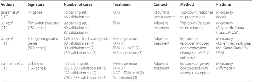
ER status has an important negative predictive value for evaluating the response to anti-estrogen therapy. Never-theless, ER expression alone is not suffi cient to predict which ER-positive tumor will respond or be resistant to diff erent modalities of endocrine therapies. Microarray-based gene expression signatures to predict outcome of tamoxifen-treated patients have been developed (Table 3). For example, a 44-gene signature, identifi ed by Jansen and colleagues [110], compared gene expression profi les in patients with advanced ER-positive breast cancers treated by tamoxifen. Other hormone sensitivity tests studying estradiol-induced genes in MCF-7 cell line culture [111] or clusters of correlated genes [112] have also been reported.
More recently, the sensitivity to endocrine therapy (SET) index was developed in a large series of ER-positive
breast cancers [113]. Th e SET index is based on the
principle that expression of genes correlated with ER may better predict response to endocrine treat ment than ER expression alone [113]. Microarray analysis of a discovery set of ER-positive tumors led to the identifi cation of 165 genes coexpressed with ER; the SET index was devised and applied to a validation cohort to defi ne three categories of sensitivity (low, intermediate, and high). Association between SET and outcome was then analyzed in three types of ER-positive cohorts receiving either adjuvant tamoxifen for 5 years or neoadjuvant chemotherapy followed by endocrine therapy (tamoxifen
or aromatase inhibition) or no adjuvant sys temic
treatment. Th e SET index was signifi cantly asso ciated with the outcome of patients receiving any type of
endocrine treatment (tamoxifen or chemo endo crine
[image:10.612.66.551.101.291.2]treatment) but had no prognostic value in untreated patients. Unlike other multigene signatures evaluating proliferation in ER-positive tumors, the SET index seems to be predictive of benefi t from endocrine therapy independently of the inherent prognosis of the tumor. Interestingly, for a potential clinical application, the SET index could identify a subset of tumors associated with an excellent prognosis and no relapse in the tamoxifen-treated group (node-negative and high-SET index tumors) and in the chemoendocrine group (high- and intermediate-SET index). Studies evaluating the clinical relevance of the SET index are warranted to expand its indications in clinical practice.
Table 2. Multigene predictors of sensitivity to chemotherapy
Number Chemosensitivity
Authors of casesa Regimen Chemotherapy evaluation Technology Method Signature NPV PPV Accuracy
Chang
et al.[116]
24 discovery 6 validation
Neoadjuvant Docetaxel Clinical response cDNA microarray
Supervised 92 genes 83% 92% 88%
Ayers
et al. [90]
24 discovery 12 validation
Neoadjuvant T/FAC pCR cDNA
microarray
Supervised 74 genes 73% 100% (3/3)
78%
Iwao-Koizumi
et al. [91]
44 discovery 26 validation
Neoadjuvant Docetaxel Clinical response High-throughput RT-PCR
Supervised 85 genes 90.9% 73.3% 80.7%
Gianni
et al. [70]
89 discovery 92 validation
Neoadjuvant TA pCR qRT-PCR/
DNA microarray
Supervised 86 genes - -
-Hess et al. [92]
82 discovery 51 validation
Neoadjuvant T/FAC pCR cDNA
microarray
Supervised 30 genes 96% 52% 76%
Thuerigen
et al. [93]
52 discovery 48 validation
Neoadjuvant G-ET pCR cDNA
microarray
Supervised 512 genes 95% 64% 88%
Farmer
et al. [103]
63 Neoadjuvant FEC pCR cDNA
microarray
Metagene approach
Stromal metagene
81% 57% 65%
aNumber of cases in discovery and validation sets. FEC, fl uorouracil, epirubicin, and cyclophosphamide; G-ET, gemcitabine, epirubicin, and docetaxel; NPV, negative
Predictors for specifi c targeted therapies
To date, only a few gene signatures have been developed to predict the response to specifi c targeted therapies in breast cancer. Recently, Loi and colleagues [114] reported promising results focusing on PIK3CA (phospho inosi tide-3-kinase, catalytic) gene mutations and the PI3K-AKT-mTOR signaling pathway targeted by PI3K/PI3K-AKT-mTOR (mammalian target of rapamycin) inhibi tors. By analysis of gene expression from 1,800 breast cancers, a gene
expression signature associated with PIK3CA mutation
was developed (PIK3CA-GS). Th e signature predicted
PIK3CA mutations in two independent datasets of breast cancers and was shown to identify good-prognosis patients in the ER-positive, HER2-negative breast cancer subgroup even in the case of highly proliferative tumors. Th e PIK3CA-GS was nega tively correlated with mTORC1 signaling, making it a potential predictor of response to PI3K/mTOR inhibitors like rapamycin, rapamycin analogs, or dual kinase inhibitors. Breast cancer cell lines with high PIK3CA-GS were confi rmed to be resistant to rapamycin [114]. Th is approach exemplifi es the potential use of microarrays as potential predictive markers for tailored therapies.
Conclusions
Microarray-based gene expression profi ling analysis has undoubtedly had a dramatic impact on our understanding of breast cancer biology by bringing the concept of the heterogeneity of breast cancer to the forefront of breast cancer research and clinical practice. In fact, it is currently inconceivable to consider positive and ER-negative breast cancers to be a single disease. However, how the information derived from the classifi cation of breast cancer into the current molecular subtypes [17] will be used for breast cancer patient management remains unclear. First-generation prognostic signatures
have led to the realization of the importance of proliferation for the prognostication of patients with ER-positive cancers [1-3]. However, despite the resources allocated to their development and valida tion, prognostic signatures have proven to add limited information to prognostic models based on clinico patho logical para-meters and standardized assessment of ER, PR, HER2, and proliferation. Gene signatures predictive of response to specifi c chemotherapy regimens have proven elusive. With the development of massively parallel sequencing techno logies, it has become possible to determine the repertoire of genetic aberrations a tumor harbors in a single experi ment. Given the successful use of genetic information as predictive markers for the use of targeted
therapies in breast cancer (for example, HER2
amplifi cation as a predictive marker for anti-HER2 agents) and tumors from other sites (for example, KIT
and PDGFRA [platelet-derived growth factor receptor alpha] mutations as predictive markers of response to imatinib mesylate in gastrointestinal stromal tumors;
EML4-ALK gene re arrange ments as predictive markers of ALK inhibitors in non-small cell lung cancer), it is plausible that the next generation of classifi ers based on sequencing information may have a greater impact on our ability to successfully stratify breast cancer patients into predictive subgroups [115]. Integrative approaches
combining genetic, trans criptomic, and proteomic
[image:11.612.64.558.99.256.2]information are likely to lead to breast cancer classifi cation systems that better refl ect the biology of the disease, and are more clinically relevant [1]. Although the deluge of high-throughput data will most certainly be a formidable challenge for the breast cancer research community, our ability to characterize tumors at an unprecedented level of detail will undoubtedly lead to novel paradigms for stratifi ed medicine and tailored therapies.
Table 3. Multigene predictors of response to endocrine treatment
Authors Signature Number of casesa Treatment Context Method Platform
Jansen et al. [110]
44 genes 48 training set, 66 validation set
TAM Recurrent breast cancer
Top-down (response vs. progression)
Microarray (local) Loi et al.
[112]
Tamoxifen predictor (181 genes)
99 training set, 69 validation set 87 validation set
TAM Adjuvant
treatment
Top-down (relapse vs. no relapse)
Microarray (Aff ymetrix, Santa Clara, CA, USA) Oh et al.
[111]
Estrogen-regulated genes
(822 genes)
Cell lines + 65 discovery set, 60 validation set (1) 90 validation set (2) 250 validation set (3)
Heterogeneous TAM (1) TAM +/− NAC (2) Heterogeneous (3)
Adjuvant treatment
Bottom-up (estrogen induced gene expression changes in MCF-7 cell lines)
Microarray
(Agilent Technologies, Inc., Santa Clara, CA, USA)
Symmans et al. [113]
SET index (165 genes)
437 training set, 225 + 298 validation set (1) 122 validation set (2) 208 + 133 validation set (3)
Heterogeneous TAM (1)
NAC + TAM or AI (2) Non-treated (3)
Adjuvant treatment
Bottom-up (genes coexpressed with estrogen receptor)
Microarray (Aff ymetrix)
Abbreviations
ALK, anaplastic lymphoma kinase; ER, estrogen receptor; FEC, fl uorouracil, epirubicin, and cyclophosphamide; FFPE, formalin-fi xed paraffi n-embedded; HER2, human epidermal growth factor receptor 2; MINDACT, Microarray In Node-negative and 1-3 positive lymph-node Disease may Avoid ChemoTherapy; mTOR, mammalian target of rapamycin; NSABP, National Surgical Adjuvant Breast and Bowel Project; pCR, pathological complete response to neoadjuvant therapy; PIK3CA, phosphoinositide-3-kinase (catalytic); PR, progesterone receptor; qRT-PCR, quantitative reverse transcriptase-polymerase chain reaction; RS, recurrence score; RT-PCR, reverse transcriptase-polymerase chain reaction; SET, sensitivity to endocrine therapy; SSP, single sample predictor.
Competing interests
The authors declare that they have no competing interests.
Acknowledgments
JSR-F and P-EC are funded in part by Breakthrough Breast Cancer. BW is funded by a Cancer Research UK postdoctoral fellowship. P-EC is funded by the Val d’Aurelle Anticancer Centre (Montpellier, France). The authors are grateful to Paul Wilkerson and Violetta Barbashina for the critical reading of the manuscript.
Published: 27 June 2011
References
1. Reis-Filho JS, Weigelt B, Fumagalli D, Sotiriou C: Molecular profi ling: moving away from tumor philately. Sci Transl Med 2010, 2:47ps43.
2. Sotiriou C, Pusztai L: Gene-expression signatures in breast cancer. N Engl J Med 2009, 360:790-800.
3. Weigelt B, Baehner FL, Reis-Filho JS: The contribution of gene expression profi ling to breast cancer classifi cation, prognostication and prediction: a retrospective of the last decade. J Pathol 2010, 220:263-280.
4. Goldhirsch A, Ingle JN, Gelber RD, Coates AS, Thurlimann B, Senn HJ: Thresholds for therapies: highlights of the St Gallen International Expert Consensus on the primary therapy of early breast cancer 2009. Ann Oncol 2009, 20:1319-1329.
5. Adjuvant! Online homepage [https://www.adjuvantonline.com]. 6. Mook S, Schmidt MK, Rutgers EJ, van de Velde AO, Visser O, Rutgers SM,
Armstrong N, van’t Veer LJ, Ravdin PM: Calibration and discriminatory accuracy of prognosis calculation for breast cancer with the online Adjuvant! program: a hospital-based retrospective cohort study. Lancet Oncol 2009, 10:1070-1076.
7. Schmidt M, Victor A, Bratzel D, Boehm D, Cotarelo C, Lebrecht A, Siggelkow W, Hengstler JG, Elsasser A, Gehrmann M, Lehr HA, Koelbl H, von Minckwitz G, Harbeck N, Thomssen C: Long-term outcome prediction by
clinicopathological risk classifi cation algorithms in node-negative breast cancer--comparison between Adjuvant!, St Gallen, and a novel risk algorithm used in the prospective randomized Node-Negative-Breast Cancer-3 (NNBC-3) trial. Ann Oncol 2009, 20:258-264.
8. Weigelt B, Reis-Filho JS: Molecular profi ling currently off ers no more than tumour morphology and basic immunohistochemistry. Breast Cancer Res 2010, 12 Suppl 4:S5.
9. Ioannidis JP, Allison DB, Ball CA, Coulibaly I, Cui X, Culhane AC, Falchi M, Furlanello C, Game L, Jurman G, Mangion J, Mehta T, Nitzberg M, Page GP, Petretto E, van Noort V: Repeatability of published microarray gene expression analyses. Nat Genet 2009, 41:149-155.
10. Ransohoff DF: Rules of evidence for cancer molecular-marker discovery and validation. Nat Rev Cancer 2004, 4:309-314.
11. Perou CM, Sorlie T, Eisen MB, van de Rijn M, Jeff rey SS, Rees CA, Pollack JR, Ross DT, Johnsen H, Akslen LA, Fluge O, Pergamenschikov A, Williams C, Zhu SX, Lonning PE, Borresen-Dale AL, Brown PO, Botstein D: Molecular portraits of human breast tumours. Nature 2000, 406:747-752.
12. Sorlie T, Perou CM, Tibshirani R, Aas T, Geisler S, Johnsen H, Hastie T, Eisen MB, van de Rijn M, Jeff rey SS, Thorsen T, Quist H, Matese JC, Brown PO, Botstein D, Eystein Lonning P, Borresen-Dale AL: Gene expression patterns of breast carcinomas distinguish tumor subclasses with clinical implications. Proc Natl Acad Sci U S A 2001, 98:10869-10874.
13. Sorlie T, Tibshirani R, Parker J, Hastie T, Marron JS, Nobel A, Deng S, Johnsen H, Pesich R, Geisler S, Demeter J, Perou CM, Lonning PE, Brown PO, Borresen-Dale AL, Botstein D: Repeated observation of breast tumor subtypes in
independent gene expression data sets. Proc Natl Acad Sci U S A 2003, 100:8418-8423.
14. Sparano JA, Paik S: Development of the 21-gene assay and its application in clinical practice and clinical trials. J Clin Oncol 2008, 26:721-728. 15. Cardoso F, Van’t Veer L, Rutgers E, Loi S, Mook S, Piccart-Gebhart MJ: Clinical
application of the 70-gene profi le: the MINDACT trial. J Clin Oncol 2008, 26:729-735.
16. Weigelt B, Reis-Filho JS: Histological and molecular types of breast cancer: is there a unifying taxonomy? Nat Rev Clin Oncol 2009, 6:718-730. 17. Parker JS, Mullins M, Cheang MC, Leung S, Voduc D, Vickery T, Davies S,
Fauron C, He X, Hu Z, Quackenbush JF, Stijleman IJ, Palazzo J, Marron JS, Nobel AB, Mardis E, Nielsen TO, Ellis MJ, Perou CM, Bernard PS: Supervised risk predictor of breast cancer based on intrinsic subtypes. J Clin Oncol 2009, 27:1160-1167.
18. Desmedt C, Haibe-Kains B, Wirapati P, Buyse M, Larsimont D, Bontempi G, Delorenzi M, Piccart M, Sotiriou C: Biological processes associated with breast cancer clinical outcome depend on the molecular subtypes. Clin Cancer Res 2008, 14:5158-5165.
19. Iwamoto T, Bianchini G, Booser D, Qi Y, Coutant C, Ya-Hui Shiang C, Santarpia L, Matsuoka J, Hortobagyi GN, Symmans WF, Holmes FA, O’Shaughnessy J, Hellerstedt B, Pippen J, Andre F, Simon R, Pusztai L: Gene pathways associated with prognosis and chemotherapy sensitivity in molecular subtypes of breast cancer. J Natl Cancer Inst 2011, 103:264-272. 20. Foulkes WD, Smith IE, Reis-Filho JS: Triple-negative breast cancer. N Engl J
Med 2010, 363:1938-1948.
21. Nielsen TO, Parker JS, Leung S, Voduc D, Ebbert M, Vickery T, Davies SR, Snider J, Stijleman IJ, Reed J, Cheang MC, Mardis ER, Perou CM, Bernard PS, Ellis MJ: A comparison of PAM50 intrinsic subtyping with immunohistochemistry and clinical prognostic factors in tamoxifen-treated estrogen receptor-positive breast cancer. Clin Cancer Res 2010, 16:5222-5232.
22. Pusztai L, Mazouni C, Anderson K, Wu Y, Symmans WF: Molecular classifi cation of breast cancer: limitations and potential. Oncologist 2006, 11:868-877.
23. Kapp AV, Tibshirani R: Are clusters found in one dataset present in another dataset? Biostatistics 2007, 8:9-31.
24. Loi S, Sotiriou C, Buyse M, Rutgers E, Van’t Veer L, Piccart M, Cardoso F: Molecular forecasting of breast cancer: time to move forward with clinical testing. J Clin Oncol 2006, 24:721-722; author reply 722-723.
25. Mackay A, Weigelt B, Grigoriadis A, Kreike B, Natrajan R, A’Hern R, Tan DS, Dowsett M, Ashworth A, Reis-Filho JS: Microarray-based class discovery for molecular classifi cation of breast cancer: analysis of interobserver agreement. J Natl Cancer Inst 2011, 103:662-673.
26. Hu Z, Fan C, Oh DS, Marron JS, He X, Qaqish BF, Livasy C, Carey LA, Reynolds E, Dressler L, Nobel A, Parker J, Ewend MG, Sawyer LR, Wu J, Liu Y, Nanda R, Tretiakova M, Ruiz Orrico A, Dreher D, Palazzo JP, Perreard L, Nelson E, Mone M, Hansen H, Mullins M, Quackenbush JF, Ellis MJ, Olopade OI, Bernard PS, Perou CM: The molecular portraits of breast tumors are conserved across microarray platforms. BMC Genomics 2006, 7:96.
27. Weigelt B, Mackay A, A’Hern R, Natrajan R, Tan DS, Dowsett M, Ashworth A, Reis-Filho JS: Breast cancer molecular profi ling with single sample predictors: a retrospective analysis. Lancet Oncol 2010, 11:339-349. 28. Haibe-Kains B, Culhane A, Desmedt C, Bontempi G, Quackenbush J, Sotiriou
C: Robusteness of breast cancer molecular subtypes identifi cation. Abstract Book of the IMPAKT 2010 Breast Cancer Conference, Brussels, Belgium, 6-8 May 2010. Ann Oncol 2010, 21:iv49-iv59.
29. Lusa L, McShane LM, Reid JF, De Cecco L, Ambrogi F, Biganzoli E, Gariboldi M, Pierotti MA: Challenges in projecting clustering results across gene expression-profi ling datasets. J Natl Cancer Inst 2007, 99:1715-1723. 30. Chang HY, Nuyten DS, Sneddon JB, Hastie T, Tibshirani R, Sørlie T, Dai H, He
YD, van’t Veer LJ, Bartelink H, van de Rijn M, Brown PO, van de Vijver MJ: Robustness, scalability, and integration of a wound-response gene expression signature in predicting breast cancer survival. Proc Natl Acad Sci USA 2005, 102:3738-3743.
31. Fan C, Oh DS, Wessels L, Weigelt B, Nuyten DS, Nobel AB, van’t Veer LJ, Perou CM: Concordance among gene-expression-based predictors for breast cancer. N Engl J Med 2006, 355:560-569.
32. Weigelt B, Geyer FC, Reis-Filho JS: Histological types of breast cancer: how special are they? Mol Oncol 2010, 4:192-208.
toward a unifi ed understanding of breast cancer subtyping and prognosis signatures. Breast Cancer Res 2008, 10:R65.
34. Peppercorn J, Perou CM, Carey LA: Molecular subtypes in breast cancer evaluation and management: divide and conquer. Cancer Invest 2008, 26:1-10.
35. Prat A, Parker JS, Karginova O, Fan C, Livasy C, Herschkowitz JI, He X, Perou CM: Phenotypic and molecular characterization of the claudin-low intrinsic subtype of breast cancer. Breast Cancer Res 2010, 12:R68. 36. Rouzier R, Perou CM, Symmans WF, Ibrahim N, Cristofanilli M, Anderson K,
Hess KR, Stec J, Ayers M, Wagner P, Morandi P, Fan C, Rabiul I, Ross JS, Hortobagyi GN, Pusztai L: Breast cancer molecular subtypes respond diff erently to preoperative chemotherapy. Clin Cancer Res 2005, 11:5678-5685.
37. de Ronde JJ, Hannemann J, Halfwerk H, Mulder L, Straver ME, Vrancken Peeters MJ, Wesseling J, van de Vijver M, Wessels LF, Rodenhuis S: Concordance of clinical and molecular breast cancer subtyping in the context of preoperative chemotherapy response. Breast Cancer Res Treat 2010, 119:119-126.
38. Perou CM, Parker JS, Prat A, Ellis MJ, Bernard PS: Clinical implementation of the intrinsic subtypes of breast cancer. Lancet Oncol 2010, 11:718-719; author reply 720-711.
39. Prat A, Perou CM: Mammary development meets cancer genomics. Nat Med 2009, 15:842-844.
40. Sorlie T: Introducing molecular subtyping of breast cancer into the clinic? J Clin Oncol 2009, 27:1153-1154.
41. Stingl J, Caldas C: Molecular heterogeneity of breast carcinomas and the cancer stem cell hypothesis. Nat Rev Cancer 2007, 7:791-799.
42. Sims AH, Howell A, Howell SJ, Clarke RB: Origins of breast cancer subtypes and therapeutic implications. Nat Clin Pract Oncol 2007, 4:516-525. 43. Lim E, Vaillant F, Wu D, Forrest NC, Pal B, Hart AH, Asselin-Labat ML, Gyorki DE,
Ward T, Partanen A, Feleppa F, Huschtscha LI, Thorne HJ, Fox SB, Yan M, French JD, Brown MA, Smyth GK, Visvader JE, Lindeman GJ: Aberrant luminal progenitors as the candidate target population for basal tumor development in BRCA1 mutation carriers. Nat Med 2009, 15:907-913. 44. Molyneux G, Geyer FC, Magnay FA, McCarthy A, Kendrick H, Natrajan R,
Mackay A, Grigoriadis A, Tutt A, Ashworth A, Reis-Filho JS, Smalley MJ: BRCA1 basal-like breast cancers originate from luminal epithelial progenitors and not from basal stem cells. Cell Stem Cell 2010, 7:403-417.
45. Teschendorff AE, Miremadi A, Pinder SE, Ellis IO, Caldas C: An immune response gene expression module identifi es a good prognosis subtype in estrogen receptor negative breast cancer. Genome Biol 2007, 8:R157. 46. Farmer P, Bonnefoi H, Becette V, Tubiana-Hulin M, Fumoleau P, Larsimont D,
Macgrogan G, Bergh J, Cameron D, Goldstein D, Duss S, Nicoulaz AL, Brisken C, Fiche M, Delorenzi M, Iggo R: Identifi cation of molecular apocrine breast tumours by microarray analysis. Oncogene 2005, 24:4660-4671.
47. Doane AS, Danso M, Lal P, Donaton M, Zhang L, Hudis C, Gerald WL: An estrogen receptor-negative breast cancer subset characterized by a hormonally regulated transcriptional program and response to androgen. Oncogene 2006, 25:3994-4008.
48. Banneau G, Guedj M, MacGrogan G, de Mascarel I, Velasco V, Schiappa R, Bonadona V, David A, Dugast C, Gilbert-Dussardier B, Ingster O, Vabres P, Caux F, de Reynies A, Iggo R, Sevenet N, Bonnet F, Longy M: Molecular apocrine diff erentiation is a common feature of breast cancer in patients with germline PTEN mutations. Breast Cancer Res 2010, 12:R63.
49. Hennessy BT, Gonzalez-Angulo AM, Stemke-Hale K, Gilcrease MZ, Krishnamurthy S, Lee JS, Fridlyand J, Sahin A, Agarwal R, Joy C, Liu W, Stivers D, Baggerly K, Carey M, Lluch A, Monteagudo C, He X, Weigman V, Fan C, Palazzo J, Hortobagyi GN, Nolden LK, Wang NJ, Valero V, Gray JW, Perou CM, Mills GB: Characterization of a naturally occurring breast cancer subset enriched in epithelial-to-mesenchymal transition and stem cell characteristics. Cancer Res 2009, 69:4116-4124.
50. Weigelt B, Kreike B, Reis-Filho JS: Metaplastic breast carcinomas are basal-like breast cancers: a genomic profi ling analysis. Breast Cancer Res Treat 2009, 117:273-280.
51. van ‘t Veer LJ, Dai H, van de Vijver MJ, He YD, Hart AA, Mao M, Peterse HL, van der Kooy K, Marton MJ, Witteveen AT, Schreiber GJ, Kerkhoven RM, Roberts C, Linsley PS, Bernards R, Friend SH: Gene expression profi ling predicts clinical outcome of breast cancer. Nature 2002, 415:530-536.
52. Paik S, Shak S, Tang G, Kim C, Baker J, Cronin M, Baehner FL, Walker MG, Watson D, Park T, Hiller W, Fisher ER, Wickerham DL, Bryant J, Wolmark N: A multigene assay to predict recurrence of tamoxifen-treated,
node-negative breast cancer. N Engl J Med 2004, 351:2817-2826.
53. Ma XJ, Wang Z, Ryan PD, Isakoff SJ, Barmettler A, Fuller A, Muir B, Mohapatra G, Salunga R, Tuggle JT, Tran Y, Tran D, Tassin A, Amon P, Wang W, Enright E, Stecker K, Estepa-Sabal E, Smith B, Younger J, Balis U, Michaelson J, Bhan A, Habin K, Baer TM, Brugge J, Haber DA, Erlander MG, Sgroi DC: A two-gene expression ratio predicts clinical outcome in breast cancer patients treated with tamoxifen. Cancer Cell 2004, 5:607-616.
54. Wang Y, Klijn JG, Zhang Y, Sieuwerts AM, Look MP, Yang F, Talantov D, Timmermans M, Meijer-van Gelder ME, Yu J, Jatkoe T, Berns EM, Atkins D, Foekens JA: Gene-expression profi les to predict distant metastasis of lymph-node-negative primary breast cancer. Lancet 2005, 365:671-679. 55. Sotiriou C, Wirapati P, Loi S, Harris A, Fox S, Smeds J, Nordgren H, Farmer P, Praz V, Haibe-Kains B, Desmedt C, Larsimont D, Cardoso F, Peterse H, Nuyten D, Buyse M, Van de Vijver MJ, Bergh J, Piccart M, Delorenzi M: Gene expression profi ling in breast cancer: understanding the molecular basis of histologic grade to improve prognosis. J Natl Cancer Inst 2006, 98:262-272.
56. Ma XJ, Hilsenbeck SG, Wang W, Ding L, Sgroi DC, Bender RA, Osborne CK, Allred DC, Erlander MG: The HOXB13:IL17BR expression index is a prognostic factor in early-stage breast cancer. J Clin Oncol 2006, 24:4611-4619.
57. Jansen MP, Sieuwerts AM, Look MP, Ritstier K, Meijer-van Gelder ME, van Staveren IL, Klijn JG, Foekens JA, Berns EM: HOXB13-to-IL17BR expression ratio is related with tumor aggressiveness and response to tamoxifen of recurrent breast cancer: a retrospective study. J Clin Oncol 2007, 25:662-668.
58. Ma XJ, Salunga R, Dahiya S, Wang W, Carney E, Durbecq V, Harris A, Goss P, Sotiriou C, Erlander M, Sgroi D: A fi ve-gene molecular grade index and HOXB13:IL17BR are complementary prognostic factors in early stage breast cancer. Clin Cancer Res 2008, 14:2601-2608.
59. van de Vijver MJ, He YD, van’t Veer LJ, Dai H, Hart AA, Voskuil DW, Schreiber GJ, Peterse JL, Roberts C, Marton MJ, Parrish M, Atsma D, Witteveen A, Glas A, Delahaye L, van der Velde T, Bartelink H, Rodenhuis S, Rutgers ET, Friend SH, Bernards R: A gene-expression signature as a predictor of survival in breast cancer. N Engl J Med 2002, 347:1999-2009.
60. Bueno-de-Mesquita JM, van Harten WH, Retel VP, van’t Veer LJ, van Dam FS, Karsenberg K, Douma KF, van Tinteren H, Peterse JL, Wesseling J, Wu TS, Atsma D, Rutgers EJ, Brink G, Floore AN, Glas AM, Roumen RM, Bellot FE, van Krimpen C, Rodenhuis S, van de Vijver MJ, Linn SC: Use of 70-gene signature to predict prognosis of patients with node-negative breast cancer: a prospective community-based feasibility study (RASTER). Lancet Oncol 2007, 8:1079-1087.
61. Buyse M, Loi S, van’t Veer L, Viale G, Delorenzi M, Glas AM, d’Assignies MS, Bergh J, Lidereau R, Ellis P, Harris A, Bogaerts J, Therasse P, Floore A, Amakrane M, Piette F, Rutgers E, Sotiriou C, Cardoso F, Piccart MJ: Validation and clinical utility of a 70-gene prognostic signature for women with node-negative breast cancer. J Natl Cancer Inst 2006, 98:1183-1192.
62. Foekens JA, Atkins D, Zhang Y, Sweep FC, Harbeck N, Paradiso A, Cufer T, Sieuwerts AM, Talantov D, Span PN, Tjan-Heijnen VC, Zito AF, Specht K, Hoefl er H, Golouh R, Schittulli F, Schmitt M, Beex LV, Klijn JG, Wang Y: Multicenter validation of a gene expression-based prognostic signature in lymph node-negative primary breast cancer. J Clin Oncol 2006,
24:1665-1671.
63. Desmedt C, Giobbie-Hurder A, Neven P, Paridaens R, Christiaens MR, Smeets A, Lallemand F, Haibe-Kains B, Viale G, Gelber RD, Piccart M, Sotiriou C: The Gene expression Grade Index: a potential predictor of relapse for endocrine-treated breast cancer patients in the BIG 1-98 trial. BMC Med Genomics 2009, 2:40.
64. Loi S, Haibe-Kains B, Desmedt C, Lallemand F, Tutt AM, Gillet C, Ellis P, Harris A, Bergh J, Foekens JA, Klijn JG, Larsimont D, Buyse M, Bontempi G, Delorenzi M, Piccart MJ, Sotiriou C: Defi nition of clinically distinct molecular subtypes in estrogen receptor-positive breast carcinomas through genomic grade. J Clin Oncol 2007, 25:1239-1246.
65. Liedtke C, Hatzis C, Symmans WF, Desmedt C, Haibe-Kains B, Valero V, Kuerer H, Hortobagyi GN, Piccart-Gebhart M, Sotiriou C, Pusztai L: Genomic grade index is associated with response to chemotherapy in patients with breast cancer. J Clin Oncol 2009, 27:3185-3191.
66. Kim C, Paik S: Gene-expression-based prognostic assays for breast cancer. Nat Rev Clin Oncol 2010, 7:340-347.