Page 1 of 11
Open Access
Research
Complementation of diverse HIV-1 Env defects through
cooperative subunit interactions: a general property of the
functional trimer
Karl Salzwedel
1,2and Edward A Berger*
1Address: 1Laboratory of Viral Diseases, National Institute of Allergy and Infectious Diseases, National Institutes of Health, Bethesda, MD 20892,
USA and 2Current address: Division of AIDS, NIAID, 6700-B Rockledge Drive, Room 4149, Bethesda, MD 20892, USA
Email: Karl Salzwedel - [email protected]; Edward A Berger* - [email protected] * Corresponding author
Abstract
Background: The HIV-1 Env glycoprotein mediates virus entry by catalyzing direct fusion between the virion membrane and the target cell plasma membrane. Env is composed of two subunits: gp120, which binds to CD4 and the coreceptor, and gp41, which is triggered upon coreceptor binding to promote the membrane fusion reaction. Env on the surface of infected cells is a trimer consisting of three gp120/gp41 homo-dimeric protomers. An emerging question concerns cooperative interactions between the protomers in the trimer, and possible implications for Env function.
Results: We extended studies on cooperative subunit interactions within the HIV-1 Env trimer, using analysis of functional complementation between coexpressed inactive variants harboring different functional deficiencies. In assays of Env-mediated cell fusion, complementation was observed between variants with a wide range of defects in both the gp120 and gp41 subunits. The former included gp120 subunits mutated in the CD4 binding site or incapable of coreceptor interaction due either to mismatched specificity or V3 loop mutation. Defective gp41 variants included point mutations at different residues within the fusion peptide or heptad repeat regions, as well as constructs with modifications or deletions of the membrane proximal tryptophan-rich region or the transmembrane domain. Complementation required the defective variants to be coexpressed in the same cell. The observed complementation activities were highly dependent on the assay system. The most robust activities were obtained with a vaccinia virus-based expression and reporter gene activation assay for cell fusion. In an alternative system involving Env expression from integrated provirus, complementation was detected in cell fusion assays, but not in virus particle entry assays.
Conclusion: Our results indicate that Env function does not require every subunit in the trimer to be competent for all essential activities. Through cross-talk between subunits, the functional determinants on one defective protomer can cooperatively interact to trigger the functional determinants on an adjacent protomer(s) harboring a different defect, leading to fusion. Cooperative subunit interaction is a general feature of the Env trimer, based on complementation activities observed for a highly diverse range of functional defects.
Published: 11 August 2009
Retrovirology 2009, 6:75 doi:10.1186/1742-4690-6-75
Received: 4 July 2009 Accepted: 11 August 2009
This article is available from: http://www.retrovirology.com/content/6/1/75
© 2009 Salzwedel and Berger; licensee BioMed Central Ltd.
Background
The envelope glycoprotein (Env) of human immunodefi-ciency virus type 1 (HIV-1) promotes virus entry by cata-lyzing direct fusion between the virion membrane and the target cell plasma membrane; similarly, Env-expressing cells can fuse with target cells to form multinucleated giant cells (syncytia). Env is synthesized as a gp160 pre-cursor protein that assembles into homo-trimeric com-plexes in the endoplasmic reticulum. During transport through the secretory pathway, gp160 is cleaved in the trans-Golgi network by a furin-like protease(s) to yield the external gp120 subunit noncovalently associated with the gp41 transmembrane subunit (derived from the N- and C-regions of gp160, respectively) [1]. The functional Env spike on mature virions of HIV-1 and the related simian immunodeficiency virus consists of a homo-trimer of gp120/gp41 hetero-dimers [2].
Env-mediated fusion involves a strict division of labor between the two subunits: gp120 is responsible for sequential binding to specific target cell receptors, first to CD4 and then to the coreceptor (a specific chemokine receptor, typically CCR5 or CXCR4); receptor binding then triggers gp41 to promote membrane fusion. These steps involve a tightly orchestrated series of conforma-tional changes in both Env subunits that drive the fusion process. The emerging understanding of the complexities of HIV Env/receptor interactions and the subsequent events leading to fusion/entry have been the central focus of numerous review articles over the past decade [3-8]. X-ray crystallographic analyses of gp120 from HIV-1 [9] and the closely related simian immunodeficiency virus [10] have revealed that CD4 binding induces a profound rear-rangement of the relatively disordered gp120 subunit to create a new surface consisting of four anti-parallel beta strands derived from discontinuous regions of the linear sequence; this highly conserved "bridging sheet", which is not present in the unliganded pre-CD4-bound state, is directly involved in binding to coreceptor [11] in conjunc-tion with the third variable loop (V3) of gp120, which determines coreceptor specificity [12,13]. Binding of gp120 to coreceptor then triggers the fusogenic activity of gp41 in a process believed to involve insertion of the gp41 N-terminal fusion peptide (FP) into the target cell plasma membrane [14,15]. Detailed structural information is not yet available for the native state of gp41, but the structure of the final post-fusion state has been determined to be a trimer of hairpins in the form of a six-helix coiled-coil bundle [16-18]. A transient intermediate conformation is thought to exist in which the gp41 subunits adopt an extended triple-helix coiled-coil with the N-terminal FPs inserted into the target cell membrane. The heptad repeat (HR) segments near the external C-terminal region (HR2) then fold to insert in anti-parallel fashion into the grooves formed by the cluster of the three N-terminal heptad repeat (HR1) segments; the resulting formation of a
6-helix bundle brings the virion and target cell plasma membranes together, and provides the driving force for membrane fusion underlying HIV entry. The molecular complexity of the HIV entry process presents a variety of targets for novel antiviral agents [19-22]; the T-20 peptide (enfuvirtide, Fuzeon) targeting the gp41 intermediate conformation is the first-in-class HIV-1 fusion inhibitor [23], and the recently approved maraviroc is the first-in-class inhibitor that binds to the CCR5 coreceptor and blocks the gp120 interaction [24].
While each gp120/gp41 hetero-dimeric complex contains all the determinants required for fusion, it is possible that molecular interactions between complexes within the trimer influence Env function. In a previous study we used a quantitative vaccinia expression-based cell fusion assay to demonstrate that individual subunits within the Env trimer can interact cooperatively during fusion [25]. By coexpressing Env proteins with defects in different essen-tial determinants, we found that functional complemen-tation could occur between subunits within a mixed trimer. In the present report, we show that subunit com-plementation is a general capacity of the HIV-1 Env trimer, though its efficiency and detectability are depend-ent on the particular defective variants examined and the assay systems employed. The results are discussed in terms of potential biological implications for Env function and HIV neutralization.
Methods
Construction and expression of Env variants
For vaccinia virus expression-based cell fusion assays, HIV-1 Envs were transiently expressed from pSC59-based plasmids under control of a strong synthetic vaccinia virus early/late promoter [26]. Previously described plasmids [27,28] were used to express wild-type Envs from the fol-lowing HIV-1 strains: LAI [29] (LAV isolate, unless indi-cated otherwise), plasmid 41; SF162, plasmid pCB-32; Ba-L, plasmid pCB-43, and CM235, plasmid pCB-52. In addition, a Kpn I-Xho I fragment encoding wild-type YU-2 Env was substituted into a variant of pCB-41 con-taining a unique Xho I site at the 3' end of Env (pKS-9) to create the plasmid pKS-10. As a negative control, an uncleaveable (Unc) mutant form of LAI (IIIB isolate) was used (plasmid pCB-16).
Page 3 of 11
14); heptad repeat mutants LAI-HR1a (plasmid pKS-15) and LAI-HR1e (plasmid pKS-16); V3 loop mutant LAI-V3 (plasmid pKS-17). A Kpn I-Bam HI fragment encoding the LAI-Δ665-856 mutant, previously referred to as Δ192 [31], was substituted into a variant of pCB-41 (pKS-8) in which a Bam HI site upstream of the promoter had been destroyed by cutting and then filling in with T4 DNA polymerase; the resulting plasmid was pKS-18. Kpn I-Xho I fragments encoding the mutants LAI-HT-1 [32], LAI-HT-2 [32], and LAI-Δ665-682 [33,34] were substi-tuted into the plasmid 8 to create the plasmids pKS-19, pKS-20, pKS-21, and pKS-22, respectively. Previous studies indicate that each of these variants is capable of Env processing (except for Unc), surface expression (except for LAI-Δ665-856, which is secreted), and CD4 binding (except for LAI-BS) [25,30,32-34].
For MAGI cell HIV infectivity assays, virus was expressed from the pNL4-3 proviral clone [35] encoding wild-type LAI Env (LAV isolate). pNL4-3 containing a frame-shift mutation at the Nhe I site within Env (pNL4-3Δenv) [33] was used as a negative control. Nhe I-Bam HI fragments encoding the LAI-FP26 and LAI-BS mutants [25] were sub-cloned into pNL4-3 to create pKS-11 (encoding NL4.3-FP26) and pKS-12 (encoding NL4.3-BS), respectively. The phenotype for each construct, both when expressed alone and in complementation experiments, was confirmed using two independent plasmid clones constructed in par-allel.
Vaccinia virus-based cell fusion assay
Env-mediated cell fusion activity was measured using a quantitative vaccinia-based reporter gene assay as described previously [36,37]. Each vaccinia virus was used at a multiplicity of infection of 10. Target cells were pre-pared by co-infecting NIH 3T3 cells with vaccinia virus recombinant vCB21R-LacZ containing the E. coli lacZ reporter gene linked to the T7 promotor [38], plus vac-cinia recombinants encoding the following cDNAs linked to vaccinia early/late promoters: CD4, vCB-3 [39] and the designated coreceptor CCR5, vHC-1 [40] or CXCR4, vCBYF1-fusin [41]. Effector cells were prepared by trans-fecting HeLa cells with the above-described plasmids con-taining the Env genes linked to a strong synthetic vaccinia early/late promoter and infecting with vaccinia recom-binant vP11T7gene1 encoding bacteriophage T7 RNA polymerase [42]. Transfection was performed with DOTAP (Boehringer Mannheim, Indianapolis, IN); the total amount of DNA was held constant at 5 μg DNA per 25 cm2 flask, in both single-transfection and cotransfec-tion experiments. Effector and target cells were incubated overnight at 31°C to allow expression of the recombinant proteins. After these cells were washed by centrifugation, they were mixed in equal numbers in duplicate wells of a 96-well plate (2 × 10⁵ of each per well) and incubated for 2.5 hr at 37°C. Fusion reactions were terminated by addi-tion of nonidet-P40 (0.5% final) and quantified by spec-trophotometric measurement of β-galactosidase activity as described previously [36]. For each data point, error bars indicate the standard errors of the mean of duplicate samples; in cases where error bars appear to be absent, the data points were so close that error bars are not visible. All experiments were repeated at least twice; representative data are shown for each experiment.
MAGI cell assays for cell fusion and virus entry
[image:3.612.60.294.279.436.2]HIV-1 entry and Env-mediated cell fusion in the context of HIV-1 provirus expression were measured using the HeLa-CD4/LTR-β-gal (MAGI) indicator target cell line [43], which was obtained from the NIH AIDS Research and Ref-erence Reagent Program (originally contributed by M. Emerman). BS-C-1 cells plated at 3 × 10⁵ per well in 6-well Mutations in HIV-1 Env
Figure 1
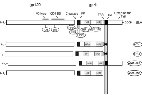
Mutations in HIV-1 Env. A schematic representation of HIV-1 Env. Functional and structural domains within the gp120 and gp41 subunits are labeled at the top: V3 loop (3rd
variable loop), CD4 BS (CD4 binding site), FP (fusion pep-tide), TRR (tryptophan-rich region), TM (transmembrane domain). For various inactivating mutants in the LAI Env (des-ignations encircled), the approximate locations of specific point mutants are indicated underneath, and the deletion mutants are indicated on the right. The specific point muta-tions are: V3 (R320G in the conserved GPGR motif at the tip of the V3 loop); BS (D368R within the CD4 binding site); var-ious positions in the fusion peptide including FP2 (Val→Glu), FP9 (Leu→Arg) and FP26 (Leu→Arg); heptad repeat muta-tions HR1e (V570R) and HR1a (I573P). HT-1 and HT-2 are chimeric LAI Env/Thy-1.1 glycoproteins that are membrane-associated via a glycosyl-phosphatidylinositol (GPI) anchor. HT-1 contains the gp41 ectodomain minus the tryptophan-rich region (K665 through I682), whereas HT-2 contains the entire gp41 ectodomain; both constructs have 22 intervening amino acid residues from the C-terminus of Thy1.1. The
Δ665-682 construct has a selective deletion of the TRR (K665 through I682) and the Δ665-856 construct has an introduced premature stop codon that results in deletion of the C-terminal 192 aa of Env, including the TM and cytoplas-mic domains. See Methods for construction and references.
HR1 HR2
NH2 665-682
NH2 HR1 HR2 HT-1
gp120 gp41
HR1 HR2
TM Cytoplasmic Tail FP
Cleavage CD4 BS
NH2 COOH
V3 loop TRR
ENV
V3
FP26 BS
FP9
FP2 HR1e HR1a
HR1 HR2
NH2 HT-2
HR1 HR2
plates the previous day were transfected (or cotransfected) with the designated pNL4-3-based proviral construct(s) using FuGENE 6 (Boehringer Mannheim, Indianapolis, IN) according to the manufacturer's protocol. The next day, cells were washed and given fresh media (2 ml per well) containing 10 mM HEPES. Three days post-transfec-tion, the supernatants were removed, filtered through a 0.45 μm filter to remove cellular debris, and stored at 4°C. For cell fusion assays, the cells were trypsinized, washed, mixed 1:10 with MAGI cells, and replated in duplicate at 1 × 10⁵ total cells per well of a 24-well plate. Cells were allowed to fuse overnight at 37°C and were then stained with X-gal. Cell fusion was quantitated by counting the total number of blue multi-nucleated syncytia per well with the aid of a grid. For the virus entry assays, p24 levels in the filtered supernatants were quantitated using the HIV-1 p24 Antigen Assay (Coulter), and supernatant vol-umes were normalized accordingly. MAGI cells were infected in duplicate with 300 μl of filtered supernatant per well of a 24-well plate and stained with X-gal 48 hrs post-infection. Virus entry was quantitated by counting the total number of blue foci per well. For complementa-tion pairs, supernatants containing up to 2.35 ng of p24 per well were used (equivalent to 628 infectious units for wild-type). This corresponds to approximately 15–20% of the total supernatant from the cells. For each data point, the standard errors of the mean of duplicate samples are shown.
Results
To test the ability of fusion-inactive Env subunits to func-tionally complement one another in the context of mixed Env trimers, we first employed a vaccinia-based quantita-tive cell fusion assay system wherein fusion between effec-tor cells expressing Env and target cells expressing the necessary receptors leads to reporter gene activation (β -galactosidase production) [36,37]. We examined comple-mentation between variants in gp120 that were inactive due to inability to interact with CD4 (CD4 BS mutation) or coreceptor (mismatched specificity, or mutation in the V3 loop), as well as variants in gp41 with mutations at dif-ferent points within the FP and HR1 regions, as well as modifications of the membrane proximal tryptophan-rich region (TRR) and the transmembrane (TM) domain (Fig. 1). Throughout these studies, target cells lacking corecep-tor served as negative controls; where indicated, an uncleaveable mutant Env (Unc) containing a mutation in the gp120/gp41 cleavage site provided an additional neg-ative control.
Complementation by Env subunits from HIV-1 primary isolates of different genetic subtypes
Previously we demonstrated complementation between Env constructs from two HIV-1 strains that were highly laboratory-adapted and both clade B: LAI (X4, i.e. CXCR4-specific) and SF162 (R5, i.e. CCR5-CXCR4-specific) [25]. To
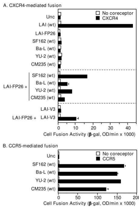
deter-mine whether complementation potential is a more gen-eral property of HIV-1 Envs, we analyzed the relative complementation efficiencies of an LAI Env mutant con-taining a defective FP (LAI-FP26) with Envs from diverse R5 isolates in a CXCR4-dependent cell fusion assay. When tested alone under these conditions, wild type LAI showed potent activity whereas the LAI-FP26 mutant and all four wild type R5 Envs were non-fusogenic (Fig. 2A, top sec-tion). In coexpression experiments that enabled mixed trimer formation, complementation of LAI-FP26 with the R5 Envs occurred not only with SF162 as shown previ-ously, but also with the laboratory-adapted Ba-L strain (clade B) and the primary YU-2 (clade B) and CM235 (CRF01_AE recombinant) isolates (Fig. 2A, middle sec-tion). The differences in the relative complementation efficiencies of the various Envs correlated roughly with their relative intrinsic fusogenicities in a CCR5-dependent assay (Fig. 2B).
Complementation by Env subunits containing a mutationally inactivated V3 loop
Our previous results [25] coupled with the data above demonstrate that an Env with a mutational defect in gp41 can complement an Env containing a gp120 subunit inca-pable of interacting with coreceptor due to mismatched coreceptor specificity. We wished to extend this finding by testing a gp120 subunit rendered inherently defective for coreceptor interaction by site-directed mutation. The V3 loop, though highly variable, contains a conserved β-turn motif at its crown (typically GPGR or GPGQ) that is essen-tial for coreceptor binding activity [12,13]. We analyzed a point mutant (LAI-V3) containing a G in place of the R residue in the GPGR motif, which has been shown previ-ously to abolish fusogenicity [44]. Our results demon-strate that the fusion-defective LAI-V3 was able to complement LAI-FP26 (Fig. 2A, bottom section). The fusion activity was in the same range observed for the coreceptor-mismatched Envs (Fig. 2A, middle section), indicating that complementation efficiency was not lim-ited by structural incompatibilities between Envs from these different strains.
Page 5 of 11
distinct gp120 receptor binding deficiencies were some-what less active than complementation between Envs containing a defective gp120 and a defective gp41 (LAI-BS + LAI-FP26) (Fig. 3). As expected, no complementation was observed upon coexpression of LAI-V3 with SF162, since the gp120 from neither Env is capable of function-ing with the CXCR4 coreceptor.
Varying complementation efficiencies of different point mutations within the gp41 FP
Our previously described data and the experiments above demonstrated functional complementation of a particular
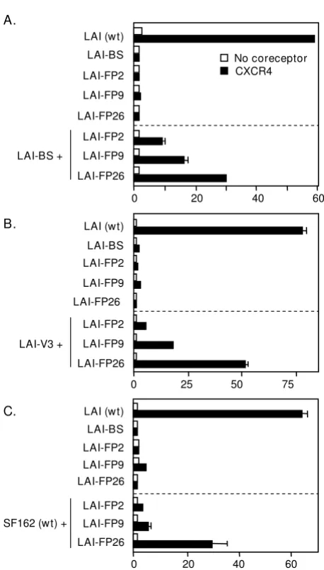
gp41 FP mutation, i.e. substitution of Arg for Leu at the 26th position from the gp41 N-terminus (LAI-FP26). To extend these analyses, we analyzed two additional FP mutations previously shown by others to abolish fusogenic activity without affecting Env processing or CD4 binding [30]: LAI-FP2 substitutes Glu for Val at the 2nd position of the FP, and LAI-FP9 substitutes Arg for Leu at the 9th position (Fig. 1). The LAI-FP2 mutant has been shown to dominantly interfere with cell fusion when coexpressed with wild-type Env, whereas the LAI-FP9 mutant reduced fusion two-fold and the LAI-FP26 mutant had no negative effect when coexpressed with wild-type Env [45]. The results of complementation experiments with these gp41 FP mutations are shown in Fig. 4. Con-sistent with previous reports, each mutation alone strongly impaired cell fusion activity compared to wild type (top sections in Figs. 4A–C). The relative efficiencies of complementation, FP26 > FP9 > FP2 was observed whether the complementation partner was LAI-BS (Fig. 4A), LAI-V3 (Fig. 4B), or SF162 (wt) (Fig. 4C).
Complementation with gp41 subunits lacking the normal membrane anchoring and membrane proximal external regions
Highly conserved regions close to the membrane are known to be critical for Env function, including the 22 amino acid TM domain that anchors Env to the surface of virions and infected cells, and the membrane-proximal external region, generally defined as the last 24 C-terminal residues of the gp41 ectodomain (L660 – K683) [15]. This Complementation with laboratory-adapted and primary Envs
[image:5.612.60.290.86.434.2]from different clades
Figure 2
Complementation with laboratory-adapted and pri-mary Envs from different clades. The vaccinia system was employed to assay cell fusion between effector cells expressing Envs and target cells expressing CD4 either with (filled bars) or without (open bars) the indicated coreceptor.
A) CXCR4-dependent fusion. Effector cells expressed the indicated Envs either individually (top section) or in combina-tion with LAI-FP26 (middle seccombina-tion). Effector cells expressed LAI-V3 individually or in combination with LAI-FP26 (bottom section). B) CCR5-dependent fusion. The indicated wt Envs were assayed for their intrinsic fusogenicity with target cells expressing CD4 and CCR5.
Cell Fusion Activity ( -gal, OD/min x 1000) B. CCR5-mediated fusion
SF162 (wt)
Ba-L (wt)
YU-2 (wt)
CM235 (wt) Unc
0 50 100 150 200
CCR5 No coreceptor A. CXCR4-mediated fusion
Cell Fusion Activity ( -gal, OD/min x 1000) SF162 (wt)
Ba-L (wt)
YU-2 (wt) CM235 (wt) LAI (wt)
SF162 (wt)
Ba-L (wt)
YU-2 (wt)
CM235 (wt) LAI-FP26
LAI-V3
LAI-V3
0 10 20 30 40
Unc CXCR4No coreceptor
LAI-FP26 +
LAI-FP26 + Complementation between gp120 variantsFigure 3
Complementation between gp120 variants. The vac-cinia system was employed to assay cell fusion between effec-tor cells expressing Envs and target cells expressing CD4 either with (filled bars) or without (open bars) CXCR4. Effector cells expressed the indicated Envs either individually (top section) or in combination with the indicated gp120-defective Envs LAI-BS or LAI-V3 (bottom section).
Cell Fusion Activity ( -gal, OD/min X 1000)
0 20 40 60
CXCR4 No coreceptor Unc
LAI (wt)
LAI-BS
LAI-FP26 LAI-V3
SF162 (wt)
LAI-FP26 LAI-V3
SF162 (wt)
[image:5.612.314.551.89.265.2]region contains the TRR (defined here as K665 – K683) and contains or overlaps the epitopes for the broadly neutral-izing 2F5 and 4E10 monoclonal antibodies. We tested five previously described defective mutants in this gp41 region for their ability to support complementation. HT-1 and HT-2 are chimeric LAI Env/Thy-1.1 glycoproteins that are membrane-associated via a glycosyl-phosphatidylinositol (GPI) anchor. HT-1 contains the gp41 ectodomain minus the tryptophan-rich region whereas HT-2 contains the
entire gp41 ectodomain; both constructs have 22 inter-vening amino acid residues derived from the C-terminus of Thy1.1 [32]. HG-1 is analogous to HT-1 except that a minimal GPI attachment signal has been used without the intervening Thy-1.1 residues (K. Salzwedel and E. Hunter, unpublished data). LAI-Δ665-856 contains a stop codon in place of the Lys at position 665, resulting in deletion of 192 amino acids from the C-terminus, including the entire tryptophan-rich, TM, and cytoplasmic domains; this protein is secreted into the medium [31]. Finally,
LAI-Δ665-682 contains an 18-amino acid deletion of the tryp-tophan-rich region (K665 – I682) [33,34].
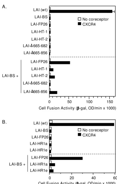
The results of complementation experiments with these mutants are shown in Fig. 5A. Each of the fusion-defective GPI-anchored Envs was capable of complementing LAI-BS. Perhaps surprisingly, even the truncated non-anchored LAI-Δ665-856 Env was able to complement the full-length LAI-BS665-856 mutant, with comparable or higher efficiency compared to the GPI-anchored con-structs.
Complementation with gp41 subunits containing heptad repeat mutations
Two previously characterized point mutations within the N-terminal heptad repeat region (HR1) of the gp41 ecto-domain (Fig. 1) were analyzed for complementation, i.e. substitution of Pro for Ile at residue 573 at the "a" posi-tion within the HR1 heptad repeat motif (LAI-HR1a) and Arg for Val at residue 570 at the "e" position within the HR1 heptad repeat (LAI-HR1e). The LAI-HR1a mutation has been shown previously to disrupt self-association of HR1 to form the trimeric coiled-coil pre-hairpin interme-diate structure [46] and the LAI-HR1e mutation is sus-pected to block association of HR2 with the HR1 trimer to form the 6-helix coiled-coil hairpin structure [47]. Inter-estingly, each of these mutants displayed some comple-mentation activity with LAI-BS Fig. 5B). However the efficiency was relatively low, and no significant comple-mentation by these mutants was observed with Envs defective in CXCR4 interaction (LAI-V3 and SF162 wt, data not shown).
Complementation requires coexpression of Env mutants within the same cell
The observed complementation activities involved coex-pression of two distinct nonfusogenic Env variants within the same cell. Although in our previous study [25] we ver-ified the formation of mixed trimers between the two var-iants, we could not rule out the possibility that the complementation activity was due to cooperative interac-tions between nonfusogenic homo-trimers of each vari-ant. While this seemed an unlikely explanation, it has been reported that cell fusion can occur when CD4 and coreceptor are expressed on separate target cells [48]. To determine whether this might also be true for Env trimers Complementation with Envs containing point mutations in
[image:6.612.62.291.84.487.2]the gp41 fusion peptide
Figure 4
Complementation with Envs containing point muta-tions in the gp41 fusion peptide. The vaccinia system was employed to assay cell fusion between effector cells express-ing Envs and target cells expressexpress-ing CD4 either with (filled bars) or without (open bars) CXCR4. Within each section, effector cells expressed the indicated Envs either individually (top section) or in combination with the indicated nonfunc-tional Envs (bottom section): A. LAV-BS, B. LAV-B3, C. SF162 (wt).
A.
0 20 40 60
LAI-BS +
LAI (wt)
LAI-BS
LAI-FP2
LAI-FP9
LAI-FP26
LAI-FP9
LAI-FP26
No coreceptor CXCR4
LAI-FP2
LAI-V3 +
0 25 50 75
LAI-FP2 LAI (wt)
LAI-BS
LAI-FP9
LAI-FP26
LAI-FP2
LAI-FP9
LAI-FP26 B.
Cell Fusion Activity ( -gal, OD/min x 1000) SF162 (wt) +
0 20 40 60
LAI-FP2 LAI (wt) LAI-BS
LAI-FP9 LAI-FP26
LAI-FP2
LAI-FP9
Page 7 of 11
expressed on separate cells, we expressed BS and LAI-FP26 in separate effector cells and asked whether mixing the two cell populations with target cells could result in fusion. As shown in Fig. 6, fusion was detected only when the constructs were cotransfected into the same effector cell population, indicating that functional complementa-tion requires both Env variants to be expressed within the same cell.
Complementation activity is dependent on the nature of the functional assay
The cell fusion assay used in the above experiments employed vaccinia virus expression technology to
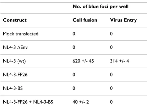
pro-duce Env and CD4 and to provide the reporter gene acti-vation system for readout. We wished to assess whether complementation could also be detected in a more bio-logically relevant situation, i.e. under conditions of HIV-1 proviral expression, using a target cell reporter system more commonly used to quantitate this process. Further-more, we asked whether complementation can be detected not only by measuring Env-mediated cell fusion, but also virion entry. To address these questions, we expressed Env from molecular variants of an HIV-1 infec-tious molecular clone and analyzed both cell fusion and virus entry using as targets the well studied HeLa-CD4/ LTR-β-gal (MAGI) indicator cell line [43]. Proviruses encoding the LAI-FP26 and the LAI-BS mutant Envs were cotransfected into BS-C-1 producer cells. This Env variant pair was selected because it consistently yielded the high-est levels of fusion complementation in the vaccinia-based system. Two alternative assays were then compared. First, the BSC-1 producer cells were used as effectors and mixed with MAGI target cells in a cell fusion assay; sec-ond, filtered supernatants from the BSC-1 producer cells containing cell-free HIV-1 virions were used to infect MAGI cells in a parallel virus entry assay. In both cases, complementation was assessed by counting the number of blue foci observed upon in situ staining with X-gal. As shown in Table 1, functional complementation was detected in the cell fusion assay: cells transfected individ-ually with either NL4-3-FP26 or NL4-3-BS infectious molecular clones were fusion-incompetent, whereas cells cotransfected with both gave significant fusion activity. By
[image:7.612.61.289.86.442.2]Complementation with Envs containing alterations in the membrane-spanning domain, the TRR, and HR1
Figure 5
Complementation with Envs containing alterations in the membrane-spanning domain, the TRR, and HR1. The vaccinia system was employed to assay cell fusion between effector cells expressing Envs and target cells expressing CD4 either with (filled bars) or without (open bars) CXCR4. Within each section, effector cells expressed the indicated Envs either individually (top section) or in com-bination with the nonfunctional LAV-BS (lower section).
LAI-FP26
LAI-FP26
LAI-HR1a LAI-HR1e LAI (wt)
LAI-BS
LAI-HR1a
LAI-HR1e
LAI-BS +
Cell Fusion Activity ( -gal, OD/min x 1000)
0 20 40 60
No coreceptor CXCR4 B.
A.
Cell Fusion Activity ( -gal, OD/min x 1000) No coreceptor CXCR4
0 50 100 150
LAI-BS +
LAI-FP26 LAI (wt)
LAI-BS
LAI-FP26
LAI-HT-1
LAI-HT-2
665-682
LAI-LAI-HT-1
LAI-HT-2
665-682
LAI-665-856
LAI-665-856
LAI-Complementation requires coexpression of Env mutants within the same cell
Figure 6
Complementation requires coexpression of Env mutants within the same cell. The vaccinia system was employed to assay cell fusion between effector cells express-ing Envs and target cells expressexpress-ing CD4 either with (filled bars) or without (open bars) CXCR4. Top section: Effecter cells expressing the indicated Envs individually. Bottom sec-tion: Effector cells either cotransfected with the indicated Envs, or infected separated and mixed 1:1.
Unc
LAI (wt)
LAI-BS + LAI-FP26: cotransfection LAI-FP26
LAI-BS + LAI-FP26: separate transfections LAI-BS
Cell Fusion Activity ( -gal, OD/min x 1000)
0 50 100
[image:7.612.319.546.450.609.2]contrast, no complementation was observed in the virus entry assay using the viruses produced from these same cells (Table 1).
Discussion
The present results extend our earlier findings [25] by demonstrating that the capacity for functional subunit complementation is a general feature of the HIV-1 Env trimer. We interpret our results to reflect cooperative sub-unit interactions within mixed heterotrimers, consistent with our previous verification that mixed heterotrimers do indeed form upon coexpression of different HIV-1 Env variants, as well as our previous reference to other exam-ples of mixed trimer formation with glycoproteins from different enveloped viruses [25]; however we cannot for-mally exclude the possibility that the observed comple-mentation activities reflect complex interactions amongst homo-trimers with different defects expressed on the same membrane. In the present work, complementation was observed upon coexpression of Envs from primary as well as laboratory-adapted HIV-1 strains of different gen-otypes, and with a wide diversity of defects within both gp120 and gp41 (Figs. 2, 3, 4, 5). Thus fusion does not require every gp120 subunit in the trimer to be competent for CD4 or coreceptor binding, nor every gp41 subunit to contain a functional fusion peptide, a normal membrane anchoring region, a native TRR, or a functional HR1 region. We also demonstrate that complementation requires coexpression of the Env variants in the same cell (Fig. 6), and provide further evidence (by virtue of com-plementation between LAI-BS and LAI-V3, Fig. 3) against the interpretation that the observed complementation requires reassortment of gp120 and gp41 subunits to form homo-trimers composed of completely functional gp120/ gp41 protomers.
We interpret complementation as a reflection of coopera-tive cross-talk between defeccoopera-tive protomers within a
mixed trimer, whereby the wild type determinants on one protomer transmit structural changes to activate wild type determinants on an adjacent protomer(s), thereby over-coming defects that are otherwise inactivating in the con-text of homo-trimers. For example when a CD4 BS mutant is coexpressed with coreceptor inactive variant, we pro-pose that CD4 binding to the subunit(s) with functional BS promotes the conformational changes required for coreceptor binding, which are then transmitted to an adja-cent gp120 subunit(s) that can then undergo the essential coreceptor interaction, thereby triggering activation of the wild type gp41 subunits. Particularly striking is the wide range of gp41 mutants capable of complementation when coexpressed with a nonfunctional gp120 variant. Nearly all tested displayed some level of activity, the only excep-tion being LAI-Δ665-682, a normally anchored form con-taining a deletion of the TRR. The mere absence of the TRR cannot be the simple explanation, since several constructs lacking this region did show complementation activity (HT-1, LAI-Δ665-856). Perhaps misalignment of the
LAI-Δ665-682 mutant with wild type gp41 is not tolerated for Env function.
[image:8.612.54.295.572.747.2]Several Env defects have been described in the literature as "dominant negative", based on their potent functional suppressive activities when coexpressed with wild type Env. Mutants in the gp120/gp41 cleavage site, which alone are completely inactive for fusion and infectivity, are reported to have strong dominant negative activities when coexpressed with wild type Env [49,50]. In our pre-vious studies complementation was not detected with mixed trimers in which Unc was one of the defective vari-ants [25]; thus dominant suppression appears to be the major functional activity for uncleaved Env. However we show here that this is not the case for all reported domi-nant negative mutations. Despite the strong inhibitory activities in fusion and infectivity assays reported for the FP2 mutation when coexpressed with wild type [45], we found that the same mutant is still able to complement fusion activity (albeit at relatively low levels) in mixed trimers with various non-functional Envs (Fig. 4). In fact the relative complementation efficiencies of the FP mutants (FP26 > FP9 > FP2) (Fig. 4) inversely correlated with their previously described inhibitory effects when coexpressed with wild type Env (FP2 > FP9 > FP26) [45]. Another example involves the TM domain, for which it has been reported that substitution of this HIV-1 Env region with its counterpart from the influenza virus hemagglutinin glycoprotein results in potent dominant inhibition [51]. The present studies indicate that this sup-pressive effect is not due simply to the absence of the native functional HIV-1 TM region, since we observed complementation with fusion-defective constructs in which the normal membrane-spanning domain was replaced by a GPI anchor, or was completely deleted (Fig. 5A). Thus the complementation analyses help distinguish
Table 1: Complementation analysis of Envs expressed from infectious HIV-1 molecular clones in assays of cell fusion and virus entry.
No. of blue foci per well
Construct Cell fusion Virus Entry
Mock transfected 0 0
NL4-3 ΔEnv 0 0
NL4-3 (wt) 620 +/- 45 314 +/- 4
NL4-3-FP26 0 0
NL4-3-BS 0 0
Page 9 of 11
between a fusion-defective Env variant that exerts a strictly dominant suppressive activity, vs. others that, though defective, can permit a low level of functionality as revealed by the ability of their active determinants to com-plement when coexpressed with a variant defective in another function.
Complementation in cell fusion assays was observed not only with the previously described robust vaccinia-based expression and reporter system (Figs. 2, 3, 4, 5, 6), but also with Env expression from an HIV-1 infectious molecular clone using the MAGI reporter cell line as targets (Table 1). However there were marked variations in complemen-tation efficiencies depending on the assay system employed. Thus, for complementation between LAI-BS and LAI-FP26, the activities in the vaccinia cell fusion sys-tem ranged from about ~30–50% relative to wild type WT, consistent with our previous findings [25]; in contrast, the relative activities were much lower with the infectious HIV/MAGI system, i.e. only ~6% in the cell fusion assay and below detection in the virus entry assay (Table 1). We believe these differences mainly reflect variations in the robustness of the functional Env-receptor interactions and the associated reporter gene activation readouts in the dif-ferent assays, rather than fundamental mechanistic dis-tinctions. Numerous variables can influence the efficiency of Env-receptor interactions leading to fusion/entry, including surface densities of the participating molecules, gp120 affinities for CD4 and coreceptor, varying receptor conformations and molecular associations, the biochem-ical environments of both effector and target membranes (lipid composition, facilitating or interfering accessory factors), etc. [52,53]. Similarly for the reporter gene read-outs, multiple parameters can influence detectability (sig-nal sensitivity, sig(sig-nal/background ratios, etc.), and different factors might be limiting for the measured read-out in alternate assay systems. Thus, while a particular assay might be quantitative in terms of yielding numeri-cally reproducible values, such data are not necessarily proportional to the inherent functional activities of the particular Env-receptor interactions involved. Therefore, some assays might reveal weak activities not detected by others, but might overestimate their relative efficiencies. Further complicating the quantitative interpretation is the fact that unlike wild type Env, for which all trimers are potentially active, the complementation activities result only from mixed trimers which presumably represent a subset (theoretically 75%) of the total; moreover upon cotransfection of two nonfunctional Env variants (A & B), the relative functionalities of the two possible mixed trim-ers (AAB and ABB) might be very different. Thus the reported absence of Env complementation in assays of both reporter virus entry and cell fusion [54] could reflect the absence of functional interactions between the
partic-ular mutant Env constructs examined (different from those tested in this report), or limited sensitivities in the assays used. Another point worth noting is that our approach to studying cooperativity within the Env trimer involved complementation analyses between Env mutants that were inactive when expressed alone; thus functional activity was detected despite the presence of a fusion-impairing determinant in every protomer of the mixed trimer. It seems reasonable to propose that the con-tributions of subunit cooperativity to Env function might be greater with wild type native Env molecules, in which all subunits are fully functional.
Given the experimental complexities, assessing the bio-logical significance of subunit cooperativity for HIV entry is a challenging problem. It is well known that there are functional constraints on subunits within the trimer com-pared to their monomeric counterparts. A particularly striking example is the interaction of soluble CD4 (sCD4) with gp120; the comparably high affinity of sCD4 for sol-uble monomeric gp120 from primary and T cell line-adapted HIV-1 isolates stands in marked contrast to the relatively weak binding and neutralization activities of sCD4 for native trimeric Env on the former compared to the latter [61]. Cooperative subunit interactions, whereby binding of gp120 to CD4 on one protomer in the trimer initiates fusion-related conformational changes in the other protomers, might thus enhance Env fusogenic activ-ity, particularly toward target cells containing low densi-ties of CD4 and coreceptor. Another consideration involves the extensively studied phenomenon of epitope masking within the HIV-1 Env trimer [62]. For example, some highly conserved epitopes are freely accessible on monomeric gp120 but are masked in the trimer prior to CD4 binding; cooperative subunit interactions may facil-itate exposure of such epitopes on subunits within the trimer that have not yet engaged CD4. With questions such as these in mind, the combination of detailed func-tional and structural studies will potentially delineate the molecular basis for subunit cooperativity within the native HIV-1 Env trimer, and help define its biological sig-nificance.
Conclusion
The data presented herein demonstrate that every subunit within the Env trimer need not be competent for all criti-cal activities. Cooperatvie cross-talk occurs between subu-nits, thereby enabling adjacent protomers to complement different functional defects. The diversity of defects that can be complemented illustrates the general nature of cooperative subunit interactions within the HIV-1 trimer. Cooperativity may have important implications for Env function and sensitivity to neutralization.
Abbreviations
HIV: human immunodeficiency virus; Env: envelope glyc-oprotein; V3: third variable loop of gp120; FP: fusion pep-tide; HR: heptad repeat; BS: CD4 binding site; TRR: tryptophan-rich region: TM: transmembrane.
Competing interests
The authors declare that they have no competing interests.
Authors' contributions
KS designed and performed experiments, and contributed to data analysis and interpretation, and to writing of the manuscript. EB contributed to data analysis and interpre-tation, and to the writing of the manuscript.
Acknowledgements
We thank C. Broder for supplying the vHC-1 vaccinia virus recombinant, and Paul Kennedy for outstanding technical assistance. K. S. was supported in part by a National Research Council-National Institutes of Health research associateship. This research was funded in part by the Intramural Program of the NIH, NIAID, including the NIH Intramural AIDS Targeted Antiviral Program.
References
1. Byland R, Marsh M: Trafficking of viral membrane proteins.
Curr Top Microbiol Immun 2004, 285:219-54.
2. Roux KH, Taylor KA: AIDS virus envelope spike structure. Curr Opin Struct Biol 2007, 17:244-52.
3. Wyatt R, Sodroski J: The HIV-1 envelope glycoproteins: fusogens, antigens, and immunogens. Science 1998, 280:1884-8. 4. Eckert DM, Kim PS: Mechanisms of viral membrane fusion and
its inhibition. AnnuRevBiochem 2001, 70:777-810.
5. Gallo SA, Finnegan CM, Viard M, Raviv Y, Dimitrov A, Rawat SS, et al.: The HIV Env-mediated fusion reaction. Biochim Biophys Acta
2003, 1614:36-50.
6. Alkhatib G, Berger EA: HIV coreceptors: from discovery and designation to new paradigms and promise. Eur J Med Res
2007, 12:375-84.
7. White JM, Delos SE, Bretcher M, Schornberg K: Structure and mechanisms of viral membrane fusion proteins: multiple variations on a common theme. Crit Rev Biochem Mol Biol 2008, 43:189-219.
8. Melikyan GB: Common principles and intermediates of viral protein-mediated fusion: the HIV-1 paradigm. Retrovirology.
2008, 5:111.
9. Kwong PD, Wyatt R, Robinson J, Sweet RW, Sodroski J, Hendrickson WA: Structure of an HIV gp120 envelope glycoprotein in complex with the CD4 receptor and a neutralizing human antibody. Nature 1998, 393:648-59.
10. Chen B, Vogan EM, Gong HY, Skehel JJ, Wiley DC, Harrison SC: Structure of an unliganded simian immunodeficiency virus gp120 core. Nature 2005, 433:834-41.
11. Rizzuto CD, Wyatt R, Hernandez-Ramos N, Sun Y, Kwong PD, Hen-drickson WA, et al.: A conserved HIV gp120 glycoprotein struc-ture involved in chemokine receptor binding. Science 1998, 280(5371):1949-1953.
12. Zolla-Pazner S: Identifying epitopes of HIV-1 that induce pro-tective antibodies. Nature Rev Immunol 2004, 4:199-210. 13. Hartley O, Klasse PJ, Sattentau QJ, Moore JP: V3: HIV's
switch-hit-ter. AIDS Res Hum Retrovir 2005, 21:171-89.
14. Weiss CD: HIV-1 gp41: mediator of fusion and target for inhi-bition. AIDS Rev 2003, 5:214-21.
15. Montero M, van Houten NE, Wang X, Scott JK: The membrane-proximal external region of the human immunodeficiency virus type 1 envelope: dominant site of antibody neutraliza-tion and target for vaccine design. Microbiol Mol Biol Rev 2008, 72:54-84.
16. Chan DC, Fass D, Berger JM, Kim PS: Core structure of gp41 from the HIV envelope glycoprotein. Cell 1997, 89:263-73. 17. Weissenhorn W, Dessen A, Harrison SC, Skehel JJ, Wiley DC:
Atomic structure of the ectodomain from HIV-1 gp41.
Nature 1997, 387:426-30.
18. Tan KM, Liu JH, Wang JH, Shen S, Lu M: Atomic structure of a thermostable subdomain of HIV-1 gp41. Proc Natl Acad Sci USA
1997, 94:12303-8.
19. Moore JP, Doms RW: The entry of entry inhibitors: a fusion of science and medicine. Proc Natl Acad Sci USA 2003, 100:10598-602.
20. Pohlmann S, Reeves JD: Cellular entry of HIV: evaluation of therapeutic targets. Curr Pharm Des 2006, 12:1963-73.
21. Sterjovski J, Churchill MJ, Wesselingh SL, Gorry PR: HIV-1 entry inhibitors: classes, applications and factors affecting potency.
Curr HIV Res 2006, 4:387-400.
22. Este JA, Telenti A: HIV entry inhibitors. Lancet 2007, 370:81-8. 23. Matthews T, Salgo M, Greenberg M, Chung J, DeMasi R, Bolognesi D:
Enfuvirtide: the first therapy to inhibit the entry of HIV-1 into host CD4 lymphocytes. Nat Rev Drug Discov 2004, 3:215-25. 24. Kuritzkes D, Kar S, Kirkpatrick P: Fresh from the pipeline –
Page 11 of 11
25. Salzwedel K, Berger EA: Cooperative subunit interactions within the oligomeric envelope glycoprotein of HIV-1: func-tional complementation of specific defects in gp120 and gp41. Proc Natl Acad Sci USA 2000, 97:12794-9.
26. Chakrabarti S, Sisler JR, Moss B: Compact, synthetic, vaccinia virus early/late promoter for protein expression. BioTech-niques 1997, 23:1094-7.
27. Broder CC, Berger EA: Fusogenic selectivity of the envelope glycoprotein is a major determinant of human immunodefi-ciency virus type 1 tropism for CD4+ T-cell lines vs. primary macrophages. Proc Natl Acad Sci USA 1995, 92:9004-8.
28. Salzwedel K, Smith ED, Dey B, Berger EA: Sequential CD4-core-ceptor interactions in human immunodeficiency virus type 1 Env function: soluble CD4 activates Env for coreceptor-dependent fusion and reveals blocking activities of antibod-ies against cryptic conserved epitopes on gp120. J Virol 2000, 74:326-33.
29. Wain-Hobson S, Vartanian JP, Henry M, Chenciner N, Cheynier R, Delassus S, et al.: LAV revisited – origins of the early HIV-1 iso-lates from Institut-Pasteur. Science 1991, 252:961-5.
30. Freed EO, Myers DJ, Risser R: Characterization of the fusion domain of the human immunodeficiency virus type 1 enve-lope glycoprotein gp41. Proc Natl Acad Sci USA 1990, 87:4650-4. 31. Dubay JW, Roberts SJ, Hahn BH, Hunter E: Truncation of the
human immunodeficiency virus type-1 transmembrane glyc-oprotein cytoplasmic domain blocks virus infectivity. J Virol
1992, 66:6616-25.
32. Salzwedel K, Johnston PB, Roberts SJ, Dubay JW, Hunter E: Expres-sion and characterization of glycophospholipid-anchored human immunodeficiency virus type-1 envelope glycopro-teins. J Virol 1993, 67:5279-88.
33. Salzwedel K, West JT, Hunter E: A conserved tryptophan-rich motif in the membrane-proximal region of the human immunodeficiency virus type 1 gp41 ectodomain is impor-tant for env-mediated fusion and virus infectivity. J Virol 1999, 73:2469-80.
34. Munoz-Barroso I, Salzwedel K, Hunter E, Blumenthal R: Role of the membrane-proximal domain in the initial stages of human immunodeficiency virus type 1 envelope glycoprotein-medi-ated membrane fusion. J Virol 1999, 73:6089-92.
35. Adachi A, Gendelman HE, Koenig S, Folks T, Willey R, Rabson A, et al.: Production of acquired immunodeficiency syndrome-associated retrovirus in human and nonhuman cells trans-fected with an infectious molecular clone. J Virol 1986, 59:284-91.
36. Nussbaum O, Broder CC, Berger EA: Fusogenic mechanisms of enveloped-virus glycoproteins analyzed by a novel recom-binant vaccinia virus-based assay quantitating cell fusion-dependent reporter gene activation. J Virol 1994, 68:5411-22. 37. Dey B, Berger EA: Vaccinia-based reporter gene cell-fusion
assays to quantitate functional interactions of HIV envelope glycoprotein with receptors. In Curr Prot Immunol, Unit 12Volume 10. Issue Suppl 54 Edited by: Coligan JE, Kruisbeek AM, Margulies DH, Shevach EM, Strober W. New York: John Wiley & Sons, Inc; 2003:12.0.1-.0.20.
38. Alkhatib G, Broder CC, Berger EA: Cell type-specific fusion cofactors determine human immunodeficiency virus type 1 tropism for T-cell lines versus primary macrophages. J Virol
1996, 70:5487-94.
39. Broder CC, Dimitrov DS, Blumenthal R, Berger EA: The block to HIV-1 envelope glycoprotein-mediated membrane fusion in animal cells expressing human CD4 can be overcome by a human cell component(s). Virology 1993, 193:483-91.
40. Xiao XD, Wu L, Stantchev TS, Feng Y-R, Ugolini S, Chen H, et al.: Constitutive cell surface association between CD4 and CCR5. Proc Natl Acad Sci USA 1999, 96:7496-501.
41. Feng Y, Broder CC, Kennedy PE, Berger EA: HIV-1 entry cofactor: functional cDNA cloning of a seven-transmembrane, G pro-tein-coupled receptor. Science 1996, 272:872-7.
42. Alexander WA, Moss B, Fuerst TR: Regulated expression of for-eign genes in vaccinia virus under the control of bacteri-ophage T7 RNA polymerase and the Escherichia coli lac repressor. J Virol 1992, 66:2934-42.
43. Kimpton J, Emerman M: Detection of replication-competent and pseudotyped human immunodeficiency virus with a
sen-sitive cell line on the basis of activation of an integrated beta-galactosidase gene. J Virol 1992, 66:2232-9.
44. Freed EO, Myers DJ, Risser R: Identification of the principal neu-tralizing determinant of human immunodeficiency virus type 1 as a fusion domain. J Virol 1991, 65:190-4.
45. Freed EO, Delwart EL, Buchschacher J, GL , Panganiban AT: A muta-tion in the human immunodeficiency virus yype-1 trans-membrane glycoprotein-gp41 dominantly interferes with fusion and infectivity. Proc Natl Acad Sci USA 1992, 89:70-4. 46. Dubay JW, Roberts SJ, Brody B, Hunter E: Mutations in the
leu-cine zipper of the human immunodeficiency virus type-1 transmembrane glycoprotein affect fusion and infectivity. J Virol 1992, 66:4748-56.
47. Weng YK, Weiss CD: Mutational analysis of residues in the coiled-coil domain of human immunodeficiency virus type 1 transmembrane protein gp41. J Virol 1998, 72:9676-82. 48. Speck RF, Esser U, Penn ML, Eckstein DA, Pulliam L, Chan SY, et al.:
A trans-receptor mechanism for infection of CD4-negative cells by human immunodeficiency virus type 1. Curr Biol 1999, 9:547-50.
49. Iwatani Y, Kawano K, Ueno T, Tanaka M, Ishimoto A, Ito M, et al.: Analysis of dominant-negative effects of mutant env proteins of human immunodeficiency virus type 1. Virology 2001, 286:45-53.
50. Herrera C, Klasse PJ, Kibler CW, Michael E, Moore JP, Beddows S: Dominant-negative effect of hetero-oligomerization on the function of the human immunodeficiency virus type 1 enve-lope glycoprotein complex. Virology 2006, 351:121-32. 51. Welman M, Lemay G, Cohen EA: Role of envelope processing
and gp41 membrane spanning domain in the formation of human immunodeficiency virus type I (HIV-1) fusion-compe-tent envelope glycoprotein complex. Virus Res 2007, 124:103-12.
52. Doms RW: Beyond receptor expression: the influence of receptor conformation, density, and affinity in HIV-1 infec-tion. Virology 2000, 276:229-37.
53. Freed EO: HIV-1 and the host cell: an intimate association.
Trends In Microbiology 2004, 12:170-7.
54. Yang XZ, Kurteva S, Ren XP, Lee S, Sodroski J: Subunit stoichiom-etry of human immunodeficiency virus type 1 envelope glyc-oprotein trimers during virus entry into host cells. J Virol 2006, 80:4388-95.
55. Zhu P, Chertova E, Bess J, Lifson JD, Arthur LO, Liu J, et al.: Electron tomography analysis of envelope glycoprotein trimers on HIV and simian immunodeficiency virus virions. Proc Natl Acad Sci USA 2003, 100:15812-7.
56. Zhu P, Liu J, Bess J, Chertova E, Lifson JD, Grise H, et al.: Distribu-tion and three-dimensional structure of AIDS virus envelope spikes. Nature 2006, 441:847-52.
57. Yang XZ, Kurteva S, Ren XP, Lee S, Sodroski J: Stoichiometry of envelope glycoprotein trimers in the entry of human immu-nodeficiency virus type 1. J Virol 2005, 79:12132-47.
58. Klasse PJ: Modeling how many envelope glycoprotein trimers per virion participate in human immunodeficiency virus infectivity and its neutralization by antibody. Virology 2007, 369:245-62.
59. Magnus C, Rusert P, Bonhoeffer S, Trkola A, Regoes RR: Estimating the stoichiometry of human immunodeficiency virus entry. J Virol 2009, 83:1523-31.
60. Miyauchi K, Kim Y, Latinovic O, Morozov V, Melikyan GB: HIV enters cells via endocytosis and dynamin-dependent fusion with endosomes. Cell 2009, 137:433-44.
61. Sattentau QJ, Moore JP: The role of CD4 in HIV binding and entry. Philos Trans R Soc Lond [Biol] 1993, 342:59-66.