JOURNAL OFVIROLOGY, June 2006, p. 5958–5967 Vol. 80, No. 12 0022-538X/06/$08.00⫹0 doi:10.1128/JVI.00181-06
The NS2 Protein of Human Respiratory Syncytial Virus Suppresses the
Cytotoxic T-Cell Response as a Consequence of Suppressing
the Type I Interferon Response
Alexander Kotelkin,
1Igor M. Belyakov,
2Lijuan Yang,
1Jay A. Berzofsky,
2Peter L. Collins,
1and Alexander Bukreyev
1*
Laboratory of Infectious Diseases, National Institute of Allergy and Infectious Diseases,1and Molecular Immunogenetics and
Vaccine Research Section, Vaccine Branch, National Cancer Institute,2National Institutes of Health, Bethesda, Maryland 20892-8007
Received 26 January 2006/Accepted 31 March 2006
The NS1 and NS2 proteins of human respiratory syncytial virus (HRSV) have been shown to antagonize the type I interferon (IFN) response, an effect subject to host range constraints. We have now found that the HRSV NS2 protein strongly controls IFN induction in mouse cells in vitro, validating the use of the mouse model to study the consequences of these gene deletions on host immunity. We evaluated the effects of deleting the NS1 and/or NS2 gene on the induction of HRSV-specific pulmonary cytotoxic T lymphocytes (CTL) in BALB/c and 129S6 mice in response to intranasal infection with HRSV lacking the NS1 and/or NS2 gene and subsequent challenge with wild-type (wt) HRSV. In mice infected with HRSV lacking the NS2 gene (⌬NS2) or lacking the NS2 gene in combination with the NS1 gene (⌬NS1/2 HRSV), the magnitude of the pulmonary CTL response was substantially elevated compared to that of mice infected with wt HRSV or the ⌬NS1 mutant, whether measured by binding of CD8ⴙcells to an HRSV-specific major histocompatibility complex class I tetramer, by measurement of CD8ⴙcells secreting gamma interferon (IFN-␥) in response to specific in vitro stimulation, or by a standard chromium release cell-killing assay. In contrast, in STAT1 knockout mice, which lack responsiveness to type I IFN, the level of IFN-␥-secreting CD8ⴙcells was not significantly different for HRSV lacking the NS2 gene, suggesting that the increase in CTL observed in IFN-responsive mice is type I IFN dependent. Thus, the NS2 protein of HRSV suppresses the CTL component of the adaptive immune response, and this appears to be a consequence of its suppression of type I IFN.
The interferon (IFN) system is an important component of host defense against viral pathogens. Type I IFNs include at least 14 species of alpha interferon (IFN-␣) and a single spe-cies of IFN-, and type II IFN is represented by a single species of IFN-␥. More recently, type III IFNs have been described, represented by IFN-1, -2, and -3 (also known as interleukin 29 [IL-29], 28A, and 28B, respectively), which ap-pear to have a number of similarities with type I IFNs (29). Type I and III IFNs are produced in response to viral infec-tions or other stimuli by many types of cells, with type I IFN being produced most abundantly by plasmacytoid dendritic cells (4), whereas type II IFN is mostly secreted by activated T cells and NK cells. Type I IFNs bind to a specific cell surface receptor that activates JAK-STAT signal transduction, result-ing in transcriptional activation of a broad panel of cellular genes. Some of these activated genes encode proteins such as Mx, PKR, and OAS that inhibit viral replication by various mechanisms, such as inhibition of intracellular trafficking of viral nucleocapsids, blocking of translation of viral proteins, and enzymatic degradation of viral RNA (reviewed in refer-ences 15 and 37). Type I IFNs are also involved in regulation of adaptive immune responses, including CD8⫹ cytotoxic T lymphocytes (CTL) (reviewed in reference 45).
Viruses exhibit a number of mechanisms that interfere with host IFN response. Some, like vesicular stomatitis virus,
influ-enza A virus, poliovirus, and bunyamwera virus, reduce the synthesis of IFN by interfering globally with host cell gene expression at one or more steps in the transcription, process-ing, transport, and translation of cellular mRNAs (26, 44). Other viruses, including numerous paramyxoviruses, filovi-ruses, influenza A virus, rabies virus, and rotavirus, specifically interfere with IFN gene induction, most commonly by inter-fering at various steps in the activation, stability, and nuclear translocation of IFN regulatory factor 3 (IRF-3). In many cases, these viruses also interfere with JAK-STAT signaling, most commonly by targeting STAT1 or STAT2 for degradation or by interfering with their activation. As another strategy, the NS3/4A protease of hepatitis C virus interferes with the IFN response by direct proteolyic attack on components of the signaling pathways (24). Human respiratory syncytial virus (HRSV) and bovine RSV (BRSV) encode two proteins, NS1 and NS2, that can act both independently and cooperatively to interfere with the activation of IRF-3 and to block sig-naling through the JAK/STAT pathway (25, 33, 36, 40). The NS1 and NS2 proteins appear to be determinants of the host range difference between BRSV and HRSV in vitro (7). In addition, there are host-specific differences in their activi-ties: whereas BRSV NS2 plays the major role in blocking IFN induction in the bovine system, HRSV NS1 plays the greater role in humans (40).
While many cases of virally mediated suppression of the type I IFN response have been described, in most cases the possible effects on the adaptive immune response to the viral infection have not been studied or are unclear. In the case of BRSV, a * Corresponding author. Mailing address: Building 50, Room 6505,
NIAID, NIH, 50 South Dr., MSC 8007, Bethesda, MD 20892-8007. Phone: (301) 594-1854. Fax: (301) 496-8312. E-mail: [email protected].
5958
on November 8, 2019 by guest
http://jvi.asm.org/
Downloaded from
on November 8, 2019 by guest
http://jvi.asm.org/
Downloaded from
on November 8, 2019 by guest
http://jvi.asm.org/
study with engineered mutants lacking the nonstructural (NS) proteins suggested that deletion of NS2 was associated with higher virus-specific antibody titers, increased stimulation of activated CD4 cells, and increased protection against challenge in calves (46). However, similar studies with comparable HRSV mutants in chimpanzees yielded no evidence of in-creased immunogenicity or protection (43, 49). In the present study, we focused on evaluating the magnitude of the CTL response in inbred mice to HRSV mutants lacking either or both NS proteins. We demonstrate that, in the murine system, the NS2 protein of HRSV is a suppressor of CTL response and provide evidence that this effect is a consequence of suppres-sion of the type I IFN response.
MATERIALS AND METHODS
Mice, viruses, and cells.Seven- to 12-week-old BALB/c mice (Charles River Laboratories, Wilmington, MA) were used in all experiments except for those shown in Fig. 7 through 9, which employed 7- to 11-week-old STAT1 gene knockout (KO) mice on an inbred 129S6/SvEv background or the control 129S6 mice (Taconic, Germantown, NY). Construction of the recombinant wild-type (wt) HRSV and the viruses lacking the NS1 and/or NS2 gene was described earlier (11, 39, 42, 43); apart from the presence or absence of the NS genes, these viruses have identical genome sequences. Construction of the virus lacking the M2-2 protein, which was used as an attenuated HRSV control, was previously reported (6). The viruses were propagated and titrated in HEp-2 and/or Vero cell monolayers (American Type Culture Collection, Manassas, VA); for
quan-titation of IFN-␣following the infection, NIH 3T3 mouse fibroblast cells were
used (American Type Culture Collection).
Virus replication in mice.Mice were infected intranasally under
methoxyflu-rane anesthesia with 106
PFU of the indicated viruses in a 100-l inoculum. On
the indicated days after infection, animals were sacrificed with carbon dioxide, the nasal turbinates and lung tissues were isolated and homogenized, and viruses were titrated by plaque immunostaining in HEp-2 or Vero cell monolayers (31).
Analysis of HRSV-specific CD8ⴙCTL.Kinetics of the virus-specific CTL response have been determined in previous studies (1). Mice were infected
intranasally with 104
or 106
PFU of the indicated viruses as noted in the figure legends. On the indicated days, animals were sacrificed and total pulmonary mononuclear cells (PMC) were isolated from mouse lungs as described previ-ously (8). For quantitation of cells bearing T-cell receptors specific for HRSV in BALB/c mice, PMC were stained with optimized amounts of
phycoerythrin-conjugated complexes of major histocompatibility complex (MHC) class I H-2Kd
tetramer bearing a peptide, SYIGSINNI, from the M2-1 protein (amino acids 82
to 90) that is the immunodominant CTL epitope in the H-2Kdbackground (22,
23) (provided by the NIAID Tetramer Facility, Yerkes Regional Primate Re-search Center, Atlanta, GA) and fluorescein isothiocyanate-conjugated rat
anti-mouse CD8␣monoclonal antibody, clone 53-6.7 (BD Biosciences).
For quantitation of pulmonary CTL (from BALB/c mice) that secrete IFN-␥
in response to HRSV-specific stimulation, PMC were washed twice with phos-phate-buffered saline containing 2% fetal bovine serum and resuspended in RPMI 1640 medium (Invitrogen, Carlsbad, CA) containing 10% fetal bovine
serum, 100 U/ml of penicillin, 100g/ml of streptomycin sulfate and 20 mM of
HEPES (Invitrogen) and incubated overnight with 1M of the SYIGSINNI
peptide in the presence of GolgiStop (BD Biosciences). For quantitation of pulmonary CTL (from 129S6 mice or STAT1 KO mice [129S6/SvEv
back-ground]) that secrete IFN-␥in response to HRSV-specific stimulation, the
stim-ulation involved overnight incubation with 1 M of NAITNAKEE peptide,
representing a recently described H-2Db-restricted CD8⫹CTL epitope located
in the HRSV M protein (amino acids 187 to 195) (35), in the presence of
brefeldin A (Sigma, St. Louis, MO) at 3g/ml, and hamster anti-mouse CD28
monoclonal antibody (clone 37.51, BD Biosciences) and rat anti-mouse CD49d
monoclonal antibody (clone R1-2, BD Biosciences) at 0.3g/ml each, as
previ-ously described (34). Following either protocol for stimulation, the PMC were washed twice, incubated with Fc Block (BD Biosciences) to block Fc receptors,
stained with the fluorescein isothiocyanate-conjugated anti-mouse CD8␣
mono-clonal antibody, fixed and permeabilized with Cytofix/Cytoperm (BD
Bio-sciences), and stained with allophycocyanin-conjugated rat anti-mouse IFN-␥
antibody, clone XMG1.2 (BD Biosciences). Flow cytometry analysis was per-formed using a FACSCalibur flow cytometer (BD Biosciences). A total of 30,000 cells were analyzed per sample.
Analysis of HRSV-specific cytolytic activity of PMC from BALB/c mice was
performed by a51
Cr release assay using target P815 mouse mastocytoma cells
(American Type Culture Collection) pulse-labeled with 1M of the peptide
SYIGSINNI, as previously described (5, 8). For measurement of HRSV-specific cytolytic activity of PMC from the STAT1 KO mice, target EL4 cells (American
Type Culture Collection) pulse-labeled with 1M of the peptide NAITNAKEE
were used.
Quantitative analysis of IFN-␣was performed by enzyme-linked
immunosor-bent assay (ELISA) using a mouse IFN-␣ELISA kit (PBL Biomedical
Labora-tories, Piscataway, NJ). As a positive control for stimulation of IFN-␣, cells were
transfected with 12g per 106cells of poly(I)-poly(C) (Amersham Biosciences,
Piscataway, NJ) using Lipofectamine 2000 reagent (Invitrogen).
Statistical analysis of data.Differences were evaluated by the Studentttest and
considered significant when thePvalue was⬍0.05. Data are shown as means⫾
standard errors (SE) of the means.
RESULTS
Level of replication in BALB/c mice of recombinant HRSVs lacking NS1 and/or NS2.BALB/c mice were infected intrana-sally with 106PFU of wt HRSV or the virus lacking the NS1
gene (⌬NS1) or⌬NS2 or⌬NS1/2, and the level of virus repli-cation was evaluated by sacrificing animals on days 4 and 5 after infection and measuring the titers of infectious virus in the nasal turbinates and lungs (Fig. 1). We found that the
⌬NS2 virus replicated to a titer similar to or slightly lower than that of wt HRSV and the⌬NS1 and⌬NS1/2 viruses replicated to titers that were 10- to 100-fold lower than wt HRSV. Thus, deletion of the NS1 gene, either alone or in combination with the NS2 gene, was associated with reduced virus replication, whereas deletion of NS2 had only a minimal effect.
Infection with HRSVs lacking NS2 is associated with an increased level of HRSV-specific pulmonary CD8ⴙ CTL.
BALB/c mice were infected intranasally with 104 PFU of wt
HRSV or the⌬NS1,⌬NS2, or⌬NS1/2 virus or the⌬M2-2 virus (an attenuated HRSV lacking the M2-2 gene as a control) and were challenged 28 days later with wt HRSV. The induction of HRSV-specific pulmonary CTL was measured on days 10 and 12 following the first infection and day 6 following the chal-lenge. Total PMC were harvested and analyzed for CTL spe-cific to the peptide SYIGSINNI, which represents amino acids 82 to 90 in the HRSV M2-1 protein and is an immunodomi-nant CTL epitope in the H-2Kdbackground. The CTL were
characterized by three independent methods: (i) staining with MHC class I tetramer loaded with the SYIGSINNI peptide followed by flow cytometry; (ii) specific stimulation in vitro with the SYIGSINNI peptide followed by intracellular IFN-␥ staining and flow cytometry; and (iii) detection of virus-specific cytotoxicity by a standard in vitro 51Cr release assay using
target cells loaded with the SYIGSINNI peptide. This was done because the methods can yield somewhat different re-sults. For example, a significant number of CTL specific to particular epitopes of HRSV or human immunodeficiency vi-rus detected by tetramer binding did not produce IFN-␥(10, 16). Human immunodeficiency virus-specific CD8⫹cells that produce antiviral cytokines can be impaired in cytolytic func-tion (2). Moreover, secrefunc-tion of IFN-␥ by CD8⫹ CTL and cytotoxicity are regulated independently (38).
We first compared the percentages of PMC that were pos-itive for CD8 and bound to the MHC class I tetramer bearing the SYIGSINNI peptide between mice infected with the wt and the mutant HRSVs. This showed that, in mice infected
on November 8, 2019 by guest
http://jvi.asm.org/
with wt RSV, 2.8% of the total PMC were tetramer⫹CD8⫹on day 10 postinfection, a value that diminished to 1.0% on day 12 (Fig. 2A and B). Surprisingly, in PMC from mice infected with the ⌬NS2 or ⌬NS1/2 virus, the number of tetramer⫹CD8⫹ cells was increased substantially compared to wt HRSV and the ⌬NS1 and⌬M2-2 viruses, indicating that the absence of NS2 was associated with a substantial increase in the CTL response (Fig. 2). To isolate the effect of deleting NS2, we compared the magnitude of CTL induction by the⌬NS2 virus to that of wt HRSV and compared that by the⌬NS1/2 virus to that of⌬NS1. These pair-wise comparisons were made because the two viruses in each comparison (⌬NS2 versus wt HRSV and ⌬NS1/2 versus ⌬NS1) differed only by the presence or absence of NS2. Also, importantly, the two viruses in each comparison replicated to approximately the same pulmonary titer, as seen in Fig. 1. We have found that the magnitude of the CTL response can be substantially affected by the pulmonary virus titer (unpublished data), a potential complication that is avoided by comparing viruses that replicate to similar titers. Thus, on day 10 postinfection, the number of tetramer⫹CD8⫹cells for the⌬NS2 virus was 123% greater than that of wt HRSV and that for the⌬NS1/2 virus was 333% greater than that of the
⌬NS1 virus. On day 12 postinfection, these differences were 64% and 178%, respectively. After the challenge with wt HRSV (day 6 postchallenge), the numbers of tetramer⫹CD8⫹ cells were not significantly different between the viruses (Fig. 2B). The dose of 104PFU of HRSV was used for characterization
of the CTL response in the BALB/c model because a higher
dose resulted in a massive response to the immunodominant M2 peptide that might have been at saturation, as also has been suggested in other studies (47, 48). Specifically, when the dose of 106PFU was used, tetramer⫹CD8⫹cells specific to the M2
peptide made up as much as 8.3% of total PMC on day 9 (9), and we were concerned that a saturated response might be less responsive to changes in the infecting virus. Both doses were used in subsequent experiments below, performed with a sec-ond strain of mice.
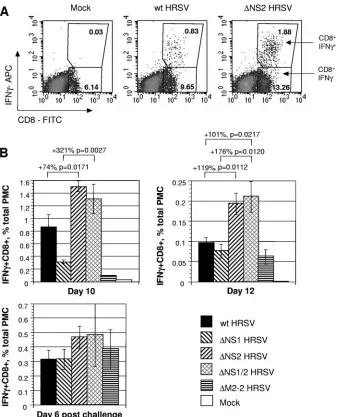
We then compared the percentage of PMC that were posi-tive for CD8 and which secreted IFN-␥⫹in response to stim-ulation in vitro with the SYIGSINNI peptide in mice infected with wt HRSV and the mutant viruses. For wt HRSV, the percentage of IFN-␥⫹CD8⫹ cells was 0.9% on day 10 and diminished to 0.1% on day 12 (Fig. 3A and B). These lower percentages than the values for tetramer⫹CD8⫹cells described above are in accordance with the previously published results (10) and indicate that the HRSV-specific IFN-␥⫹CD8⫹cells con-stitute a fraction of tetramer⫹CD8⫹cells. Similar to the results of the tetramer staining, a higher level of IFN-␥⫹CD8⫹CTL was detected for the⌬NS2 and⌬NS1/2 viruses than for their counterparts that lack the NS2 gene and replicate to the same titer, namely, wt HRSV and⌬NS1 HRSV, respectively (Fig. 3). No significant difference was observed after the challenge with wt HRSV (Fig. 3B).
We also analyzed the PMC for HRSV-specific cytolytic ac-tivity directly ex vivo, without antigen stimulation, measured on days 10 and 12 after the primary infection with the various FIG. 1. Replication in BALB/c mice of recombinant wt HRSV and the gene deletion viruses lacking NS1 and/or NS2. Groups of mice (five animals per virus per day) were inoculated intranasally with 106PFU of the indicated viruses and sacrificed on days 4 and 5. Titers of the viruses
in nasal turbinates and lungs were determined by plaque assay of the tissue homogenates and are shown as means⫾SE. The data shown are from a representative experiment from a total of three independent experiments. Significant differences and correspondingPvalues are indicated above the graphs.
5960 KOTELKIN ET AL. J. VIROL.
on November 8, 2019 by guest
http://jvi.asm.org/
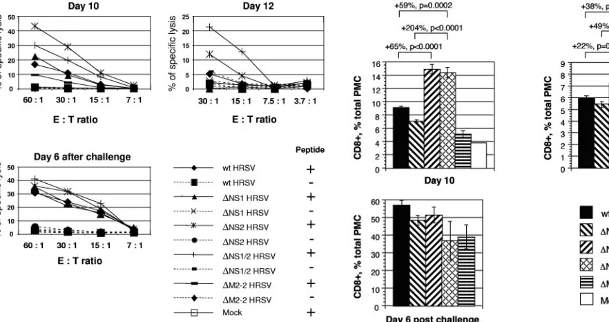
viruses and on day 6 after the challenge with wt HRSV. This demonstrated elevated levels of cell-killing activity for PMC isolated on days 10 and 12 from mice infected with ⌬NS2 HRSV and⌬NS1/2 HRSV compared to wt HRSV and⌬NS1 HRSV, respectively, whereas after challenge with wt HRSV the levels of cytotoxicity were not significantly different be-tween the viruses (Fig. 4).
HRSV NS2 suppresses the production of total CD8ⴙcells.
We also evaluated the level of total pulmonary CD8⫹cells in the lungs of mice that were mock infected or infected with wt HRSV, the recombinant viruses lacking the NS1 and/or the NS2 protein, or the⌬M2-2 control virus. On day 10 after the primary infection, we found a substantial increase in the num-ber of CD8⫹ cells in mice infected with ⌬NS2 HRSV and FIG. 2. Analysis of the pulmonary HRSV-specific CTL response in BALB/c mice by tetramer staining and flow cytometry. Groups of mice were mock infected or infected with 104PFU of wt HRSV or the⌬NS1,⌬NS2,⌬NS1/2, or⌬M2-2 mutant and, 28 days later, were challenged with 106
PFU of wt HRSV. Five animals from each group were sacrificed on days 10 and 12 following the first infection or on day 6 following the challenge, and total PMC were isolated and analyzed by staining for CD8 and binding to an MHC class I H-2Kdtetramer bearing the HRSV-specific
immunodominant peptide SYIGSINNI. (A) Examples of primary data for individual mice on day 10, with the percentages of each cell population indicated. (B) Tetramer⫹CD8⫹cells for the groups, expressed as a percentage of total PMC⫾SE. Increases in percentages between pair-wise comparisons are shown, with theP values indicated. These data are from a representative experiment out of a total of three independent experiments.
on November 8, 2019 by guest
http://jvi.asm.org/
⌬NS1/2 HRSV compared to wt HRSV and⌬NS1 HRSV, respec-tively, or compared to the mock-infected mice or the ⌬M2-2 control virus (Fig. 5). On day 12, the increase in the number of cells was also observed in mice infected with the viruses lacking the NS2 protein, but the magnitude of the increase was more modest. No significant difference between the groups was ob-served on day 6 after the challenge with wt HRSV. The increase in the percentages of the total pulmonary CD8⫹cells in⌬NS2 HRSV and⌬NS1/2 HRSV groups is likely to be due in part to CTL specific to the M2 immunodominant epitope and in part to other HRSV CTL epitopes.
HRSV NS2 suppresses production of type I IFN in mouse cells.Since the ability of the NS1 and NS2 proteins to interfere with the IFN response can be species specific, we evaluated whether NS1 and NS2 of HRSV were capable of blocking the IFN response in mouse cells. First, we used recombinant wt HRSV expressing the enhanced green fluorescent protein (HRSV/GFP) (52) to determine whether the mouse cells sup-ported efficient infection by HRSV. We found that essentially all of the cells in a monolayer of mouse fibroblast NIH 3T3 cells could be infected with HRSV/GFP if the monolayer was at a low cell density (⬃50% confluent) (Fig. 6A). Robust ex-FIG. 3. Analysis of the pulmonary HRSV-specific CTL response in BALB/c mice by intracellular IFN-␥staining and flow cytometry. Groups of mice were mock infected or infected with 104PFU of wt HRSV or the⌬NS1,⌬NS2,⌬NS1/2, or⌬M2-2 mutant and, 28 days later, were challenged with 106
PFU of wt HRSV. Five animals from each group were sacrificed on days 10 and 12 following the first infection or on day 6 following the challenge. Total PMC were isolated and stimulated in vitro with the peptide SYIGSINNI. After the stimulation, the cells were stained for CD8, fixed, permeabilized, and stained for IFN-␥. (A) Examples of primary data for individual mice on day 10, with the percentages of each cell population indicated. (B) IFN-␥⫹CD8⫹ cells for the groups, expressed as a percentage of total PMC⫾SE. Increases in percentages between pair-wise comparisons are shown, with thePvalues indicated. These data are from a representative experiment out of a total of three independent experiments. APC, allophycocyanin; FITC, fluorescein isothiocyanate.
5962 KOTELKIN ET AL. J. VIROL.
on November 8, 2019 by guest
http://jvi.asm.org/
[image:5.585.124.463.69.486.2]pression of GFP depends on extensive intracellular viral RNA replication and gene expression, and the extent of expression observed in the mouse cells was comparable to that observed in permissive human cells.
Next, we investigated whether IFN was induced in the mouse cells in response to infection by wt HRSV or the dele-tion mutants. Duplicate monolayers were infected at a multi-plicity of infection of 2 PFU/cell, and as a positive control, additional monolayers were transfected with poly(I)-poly(C). Aliquots were taken from the overlying medium at 16, 20, 24, and 28 h postinfection and analyzed by ELISA for the presence of IFN-␣ (Fig. 6B). In the media of cells infected with wt HRSV, IFN-␣was not detected at 16 and 20 h after infection, whereas low to moderate amounts were detected at 24 and 28 h postinfection (14 and 45 pg/ml, respectively). In media of cells infected with ⌬NS1 HRSV, a low level of IFN-␣ was detected at 16, 20, and 24 h but not at 28 h postinfection. In contrast, high concentrations of IFN-␣compared to the posi-tive control were detected at all time points in the media of cells infected with the⌬NS2 and⌬NS1/2 viruses, with the peak at 20 h postinfection (150 and 190 pg/ml, respectively). These results indicate that wt HRSV suppresses induction of type I IFN in mouse cells and that this effect is greatly alleviated by deletion of the NS2 protein. At most of the time points, a higher concentration of IFN-␣was detected following the in-fection with⌬NS1/2 HRSV than with⌬NS2 HRSV, suggesting a possible small additive effect of the NS1 protein to the sup-pression of type I IFN.
Suppression of the CTL response by HRSV NS2 is mediated by suppression of type I IFN.Since the NS2 protein was found to suppress both the CTL and IFN responses to HRSV infec-tion, and since IFN-regulated genes have the potential to en-hance immune responses, we evaluated whether suppression of the CTL response by the NS2 protein is a consequence of suppression of type I IFN. Specifically, we compared the mag-nitude of the CTL response to wt HRSV or⌬NS1/2 HRSV in STAT1 gene KO mice (30), which lack responsiveness to type I IFNs (13), by quantitation of pulmonary CD8⫹cells secreting IFN-␥in response to HRSV-specific stimulation. These KO mice are based on the 129S6 strain (H-2Db), and in this case
stimulation was performed using a recently described H-2Db
-restricted CD8⫹ CTL epitope from the HRSV M protein (amino acids 187 to 195) that is represented by the peptide NAITNAKEE (35).
The wt and ⌬NS1/2 mutant viruses replicated to similar titers in the wt 129S6 mice, as evaluated by the titers of virus in the lungs on day 4 following infection with 106PFU of virus per
animal (Fig. 7). The pulmonary titers of wt and⌬NS1/2 viruses in the STAT1 KO mice were at least 10-fold higher than in the wt 129S6 mice, which is consistent with a previously published study (20), but the titers of the two viruses were similar (Fig. 7). This difference between the STAT1 KO and 129S6 mice pre-sumably represents the contribution of the IFN-mediated anti-viral state in controlling HRSV replication. In preliminary experiments (not shown), we found that the induction of pul-monary CD8⫹cells secreting IFN-␥could not be reproducibly detected in wt 129S6 mice following infection with 104PFU per
animal (the dose that had been used in the previous experi-FIG. 4. Levels of HRSV-specific cytotoxicity by PMC isolated from
BALB/c mice that were infected with 104PFU of wt HRSV or⌬NS1,
⌬NS2,⌬NS1/2, or⌬M2-2 mutant or mock infected and, 28 days later, challenged with 106PFU of wt HRSV. Five animals per group per day
were sacrificed on days 10 and 12 following the initial infection or day 6 following the challenge with wt HRSV, and the cells from each group and day were pooled and analyzed in a standard51Cr release assay, directly ex
[image:6.585.48.489.70.303.2]vivo without stimulation, using mouse P815 cells pulse-labeled with the peptide SYIGSINNI. E:T ratio, effector-to-target cell ratio.
FIG. 5. Comparison of the total numbers of pulmonary CD8⫹cells from infected BALB/c mice that were isolated and quantified during the experiment presented in Fig. 2, expressed as mean percentages⫾SE of the total PMC. Increases in percentages between pair-wise comparisons are shown, with thePvalues indicated. These data are from a represen-tative experiment out of a total of three independent experiments.
on November 8, 2019 by guest
http://jvi.asm.org/
ments in BALB/c mice). This probably reflects the lower mag-nitude of the HRSV M-specific CTL response in H-2Dbmice
than the immunodominant M2-specific response in H-2Kd
mice (35). Therefore, these experiments were performed with a dose of 106PFU per animal. In 129S6 mice infected with wt
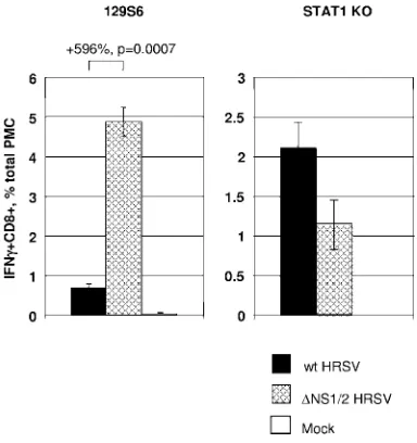
HRSV, the response of HRSV-specific CD8⫹cells secreting IFN-␥was relatively low, whereas the response was increased sevenfold in wt 129S6 mice infected with the⌬NS1/2 mutant (Fig. 8). We confirmed that this effect was specific to deletion of NS2 and not NS1 (not shown). Thus, as was the case in BALB/c mice, deletion of NS2 resulted in an increased CTL response. However, this was not the case in the STAT1 KO
mice, in which the response of CD8⫹IFN-␥⫹ cells was not greater for the⌬NS1/2 virus than for wt HRSV (Fig. 8).
We also compared PMC from the STAT1 KO mice for HRSV-specific cytolytic activity ex vivo, which was measured (with no antigen stimulation) on day 10 after primary infection with 104and 106 PFU of wt HRSV or ⌬NS1/2 HRSV. The
[image:7.585.136.452.67.479.2]magnitude of cell killing was substantially higher with the higher dose, providing confirmation of the effect. There was no increase in cell-killing activity for⌬NS1/2 HRSV compared to wt HRSV in this STAT1 KO background (Fig. 9). Thus, the augmented CTL response to infection with the⌬NS1/2 virus depended on the ability of the mouse to respond to IFN. FIG. 6. Infection of murine NIH 3T3 cells with wt HRSV or the NS gene deletion mutants and measurement of secreted IFN-␣. (A) Infection of NIH 3T3 cells with HRSV. Monolayer cultures were infected with HRSV/GFP (top) or mock infected (bottom) and, at 48 h postinfection, the same field in each culture was visualized by light (left) and fluorescence (right) microscopy. (B) Secretion of IFN-␣in the media of NIH 3T3 cells measured at the indicated times following infection with wt HRSV or the⌬NS1,⌬NS2, or⌬NS1/2 mutant or transfection with poly(I)-poly(C) as a positive control (shown in the same left-to-right order for each time point). For each virus and each time point, the concentration of IFN-␣was determined by ELISA in duplicate cell cultures. The duplicate cultures yielded very similar values; mean values are indicated. Values below the detection level (8 pg/ml, indicated by the dashed line) are indicated by asterisks. Additional cultures were mock infected and used as negative controls or infected with HRSV lacking the M2-2 coding sequence; these values were below the detection limit at all time points and therefore are not shown. These data are from a representative experiment out of a total of two independent experiments. Poly IC, poly(I)-poly(C).
5964 KOTELKIN ET AL. J. VIROL.
on November 8, 2019 by guest
http://jvi.asm.org/
DISCUSSION
Viruses often interfere with the type I IFN response of the host, but the consequences of this for the adaptive immune response have been largely unexplored. As noted in the intro-duction, this had been investigated previously for HRSV and BRSV, but the evidence was mixed. One previous study pro-vided evidence that the deletion of the NS protein IFN antag-onists in BRSV was associated with an increased virus-specific antibody response in calves (46), but comparable experiments
with HRSV NS deletion mutants in the highly permissive chim-panzee failed to provide any evidence of enhanced immuno-genicity. In the present study, focusing on the CTL response and using more refined immunological reagents possible in inbred mice, we showed that HRSV suppresses the CTL re-sponse, that this is mediated primarily by the NS2 protein, and that this is a consequence of suppression of the type I IFN response by NS2. Although connections between type I IFNs and the CTL response have been established (references be-low), the present data provide in vivo evidence that a viral IFN antagonist can have the effect of suppressing the adaptive immune response.
[image:8.585.71.255.68.211.2]A number of factors might contribute to the elevated CTL response to HRSV lacking the NS2 protein. First, suppression of the type I IFN response by HRSV NS2 might result in reduced production of factors important for the activation of CTL, such as MHC class I molecules whose expression is up-regulated by IFN, the chemokine CXCR3 that is required for CD8⫹ T-cell activation (32), and the double-stranded RNA-dependent protein kinase (PKR) that is regulated by type I IFN (19) and was shown to be involved in an induction of human immunodeficiency virus-specific CTL (12). Second, suppression of the IFN response by the NS2 protein might result in a more rapid death of HRSV-specific CD8⫹T cells, since incubation of activated CD8⫹and CD4⫹T cells with type I IFNs results in a strong inhibition of their death (27). Third, type I IFN signaling contributes positively to the maturation of dendritic cells induced by viral infection (17, 18) which, to-gether with plasmacytoid dendritic cells, contribute to the CTL response (51). A recently published study showed that RSV F protein expressed by a recombinant Newcastle disease virus induced a CTL response in mice that exceeded the response induced by RSV and the effect was accompanied by an in-creased type I interferon response and more-efficient matura-tion of dendritic cells (28). Therefore the suppression of virus-specific CTL in mice infected with the viruses expressing NS2 may be a consequence of a reduced maturation of pulmonary FIG. 7. Replication of recombinant wt HRSV and⌬NS1/2 HRSV
in 129S6 mice (left panel) and STAT1 KO mice (right panel). Groups of mice (four to six animals per virus) were inoculated intranasally with 106PFU of the indicated viruses and sacrificed on day 4. Titers of the
[image:8.585.68.260.423.627.2]viruses in lungs were determined by plaque assay of the tissue homog-enates and are shown as means⫾SE.
FIG. 8. Analysis of the pulmonary HRSV-specific CTL response in 129S6 mice (left panel) or STAT1 KO mice (right panel). Groups of mice (four to eight animals per virus) were infected with 106PFU of wt
HRSV or⌬NS1/2 HRSV or mock infected. Total PMC were isolated 10 days later and stimulated in vitro with the peptide NAITNAKEE, which represents an H-2Db-restricted HRSV CTL epitope. Following
stimulation, the cells were stained for CD8, fixed and permeabilized, stained for IFN-␥, and analyzed by flow cytometry. IFN-␥⫹CD8⫹cells are expressed as percentages⫾SE of the total PMC: the difference in percentages between the two groups of 129S6 mice is shown together with thePvalue.
FIG. 9. Levels of HRSV-specific cytotoxicity by PMC isolated from STAT1 KO mice after infection with 104PFU (left panel) or 106PFU
(right panel) of wt HRSV or the⌬NS1/2 mutant. Five animals per group per day were sacrificed on day 10 after infection and the cells from each group were pooled and analyzed in a standard51Cr release
assay without restimulation directly ex vivo, using the mouse EL4 cells pulse-labeled with the peptide NAITNAKEE. E:T ratio, effector-to-target cell ratio.
on November 8, 2019 by guest
http://jvi.asm.org/
dendritic cells and a reduced presentation of viral antigens. Fourth, it has been recently shown that type I IFNs act directly on CD8 T cells to allow clonal expansion and memory forma-tion in response to viral infecforma-tion (21).
In the bovine system, it is the BRSV NS2 protein that plays the major role in inhibiting the induction of IFN in bovine cells (46). In contrast, in the human system, it is the HRSV NS1 protein that plays the greater role in inhibiting IFN induction in human cells (40). Unexpectedly, in mouse cells, the present data show that it is the HRSV NS2 protein that plays the greater role. Thus, in comparing effects of HRSV in human versus mouse cells, it appears to be the host species that de-termines whether NS1 versus NS2 plays the greater antagonist role. The exact role(s) of the NS proteins in inhibiting IFN induction is not known except that inhibition occurs at a step prior to phosphorylation and dimerization of cytoplasmic IRF-3. Presumably, the NS proteins interact with and inhibit one or more cellular factors in the process of interrupting this signaling pathway. It might be that NS1 and NS2 act at the same step, and whether NS1 or NS2 exerts the greater effect might depend on species-specific differences in that cellular factor that confer greater affinity for one NS versus the other: in the human cell, HRSV NS1 would have the greater affinity, whereas in the murine cell, it would be HRSV NS2.
Although the murine CTL response was substantially greater when the NS2 gene was absent, this was not associated with a reduction in pulmonary virus replication. Replication of HRSV in the mouse is highly restricted due to the semiper-missive nature of the host. The peak of virus replication occurs early, at day 4, which is substantially before the peak of the CTL response on days 8 to 12. The host immune response does play a role in restricting and clearing the infection, but most of the restriction in mice (i.e., the greatly reduced magnitude of replication and the early resolution of infection) is a host range effect. For example, even in BALB/c mice that have been severely immunosuppressed with cyclophosphamide, HRSV replicates to a peak titer of only approximately 104PFU/g lung
tissue and is essentially cleared by day 7 (41). Given the severe restriction imposed by the host difference and the fact that the virus is cleared days before the peak of the CTL response, it perhaps is not surprising that an increase in the magnitude of that peak on day 10 was not associated with more rapid clear-ance on days 4 and 5. In the natural human host, where virus replication is of much higher magnitude and shedding can sometimes be detected for weeks, an increased CTL response likely would help restrict and clear the viral infection.
One of the factors that can be involved in restricting a virus in a nonnatural host is the inability of the virus to control the heterologous IFN system. The observation that the NS protein is effective in controlling the murine IFN system, coupled with the observation that the⌬NS2 virus is not substantially atten-uated compared to wt HRSV, indicates that the IFN system is not a major determinant of the host range restriction in the specific situation of HRSV in the mouse. Unexpectedly, dele-tion of the NS1 gene, either alone or in combinadele-tion with NS2, attenuated the virus in BALB/c mice. The basis for this effect is not known, and this effect was not evident in the 129S6 mice. The NS1 protein has been implicated in effects on viral RNA synthesis from both the genomic and antigenomic promoters
(3), and thus, its deletion might have effects beyond those involving IFN.
HRSV strains from which the NS1 or NS2 gene has been deleted are being developed and evaluated as vaccine candi-dates. For example, several candidates that combined the NS2 deletion with various point mutations in other genes have been evaluated in adults and seropositive and seronegative children and were found to be either overattenuated or underattenu-ated (50). This indicates that deletion of NS2 on its own is insufficiently attenuating in humans but that the combination of the NS2 deletion with one or more point mutations might yield an appropriately attenuated derivative. Studies with chimpanzees indicated that the⌬NS1 virus is more attenuated in this relevant experimental animal than the⌬NS2 virus (43, 49), and therefore the⌬NS1 virus is presently being developed as is for clinical evaluation. The deletion of IFN antagonists can be expected to result in an enhanced IFN response during immunization. Studies with mice suggest that the production of IFN during infection favors a Th1-biased immune response and reduced pathogenesis (14). The results of the present study describe another consequence of increased IFN produc-tion, namely, a heightened CTL response. The increased IFN and CTL responses likely contribute to the attenuation phe-notypes of these vaccine candidates.
ACKNOWLEDGMENTS
We thank Barney Graham and members of his group for generously providing information and advice regarding the H-2Db-restricted
NAITNAKEE CTL epitope and Elaine Lamirande for technical as-sistance in the early part of the study. We are grateful to Brian Murphy for critical reading of the manuscript.
This project was funded as a part of the NIAID Intramural Program. The authors have no conflicting financial interests.
REFERENCES
1.Anderson, J. J., J. Norden, D. Saunders, G. L. Toms, and R. Scott.1990. Analysis of the local and systemic immune responses induced in BALB/c mice by experimental respiratory syncytial virus infection. J. Gen. Virol.
71:1561–1570.
2.Appay, V., D. F. Nixon, S. M. Donahoe, G. M. Gillespie, T. Dong, A. King, G. S. Ogg, H. M. Spiegel, C. Conlon, C. A. Spina, D. V. Havlir, D. D. Richman, A. Waters, P. Easterbrook, A. J. McMichael, and S. L. Rowland-Jones.2000. HIV-specific CD8(⫹) T cells produce antiviral cytokines but are
impaired in cytolytic function. J. Exp. Med.192:63–75.
3.Atreya, P. L., M. E. Peeples, and P. L. Collins.1998. The NS1 protein of human respiratory syncytial virus is a potent inhibitor of minigenome
tran-scription and RNA replication. J. Virol.72:1452–1461.
4.Barchet, W., M. Cella, B. Odermatt, C. Asselin-Paturel, M. Colonna, and U. Kalinke.2002. Virus-induced interferon alpha production by a dendritic cell
subset in the absence of feedback signaling in vivo. J. Exp. Med.195:507–516.
5.Belyakov, I. M., J. Wang, R. Koka, J. D. Ahlers, J. T. Snyder, R. Tse, J. Cox, J. S. Gibbs, D. H. Margulies, and J. A. Berzofsky.2001. Activating CTL precursors to reveal CTL function without skewing the repertoire by in vitro
expansion. Eur. J. Immunol.31:3557–3566.
6.Bermingham, A., and P. L. Collins. 1999. The M2-2 protein of human respiratory syncytial virus is a regulatory factor involved in the balance between RNA replication and transcription. Proc. Natl. Acad. Sci. USA
96:11259–11264.
7.Bossert, B., and K. K. Conzelmann.2002. Respiratory syncytial virus (RSV) nonstructural (NS) proteins as host range determinants: a chimeric bovine RSV with NS genes from human RSV is attenuated in interferon-competent
bovine cells. J. Virol.76:4287–4293.
8.Bukreyev, A., I. M. Belyakov, J. A. Berzofsky, B. R. Murphy, and P. L. Collins.2001. Granulocyte-macrophage colony-stimulating factor expressed by recombinant respiratory syncytial virus attenuates viral replication and
increases the level of pulmonary antigen-presenting cells. J. Virol.75:12128–
12140.
9.Bukreyev, A., I. M. Belyakov, G. A. Prince, K. C. Yim, K. K. Harris, J. A. Berzofsky, and P. L. Collins.2005. Expression of interleukin-4 by recombi-nant respiratory syncytial virus is associated with accelerated inflammation
5966 KOTELKIN ET AL. J. VIROL.
on November 8, 2019 by guest
http://jvi.asm.org/
and a nonfunctional cytotoxic T-lymphocyte response following primary
in-fection but not following challenge with wild-type virus. J. Virol.79:9515–
9526.
10.Chang, J., and T. J. Braciale.2002. Respiratory syncytial virus infection
suppresses lung CD8⫹T-cell effector activity and peripheral CD8⫹T-cell
memory in the respiratory tract. Nat. Med.8:54–60.
11.Collins, P. L., M. G. Hill, E. Camargo, H. Grosfeld, R. M. Chanock, and B. R. Murphy.1995. Production of infectious human respiratory syncytial virus from cloned cDNA confirms an essential role for the transcription
elongation factor from the 5⬘proximal open reading frame of the M2 mRNA
in gene expression and provides a capability for vaccine development. Proc.
Natl. Acad. Sci. USA92:11563–11567.
12.De Lucca, F. L., V. S. Sales, L. R. Souza, and M. A. Watanabe.2002. Evidence for the involvement of the RNA-dependent protein kinase (PKR) in the induction of human cytotoxic T lymphocytes against a synthetic
pep-tide of HIV-1 by regulatory RNA. Mol. Cell. Biochem.238:19–26.
13.Durbin, J. E., R. Hackenmiller, M. C. Simon, and D. E. Levy.1996. Targeted disruption of the mouse Stat1 gene results in compromised innate immunity
to viral disease. Cell84:443–450.
14.Durbin, J. E., T. R. Johnson, R. K. Durbin, S. E. Mertz, R. A. Morotti, R. S. Peebles, and B. S. Graham.2002. The role of IFN in respiratory syncytial
virus pathogenesis. J. Immunol.168:2944–2952.
15.Garcia-Sastre, A.2004. Identification and characterization of viral antago-nists of type I interferon in negative-strand RNA viruses. Curr. Top.
Micro-biol. Immunol.283:249–280.
16.Goepfert, P. A., A. Bansal, B. H. Edwards, G. D. Ritter, Jr., I. Tellez, S. A. McPherson, S. Sabbaj, and M. J. Mulligan.2000. A significant number of human immunodeficiency virus epitope-specific cytotoxic T lymphocytes de-tected by tetramer binding do not produce gamma interferon. J. Virol.
74:10249–10255.
17.Honda, K., T. Mizutani, and T. Taniguchi.2004. Negative regulation of
IFN-␣/signaling by IFN regulatory factor 2 for homeostatic development
of dendritic cells. Proc. Natl. Acad. Sci. USA101:2416–2421.
18.Honda, K., S. Sakaguchi, C. Nakajima, A. Watanabe, H. Yanai, M. Matsumoto, T. Ohteki, T. Kaisho, A. Takaoka, S. Akira, T. Seya, and T. Taniguchi.2003. Selective contribution of IFN-alpha/beta signaling to the maturation of dendritic cells induced by double-stranded RNA or viral infection. Proc. Natl. Acad. Sci.
USA100:10872–10877.
19.Hovanessian, A. G.1989. The double stranded RNA-activated protein kinase
induced by interferon: dsRNA-PK. J. Interferon Res.9:641–647.
20.Johnson, T. R., S. E. Mertz, N. Gitiban, S. Hammond, R. Legallo, R. K. Durbin, and J. E. Durbin.2005. Role for innate IFNs in determining
respi-ratory syncytial virus immunopathology. J. Immunol.174:7234–7241.
21.Kolumam, G. A., S. Thomas, L. J. Thompson, J. Sprent, and K. Murali-Krishna.2005. Type I interferons act directly on CD8 T cells to allow clonal expansion and memory formation in response to viral infection. J. Exp. Med.
202:637–650.
22.Kulkarni, A. B., P. L. Collins, I. Bacik, J. W. Yewdell, J. R. Bennink, J. E. Crowe, Jr., and B. R. Murphy.1995. Cytotoxic T cells specific for a single peptide on the M2 protein of respiratory syncytial virus are the sole medi-ators of resistance induced by immunization with M2 encoded by a
recom-binant vaccinia virus. J. Virol.69:1261–1264.
23.Kulkarni, A. B., M. Connors, C. Y. Firestone, H. C. Morse III, and B. R. Murphy.1993. The cytolytic activity of pulmonary CD8⫹lymphocytes, in-duced by infection with a vaccinia virus recombinant expressing the M2 protein of respiratory syncytial virus (RSV), correlates with resistance to
RSV infection in mice. J. Virol.67:1044–1049.
24.Li, K., E. Foy, J. C. Ferreon, M. Nakamura, A. C. Ferreon, M. Ikeda, S. C. Ray, M. Gale, Jr., and S. M. Lemon.2005. Immune evasion by hepatitis C virus NS3/4A protease-mediated cleavage of the Toll-like receptor 3 adaptor
protein TRIF. Proc. Natl. Acad. Sci. USA102:2992–2997.
25.Lo, M. S., R. M. Brazas, and M. J. Holtzman.2005. Respiratory syncytial virus nonstructural proteins NS1 and NS2 mediate inhibition of Stat2
ex-pression and alpha/beta interferon responsiveness. J. Virol.79:9315–9319.
26.Lyles, D. S.2000. Cytopathogenesis and inhibition of host gene expression by
RNA viruses. Microbiol. Mol. Biol. Rev.64:709–724.
27.Marrack, P., J. Kappler, and T. Mitchell.1999. Type I interferons keep
activated T cells alive. J. Exp. Med.189:521–530.
28.Martinez-Sobrido, L., N. Gitiban, A. Fernandez-Sesma, J. Cros, S. E. Mertz, N. A. Jewell, S. Hammond, E. Flano, R. K. Durbin, A. Garcia-Sastre, and J. E. Durbin.2006. Protection against respiratory syncytial virus by a
recom-binant Newcastle disease virus vector. J. Virol.80:1130–1139.
29.Meager, A., K. Visvalingam, P. Dilger, D. Bryan, and M. Wadhwa.2005. Biological activity of interleukins-28 and -29: comparison with type I
inter-ferons. Cytokine31:109–118.
30.Meraz, M. A., J. M. White, K. C. Sheehan, E. A. Bach, S. J. Rodig, A. S. Dighe, D. H. Kaplan, J. K. Riley, A. C. Greenlund, D. Campbell, K. Carver-Moore, R. N. DuBois, R. Clark, M. Aguet, and R. D. Schreiber.1996. Targeted disruption of the Stat1 gene in mice reveals unexpected physiologic
specificity in the JAK-STAT signaling pathway. Cell84:431–442.
31.Murphy, B. R., A. V. Sotnikov, L. A. Lawrence, S. M. Banks, and G. A.
Prince.1990. Enhanced pulmonary histopathology is observed in cotton rats immunized with formalin-inactivated respiratory syncytial virus (RSV) or purified F glycoprotein and challenged with RSV 3–6 months after
immu-nization. Vaccine8:497–502.
32.Ogasawara, K., S. Hida, Y. Weng, A. Saiura, K. Sato, H. Takayanagi, S. Sakaguchi, T. Yokochi, T. Kodama, M. Naitoh, J. A. De Martino, and T. Taniguchi.2002. Requirement of the IFN-alpha/beta-induced CXCR3
che-mokine signalling for CD8⫹T cell activation. Genes Cells7:309–320.
33.Ramaswamy, M., L. Shi, M. M. Monick, G. W. Hunninghake, and D. C. Look.2004. Specific inhibition of type I interferon signal transduction by
respiratory syncytial virus. Am. J. Respir. Cell Mol. Biol.30:893–900.
34.Rutigliano, J. A., and B. S. Graham.2004. Prolonged production of TNF-alpha exacerbates illness during respiratory syncytial virus infection. J.
Im-munol.173:3408–3417.
35.Rutigliano, J. A., M. T. Rock, A. K. Johnson, J. E. Crowe, Jr., and B. S. Graham. 2005. Identification of an H-2Db-restricted CD8⫹ cytotoxic T
lymphocyte epitope in the matrix protein of respiratory syncytial virus.
Vi-rology337:335–343.
36.Schlender, J., B. Bossert, U. Buchholz, and K. K. Conzelmann.2000. Bovine respiratory syncytial virus nonstructural proteins NS1 and NS2 cooperatively
antagonize alpha/beta interferon-induced antiviral response. J. Virol. 74:
8234–8242.
37.Smith, P. L., G. Lombardi, and G. R. Foster.2005. Type I interferons and the innate immune response–more than just antiviral cytokines. Mol. Immunol.
42:869–877.
38.Snyder, J. E., W. J. Bowers, A. M. Livingstone, F. E. Lee, H. J. Federoff, and T. R. Mosmann.2003. Measuring the frequency of mouse and human cyto-toxic T cells by the Lysispot assay: independent regulation of cytokine
se-cretion and short-term killing. Nat. Med.9:231–235.
39.Spann, K. M., P. L. Collins, and M. N. Teng.2003. Genetic recombination during coinfection of two mutants of human respiratory syncytial virus.
J. Virol.77:11201–11211.
40.Spann, K. M., K.-C. Tran, B. Chi, R. L. Rabin, and P. L. Collins.2004. Suppression of the induction of alpha, beta, and lambda interferons by the NS1 and NS2 proteins of human respiratory syncytial virus in human
epi-thelial cells and macrophages. J. Virol.78:4363–4369. (Erratum,78:6705.)
41.Sudo, K., W. Watanabe, K. Konno, R. Sato, T. Kajiyashiki, S. Shigeta, and T. Yokota.1999. Efficacy of RD3-0028 aerosol treatment against respiratory syncytial virus infection in immunosuppressed mice. Antimicrob. Agents
Chemother.43:752–757.
42.Teng, M. N., and P. L. Collins.1999. Altered growth characteristics of recombinant respiratory syncytial viruses which do not produce NS2 protein.
J. Virol.73:466–473.
43.Teng, M. N., S. S. Whitehead, A. Bermingham, M. St. Claire, W. R. Elkins, B. R. Murphy, and P. L. Collins.2000. Recombinant respiratory syncytial virus that does not express the NS1 or M2-2 protein is highly attenuated and
immunogenic in chimpanzees. J. Virol.74:9317–9321.
44.Thomas, D., G. Blakqori, V. Wagner, M. Banholzer, N. Kessler, R. M. Elliott, O. Haller, and F. Weber.2004. Inhibition of RNA polymerase II
phosphorylation by a viral interferon antagonist. J. Biol. Chem.279:31471–
31477.
45.Tough, D. F.2004. Type I interferon as a link between innate and adaptive
immunity through dendritic cell stimulation. Leuk. Lymphoma45:257–264.
46.Valarcher, J.-F., J. Furze, S. Wyld, R. Cook, K.-K. Conzelmann, and G. Taylor.2003. Role of alpha/beta interferons in the attenuation and immu-nogenicity of recombinant bovine respiratory syncytial viruses lacking NS
proteins. J. Virol.77:8426–8439.
47.Wherry, E. J., M. J. McElhaugh, and L. C. Eisenlohr.2002. Generation of
CD8(⫹) T cell memory in response to low, high, and excessive levels of
epitope. J. Immunol.168:4455–4461.
48.Wherry, E. J., K. A. Puorro, A. Porgador, and L. C. Eisenlohr.1999. The induction of virus-specific CTL as a function of increasing epitope expres-sion: responses rise steadily until excessively high levels of epitope are
at-tained. J. Immunol.163:3735–3745.
49.Whitehead, S. S., A. Bukreyev, M. N. Teng, C.-Y. Firestone, M. St. Claire, W. R. Elkins, P. L. Collins, and B. R. Murphy.1999. Recombinant respira-tory syncytial virus bearing a deletion of either the NS2 or SH gene is
attenuated in chimpanzees. J. Virol.73:3438–3442.
50.Wright, P. F., R. A. Karron, S. A. Madhi, J. J. Treanor, J. C. King, A. O’Shea, M. R. Ikizler, Y. Zhu, P. L. Collins, C. Cutland, V. B. Randolph, A. M. Deatly, J. G. Hackell, W. C. Gruber, and B. R. Murphy.The interferon antagonist NS2 protein of respiratory syncytial virus is an important
viru-lence determinant for humans. J. Infect. Dis.193:573–581.
51.Yoneyama, H., K. Matsuno, E. Toda, T. Nishiwaki, N. Matsuo, A. Nakano, S. Narumi, B. Lu, C. Gerard, S. Ishikawa, and K. Matsushima.2005. Plas-macytoid DCs help lymph node DCs to induce anti-HSV CTLs. J. Exp. Med.
202:425–435.
52.Zhang, L., M. E. Peeples, R. C. Boucher, P. L. Collins, and R. J. Pickles.
2002. Respiratory syncytial virus infection of human airway epithelial cells is polarized, specific to ciliated cells, and without obvious cytopathology. J.
Vi-rol.76:5654–5666.
on November 8, 2019 by guest
http://jvi.asm.org/
JOURNAL OFVIROLOGY, Oct. 2006, p. 10286 Vol. 80, No. 20 0022-538X/06/$08.00⫹0 doi:10.1128/JVI.01773-06
ERRATUM
The NS2 Protein of Human Respiratory Syncytial Virus
Suppresses the Cytotoxic T-Cell Response as a Consequence of
Suppressing the Type I Interferon Response
Alexander Kotelkin, Igor M. Belyakov, Lijuan Yang, Jay A. Berzofsky,
Peter L. Collins, and Alexander Bukreyev
Laboratory of Infectious Diseases, National Institute of Allergy and Infectious Diseases, and Molecular Immunogenetics and Vaccine Research Section, Vaccine Branch, National Cancer Institute, National Institutes of Health,
Bethesda, Maryland 20892-8007
Volume 80, no. 12, p. 5958–5967, 2006. Page 5959, column 1, last paragraph, line 10: “NAITNAKEE” should read “NAIT NAKII.”
Page 5959, column 2, line 6: “NAITNAKEE” should read “NAITNAKII.”
Page 5963, column 2, last line of the first paragraph: “NAITNAKEE” should read “NAITNAKII.” Page 5965, legend to Fig. 8, line 5: “NAITNAKEE” should read “NAITNAKII.”
Page 5965, legend to Fig. 9, line 7: “NAITNAKEE” should read “NAITNAKII.” Page 5966, Acknowledgments, line 3: “NAITNAKEE” should read “NAITNAKII.”