RESEARCH NOTE
Evaluation of two DNA extraction
methods for the PCR-based detection
of eukaryotic enteric pathogens in fecal samples
Estelle Menu
1, Charles Mary
2, Isabelle Toga
2, Didier Raoult
1, Stéphane Ranque
2and Fadi Bittar
1*Abstract
Objective: Efficient and easy-to-use DNA extraction and purification methods are critical in implementing PCR-based diagnosis of pathogens. In order to optimize the routine clinical laboratory diagnosis of eukaryotic enteric pathogens, we compare, via quantitative PCR cycle threshold (Ct) values, the efficiency of two DNA extraction kits: the semi-automated EZ1® (Qiagen) and the manual QIAamp® DNA Stool Mini Kit (Qiagen), on six protozoa: Blastocystis spp., Cryptosporidium parvum/hominis, Cyclospora cayetanensis, Dientamoeba fragilis, Giardia intestinalis and Cystoisos-pora belli and one microsporidia: Enterocytozoon bieneusi.
Results: Whereas EZ1® (Qiagen) and QIAamp® DNA Stool Mini Kit (Qiagen) yielded similar performances for the detection of Cryptosporidium spp. and D. fragilis, significant lower Ct values (p < 0.002) pointed out a better perfor-mance of EZ1® on the five remaining pathogens. DNA extraction using the semi-automated EZ1® procedure was faster and as efficient as the manual procedure in the seven eukaryotic enteric pathogens tested. This procedure is suitable for DNA extraction from stools in both clinical laboratory diagnosis and epidemiological study settings. Keywords: Enteric parasites, Protozoa, Microsporidia, qPCR, DNA extraction
© The Author(s) 2018. This article is distributed under the terms of the Creative Commons Attribution 4.0 International License (http://creativecommons.org/licenses/by/4.0/), which permits unrestricted use, distribution, and reproduction in any medium, provided you give appropriate credit to the original author(s) and the source, provide a link to the Creative Commons license, and indicate if changes were made. The Creative Commons Public Domain Dedication waiver (http://creativecommons.org/ publicdomain/zero/1.0/) applies to the data made available in this article, unless otherwise stated.
Introduction
Human diseases caused by eukaryotic enteric pathogens are a major public health concern [1]. Of these, proto-zoa are the most prevalent [2]. In order to increase the specificity and sensitivity of detection of these parasites, molecular tools have been developed over the last 10 years. PCR-based detection of eukaryotic enteric patho-gens is particularly dependent upon the quality and purity of the initial DNA material. Thus, choosing an appropriate DNA extraction method is critical: it needs to (i) be highly efficient for DNA recovery from micro-organisms that are frequently in oocyst form and in low abundance compared to bacterial communities and (ii) remove the many PCR inhibitors (such as bile salts, urea, hemoglobin and heparin) that are present in stools. The relatively recent introduction and rapidly increasing use
of PCR-based diagnosis for eukaryotic enteric patho-gen diseases requires the use of a DNA extraction pro-cedure that should be efficient and standardized and which could be applied to the detection of all eukaryotic enteric pathogens species in order to simplify laboratory work-flow and avoid the heterogeneity of inter-labora-tory results [3]. Many commercialized DNA extraction and purification kits, using chemical, enzymatic and/ or mechanical lysis, are replacing in-house methods (i.e. phenol–chloroform) [4]. Studies have already been conducted to evaluate a specific combination of perfor-mance, cost-effectiveness, and simplicity of various DNA extraction kits according to the pathogen of interest [5–
7], but none of these studies have directly compared these methods. In this study, we compared the performance of two commercial kits: a semi-automated EZ1® procedure
(Qiagen GmbH, Hilden, Germany) and a manual tech-nique, the QIAamp® DNA Stool Mini Kit (Qiagen), for
the DNA extraction and purification of the most signifi-cant eukaryotic enteric pathogens [2] from stool samples.
Open Access
*Correspondence: [email protected]
1 Aix Marseille Univ, IRD, APHM, MEPHI, IHU-Méditerranée Infection, 19-21
Boulevard Jean Moulin, 13005 Marseille, France
Page 2 of 6 Menu et al. BMC Res Notes (2018) 11:206
Main text
Materials and methods
The comparison was carried out by direct elution of DNA from the same parasite-positive fecal samples and then by comparing the detectability of the eluted DNA by quantitative PCR (qPCR).
This study included 24 positive fecal samples col-lected between January and July 2017 as part of routine laboratory diagnosis at the Parasitology–Mycology Lab-oratory at Marseille’s University Hospital (France). The presence of targeted eukaryotic enteric pathogens was confirmed by microscopic examination and/or routine PCR methods in our laboratory. The fecal samples were fixed in absolute ethanol and stored at 4 °C. To assess the differences in sensitivity between both extraction procedures, positive stools were serially diluted with PCR-negative stool samples. No written consent was needed for this work in accordance with the French law on bioethics (LOI No 2004-800 relative à la bioéthique) published in the “Journal Officiel de la République Fran-çaise” on August 6, 2004, since no additional samples were taken for the study. According to this law, patients were informed that anonymized stool specimens could be used for further studies. A semi-automated procedure (EZ1®, Cat. No. 953034, Qiagen) and a manual procedure
(QIAamp® Stool Mini Kit, Cat. No. 51504, Qiagen) were
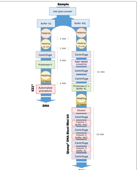
used in parallel. For each procedure, the manufacturer’s recommendations were modified by adding mechani-cal, chemical and enzymatic pretreatment steps (Fig. 1). The semi-automated extraction procedure was adapted for stool processing as follows: 200 mg of stool sample was added to 350 µL of G2 lysis buffer (Qiagen) in a tube with glass powder (acid washed glass beads 425–600 µm, Sigma-Aldrich, Saint Quentin Fallavier, France) and then disrupted in a FastPrep BIO 101 apparatus (Qbio-gene, Strasbourg, France) at maximum power for 40 s. After 10 min of incubation at 100 °C to allow for com-plete lysis, tubes were centrifuged at 10,000g for 1 min. Subsequently, 200 µL of supernatant was enzymatically digested using 20 µL of proteinase K (20 mg/mL, Qia-gen), and incubated overnight at 56 °C. The automated procedure using the EZ1 Advanced XL extractor with the V 1.066069118 Qiagen DNA bacteria card and the EZ1®
DNA Tissue Kit (Qiagen, Courtaboeuf, France) was then performed for between 15 and 30 min without the inter-vention of an operator, as described by the manufacturer. The manual procedure, based on the use of the QIAamp®
Stool Mini Kit (Qiagen), which is considered as a ref-erence method in the literature [8], was used (Fig. 1). Briefly, 200 mg of stool sample was added to 1.3 mL of ASL lysis buffer (Qiagen) in a tube with glass powder and then disrupted in a FastPrep BIO 101 apparatus at maximum power for 40 s and heated for 10 min at 95 °C.
Impurities and PCR inhibitors were removed by adding inhibitEX Tablets (Qiagen) to each sample. The obtained supernatants were then enzymatically digested using 20 µL of proteinase K (20 mg/mL, Qiagen) in Buffer AL (200 µL) and incubated overnight at 56 °C. The released DNA was absorbed onto silica membranes in QIAamp mini spin columns (Qiagen). After extensive washing with AW1/AW2 Buffers, the retained DNA was eluted in AE Buffer. Nucleic acids concentration was estimated, by measuring the absorbance at 260 nm (A260), using a NanoDrop™ 2000 spectrophotometer (Thermo Scien-tific, Villebon-sur-Yvette, France). Then, DNA purity was assessed by determining the ratio of spectrophotometric absorbance of each extracted sample at 260 nm to that of 280 nm (A260/A280 ratio; an indicator of protein or phe-nol contamination). Indeed, pure DNA extraction should have an A260/A280 ratio ≥ 1.8 [5].
Total extracted DNA was used as a template for the sin-gleplex qPCR targeting either Blastocystis spp., Crypto-sporidium parvum/hominis, Cyclospora cayetanensis,
Cystoisospora belli, Dientamoeba fragilis, Enterocytozoon bieneusi, Giardia intestinalis or E. bieneusi using spe-cific primers and probes (Table 1), as described by Sow et al. [9]. The qPCR reactions were performed in a 20 µL total volume with 10 µL Master mix (Roche Diagnostics GmbH, Mannheim, Germany), 0.5 µL of each primer, 0.5 µL of probe, 3 µL of distilled water, 0.5 µL of Uracil-DNA glycosylase (UDG) and 5 µL of Uracil-DNA. Analyses were performed using a CFX96™ Real-Time PCR detection system (BIO-RAD Life Science, Marnes-la-Coquette, France). Amplification reactions were performed as follows: 2 min at 50 °C, 5 min at 95 °C, followed by 40 cycles of 5 s at 95 °C and 30 s at 60 °C. qPCR results were considered negative when the cycle threshold (Ct) value exceeded 37 or no amplification curve was obtained, as described in previous studies [10]. To control for extrac-tion quality and the absence of PCR inhibitors, univer-sal eubacterial primers and probes [11] were used to amplify 16S rRNA bacterial genes, with qPCR named “all bacteria”, performed on all specimens. The statistical comparison of Ct values obtained with both extraction procedures was performed with GraphPad Prism, ver-sion 6.0 (La Jolla, CA). Normal distribution was assessed by the Kolmogorov–Smirnov test. The Student t test was used to compare the DNA extraction procedures.
Results and discussion
Fig. 1 A schematic flow diagram showing the two extraction procedures: EZ1® and QIAamp® DNA Stool Mini Kit and time required for each step.
[image:3.595.60.540.84.681.2]Page 4 of 6 Menu et al. BMC Res Notes (2018) 11:206
pathogens [4]. Thus, we increased the proteinase K incu-bation time by 12 h and added a mechanical lysis step. We objectively quantified and compared the hands-on time required to process an individual sample with each commercial kit by totaling the respective incubation times and the duration of each centrifugation step. The EZ1® (Qiagen) procedure required 752 min per sample (12 h 32 min), on the other hand the QIAamp® DNA Stool Mini Kit (Qiagen) procedure required 756 min (12 h 36 min). Furthermore, if we are only interested in the steps that require the operator’s intervention, the EZ1 procedure takes 15 min but this is not the case for manual technique which takes 36 min, Fig. 1. The higher the number of samples, the more complex and long the manual procedure is. We considered the EZ1® (Qiagen) procedure to be superior because of its higher through-put, shorter hands-on time, and lower contamination risk, associated with fewer manual preparation steps, than with the QIAamp® DNA Stool Mini Kit (Qiagen).
Then, nucleic acids yield and purity were assessed in both procedures; EZ1® extraction method pro-vided a higher concentration of nucleic acids (mean value ± standard deviation = 29.61 ± 18.46 ng/µL)
and lower levels of contaminating compounds (A260/ A280 = 2.34 ± 0.41) compared to QIAamp® DNA Stool Mini Kit (15.31 ± 18.78 ng/µL, A260/A280 = 1.98 ± 0.17). Moreover, all extracted samples gave positive results using the universal bacterial qPCR (the average Ct for all samples was 19.92 ± 3.97) indicating the absence of PCR inhibiters.
The qPCR Ct values generated for each DNA template obtained with each extraction method is a function of the initial amount of parasite DNA in the amplification reactions. We use the Ct values as a surrogate marker of DNA purification efficiency. Each positive sample was extracted at the same time with these two methods, and qPCR detection was performed in the same CFX96™ run. No negative controls were amplified in any experiments. Figure 2 shows the Ct value distribution in PCR-positive samples obtained with both commercial methods. For C. cayetanensis, G. intestinalis and E. bieneusi, the Cts were significantly lower (p < 0.0001) with the EZ1® compared to the QIAamp® DNA Stool Mini Kit. Similarly, the Cts were significantly lower (p < 0.002) using the EZ1® kit for
Blastocystis spp. and C. belli. In contrast, there was no statistically significant difference between either method
Table 1 List of primers and probes used in this study
Organism Name Primers/probes Target region
Blastocystis sp. Blasto FWD F5 5′-GGT CCG GTG AAC ACT TTG GATTT-3′ 18S
Blasto R F2 5′-CCT ACG GAA ACC TTG TTA CGA CTT CA-3′
Blasto probe 5′-FAM-TCG TGT AAA TCT TAC CAT TTA GAG GA-MGBNFQ-3′
Cryptosporidium parvum/hominis 1PS_F 5′-AAC TTT AGC TCC AGT TGA GAA AGT ACTC-3′ Hsp70 gene
1PS_R 5′-CAT GGC TCT TTA CCG TTA AAG AAT TCC-3′
Crypt_P 5′-FAM-AAT ACG TGT AGA ACC ACC AAC CAA TAC AAC ATC-TAMRA-3′
Cyclospora cayetanensis Cyclo250F 5′-TAG TAA CCG AAC GGA TCG CATT-3′ 18S
Cyclo350R 5′-AAT GCC ACG TAG GCC AAT A-3′
Cyclo281T 5′-FAM-CCG GCG ATA GAT CAT TCA AGT TTC TGACC-TAMRA-3′
Cystoisospora belli Ib-40F 5′-ATA TTC CCT GCA GCA TGT CTG TTT -3′ ITS2
Ib-129R 5′-CCA CAC GCG TAT TCC AGA GA-3′
Ib-81Taq 5′-FAM-CAA GTT CTG CTC ACG CGC TTC TGG -TAMRA-3′
Dientamoeba fragilis Df-124F 5′-CAA CGG ATG TCT TGG CTC TTTA-3′ 18S
Df-221R 5′-TGC ATT CAA AGA TCG AAC TTA TCA C-3′
Df-172revT 5′-VIC CAA TTC TAG CCG CTTAT-BHQ1-3′
Enterocytozoon bieneusi FEB1 5′-CGC TGT AGT TCC TGC AGT AAA CTA TGCC-3′ 18S
REB1 5′-CTT GCG AGC GTA CTA TCC CCA GAG -3′
PEB1 5′-FAM-ACG TGG GCG GGA GAA ATC TTT AGT GTT CGG G-TAMRA-3′
Giardia intestinalis Giardia-80F 5′-GAC GGC TCA GGA CAA CGG TT-3′ 18S
Giardia-127R 5′-TTG CCA GCG GTG TCCG-3′
Giardia-105T 5′-FAM-CCC GCG GCG GTC CCT GCT AG-BHQ1-3′
TTB (all bacteria) TTB_16S_F 5′- AGA GTT TGATCMTGG CTC AG-3′ 16S
TTB_16S_R 5′- TTA CCG CGGCKGCT GGC AC-3′
[image:4.595.59.542.101.422.2]for the detection of Cryptosporidium spp. and D. fragilis
DNA.
Conclusion
A universal, reproducible, simple, efficient and robust extraction method is particularly valuable for PCR-based diagnosis which is increasingly used in both clinical labo-ratories and epidemiological studies [9, 12, 13]. To date, the EZ1® procedure has been validated for the DNA extraction of viruses and bacteria [14] but also for fas-tidious microorganisms such as archaea [15]. Therefore, and in line with our findings, we recommend using the EZ1® kit-based procedure, as described herein, for the PCR-based detection of eukaryotic intestinal pathogens in stool samples.
Limitations
The main limitation of this study is the sample size that may have been too small due to the low number of posi-tive stools in France. In fact, this work was carried out as a part of an epidemiological survey in order to select the most suitable extraction kit; the positive samples were harvested prospectively, which is why they are in lim-ited numbers. Moreover, in this study, we focused on the eukaryotic enteric pathogens most found in France. In further investigations, it would be useful to test a larger number of eukaryotic enteric pathogens.
Abbreviations
qPCR: quantitative PCR; Ct: cycle threshold; A260: the spectrophotometric absorbance at 260 nm; A260/A280: the ratio of spectrophotometric absorb-ance of each extracted sample at 260 nm to that of 280 nm; UDG: uracil-DNA glycosylase.
Authors’ contributions
All cited authors qualify for authorship according to the ICMJE guidelines. EM, CM and FB carried out the molecular studies. IT carried out the sample recruitment. DR, SR and FB carried out supervision, writing, review editing. All authors read and approved the final manuscript.
Author details
1 Aix Marseille Univ, IRD, APHM, MEPHI, IHU-Méditerranée Infection, 19-21
Boulevard Jean Moulin, 13005 Marseille, France. 2 Aix Marseille Univ, IRD,
APHM, VITROME, IHU-Méditerranée Infection, Marseille, France.
Acknowledgements
Not applicable.
Competing interests
The authors declare that they have no competing interests.
Availability of data and materials
All data generated or analyzed during this study are included in this published article.
Consent for publication
Not applicable.
Ethics approval and consent to participate
Not applicable.
Funding
This research did not receive any specific grant from funding agencies in the public, private, or not-for-profit sectors.
Fig. 2 Ct values dot plots of PCR-positive samples for the seven eukaryotic enteric pathogens: Blastocystis spp. (n = 9), Cryptosporidium parvum/hominis (n = 8), Cyclospora cayetanensis (n = 10), Dientamoeba fragilis (n = 9), Giardia intestinalis (n = 8), Cystoisospora belli (n = 10) and Enterocytozoon bieneusi (n = 7) obtained with the two extraction procedures evaluated in the present study: EZ1® (EZ1) and QIAamp® DNA Stool
Mini Kit® (QA). Mean values and standard deviation ranges for each pathogen are represented by large and short horizontal bars, respectively.
[image:5.595.55.540.88.295.2]Page 6 of 6 Menu et al. BMC Res Notes (2018) 11:206
• We accept pre-submission inquiries
• Our selector tool helps you to find the most relevant journal
• We provide round the clock customer support
• Convenient online submission
• Thorough peer review
• Inclusion in PubMed and all major indexing services
• Maximum visibility for your research
Submit your manuscript at www.biomedcentral.com/submit
Submit your next manuscript to BioMed Central
and we will help you at every step:
Publisher’s Note
Springer Nature remains neutral with regard to jurisdictional claims in pub-lished maps and institutional affiliations.
Received: 5 February 2018 Accepted: 20 March 2018
References
1. World Health Organization. Accelerating work to overcome the global impact of neglected tropical diseases: a roadmap for implementation: executive summary. Geneva: World Health Organization; 2012. 2. Fletcher SM, Stark D, Harkness J, Ellis J. Enteric protozoa in the developed
world: a public health perspective. Clin Microbiol Rev. 2012;25(3):420–49. 3. Schrader C, Schielke A, Ellerbroek L, Johne R. PCR inhibitors—occurrence,
properties and removal. J Appl Microbiol. 2012;113(5):1014–26. 4. Babaei Z, Oormazdi H, Rezaie S, Rezaeian M, Razmjou E. Giardia
intesti-nalis: DNA extraction approaches to improve PCR results. Exp Parasitol. 2011;128(2):159–62.
5. Paulos S, Mateo M, de Lucio A, Hernández-de Mingo M, Bailo B, Saugar JM, et al. Evaluation of five commercial methods for the extraction and purification of DNA from human faecal samples for downstream molecular detection of the enteric protozoan parasites Cryptosporidium
spp., Giardia duodenalis, and Entamoeba spp. J Microbiol Methods. 2016;127:68–73.
6. Mary C, Chapey E, Dutoit E, Guyot K, Hasseine L, Jeddi F, et al. Multi-centric evaluation of a new real-time PCR assay for quantification of
Cryptosporidium spp. and identification of Cryptosporidium parvum and
Cryptosporidium hominis. J Clin Microbiol. 2013;51(8):2556–63.
7. Yoshikawa H, Dogruman-Al F, Dogruman-Ai F, Turk S, Kustimur S, Balaban N, et al. Evaluation of DNA extraction kits for molecular diagnosis
of human Blastocystis subtypes from fecal samples. Parasitol Res. 2011;109(4):1045–50.
8. Hamad I, Abou Abdallah R, Ravaux I, Mokhtari S, Tissot-Dupont H, Michelle C, et al. Metabarcoding analysis of eukaryotic microbiota in the gut of HIV-infected patients. PLoS ONE. 2018;13(1):e0191913.
9. Sow D, Parola P, Sylla K, Ndiaye M, Delaunay P, Halfon P, et al. Performance of real-time polymerase chain reaction assays for the detection of 20 gastrointestinal parasites in clinical samples from Senegal. Am J Trop Med Hyg. 2017;97(1):173–82.
10. Mejia R, Vicuña Y, Broncano N, Sandoval C, Vaca M, Chico M, et al. A novel, multi-parallel, real-time polymerase chain reaction approach for eight gastrointestinal parasites provides improved diagnostic capa-bilities to resource-limited at-risk populations. Am J Trop Med Hyg. 2013;88(6):1041–7.
11. Dridi B, Henry M, El Khéchine A, Raoult D, Drancourt M. High prevalence of Methanobrevibacter smithii and Methanosphaera stadtmanae detected in the human gut using an improved DNA detection protocol. PLoS ONE. 2009;4(9):e7063.
12. Verweij JJ, Blange RA, Templeton K, Schinkel J, Brienen EA, van Rooyen MA, et al. Simultaneous detection of Entamoeba histolytica, Giardia lamblia, and Cryptosporidium parvum in fecal samples by using multiplex real-time PCR. J Clin Microbiol. 2004;42:1220–3.
13. Shin J-H, Lee S-E, Kim TS, Ma D-W, Chai J-Y, Shin E-H. Multiplex-Touch-down PCR to Simultaneously Detect Cryptosporidium parvum, Giardia lamblia, and Cyclospora cayetanensis, the Major Causes of Traveler’s Diar-rhea. Korean J Parasitol. 2016;54(5):631–6.
14. Persson S, de Boer RF, Kooistra-Smid AMD, Olsen KEP. Five commercial DNA extraction systems tested and compared on a stool sample collec-tion. Diagn Microbiol Infect Dis. 2011;69(3):240–4.