SCREENING OF TERM BIRTH
ASPHYXIATED INFANTS FOR
HEARING LOSS USING
OTOACOUSTIC EMISSION
DISSERTATION SUBMITTED FOR
M.D. (BRANCH VII) PAEDIATRICS
SEPTEMBER 2006
MADURAI MEDICAL COLLEGE, MADURAI
THE TAMILNADU Dr. M.G.R. MEDICAL UNIVERSITY
CERTIFICATE
This is to certify that the dissertation entitled “Screening of term
birth asphyxiated infants for hearing loss using otoacoustic emission”
submitted by Dr. P. Jagadeesan to the Faculty of Paediatrics , The
Tamilnadu Dr. M.G.R. Medical university, Chennai in partial fulfillment
of the requirement for the award of M.D. Degree Branch VII
( Paediatrics) is a bonafide research work carried out by him under our
direct supervision and guidance.
Dean, Professor and Head
Govt. Rajaji Hospital, Institute of Child Health and
Madurai Medical College, Research Centre,
Madurai Govt. Rajaji Hospital,
Madurai Medical College,
ACKNOWLEDGEMENT
I have immense pleasure in expressing my heart felt gratitude
and sincere thanks to Prof Dr. M.L. Vasnatha kumari MD,DCH
Professor and Head of Department of Paediatrics and Prof
Dr. N. Raghavan MD, DCH former professor and Head of Department
of paediatrics for their constant encouragement and support while
undertaking this study.
I express my sincere thanks to Prof Dr. D. MEIKANDAN MD,
DM (Neuro) Prof Dr. P. Amutha Rajeswari MD DCH for their kind
support and guidance during the study.
I also express my sincere thanks to Dr. G. Krishnan MD DCH,
The Registrar, Department of Pediatrics for his kind advices &
guidance.
I wish to express my heartful thanks to Assistant professors
Dr. S. Rajasekaran MD., DCH., Dr. S. Nataraja Rathinam MD.,
DCH., Dr. T. Nagarajan MD DCH & Dr. S. Sambath MD DCH for their
unflinching support, sound advices and guidance in successfully
I thank the Dean Madurai Medical College Madurai for
permitting me to undertake this study in Government Rajaji Hospital.
I thank Dr. K.S. Rajaganesh ENT surgeon and Mr. R. Aravind
Kumar M.Sc., Audiologist, Affordable Hearing Aid project for their
valuable help in doing oto acoustic emission testing.
It is my pleasure to thank all my fellow Post Graduates for their
assistance.
I thank all the parents and children who have lent themselves
to undergo this study without whom this study would not have been
possible.
Finally I thank my family members who have always been there
CONTENTS
Chapter
No.
Title
Page
No.
1. Introduction
1
2. Review
of
Literature
2
3.
Aim of the Study
9
4. Otoacoustic
Emission
10
5.
Materials and Methods
31
6. Results
and
Observations
33
7. Discussion
44
8. Limitation
48
9. Conclusion
49
10 Recommendation
50
Bibliography
Specimen proforma
INTRODUCTION
INTRODUCTION
The agony and handicap caused by hearing impairment to a child is far beyond hearing alone, as we all know that a good hearing is essential for normal development of speech, language and cognitive functions of the child. So early diagnosis of hearing impairment is essential for early initiation of rehabilitative measures in a child which is important for future speech, language and cognitive development.
Most of the tests used for assessing the hearing status in a individual requires the cooperation of the subjects, which is obviously not possible in an infant.
REVIEW OF
REVIEW OF LITERATURE
White KR, Vohr BR, Maxon AB et al have published a paper in International Journal of pediatric otorhinolaryngology stating that Transient evoked oto acoustic emission is a promising technique for screening newborns for hearing loss and it could be used in a wide basis.
C. Yoshinaga itano, A.L.Gedey & DK Coutler et al have done a study on language development in hearing impaired. Their finding was that in children in whom the hearing loss was identified early ie by six months of age and appropriate rehabilitative measures were started, had a better language scores than those who were identified later than six months of age.2
Behrens TR, Vohr BR & White KR et al have published a report quoting the usefulness of Transient evoked oto acoustic emission in universal screening3 of newborn infants for hearing
Kemp DT & Ryans have published a paper quoting the use of Transient evoked oto acoustic emission in neonatal screening programme.4
A similar report was also published by Johnson AJ & Maxon AB et al.5
Fortnum H, Framworth A & Davis A et al have done a study on the feasibility of evoked oto acoustic emission on inpatient hearing check after meningitis.6
Dr. Owen et al from Department of pediatrics Gloucestershire have studied the possibility of community based universal neonatal screening by health visitors7 using oto
acoustic emission. Health visitors were able perform OAE in local health centres. They were able to achieve high population coverage rates.
following. A pass in one ear is enough not required to get pass result in both ears. Perform the testing after the second post partum day. A single testing is not enough and it is a must to perform oto acoustic emission testing atleast twice to minimize the false positive results.
Stevens Jc, Webb HB, Hutchinson J & connell J et al have published a report on comparison between click evoked oto acoustic emission and auditory Brain stem evoked Response9
which states that the results by both tests are comparable.
Hunter M, Kimml, Cafarelli Dees D et al have published a report stating the feasibility of oto acoustic emission detection followed by Auditory Brain stem evoked response audiometry10
in universal screening of neonates for hearing impairment.
Heinemann & Bohnert A have published a paper quoting the comparative studies and cost analysis with different instruments in screening for hearing impairment in children11.
children and then in those who fail the test Auditory Brain stem evoked response audiometry can be done.
Doyle KJ Burggruff B, Fujikawa S & Kim J have compared the utility of oto acoustic emission testing and auditory brainstem evoked response audiometry12. Their inference is that in both the
testing modalities there is no obvious difference in test results.
Kennedt CR & Kimml et al have also published a similar report13 in archives of diseases of child hood.
Alex R. Kemper & Stephen M. Downs et al have done a cost effect analysis of newborn hearing screening strategies comparing the universal screening with oto acoustic emission followed by BERA and Targeted screening of High risk, infants for hearing loss in two stage process 14. The result of their study was
Sun JH, Li J Huang P et al from shanghai medical university
15 have published a report stating that critically ill neonates with
some specific high risk factors had a significantly high incidence of hearing impairment and therefore early hearing screening is necessary for neonates who are discharged from Neonatal intensive case unit.
Joint committee on infant hearing have given some guidelines16 for early defection of hearing impairment. They
have adviced hearing screening for infants with.
1. Family History of hereditary childhood sensorineural hearing loss.
2. In utero infections (TORCH, Syphilis)
3. Cranio facial anomalies involving pinna and ear canal.
4. Birth weight less than 1.5kg
5. APGAR scores of 0 to 4 at one minute & 0 to 6 at 5 minutes.
8. Ototoxic medications – multiple courses of aminoglycosides and loop diuretics.
9. Stigmata associated with a syndrome known to include hearing loss.
10. Head trauma associated with loss of consciousness or skull fractures.
11. Bacterial meningitis.
12. Recurrent or persistent otitis media with effusion for atleast 3 months.
13. Parental concern regarding hearing or development delay
American Academy of pediatrics, Task force on newborn infant hearing loss detection and intervention17 has also
proposed similar guidelines for hearing screening.
Christiane Meyer, Jan witte, Agner Hildman et al have published a report on neonatal screening for hearing impairment in which they have considered some other factors18
and maternal drug abuse, persistent pulmonary hypertension in neonate, intracranial hemorrhage of Grade III and above and periventricular leucomalacia and they have found to have a positive correlation.
Cone Wesson, Barbara Betty & Rsinger et al in their study on identification of neonatal hearing screening have stated that it is essential to do a universal screening 19 rather than a selective
high risk screening.
Wessex universal neonatal hearing screening trial group20
AIM OF THE STUDY
1. To screen for hearing impairment in term birth asphyxiated hypoxic ischemic encephalopathy stage 2 infants using oto acoustic emission.
OTO
ACOUSTIC
OTO
ACOUSTIC EMISSION (OAE)
Oto acoustic emissions were first discovered by Dr. David Kemp in 1978. The first commercial equipment for recording OAE was produced in USA by 1988. Since then oto acoustic emission testing is used for screening hearing impairment.
What is Oto Acoustic Emission:-
called so because their appearance resembles small hair follicles. A closer look at hair cells show that they are arranged in rows.
The inner hair cells are arranged in single row and the outer hair cells are arranged in multiple rows. (Three to four)
When the basilar membranes vibrate the hair cells are set into motion and an electro mechanical response is elicited, while an afferent signal is transmitted to the brain an efferent signal is also emitted by the outer hair cells. These efferent signals we call by the name oto acoustic emissions. (OAE). The OAE travels in the reverse direction from cochlea through the ossicular chains vibrating the tympanic membrane to the external auditory canal. When we use special sensitive equipment with a probe in auditory canal these oto acoustic emissions can be recorded.
There are four types of oto acoustic emissions
3. Distortion product Oto acoustic emission (DPOAE) 4. Sustained frequency Oto acoustic emission(SFOAE)
Spontaneous Oto acoustic emissions:-
These are sounds produced without any auditory stimuli. These non evoked response usually is measured in narrow bands (< 30 Hz bandwidth) of frequencies. Obtain multiple recordings
to ensure replicability and to distinguish the response from the noise floor. SOAE recordings usually span 500 to 7000 Hz frequency range.
Transient Evoked Oto Acoustic emissions:-
do not correlate are considered noise. When present TEOAE generally occur at frequencies of 500 to 4000 HZ. Data in the time domain then are converted to the frequency domain, usually in octave band analysis.
Distortion product Oto acoustic emissions:-
In this stimuli consists of two pure tones at two frequencies (f1,f2 [f2>f1]) and two intensity levels (ie L1 & L2). The relationship
between L1 – L2 and f1/ f2 dictates the frequency response. An
f1/f2 ratio yields the greatest DPOAE at 1:2 for low and high
frequencies and at 1.3 for medium frequencies. To yield an optimal response, set intensities so that L1 equals or exceeds L2.
Lowering the absolute intensity of the stimulus renders the DPOAE s more sensitive to abnormality. A setting of 65/55 dB SPL L1/L2 is frequently used. Responses are usually most robust and
recorded at the emitted frequency of 2fl-f2 however, they
generally are charted according to f2 because that region
Sustained frequency oto acoustic emissions:-
SFOAEs are responses recorded to a continuous tone. Because the stimulus and emission overlap in the ear canal, the recording microphone detects both. Therefore interpretation depends on reading a complicated series of rippler in the recording. At present SFOAE s are not used clinically.
In clinical practice TEOAE and DPOAE are most commonly used. In out study we have used TEOAE for screening the infants.
Prerequisites for obtaining oto acoustic emissions:-
1. Un Obstructed outer ear canal (like wax) 2. Hermetic seal of the ear canal with the probe. 3. Optimal positioning of the probe.
4. Absence of middle ear pathology. 5. Functioning Cochlear outer hair cells.
6. A quiscent patient. Excessive movement or vocalization may preclude recording.
Nonpathological problems that can cause absence of OAEs
1. Poor probe tip placement or poor seal. Most current equipments alerts clinicians to these problems.
2. Standing waves - most current equipments alerts clinicians to standing waves.
3. Cerumen occluding the canal or blocking a probe port 4. Debris and foreign objects in the outer ear canal.
5. Vernix caseosa in neonates. This is common immediately after birth.
6. Un cooperative patient. Usually, recordings simply are not obtained
Pathological problems that can cause absence of OAEs
1. Stenosis of ear canal. 2. Otitis externa
3. Cysts in ear canal
4. Abnormal middle ear pressure 5. Otitis media
6. Oto sclerosis
Advantages of OAE:
1. Objective test does not require the cooperation of infants
2. Less time consuming. 3. Less Costly.
4. The probes are less invasive than electrodes required for electrical responses.
5. Can be done in a sleeping child. 6. Less distressing for the parents.
7. All frequencies are tested unlike Brain stem evoked response audiometry
8. Response can be obtained even in the presence of tympanostomy tube.
9. Does not require a sound booth. Can be done in any quiet environment
10. Child needs to be quite and still only for 2 to 5 minutes
Disadvantages:-
1. Cannot be recorded in presence of secretory Otitis media.
2. Requires the child to be completely quiet without noisy breathing or sucking.
3. Can identify only hearing loss more than 30 dB. 4. Gives no indication of the severity of any hearing
HEARING OR
HEARING OR AUDITION
Hearing or audition is the function of ear. Among the special senses inner ear is the first to be fully formed in Humans. It has been proved by various studies that human fetus is able to hear from 27th week of gestation onwards.
Except for the pinna the entire ear is encased in the temporal bone. Anatomically the ear has three parts.
1. External ear is composed of pinna and external auditory canal. Sound waves funnelled by pinna into external auditory meatus. The canal acts as an open pipe resonator.
3. Inner ear consists of cochlea and the vestibule. The vestibule contains the semicircular canals, saccule and utricle. The foot plate of stapes is attached to oval window through which sound is conducted to the inner ear.
Embryology of Ear:-
1. External auditory canal develops from the Ist Branchial groove.
2. Pinna develops from six auditory hillock around the first Branchial grove therefore from Ist and IInd Branchial arches.
3. Middle ear develops from tubotympanic recess from the dorsal part of first pharyngeal pouch.
a. Tympanic membrane develops from apposition of Tubotympanic recess and Ist Branchial groove.
b. Malleus and Incus from Ist arch cartilage. Stapes develops from IInd arch cartilage.
Embryological time table
Time in
weeks External & Middle ear Inner ear
3 First Pharyngeal Pouch Otic Placode 4 Primitive Meatus Otic Vesicle
6. Auditory Hillocks Endolymphatic Sac & duct
8 Solid epithelial core from primitive meatus towards tympanum
Cartilagenous oocyst
12 Hillocks fuse, ossicles differentiate Tympanic ring ossifies
Organ of corti
16 Ossicles fully formed in cartilage begin to ossify, external ear developed
Fistula antifenestrum appears, ossification of labyrinthine
capsule begins. 23 Pneumatisation of upper half of
tympanic antrum appears
Ossification of
Labyrinthinecapsule nearly complete. 28 Solid Epithelial meatal core
canalize
35 Pneumatisation of cells begin around antrum
Birth Pneumatisation accelerates mastoid process appears
Puberty Oseous meatus complete
Cochlea:-
Cochlea is buried in hardest bone of the body, the petrous part of temporal bone. Cochlea contains the receptors for hearing. Cochlea is snail shaped. It is 3cm long and makes 2 ¾ turns around the central axis called modiolus. The canal is divided by two membranes i.e. Basilar and Reisners membrane into three compartments, the upper scala vestibuli, the middle scala media, the lower scala tympani scala media contains the endolymph. The scala vestibuli and scala tympani contain the perilymph. The receptors for hearing the hair cells are located on the basilar membrane in scala media.
Auditory pathway:-
Auditory pathway begins with auditory nerve endings at base of hair cells. The cell bodies of which are in spiral ganglion in Rossenthals canals.
On entering the brain stem auditory fibres bifurcate into upper division and the lower division. The upper division ends in dorsal cochlea nuclei on both sides. So they form a cross over in the midline. This cross over forms the acoustic striae. The lower division ends in ventral cochlear nucleus. Second order neurons from the ventral cochlea nucleus ends in superior olivary nucleus on both sides. This cross over is called by the name trapezoid body.
Second order neurons from dorsal cochlear nucleus ascends in Lateral Lemniscus to relay at the inferior cochlear nucleus.
a cross over between fibres of Nucleus of lateral lemniscus of both sides which terms the commisure of probst.
There is a cross over of fibres between inferior colliculus on both side which forms the inter collicular commisure.
From the inferior colliculus the fibres ascend to the medial Geniculate body. From the medial geniculate body fibres are projected to the Auditory Cortex (area 41 & 42)
Area 41 the Heschl’s gyrus is the primary auditory area where pitch and intensity discrimination occurs. Area 42 is auditory association area where complex synthesis of sound occurs. In Auditory area of brain there is cochleotopic representation as if cochlea is unwinded on cortex with apex represented on outer aspect and base of cochlea on inner aspect.
Sound perception involves:-
1. Conduction of sound waves through external, middle and inner ear.
2. Stimulation of receptors (ie) the hair cells of cochlea. 3. Generation of impulse in auditory nerve.
4. Transmission of nerve impulse through auditory pathway. 5. Final processing in cerebral cortex.
Etiology of hearing loss:-
Hearing loss can be central or peripheral in origin. The peripheral hearing loss is further divided into
1. Conductive hearing loss 2. Sensory neural hearing loss 3. Mixed Hearing loss.
1.Conductive hearing loss:-
A. Congenital:-
(i) Anomalies of pinna, external ear canal, tympanic membrane and ossicles. (Most common cause of congenital conductive hearing loss)
(ii) Genetic conditions
a) Pierre Robbin’s syndrome b) Treacher Collins syndrome c) Klippel-feil syndrome d) Crouzon’s syndrome
(iii) Congenital Cholesteatoma (very rarely)
B. Acquired
(i) Otitis media both acute & chronic variety and its complications like effusion, Cholesteatoma, tympano sclerosis & adhesive otitis.
(ii) Impacted wax or cerumen. (iii) Impacted foreign body.
(iv) Tympanic membrane perforation (due to trauma or otitis media)
(v) Oto sclerosis.
(vii) Osteopetrosis
(viii) Tumors in the ear canal or middle ear (Osteomas, easinophilic granuloma, rhabdomyosarcorna)
2. Sensorineural hearing loss:-
It is the type of hearing loss where the inner ear or the Eighth cranial nerve is involved resulting in impairment of sound perception in the cochlea and higher centre. Sensorineural hearing loss can be because of congenital or acquired causes.
A. Congenital:-
(i) Genetic Causes
(a) Autosommal Recessive syndromes
a. Usher syndrome b. Pendred syndrome
c. Jervell Nielsen syndrome ( a form of the long Q.T interval syndrome)
(b) Autosommal Dominant
(c) Sex linked syndrome
a. Alport syndrome b. Norrie syndrome
(d) Chromosomal Abnormalities:-
a. Downs syndrome b. Turner’s syndrome c. Trisomy 18 & 13
(ii) Infection (intrauterine infections)
1) Rubella
2) Cytomegalovirus 3) Toxoplasmosis 4) Syphilis
(iii) Teratogenic
1. Thalidomide 2. Quinine
B. Acquired
1. Perinatal asphyxia – very important cause of hearing loss in
infants in the absence of any congenital causes of hearing loss
2. Kernicterus 3. Prematurity 4. Infections
a. Bacterial Meningitis
i) Pneumococcus ii) Hemophilus influenza iii) Meningococcus
B. Viral Infections
i) Measles ii) Mumps iii) Rubella iv) Varicella
5. Ototoxic dings:-
i) Quinine
v) Salicylates.
6. Traumatic Causes
(i) Fracture Temporal bone (ii) Head injury
(iii) Barotramma
(iv) Noise (acoustic trauma)
Central Causes:-
Auditory deficits originating along the central auditory nervous system pathways from the proximal eighth nerve to the cerebral cortex are generally considered central hearing loss.
Head to foot examination of a case with hearing loss
1. Face and head
Look for any abnormalities in shape, symmetry & presence of any skin tags.
2. Eyes
3. Ears
o Look for preauricular pits, skin tags, shape of pinna
any abnormality in ear canal, patency & size.
o Downward slanting palpebral fissures, coloboma of
lower eyelid, malar hypoplasia, malformation of external ear with or without atresia of ear canal, preauricular skin tags, dental malocclusion, teeth hypoplasia & cleft palate are features of Treacher collins syndrome.
o Anterior lenticonus is present in Alports syndome.
o Myopia, cataract, retinal detachment, arthropathy,
cleft palate and micrognathia in Sticklers syndrome.
o Retinitis pigmentosa is present in Ushers syndrome
o Bilateral acoustic neuroma, café aulait spots and sub
4. Hair
Look for texture, colour & white forelock.
White forelock, premature graying of hair heterochromidia iris, hypertelorism & partial albinism are features of waardenburg syndrome.
5. Neck
Look for sinus tracts, Thyromegaly
Thyroid enlargement can occur in Pendreds syndrome.
Branchial clefts, fistula and cysts with malformed pinna preauricular pits & renal anomalies occur in Branchio otorenal syndrome.
6. Skin
Look for cafeaulait spots, hypopigmentation, hyper-pigmentation and axillary freckling can occur with Neurofibromatosis type I.
7. Balance & gait
MATERIALS &
MATERIALS & METHODS
Study design
Prospective longitudinal study
Study population
Term birth asphyxiated Hypoxic ischemic encephalopathy stage 2 infants attending the well baby clinic in Institute of Child Health and Research Centre in Government Rajaji Hospital attached to Madurai Medical College.
Study period
From August 2004 to January 2006.
Inclusion Criteria:-
2. Term babies
Exclusion Criteria:-
1. Preterm babies.
2. Babies with severe neurological impairment.
3. Babies with other risk factors like Hyperbilirubinemia 4. Babies with other congenital anomalies.
5. Babies with family history of hearing loss. 6. Very low birth weight babies.
Method:
RESULTS AND
RESULTS AND OBSERVATIONS
A Table 1
Follow up of children enrolled
Children
No %
Children followed up 176 62.4 Children lost to follow up 106 37.6 Total Children enrolled 282 100
Table 2 Bell test
Among the 176 children followed up, 48 (27.3%) children were suspected of having defect in the Bell test.
Children Bell test result
No %
Children found normal 128 72.7 Children suspected to be
defective
48 27.3
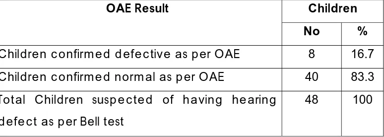
Table 3: Bell test and OAE test.
Children OAE Result
No %
Children confirmed defective as per OAE 8 16.7 Children confirmed normal as per OAE 40 83.3 Total Children suspected of having hearing
defect as per Bell test
48 100
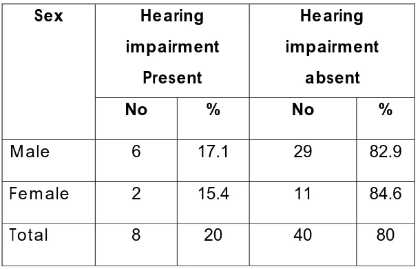
Table: 4 Sex wise distribution
Hearing impairment
Present
Hearing impairment
absent Sex
No % No %
Male 6 17.1 29 82.9 Female 2 15.4 11 84.6 Total 8 20 40 80
p= 0.6266
Table 5 Obstetric history
Hearing impairment
Present
Hearing impairment Absent Obstetric History
No % No %
B.O.H. (n=10) 2 20 8 80 Normal (n=38) 6 15.8 32 84.2 Total (n=48) 8 20 40 80
P=0.5359
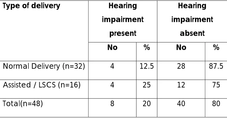
Table 6: Type of delivery
Hearing impairment
present
Hearing impairment
absent Type of delivery
No % No %
Normal Delivery (n=32) 4 12.5 28 87.5 Assisted / LSCS (n=16) 4 25 12 75
Total(n=48) 8 20 40 80
p = 0.2424
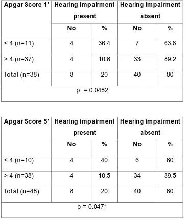
Table 7 : Apgar score
There is a significant correlation between APGAR score and hearing defect. When the APGAR score was less than 4 at 1 or 5 minutes. (ie those with severe birth asphyxia) incidence of hearing defects increases significantly.
Hearing impairment present
Hearing impairment absent
Apgar Score 1’
No % No %
< 4 (n=11) 4 36.4 7 63.6 > 4 (n=37) 4 10.8 33 89.2
Total (n=38) 8 20 40 80 p = 0.0482
Hearing impairment present
Hearing impairment absent
Apgar Score 5’
No % No %
< 4 (n=10) 4 40 6 60 > 4 (n=38) 4 10.5 34 89.5 Total (n=48) 8 20 40 80
Table 8 Birth Weight
Hearing impairment
present
Hearing impairment
absent Birth Weight
No % No %
< 2.5 Kg(n=6) 1 16.7 5 83.3 2.5 – 3.0 Kg (n=34) 6 17.6 28 82.4 > 3.0 Kg (n=8) 1 12.5 7 87.5
Total 8 20 40 80
p=0.6872
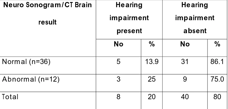
Table 9 Neurosonogram /CT Brain Results
Hearing impairment
present
Hearing impairment
absent Neuro Sonogram/CT Brain
result
No % No %
Normal (n=36) 5 13.9 31 86.1 Abnormal (n=12) 3 25 9 75.0
Total 8 20 40 80
P=0.3137
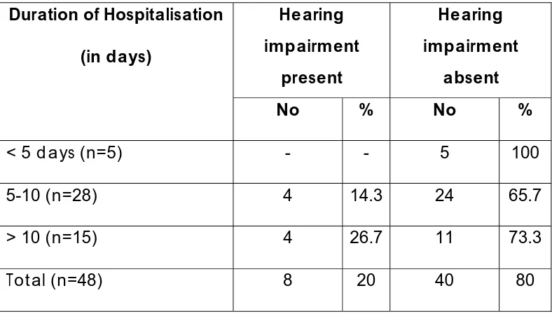
Table 10 Duration of Hospitalisation
Hearing impairment
present
Hearing impairment
absent Duration of Hospitalisation
(in days)
No % No %
< 5 days (n=5) - - 5 100 5-10 (n=28) 4 14.3 24 65.7 > 10 (n=15) 4 26.7 11 73.3 Total (n=48) 8 20 40 80
P=0.4034
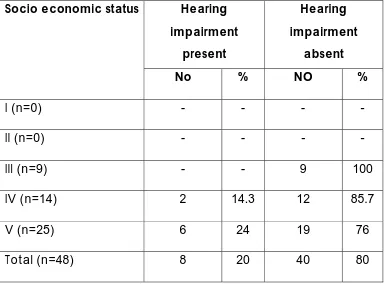
Table 11 Socio economic Status
Hearing impairment
present
Hearing impairment
absent Socio economic status
No % NO %
I (n=0) - - - -
II (n=0) - - - - III (n=9) - - 9 100
IV (n=14) 2 14.3 12 85.7 V (n=25) 6 24 19 76 Total (n=48) 8 20 40 80
P=0.1509
Discussion
Discussion
found to have doubtful turning to sound in bell test they were subjected to oto acoustic emission testing. Since the mean age of turning to sound is around 5.8 months we have taken 6 month as cut off point and screened all infants at 6th months while they
are on follow up.
A total of 282 cases of term birth asphyxiated HIE state II infants were registered for study and out of which 106 cases were lost to follow up for various reasons. Of the remaining 176 cases who were on regular follow up 48 infants had doubtful turning to sound when they were tested by bell method. Of these 48 cases 35 were males 13 were females. These 48 cases were subjected to screening by oto acoustic emission testing.
Of the 8 cases mothers of 2 cases had previous bad obstetric history and in the remaining 6 cases the obstetric history was normal so previous had obstetric history does not affect the out come of hearing significantly.
When comparing the hearing outcome in various mode of delivery we could find that the percentage of infants with hearing impairment in those with assisted delivery was twice as compared to babies delivered by labour natural. But the confounding factor here in that in cases which required assisted delivery already they were in a state of prolonged labour which may itself contribute to perinatal asphyxia.
Very low birth weight infants have been excluded from the study. In this study group were the birth weight ranged from 2.0kg to 3.5 kg there was no obvious significant difference in incidence in any particular weight categories of infants.
incidence of hearing impairment could not be assessed in all social classes. But among this cases no infants was found to be hearing impaired in class III and the percentage of hearing impairment was slightly higher in those belonging to class V when compared to class IV socio economic strata. But a conclusion cannot be reached on this point as this is not a population based study and most of the cases attending the government hospital belonged to lower socio economic strata.
There was a significant correlation between APGAR score and the incidence of hearing impairment. The incidence of hearing impairment was significantly higher in those infants with severe birth asphyxia i.e. infants with 5 minute APGAR score of less than 4 when compared with those of APGAR score of more than 4 at 5 minutes. So the incidence of hearing impairment is directly proportional to the severity of asphyxia.
neuro imaging finding can not be taken as a positive collaborative evidence.
LIMITATIONS
1. Oto acoustic emission was not done for all cases.
2. The testing was done only once and was not repeated.
CONCLUSION
1. Birth Asphyxia can cause hearing impairment in infants.
2. The incidence of hearing impairment is directly proportional to the severity of asphyxia.
3. The incidence of hearing impairment is more in those who required longer duration of inpatient care for control of seizures.
4. The incidence of hearing impairment is higher in those who required assistance during delivery than those who were delivered by labour natural.
RECOMMENDATIONS
1. Universal screening of hearing impairment is essential in all newborns as this can detect hearing impairment at an early stage and early referral for rehabilitation.
BIBLIOGRAPHY
1. White KR Vohr BR, maxon AB, Behrens TR et al article on use of transient evoked oto acoustic emission from International Journal on pediatric oto rhino laryngology 1994 June 29(3) 203-17.
2. Language development in early and late identified children with hearing loss by C.Yoshinaga – itano, A.C.Gedey, D.K.Coutler and A.L.Mehl from Archives of Diseases in child Novl 1998 102 (5): 1161-1171.
3. Universal newborn infant heaving screening using transient evoked oto acoustic emission Results of Rhode Island hearing assessment project by Behrens TR , Vohr BR, White KR from seminars in hearing 1993 14:18-29.
4. The use of transient evoked oto acoustic emission in neonatal screening programs by Kemp DT & Ryans from seminars in hearing 1993 14: 30 to 45.
Behrens TR et al from British Journal of Audiology 1993 27:149-53.
6. Inpatient hearing check of the meningitis using transient evoked oto acoustic emission by Fortnum H, Fransworth A and Davis A. British Journal of Audiology 1993 27 227-32.
7. Community based universal neonatal screening by visitors using oto acoustic emission by Dr. Owen et al Department of pediatrics Gloucestershire Royal NHST from Archives of Diseases of child Ed 2001.84 F157-162. 8. Optimising hearing screening by transient evoked oto
acoustic emission in newborn infants by Welzl Muller K. Boheim K, Stephan K, Schlogel H, Stadimann A, Nekahm D from springer link article 1997 April 45 (4) 227-32.
10. Feasibility of oto acoustic emission detection followed by Auditory Brainstem evoked response audiometry as a universal neonatal screening test for hearing impairment by Hunter MH, Kimml, Cafarelli Dees D, Kennedy CR, Thornton ARD from British Journal of Audiology 1994 28:47-51.
11. Hearing screening in newborn infants comparative study and cost analysis with different instruments by Heinemann M and Bonnert A from International Journal on otorhinolryngology 2000 August 79 (8) 453-8.
12. Newborn hearing screening by oto acoustic emission and ABER by Doyle KJ, Burggruff B, Fujikawas, kim J from International Journal of pediatric otorhinolaryngology 1997 August 20. 41(2):111-9.
13. The use of oto acoustic emission and ABER in newborn by Kennedt CR, Kimml, Cafarelli Dees D, Evann pp et al from Archives of Diseases of childhood 1991:66:1124-9.
targeted screening of High risk infants by Alex R. Kemper MD,MDH, stephen M. Downs MD.MS from Archives of pediatrics and Adolescent medicine 2000;154:484-488.
15. Early hearing screening for infants discharged from NICU by SUNJA, LiJ, Huang P, Buj et al NICU and Department of ENT shangai second medical university from Zhanghna Er keza.zhi 2003 May 41(5):357-9.
16. Joint committee on infant hearing year 2000 position statement principles and guidelines for early hearing detection and intervention programme. American Journal of Audiology 2000 9:9-29.
17. American academy of pediatrics, Task force on newborn infant and hearing loss detection and intervention Archives of diseases of child 1999:103:527-53.
19. Identification of neonatal hearing impairment by con wesion, Barbara, Vehr, Betty, Rsinger et al from Journal of ear and hearing 21(5) 488-597 oct 2000.
20. Wessex universal neonatal hearing screening trial group controlled trial of universal neonatal screening for early identification of permanent childhood hearing impairment.
21. Hall J.W. & Mueller (2000) Handbook of oto acoustic emission son Diago A. Singualar Thomson learning. 22. Watkin pm Neonatal oto acoustic emission screening
and the identification of Deafness Archives of Diseases in child 1996 74:F16-F25.
23. Hack M, Friedman H et al outcomes of extremely low birth weight infants 1996:98 931-937.
24. E medicine article-oto acoustic emission by kathlees cm cambell.phd Director of Audiology southern Illinois university school of medicine july 2002.
25. Nelsons textbook of pediatrics 17th Edition.
26. Manual of Neonatal care John P. Cloherty 5th Edition.
27. Care of Newborn by Meharban singh 5th Edition.
29. Forfar and Arneil’s Textbook of pediatrics 6th Edition.
30. Harold Ludman & Tony wright diseases of the ear 6th
PROFORMA
Screening of term birth asphyxiated infants for hearing loss using oto acoustic emission
Name :
Age : Sex :
Mother : Father :
Address :
Date of Admission :
Date of discharge :
O/P No. :
Family History :
Consanguinity
Other Siblings
Family History of hearing loss
Antenatal history :
H/O exanthematous fever
H/O drug intake
H/O radiation exposure
Natal & Postnatal History :
Mode of Delivery Birth weight
H/O Birth asphyxia
Apgar 1’
5’
H/O Neonatal convulsions
H/O Neonatal Hyperbilirubinemia
H/O Hospitalisation
H/O Seizures & Treatment
Developmental History :
Socio Economic History :
General Examination :
Alertness
Neurocutaneous markers
Abnormal Facies
Developmental anomalies
Vitals :
HR
RR
CRT
Height
Head circumference
CNS :
Consciousness
AF
Pupils
EOM
Facial Nerve
Response to Sound :
Turning to bell
Startle response
Nasal regurgitation
R L
CVS :
S1 S2
Murmur
RS
Trachea
Air Entry
Breath sounds
Abdomen
Soft
Organomegaly
INVESTIGATION
Hb
TC
DC
EEG
Neurosonogram/CT Brain
Oto acoustic emission
Follow up of children enrolled
62% 38%
Bell Test
73% 27%
Bell Test and OAE Test
17%
83%
Sex wise distribution
6
29
2
11
0 5 10 15 20 25 30 35
Obstetric History
Hearing impairment Present
Hearing impairment absent 2
6 8
32
0 5 10 15 20 25 30 35
Type of Delivery
4
28
4
12
0 5 10 15 20 25 30 Hearing impairment
Present Hearing impairment
absent
Birth Weight 1 7 6 28 1 5 0 5 10 15 20 25 30 Hearing impairment Present Hearing impairment absent
< 2.5 Kg(n=6)
2.5 – 3.0 Kg (n=34) > 3.0 Kg (n=8)
Neuro Sonogram/CT Brain result
5 3
31
9
0 5 10 15 20 25 30 35
Hearing impairment Present Hearing impairment absent
Socio Economic Status 0 9 2 12 6 19
0 0 0 0
0 2 4 6 8 10 12 14 16 18 20
Duration of Hospitalisation
Hearing impairment Present
Hearing impairment absent 0
4 4
5 24
11
0 5 10 15 20 25 30 35 40
Apgar Score 1’
4 7
4
33
0 5 10 15 20 25 30 35 Hearing impairment
Present Hearing impairment
absent
< 4 (n=11)> 4 (n=37)
4 6
4
34
0 5 10 15 20 25 30 35 40 Hearing impairment
Present Hearing impairment
absent
< 4 (n=12)> 4 (n=36)
MASTER CHART
APGAR Name OP No. Sex BOH LN/Assisted
or LSCS B.W 1’ 5’ Duration of Hospitalisation Socio economic Status Neuro Sonogr / CT Br
B/o Jeyanthi 308/04 M B A 2.5 3 4 12 V A /O Mariam Beevi 327/04 F N LN 3.1 5 6 4 III N O Mahabaha Beevi 332/04 M B A 2.5 3 4 8 IV A
APGAR Name OP No. Sex BOH LN/Assisted
or LSCS B.W 1’ 5’ Duration of Hospitalisation Socio economic Status Neuro Sonogr / CT Br
B/o Laxmi 23/05 F N LN 2.9 5 6 12 IV N B/o Sameema 29/05 M B A 2.4 4 5 14 IV N B/o Sasikala 38/05 M N LN 3.1 6 6 9 III N B/o Thiravium 43/05 F N LN 2.5 5 6 9 IV N
B/o Puspham 54/05 M N LN 2.8 4 5 7 V N B/o Backialaxmi 62/05 M B LN 2.8 3 4 13 V N B/o Sakeela 76/05 M B A 2.7 5 6 12 V A B/o Mayil 85/05 F N LN 2.3 5 6 8 IV N B/o Punitha 93/05 M N A 2.6 5 6 8 V N B/o Vijayalaxmi 101/05 M N A 3.1 5 5 14 III N B/o Nafeesa 130/05 M N LN 2.5 4 5 6 V N /o Regina Dhoni 146/05 F N LN 2.5 5 6 4 IV N /o Azhagumeena 155/05 F N LN 2.9 3 3 8 V N B/o Deepa 162/05 M N LN 2.7 4 6 9 V N B/o Alamely 171/05 M N LN 3.2 4 6 8 III N B/o Amutha 178/05 M N LN 2.8 5 6 13 V N B/o Laxmi 193/05 M N LN 2.9 4 5 8 IV N B/o Mahadevi 209/05 F B A 2.5 3 4 7 V A B/o Kurinji malar 220/05 M B LN 2.3 5 6 4 V A
APGAR Name OP No. Sex BOH LN/Assisted
or LSCS
B.W
1’ 5’
Duration of Hospitalisation
Socio economic
Status
Neuro Sonogr / CT Br