AND NON-DIABETICS-A COMPARATIVE STUDY
DISSERTATION SUBMITTED FOR M.D GENERAL MEDICINE
BRANCH – I
APRIL 2016
THE TAMILNADU DR.M.G.R. MEDICAL UNIVERSITY CHENNAI,
This is to certify that the dissertation entitled “PULMONARY FUNCTION TEST IN TYPE-2 DIABETICS AND NON-DIABETICS-A
COMPARATIVE STUDY” is the bonafide work of Dr. ILAMARAN.M, in partial fulfillment of the university regulations of the Tamil Nadu Dr. M.G.R. Medical University, Chennai, for M.D General Medicine Branch I examination to be held in April 2016.
Dr. S. VADIVEL MURUGAN, M.D
DEAN,
KANYAKUMARI GOVERNMENT MEDICAL COLLEGE
This is to certify that the dissertation entitled “PULMONARY FUNCTION TEST IN TYPE-2 DIABETICS AND NON-DIABETICS-A
COMPARATIVE STUDY” is the bonafide work of Dr. ILAMARAN. M., in partial fulfillment of the university regulations of the Tamil Nadu Dr. M.G.R. Medical University, Chennai, for M.D General Medicine Branch I examination to be held in April 2016.
Dr. V. ANTONY DAVID DEVADHAS,M.D
PROFESSOR AND HOD,
DEPARTMENT OF GENERAL MEDICINE,
KANYAKUMARI GOVERNMENT MEDICAL COLLEGE
This is to certify that the dissertation entitled “PULMONARY FUNCTION TEST IN TYPE-2 DIABETICS AND NON-DIABETICS-A
COMPARATIVE STUDY” is the bonafide work of Dr. ILAMARAN.M, in partial fulfillment of the university regulations of the Tamil Nadu Dr. M.G.R. Medical University, Chennai, for M.D General Medicine Branch I examination to be held in April 2016.
Dr. M. CHRISTOPHER NESAMONY, M.D
PROFESSOR,
DEPARTMENT OF GENERAL MEDICINE,
KANYAKUMARI GOVERNMENT MEDICAL COLLEGE
I, Dr.ILAMARAN.M, solemnly declare that, this dissertation
“PULMONARY FUNCTION TEST IN TYPE-2 DIABETICS AND
NON-DIABETICS-A COMPARATIVE STUDY” is a bonafide record of work done by me at the Department of General Medicine, Kanyakumari government medical college hospital, Asaripallam, under the guidance of Dr. M. CHRISTOPHER NESAMONY, M.D, Professor, Department of General Medicine, Kanyakumari Govt Medical college, Asaripallam. This dissertation is submitted to The Tamil Nadu Dr. M. G. R. Medical University, Chennai in partial fulfillment of the rules and regulations for the award of M.D Degree General Medicine Branch-I;
examination to be held in April 2016.
Place: ASARIPALLAM Date:
ACKNOWLEDGEMENT
I would like to thank Dr.S.VADIVEL MURUGAN M.D GENERAL MEDICINE Dean, Kanyakumari Medical College, for permitting me to utilize the facilities of Kanyakumari Medical College and Hospital facilities for this dissertation.
I wish to express my respect and sincere gratitude to my beloved
teacher and Head of the Department, Prof. Dr.V.ANTONY DAVID DEVADHAS M.D., Professor of Medicine for his valuable guidance and encouragement during the study and also throughout my course period.
I would like to express my deep sense of gratitude, respect and thanks to my beloved Unit Chief and Professor of Medicine, Prof. Dr. M. CHRISTOPHER NESAMONY, M.D., for his valuable suggestions, guidance and support throughout the study and also throughout my course period.
I am greatly indebted to my beloved Professors, Dr. PRINCE SREEKUMAR PIUS, M.D., Dr.VIJAYARAJU, M.D., for their help throughout the study.
throughout the study period.
I am extremely thankful to Assistant Professor of Medicine of my Unit,
Dr. G. SANTHLEDGE M.D.,D.M, for their valid comments and suggestions. I sincerely thank the Assistant Professors of Thoracic Medicine, Dr. S. MUTHUKUMAR, M.D chest medicine, Dr.T. JOSEPH PRATHEEBAN
M.D chest medicine for their guidance and suggestions in my dissertation work.
I sincerely thank all the staffs of Department of Medicine and Department of Thoracic Medicine for their timely help rendered to me, whenever and wherever needed.
I extend my thanks to all my friends, batch mates and my junior and senior colleagues who have stood by me and supported me throughout my study and course period.
Finally, I thank all the patients, who form the most vital part of my work, for their extreme patience and co-operation without whom this project would have been a distant dream and I pray God, for their speedy recovery.
CONTENTS
S.NO CONTENTS PAGE.NO
1. INTRODUCTION 1-4
2. REVIEW OF LITERATURE 5-57
3. AIM OF STUDY 58
4. MATERIALS AND METHODS 59-63
5. RESULTS AND INTERPRETATION 64-71
6. DISCUSSION, LIMITATION 72-75
7. CONCLUSION 75-76 8. SUMMARY 76-77 9. ANNEXURES 78-96
BIBLIOGRAPHY PROFORMA MASTER CHART
ETHICAL COMMITTEE APPROVAL LETTER ANTI PLAGIARISM CERTIFICATE
INTRODUCTION
The management of endocrine disorders requires the understanding of intermediary metabolism, reproductive physiology, bone metabolism, and growth. The practice of endocrinology is intimately linked to conceptional framework for understanding hormone secretion, hormone action and principles of feedback control.
Thyroid hormone controls about 25% of basal metabolism of most of the body tissues, cortisol exerts a permissive action for many hormones in addition to its own direct effects, PTH regulates calcium and phosphorus levels, vasopressin regulates serum osmolality by controlling water balance, mineralocorticoids control minerals metabolism, insulin maintains euglycemia in the fed and fasted states.
The causes of endocrine dysfunctions are mainly due to
1.Hyperfunction:
-neoplastic
-autoimmune
-iatrogenic
-infectious
- activating receptor mutation.
2.Hypofunction:
-autoimmune
-iatrogenic
-hormone mutations
-enzyme effects
-develepmental defects
-nutritional or vitamin deficiency
-hemorrhage or infarction
3. Hormone resistance:
-receptor mutation
-signalling mutation
-post receptor mutation
Type 2 diabetes mellitus is nothing but a persistent hyperglycemia and altered metabolism of lipids, carbohydrates and proteins. Several distinct types of diabetes mellitus are caused by complex interactions between genetics and environmental factors. These are result from impaired insulin secretion and insulin resistance or combination of both of these mechanisms.
Type 2 diabetes mellitus is associated with chronic tissue damage, reduction in function, failure of multiple organs and its complications are preferably caused by macrovascular and microvascular damages and all are due to the metabolic dysregulation of the diabetes mellitus.
Though great attention was centered on the diabetic complications which had cardiovascular nature, nephropathy, retinopathy and neuropathy, the pulmonary complications of type2 diabetes mellitus have been poorly characterized. Of late the concept of the lung as a target organ for diabetic microangiopathy is receiving continuing attention. The aim of the study was to assess the effects of chronic hyperglycemia on lung functions, which focused on mechanical aspects of lung dysfunction, maximal forced spirometric pulmonary function tests like FVC, FEV1, FEV1/FVC to be specific1.
The pulmonary complications of type 2 diabetes mellitus have been poorly characterized1, 2 .The complications are affect the lungs silently and may produce increased morbidity because of lung dysfunction2.
Relatively few studies have been done on pulmonary mechanical function. Our study mainly concentrating on mechanical dysfunction of lungs due to diabetes mellitus mainly maximal forced spirometric PFTs to be specific. Most of the studies were done on type1diabetics3. The present study was done on type 2 diabetics.
REVIEW OF LITERATURE
DIABETES MELLITUS:
Diabetes mellitus is characterized by chronic hyperglycemia due to defects in insulin secretion, peripheral insulin action or both which leading to alteration in the fat, proteins and carbohydrate metabolism of the individual5.
Type 2 diabetes affects all types of ethnicity, social and economic levels of the society. Diabetes is among the 5 leading cause of death in most countries. Better and early disease detection, changing life styles and changes in the diagnostic criteria have led to this increase.
Death and disability associated with diabetes poses a serious challenge to physicians and the health system at large.
ETIOLOGIC CLASSIFICATION OF DIABETES:
1) Type 1 diabetes (Beta cell destruction, leading to absolute insulin deficiency)6
a) Immune mediated
b) Idiopathic
3) Other specific types6
a) Genetic defects of beta cell function
1. Maturity onset diabetes of the young 1-6
2. Mitochondrial DNA
3. Mutant insulins
4. Hyperinsulinemia
5. Others
b) Genetic defects in insulin action
1. Type A insulin resistance
2. Leprechaunism
3. Rabson-Mendenhall syndrome
4. Lipoatropic diabetes
5. Others
c) Diseases of exocrine pancreas
1. Pancreatitis
2. Trauma / Pancreatectomy
4. Cystic fibrosis
5. Hemochromatosis
6. Fibro-calculous pancreatopathy
7. Others
d) Endocrinopathies
e) Drug or chemical induced
1. Vacor
2. Pentamidine
3. Nicotinic acid
4. Glucocorticoids
5. Thyroid hormone
6. Diazoxide
7. Beta adrenergic agonists
8. Thiazides
9. Phenytoin
10. Alpha interferon
12. Atypical antipsychotics
f) Infections
1. Congenital rubella
2. Cytomegalovirus
3. Others
g) Uncommon forms of immune mediated diabetes
1. “Stiff man syndrome”
2. Anti-insulin receptor antibodies
3. Others
h) Other genetic syndromes associated with diabetes
1. Down’s syndrome
2. Klinefilter’s syndrome
3. Wolfram’s syndrome
4. Friederich’s ataxia
5. Huntington’s chorea
6. Laurence Moon Biedel syndrome
8. Porphyria
9. Prader willi syndrome
10. Others
4) Gestational diabetes mellitus5, 6
CRITERIA FOR DIAGNOSIS OF DIABETES MELLITUS:5, 6, 7
1. Symptoms of diabetes with casual random blood glucose – 200 mg/dl (11.1 mmol/L)
Random is defined as any time of delay without time of the last meal.
The symptoms of diabetes are polyuria, polydipsia, and unexplained weight loss
OR
2. Fasting blood glucose > 126 mg/dl (7.0 mmol/L)
Fasting is defined as no caloric intake for atleast 8 hour
OR
A1C > 6.5%
3. 2- hr postprandial blood glucose >200 mg/dl (11.1 mmol/L) during an OGTT. The test should be performed as described by WHO, using a glucose load containing the equivalent of 75g of anhydrous glucose dissolved in water.
SCREENING OF DIABETES MELLITUS:
Widespread use of fasting plasma glucose and A1C as a screening for diabetes mellitus is recommended because
1. A large number of individual who met the criteria foe diagnosis are mostly asymptomatic only and unaware of that they having the disorder.
2. Epidemiologic studies suggested that type 2 diabetes mellitus may be present for up to the decade before diagnosis.
3. Some people having one or more diabetic complication at the time of diagnosis itself.
4. The treatment of diabetes mellitus may alter the natural history of the disease.
RISK FACTORS FOR TYPE 2 DIABETES MELLITUS:
1) History of diabetes mellitus in family members
2) Obese ( Body mass index > 25/meter square)9
3) Sedantary life 9
4) Past history of Impaired fasting glucose or Impaired glucose tolerance or and A1c of 5.7 – 6.4% 9
5) Past History of gestational diabetes mellitus or delivery of a baby of > 4 kilograms 9
6) High density cholesterol < 35 milligrams/dl (0.90 mmol/L) and /or a triglyceride > 250milligrams/dl (2.82mmol/L) 9
7) Polycystic ovarian disease 8, 9
8) History of vascular disease 9
9) History of cardiac disease 8, 9
PATHOGENESIS FOR TYPE 2 DIABETES MELLITUS:
Earliest abnormality seen in type 2 diabetes is impairment in tissue sensitivity to insulin. This results in an increase in demand on the beta cell to maintain a sufficiently high rate of insulin secretion to the offset of insulin resistance. After a certain time, when the insulin secretion fails to meet the insulin demand, overt diabetes occurs.
There is a very clear understanding for the role of genetic factors in development of type 1 diabetes however; it is not the case with type 2 diabetes. Type 2 diabetes is caused by interactions between the environmental factors and genetic factors.
DEFECTIVE 1ST PHASE INSULIN SECRETION:
This phase of insulin secretion helps in priming the insulin target tissues to maintain the normal glucose homeostasis. It is one of the early manifestations, which is found to occur when fasting glucose rise to 115-120mg% 10
DEFECTIVE PULSATILE INSULIN SECRETION:
FACTORS AFFECTING BETA CELL FUNCTION:
• Chemical toxins like alcohol
• Malnutrition
• Chronic pancreatitis
• Amylin accumulation
• Intrauterine environment
• Drugs like thiazides, beta blockers etc…
• Glucose transporter 2 defect
• Decreased glucokinase activity
• Defects in phosphoinositol
ROLE OF LIVER IN GLUCOSE METABOLISM:
1. Liver is the organ of glucose production and glucose consumption
2. It is exposed to insulin concentration in the portal circulation which is 3 – 10 times more the systemic circulation11.
3. Sole site of glycol regulatory action of glucose11, 12.
Liver has storage of glucose and glycogen about 70gm at a time. 75% of hepatic glucose output comes from gluconeogenesis11, 12.
Contribution of liver in glucose homeostasis depends on the following factors:
1. Sensitivity of hepatocytes to small increments in insulin levels
2. Ratio of insulin to glucagon
3. Responsiveness of glycogenolysis and gluconeogenesis to hormonal modulation11.
PRESENTATION OF DIABETES MELLITUS:
Diabetes can be detected in one of the following ways. Some patients are found to have excess of glucose or sugar in urine incidentally on routine checkup without any complaints or physical signs.
Some patients are found to have diabetes while investigating for an associated complaint like, ischemic heart disease, hypertension, eye diseases, kidney disease, non- healing foot ulcers etc12.
Some of the patients often present with classical symptoms of diabetes. Eg., excessive thirst, frequent micturition, increased appetite, weight loss, severe weakness, repeated infections, itching in genitals, diminished vision, numbness in limbs and occasionally impotence13.
COMPLICATIONS OF DIABETES MELLITUS:
DIABETIC METABOLIC EMERGENCIES:
The two main metabolic complications of diabetes are diabetic ketoacidosis and hyperosmolar state12. Initially DKA was considered as the main complication of Type 1 diabetes mellitus. Both are associated with relative insulin deficiency, volume depletion and acid base abnormalities.
PATHOPHYSIOLOGY:
1. Non-enzymatic glycosylation of proteins, e.g. Hemoglobin, collagen, LDL and tubulin in peripheral nerves14.
2. Polyol pathway13, 14.
3. Abnormal microvascular blood flow to the peripheries14.
4. Reactive oxygen species and growth factors stimulation (TGF -J3) and vascular endothelial growth factor (VEGF) 14.
MACROVASCULAR COMPLICATIONS:
Diabetes is a risk for the development of atheroma.
This includes
a) Ischemic heart disease
b) Peripheral vascular disease
c) Cerebrovascular accidents
The cardiovascular complications are more in diabetes mellitus. So the patient with diabetes mellitus with suspected cardiovascular risk factors has to be started with an anti-hypertensive mainly ACE inhibitor, a statin and a low dose aspirin for all the patients15 unless otherwise contraindicated.
MICROVASCULAR COMPLICATIONS:
Small blood vessels all over the body are affected but the disease process is of danger in 3 sites:
a) Eye – Retionopathy
b) Neuropathy –(mono and polyneuropathy)
DIABETIC RETINOPATHY:
Diabetic retinopathy is divided into proliferative diabetic retinopathy (PDR) and non-proliferative diabetic retinopathy (NPDR). Hemorrhages or micro aneurysms, cotton wool spots, hard exudates, intra retinal microvascular abnormalities, venous caliber abnormalities like venous loops, venous bleeding and venous tortuosity are some of the findings associated with early and progressive diabetic retinopathy. Micro aneurysms and saccular out-pouchings of the capillary walls can leak fluid and results in intra edema and hemorrhages16. These intra retinal hemorrhages are flame shaped or dot blot like appearance, reflecting the architecture of the layer of the retina in which they occur. Flame shaped hemorrhages occur in inner retina closer to the vitreous and dot blot hemorrhages occur deep in the retina. Intra retinal microvascular abnormalities are either new vessel growth within retinal tissue or shunt vessels through areas of poor vascular perfusion.
DIABETIC NEUROPATHY:
Diabetic neuropathy has a number of clinical syndromes with subclinical or clinical manifestations depending upon the class of nerve fibres involved, it can manifest as polyneuropathy, mono neuropathy or autonomic neuropathy.
Distal symmetrical polyneuropathy is the most common form of diabetic neuropathy, where patients frequently present with distal loss of sensation, hyperesthesia, paresthesia and dysthesia. Symptoms may include a sensation of numbness, tingling, sharpness that begins in feet and spreads proximally. As the diabetes progresses, the pain subsides and eventually disappears, but a sensory deficit in lower limbs persist.
Diabetic poly-radiculopathy is a syndrome characterized by severe disabling pain in the distribution of one or more nerve roots, which can be accompanied by motor weakness. There can be severe pain in hip and thigh due to involvement of lumbar plexus. Fortunately this condition is usually self-limited and resolves in 6 -12 minutes.
Autonomic neuropathy in diabetes can involve multiple systems like cardiovascular, gastrointestinal, genito-urinary17 etc. patient can have resting tachycardia and orthostatic hypotension when cardiovascular system is involved. Gastropathy and bladder dysfunction are caused by autonomic neuropathy of gastrointestinal tract and genitourinary tract.
Increased sweating of upper extremities and decreased sweating of lower extremities can occur due to sympathetic nervous system dysfunction. There is a increased chance of foot ulcers due to anhidrosis of lower extremities which causes dry skin and cracking.
Autonomic neuropathy can produce hypoglycemic unawareness, that means it decreases the counter regulatory hormones of glucose metabolism in the period of hypoglycemia. So the hypoglycemia episodes are more common with diabetic neuropathy.
NEPHROPATHY:
Nephropathy secondary to diabetic glomerulopathy occurs after diagnosis is about 15-20years and 25-35% of patients detected under the age of 30 years.
It may the leading cause of premature death in diabetic patients in the young individuals.
Diabetic nephropathy characterized by hypertension, proteinuria and renal impairment.
The diabetes affects kidney by: • Glomerular insult
• Ischemia from hypertrophy of afferent and efferent arterioles
• Ascending mode of infection.
The earliest abnormality of the diabetic kidney has the renal hypertrophy associated with an increased glomerular filtration rate. This appears immediately after diagnosis and is related to poor diabetic control.
Stage Description GFR (mL/min/1.73 m2)
1. Kidney damage with normal or elevated Glomerular filtration rate >90
2. Kidney damage with mildly decreased Glomerular filtration rate 60–89
3. Moderately decreased Glomerular filtration rate 30–59
4. Severely decreased Glomerular filtration rate 15–29
5. Kidney failure <less than 15 (or dialysis)
Stage 1:
Here glomerular filtration rate is elevated on an average by 20-40% above that of age matched normal controls in both adults and children with type 1 DM resulting in glomerular enlargement.
Stage 2 :
normal albumin excretion rate despite structural changes. Most patients remain in stage 2, however 30-40% progress to subsequent stages.
Stage 3:
Here patients will have microalbuminuria detected. Patients who have albumin excretion rates >30 microgm/min are more likely to develop clinical diabetic nephropathy than those with less than 30 microgm/min.
Stage 4:
In this stage, patients develop overt nephropathy. By definition, patients have persistent clinical proteinuria with albumin excretion rate of > 250 microgm/min in 24 hrs, hypertension and a decrease in GFR. Once proteinuria is persistently present, development of end stage renal disease or death occurs in 3 – 4.8 yrs.
Stage 5:
This is a stage of advanced renal failure. In contrast to non-diabetic patients, diabetics with end stage renal disease usually have other systemic manifestations in addition to their renal diseases.
can induce a transient nephrotic syndrome, anasarca and decreased albumin level.
Patients with nephropathy show in peripheral smear normochromic normocytic anemia and an elevated erythrocyte sedimentation rate (ESR).
Development of hypertension may itself damage the kidney still further.
Effective treatment of blood pressure in a target of less than 130/80 mmHg has been shown to delays the progression of renal failure considerably.
ACE inhibitors or angiotensin receptor blockers are the drugs of choice.
For evaluating diabetic nephropathy initially have to do microalbuminuria test as a spot collection and excludes the conditions that causes the increase albumin excretion, and repeat it every 3-6 months period. If two of the three microalbuminuria test have positive results then we have to start the treatment with either ACE inhibitors or angiotensin receptor blockers.
PULMONARY COMPLICATIONS:
Major consequences of hyperglycemia are excessive non enzymatic glycosylation of various body proteins including albumin1, 3, 7, collagen and elastin. In type 2 diabetics with uncontrolled disease there is decreased pulmonary functions have been noted. There may be increased cross linkage between polypeptides of collagen which leading on to thickening leading to restriction of lung volume and alveolar gas transport, reduced membrane diffusion capacity and pulmonary capillary blood volume15,16, 17. The possible explanations of restrictive type of lung disease are thickening of alveolar epithelium, pulmonary microangiopathy and centrilobular emphysema. So the net effect due to collagen & elastin changes and microangiopathy1`, 2 , 18, 19,.
The lung complications of type 2 diabetes mellitus are mainly affects the
OTHER COMPLICATIONS:
a) Cardio vascular
b) Gastrointestinal tract ( decreased motility, diarrhea)
c) Genitourinary tract (urological abnormalities, impotence)
d) Infections
e) dermatological
f) glaucoma
g) cataracts
h) periodontal diseases
i) hearing loss
j) lower extremity complications
k) Dyslipidemia
METABOLIC SYNDROME:
Insulin resistance and hyper insulinemia is being increasingly implicated in the pathogenesis of various metabolic disturbances. Some of them has been clubbed under a syndrome.
Diagnosis of metabolic syndrome is made in the presence of atleast three of the following –
1. Waist circumference >102 centimeter in male and >88 centimeter in females
2. Triglycerides >150mg/deci litre or patients using nicotinic acid or fibrates
3. HDL <40mg/deci litre in males and <50mg/deci liter in females
4. Systolic Blood pressure >130mmHg or diastolic Blood pressure >85 mmHg or patients using anti hypertensive drugs
5. Fasting blood sugar > 100mg/dl or patients using OHA’s.
CHILDREN, ADOLESCENTS,AND YOUNG ADULTS:
Diabetes in children and teenagers is generally ketosis prone, insulin deficient type 1 diabetes, but cases of type 2 diabetes in obese children are now occurring and the possibility of a diagnosis of maturity onset diabetes of the young should be considered, especially when there is a positive family history of diabetes. Adherence to the treatment is tough in adolescents, so the deterioration of glycemic control is common18.
MANAGEMENT OF TYPE 2 DIABETES:
LIFESTYLE INTERVENTION:
Reducing weight and physical exercise are also the important part in the management of diabetes mellitus. Increase in weight with diabetes mellitus and decreased physical activity increases the complications of diabetes mellitus and increases the mortality.
Proper exercise and diet control are the key points in the management of diabetes mellitus. But the long term complications are which are not controlled by exercise and diet control alone, it needs immediate drug management.
FOOD SELECTION:
RECOMMENDED COMPOSITION OF DIET:
Carbohydrate- 45-60% daily
Sucrose- up to 10%
Fat (total) <35%
n-6 polyunsaturated- <10%
n-3 polyunsaturated- eat oily fish once or twice weekly
monounsaturated- 10-20%
saturated- <10%
protein- 10-20% (do not exceed 1gram per kilogram body weight)
Plate model may provide a simple visual aid to show the proportions of carbohydrates and other food groups for selection at mealtimes.
PHYSICAL ACTIVITY:
Increase in the physical activity among the people with diabetes mellitus is the first main treatment in the management of diabetes mellitus. Aerobic exercise is advisable. The importance of exercise is to reduce the weight and it increases the insulin sensitivity in the target organs by increasing the receptors for the activity.
The physical activity is individualized according to the patients performance status and comorbidities and the age and the risk factors. In the patient with diabetes mellitus ADA recommends about 150minutes per week of exercise that is distributed over at least 3days, of moderate aerobic exercise20, 21. The exercise regimens also includes the resistance training.
The one of the major complication is exercise induced hypoglycemia that too more common in type 1 diabetic patients and can occur with patient on insulin or insulin analogues.
MEDICATIONS:
The goal of therapy in type 2 diabetes should not only improve the beta cell function, but also enhance the glucose utilization in the peripheral tissues. These oral drugs also have the potential to correct the hormonal and metabolic abnormalities in diabetes.
SULFONYLUREAS:
Sulfonylureas act by binding to the so called sulfonylurea receptors on the pancreatic beta cell membranes causing depolarization, calcium influx and degranulation of secretory granules with insulin release. Therefore, these drugs primarily augment second phase of insulin secretion and has very less action on first phase.
Drugs – Tolbutamide, Chlorpropamide, Glimepride, Glipizide, Glibenclamide, Gliclazide.
The more rapid onset of action of sulfonylureas, lesser is the delay in the postprandial insulin release. Glipizide results in rapid post prandial insulin release and lowers post prandial glucose. In contrast glibenclamide exerts a better effect on fasting glucose. Hence, where fasting glucose levels are high, glibenclamide may be preferred to glipizide and vice versa.
sulfonylurea therapy. Secondary failure, decresing the beta cell function and increasing the insulin resistance are important disease related factors for sulfonylurea failure besides the duration of the disease.
BIGUANIDES:
Metformin, phenformin and buformin belongs to this group. The later two drugs are withdrawn long back. Metformin is an old, but still the best agent of choice to start with in treatment of type 2 diabetes. Its efficacy, safety profile and its capacity to be associated with other anti-diabetic agents makes metformin the first line glucose lowering drug of choice in diabetes management.
The principle site of action is in the liver and muscles. The effort of metformin on liver is mediated by activation of liver kinase B1. This drug is prefered in normal weight patients, contrary to its widespread perception in our country that it is preferred in obese patients.
THIAZOLIDINEDIONES:
Thiazolidinediones are potent insulin sensitisers, that act through the PPAR gamma This PPAR gamma mediated transcriptional effects have are improves whole body sensitivity to insulin. Drugs used are Troglitazone, Rosiglitazone and pioglitazone. Among these drugs, troglitazone have been banned in the year 2000, due to its fatal hepatotoxicity after which rosiglitazone was introduced. Pioglitazone is the third drug used that has been shows to improvement of sensitivity of human body insulin.
GLP-1 ANALOGUES:
This incretin hormone GLP-1 is secreted from intestinal L cells, in the distal part of ileum and colon after food intake. The effect of subcutaneous injection of GLP-1 analogue is very short acting due to N terminal degradation by the enzyme dipeptidyl peptidase IV, restricting its cardiac use.
Drugs include Exenatide, Lexixinatide, Liraglutide, Exenatide LAR.. These drugs has beneficial effect on cardiovascular system lipid profile, obesity and also in central nervous system.
DIPEPTIDYL PEPTIDASE- IV INHIBITORS:
DPP-IV inhibitors are novel anti-diabetic drugs based on the incretin therapy. These drugs help in decreasing the degradation of endogenous incretin hormone. Gliptins are generally considered as weight neutral agents and may assist a small amount of weight loss. Drugs include – Sitagliptin, Vidagliptin, Saxagliptin, Linagliptin.
ALPHA – GLUCOSIDASE INHIBITORS:
Acarbose and Voglibose are these drugs. Mechanism of action of alpha glucosidase inhibitors involves block of the enzyme alpha glucosidase in the intestine which normally clears carbohydrates into absorbable monosaccharides.
ROLE OF INSULIN IN TYPE 2 DIABETES:
Type 2 diabetes constitutes nearly 95- 97% of all diabetics. The successful management of type 2 diabetes involves around an individual tailored nutritional plan, exercise regimen, use of oral agents and or insulin.
Tissues insensitivity to insulin and impaired insulin secretion are the pathogenetic factors underlying type 2 diabetes. The primary defect is tissue insensitivity to insulin.
INDICATIONS FOR INSULIN USE IN TYPE 2 DIABETICS:
a) Primary oral agent failure
b) Secondary oral agent failure
c) Peri-operative
d) Pregnancy
e) Acute on chronic sepsis
f) Acute medical or surgical event
g) Major organ failure
h) Glucotoxicity
WHEN TO START INSULIN?:
a) At diagnosis , fasting glucose > 200mg/dl ,post prandial glucose > 300 mg/dl with HbA1c >9%22.
b) After oral; drug failure, despite receiving optimal dose of 2 or 3 OAD’s, fasting glucose > 150 mg/dl, post prandial glucose > 200mg/dl with HbA1c levels of >8.5% 22 .
PROPERTIES OF INSULIN PREPARATIONS:
Type of preparation Time onset
(hours)
Peak level
(hours)
Duration of
action (hours)
Lispro < 0.25 0.5-1.5 3-4
Aspart <0.25 0.5-1.5 3-4
Glulisine <0.25 0.5-1.5 3-4
Regular 0.5-1.0 2-3 4-6
Detemir 1-4 Up to 24hrs
Glargine 1-4 Up to 24hrs
NPH 1-4 6-10 10-16
75/25 – 75%protamine lispro,25% lispro
50/50- 50% protamine
lispro,50lispro 70/30-70%NPH,30%
regular
0.5 -1 dual 10-16
Factors affecting the disposal of injected insulin:
1. Anatomic site 2. Exercise 3. Depth
4. Insulin concentration 5. Mixing of preparations 6. Local tissue degradation
7. Intra subject coefficient variation
8. Antibody binding and release of insulin
GESTATIONAL DIABETES MELLITUS:
Glucose metabolism is altered during the normal pregnancy. There is a continuous relationship between maternal blood glucose and risk of adverse perinatal out comes . So that there is no threshold to defines the risk. According to WHO criteria all women meeting criteria for impaired glucose tolerance or diabetes after 75grams of oral glucose tolerance test having gestational diabetes. European criteria for diagnosis for gestational diabetes are a venous blood glucose of more than 99mg/dl in fasting state and of more than 162mg/dl of 2hr postprandial24.
RISK FACTORS FOR GESTATIONAL DIABETES:
1. Obesity
2. Ethnicity (south Asian, black, Hispanic, native American) 3. Family history of type2 diabetes
4. Previous glucose abnormalities in pregnancy 5. Previous macrosomia
SPIROMETRY & PFT:
Used in the ambulatory setting, physician's office, emergency department, or inpatient setting 1 ,5 ,8 ,24.
It needs patients voluntary effort to inhale maximally beyond the tidal volume and exhale forcefully into the close circuit maneuver. Before doing the test the examiner must explain the complete procedure to reduce the faults10.
Can determine:
- Forced expiratory volume in first second (FEV1)
- Forced vital capacity (FVC)
- FEV1/FVC
- Forced expiratory flow 25%-75% (FEF25-75)
Indications for Diagnosis:
1. Evaluate the patient having sudden onset of dyspnea, exertional dyspnea, chronic cough.
2. Screen the high risk populations.
3. Monitoring pulmonary toxicities of certain drugs.
4. Abnormalities in chest x ray, acid blood gas analysis and hemoglobin. 5. Preoperative evaluation.
Indications for Prognosis:
1. Assess for severity of illness. 2. Therapy response.
3. Requirements of further treatment plan. 4. Surgery referral.
5. Disable
Contraindications for spirometer:
Relative contraindications for spirometer includes recent heart disease, unstable angina, abdominal aorta aneurysm, recent eye surgery, recent surgeries in the past, hemoptysis of unknown origin hemoptysis of unknown origin, pneumothorax, cerebral artery aneurysm, syncopal attacks, thoracic aorta aneurysm28, 29.
Spirometer is used for only the single breath. Repeated breathing is not indicated in this spirometer because repeated breathing can cause the occumulation of carbon dioxide and other air and oxygen are cannot be provided to the patients.
Computerized spirometer is the solid state does not contain drum or water column. Subjects are respire into a sophisticated transducer which is connected to the instrument of a cable.
SPIROMETRY ACCEPTIBILITY CRITERIA:
1. There is a good starting of the test without any form of hesitation
2. No interrupting coughing and the closure of the glottis- may produces the faulty procedure
3. The flow will be constant, there should not be any variability in the flow of air
4. Not to terminate very earlier. It should be more than 6seconds
5. There should not be any leakage of air through the mouth or anywhere from the instrument
6. It should be reproducible in nature.
PFT:
LUNG VOLUMES AND CAPACITIES:
LUNG VOLUMES:
TIDAL VOLUME (TV):
It is the volume of air breathed in and out of the lungs in a single normal quiet breathing. It signifies the normal depth of breathing. Normal 500ml
Inspiratory Reserve Volume (IRV):
It is an additional volume of air that can be inspired forcefully after the end of normal inspiration.
3000ml
Expiratory Reserve Volume(ERV):
It is the additional amount of air that can be expired out forcefully after normal expiration. Normal 1500ml.
Residual Volume (RV):
It is the volume of air remaining in the lungs even after forceful expiration.
Normal 1200ml
LUNG CAPACITIES:
Total Lung Capacity (TLC):
It is the volume of air present in lungs after a deep inspiration. It includes
Vital Capacity (VC):
It is the maximum volume of air that can be expelled out forcefully after a deep inspiration (60‐70 ml/kg) 5000ml.
Inspiratory Capacity (IC):
It is the maximum volume of air that is inspired after normal expiration. (2400‐3800ml).
Expiratory Capacity (EC):
TV+ ERV
Functional Residual Capacity (FRC):
It is the volume of air remaining in lungs after normal expiration (30‐35 ml/kg) 2500 ml.
MEASURING RESIDUAL VOLUME AND FUNCTIONAL RESIDUAL
CAPACITY:
1. Nitrogen washout technique. 2. Helium dilution method. 3. Body plethysmography
PROCEDURE:
FORCED VITAL CAPACITY (FVC):
\
FORCED VITAL CAPACITY INTERPRETATION30:
Interpretation should be the percentage of the predicted value : 80-120percentage of predicted Normal
70-79 percentage of predicted Mild reduction 50%-69 percentage of predicted Moderate reduction <50 percentage of predicted Severe reduction30.
FORCED EXPIRATORY VOLUME IN 1st SECOND: (FEV1)
Volume of air forcefully expired from full inflation (TLC) in the first second. Measured in liters (L) .Normal people can exhale more than 75-80% of their FVC in the first second; thus the FEV1/FVC can be utilized to characterize lung disease.
FORCED EXPIRATORY VOLUME IN 1st SECOND
INTERORETATION:
Interpretation of % predicted:
>75% of predicted Normal
60%-75% of predicted Mild obstruction 50-59% of predicted Moderate obstruction <49% of predicted Severe obstruction31
FORCED EXPIRATORY FLOW 25-75% (FEF25-75)
Mean forced expiratory flow during middle half of Forced vital, capacity
Measured in Liter/second,
May reflect effort independent expiration and the status of the small airways,
Highly variable,
Depends heavily on forced vital capacity.
FORCED EXPIRATORY FLOW 25-75%(FEF25-75) INTERPRETATION:
Interpretation of % predicted:
>60%of predicted Normal
40-60% of predicted Mild obstruction 20-40% of predicted Moderate obstruction <10% of predicted Severe obstruction31, 32
OBSTRUCTIVE DISORDERS:
The main mechanism is limitation of expiratory airflow like asthma, COPD, bronchiolitis, bronchiectasis.
Manifested by decreased level of FEV1, FEF25-75, FEV1/FVC ratio (<0.8)
REST
TRICTIVE
Manifeste
– abn
– abn dys
Decreased
E LUNG
ed by decr
normalitie
normalitie strophy) ,c
d total lun
DISEASE
reased volu
s in lung p
s in pleur chest wall
ng capacity
E:
umes of lu
parenchym
ra, neuro (e.g. scol
y, forced v
ungs becau ma (interst muscular iosis) vital capac use of: titial lung apparatu city disease)
Normal o
r increase
LARG
GE OBST
Character
TRUCTIO
rized by a
ON IN AI
truncated
IRWAY:
OBSTRUCTIVE PATTERN:
Reduced FEV1
Reduced FVC
Reduced FEV1/FVC
- <70% of the predicted value
FEV1 is used to classifies the severity of COPD33.
OBSTRUCTIVE LUNG DISEASE — DIFFERENTIAL DIAGNOSIS:
Asthma
COPD
chronic bronchitis
emphysema
Bronchiectasis
Bronchiolitis
Upper airway obstruction
The obstructive lung disorders are mainly the disease of the abnormality in expiration33. Involves both the upper and lower respiratory tract.
RESTRICTIVE PATTERN:
Decreased FEV1
Decreased FVC
There is a rapid upstroke as in the normal volume5, 8 ,23.
The plateau level is low
RESTRICTIVE LUNG DISEASE —DIFFERENTIAL DIAGNOSIS:
Chest wall
Neuromuscular
Pleural
Parenchymal
AIMS & OBJECTIVES
1.To compare the pulmonary function test in type 2 diabetics and non-diabetics a comparative study.
MATERIALS AND METHODS:
STUDY POPULATION:
This study is to be conducted among 50 people with type 2 diabetes
and 50 people with non-diabetes without having risk factors that affect the lung functions.
INCLUSION CRITERIA:
(i)People with type2 diabetes and non-diabetes in age group between 30 – 50 yrs
(ii)Both male and female
EXCLUSION CRITERIA :
(i) Patient refusal
(ii)Subjects with vertebral column or thoracic cavity anatomical abnormality
(iii) acute or chronic respiratory infections,
(iv) Neuro and muscular disease,
(v) Known cancer patients
(vi) Cardiac disease
(viii) Smokers of any duration, betel nut chewers, smoking in any form of preparation using
(ix) Those who are obese
(x) Asthma and COPD
ANTICIPATED OUTCOME:
Type 2 diabetes with uncontrolled disease pattern will cause pulmonary complications mainly restrictive pattern of disease1 ,5 ,6 ,14.
DATA COLLECTION:
The following information were collected from patients who were a known case of Diabetes mellitus and a recently noticed type 2 diabetes mellitus according to the American diabetes association criteria for diagnosing type 2 diabetes mellitus who attended medicine opd in the form of Age, Sex, Height, Weight, BMI, Random blood sugar, HBA1C value from both male and female with type 2 Diabetes mellitus those who were having symptoms of diabetes mellitus in a age between 30 to 50.
I referred all the participants to department of thoracic medicine for pulmonary function testing. Before I refer the patients I have instructed the proper way of spirometric examination and procedure of the examination. I have collected the pulmonary function test results mainly FVC, FEV1, FEV1/FVC for all participants of 50 participants with type 2diabetes mellitus and 50 participants without type 2 diabetes mellitus.
LABORATORY INVESTIGATIONS:
- Random blood sugar - HBA1C value
- FORCED VITAL CAPACITY
- FORCED EXPIRATORYVOLUME AT FIRST SECOND - FEV1/FVC
DESIGN OF STUDY:
Cross sectional and Retrospective
PERIOD OF STUDY:
One year (September 2014 to september2015)
COLLBORATING DEPARTMENTS:
Department of Biochemistry
ETHICAL CLEARANCE:
Obtained
CONSENT:
Individual written & informed consent
ANALYSIS:
Statistical Analysis
CONFLICT OF INTEREST:
Nil
FINANCIAL SUPPORT:
Nil
PARTICIPANTS:
50 persons with type 2 diabetes mellitus and 50 persons without type 2 diabetes mellitus.
STATISTICAL METHODS:
OBSERVATION AND RESULTS:
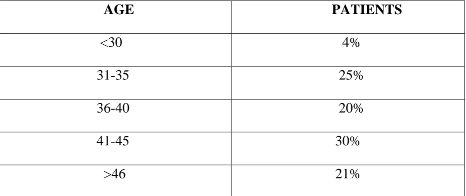
Table 1 : Age Profile
AGE PATIENTS
<30 4% 31-35 25% 36-40 20% 41-45 30% >46 21%
Fig.1 Age Profile
In our study majority of the population are in between the age group of 41 to 45. 4% 25% 20% 30% 21% 0% 5% 10% 15% 20% 25% 30% 35%
<30 31 ‐35 36 ‐40 41 ‐45 >46
Percentage
Age in Years
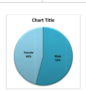
[image:72.595.87.514.224.615.2]GEND Table 46% in DER DIST 2: M F n our study
TRIBUTI
MALE
EMALE
In our st
Male par y.
ION:
Figure 2
tudy the m
rticipants Female 46%
C
gender di majority of around 54hart
Tit
stribution
f patients a
4% and fe Male 54%
le
54% 46% are males. female par . [image:73.595.147.446.229.545.2]CASE Table In our Becau E DISTRI 3: TYPE NON study the se we are
[image:74.595.110.479.147.599.2]BUTION 2 DIABE N DIABET type2 dia comparin N: ETICS TICS Figure 3 abetic patie ng the stud
Non‐DM 50%
C
3 case dist
ents and n dy so it mu
hart
Tit
tribution
non diabeti ust be equ
DM 50%
le
50
50
ics are equ ual in num
ual in num mber.
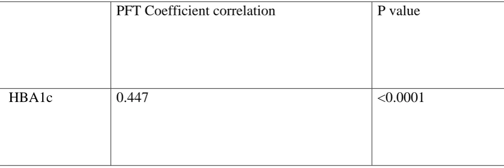
[image:74.595.181.414.302.590.2]CORRELATION OF HBA1C AND PFT:
Table 4:
PFT Coefficient correlation P value
HBA1c 0.447 <0.0001
PEARSON CORRELATION TEST
In our study correlation between HBA1C and Pulmonary function is statistically significant (p value is <0.0001)
From this pearson correlation test we had came to the conclusion that there is a relationship between the HBA1C level and the results of the pulmonary
function test. Most of the patients with the elevated levels had the abnormality in the pulmonary function tests mainly the restrictive pattern of the lung
COMPARISON OF PFT AMONG TWO GROUPS:
Table 5:
PARAMETERS
GROUP A
(TYPE 2
DIABETICS)
(n=50)
GROUP B
(NON-DIABETICS)
(n=50)
P VALUE
MEAN SD MEAN SD
FEV1 68.37 11.19 77.27 13.47 0.001
FVC 68.18 13.25 84.52 6.33 <0.0001
FEV1 / FVC 100.66 13.86 90.8 15.46 0.001
In our more t signifi This te the res study both than FEV1 icant in all
est depicts strictive pa 0 20 40 60 80 100 120 140 IND
h FEV1 an 1 so that F l the param
s that most attern of th
68.37
DM
FE
[image:77.595.137.457.115.454.2]DEPENDE
Figure 4
nd FVC a FEV1/FVC
meters acc
t of the pa he lung di
77.27 Non‐DM V1
C
ENT SAM compariso are reduced C has incre cording to atients with isease. 68.18 8 DM No FVChart
Tit
MPLE t TE
on of PFT
d, in whic eased, and
independ
h type 2 d 84.52 100 on‐DM DM
le
EST Th FVC ha d the ‘p’va dent sampl diabetes m 0.66 90.8 M Non‐D FEV/FVC ave reduce alue is le t test.
mellitus hav
M
ed
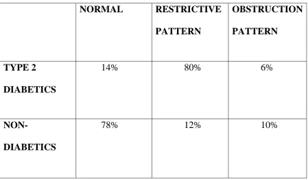
PATTERN OF PULMONARY FUNCTION:
Table 6:
NORMAL RESTRICTIVE
PATTERN
OBSTRUCTION
PATTERN
TYPE 2
DIABETICS
14% 80% 6%
NON-
DIABETICS
78% 12% 10%
Pearson’s chi square test
P<0.0001
In our study the patients with diabetes have 14% normal and 80% with restrictive lung disease and 6% with obstructive lung disease of pulmonary function tests.
In non-diabetics 78% having normal and 12% having restrictive pattern and 10 % having the obstructive pattern of lung disease.
Figure 5 pattern of pulmonary function
According to our study profile the most of the people with type 2 diabetes mellitus having restrictive type of lung disease. About 80% having restrictive pattern and about 6% having obstructive pattern and about 14% having normal pattern. The relation between type 2 diabetes mellitus and restrictive pattern of lung disease is statistically significant.
14%
80%
6% 78%
12% 10%
0% 10% 20% 30% 40% 50% 60% 70% 80% 90%
Normal Restrcition Obstruction
Chart
Title
DISCUSSION:
The present study has to assessed that type 2 diabetes was associated with reduced lung functions, by doing forced Spirometric Pulmonary Function Tests. This study clearly showed a highly statistically significant p value when the lung function tests (FVC, FEV1) were compared between type 2 diabetics and controls (age, sex and BMI matched).
In our study done in Kanyakumari medical college hospitals, about 100 participants were studied of which50 are having type 2 diabetes mellitus are cases and 50 without diabetes mellitus are controls and the males are 54 and females are 46 in number. The age distribution in our study mostly between 41to45 are 30%. Pulmonary function test has been conducted in all the persons.
There is statistically significant value in correlation between HBA1c and pulmonary function test results were noted and the p value is <0.0001.The FEV1and FVC both are reduced in type2 diabetic patients and the FEV1/FVC is increased. Mean, standard deviation are decreased in type 2 diabetics when comparing to non-diabetics. The p value foe FEV1 in type 2 diabetics is0.001 and foe FVC is <0.0001 and for FEV1/FVC is 0.001.
Recent studies which were conducted by Lange et al., indicated that the type 2 diabetic patients are having mild decrease in forced vital capacity may because of impaired immunity against environmental challenges such as
infections in diabetes and smoking. In a study which was done by Davis1 ,2 ,3 ,5. A Wendy et al., it was found that there was a decrease in mean FVC values in type 2 diabetics. In a study which was done by Robert E. Walter et. al., it was found that there was a progressive decrease in mean forced vital capacity value is 109 ml/year 33,34.
The FEV1/FVC % was increased in type 2 diabetics as compared to that in the controls and the increase was statistically significant. The increased FEV1/FVC % suggested that the impairment of pulmonary functions in type 2 diabetics was primarily restrictive in nature
Davis et al., study detected that almost all the parameters like forced vital capacity, forced expiratory volume at 1st second are decreased so that the ratio is increased almost most of the patient with uncontrolled diabetes mellitus23,27. So it is clear that type 2 diabetes mellitus affect the lung and lung may the target organ for damage and the pattern of disease is restrictive in nature1 ,4 ,5 ,9.
capacity,basal lamina thickening, and increased susceptibility to respiratory infections1,3,9,35.
The study which was done by Mario Cazzola et al., on human isolated bronchi elucidated the obstructive nature of pulmonary pathology in diabetes at a molecular level16, 18. Thus hyperglycaemia may contribute to obstruction of airways.
The lungs are affected by diabetic microangiopathy33, 34. This was evidenced autopsy findings in human diabetic subjects, which showed pulmonary microangiopathy, thickening of alveolar epithelia, pulmonary
LIMITATION:
1. Few studies showed there is no correlation between HBA1C and PFT’s.
2. Sample size is small
3. Patients refusal
4. There is no explained exact mechanisms of restrictive lung pattern of lung disease in type 2 diabetics.
CONCLUSION:
The findings from our study is nearly related with other studies that have done in the diabetics pulmonary function test. It is clearly shown that diabetes will affect the lung too that too mainly restrictive pattern of lung disease is formed but some study shows obstructive pattern also.
So it has been assigned that patients with type 2 diabetes mellitus should undergo pulmonary function tests intermittently .This will help to assess the pulmonary complication in type 2 diabetes mellitus and to detect in the earlier stages itself. But it needs studies to assess the clean mechanism behind abnormality in pulmonary function test in the patients with type 2 diabetes mellitus .
SUMMARY:
The pulmonary complications of type 2 diabetes mellitus are rare only. Our study assessed that the comparison of pulmonary function test in type 2 diabetics and non-diabetics. This study has to assess the effects of uncontrolled diabetes mellitus on lung functions.
100 participants were selected on which 50 having type 2 diabetes mellitus and 50 without diabetes mellitus in a age group between 30to50. Sex, height , weight, BMI,HBA1C,FEV1,FVC,FEV1/FVC were analysed in all the participants. 54 are males and 46 are females.
ANNEXURE
BIBLIOGRAPHY
1. Viberti GC, Rosiglitazone. Potential beneficial impact on cardiovascular disease. Int. J. ClincPract. 2003; 57 (2): 128-34
2. Boulbou MS, Gourgoulianis KI, Klisiaris VK, Tsikrikas TS, Stathakis NE, Molyvdas PA. Diabetes mellitus and lung function. Med Princ.Pract. 2003;12(2): 87-91.
3. Recommendations for a standard technique—1995 update. Am J RespirCrit Care Med.1995;152:2185-98
4. Asanuma Y, Fujiya S, Ide H, Agishi Y. Characteristics of pulmonary function in patients with diabetes mellitus. Diabetes. Res. Clin. Pract. 1985; 1(2): 95-101.
5. Lange P, Groth S, Kastrup J, Mortensen J, Appleyard M, Nyboe J, et al. Diabetes mellitus, plasma glucose and lung function in a cross-sectional population study. Eur. Respir J. 1989;2 (1):14-19.
6. Benbassat CA, Stern E, Kramer M, Lebzelter J, Blum I, Fink G. Pulmonary function in patients with diabetes mellitus. Am. J. Med. Sci. 2001;322 (3): 127-32.
7. Sreeja CK, Elizabeth Samuel, C Kesavachandran, Shankar Shashidhar. Pulmonary function in patients with Diabetes Mellitus.Indian J
8. P Lange, J Parner, P Schnohr, G Jensen. Copenhagen City Heart Study: longitudinal analysis of ventilatory capacity in diabetic and non-diabetic adults. EurRspir J. 2002;20:1406 -12.
9. Davis WA, Knuiman M, Kendall P, Grange V, Davis TM. Fremantle Diabetes Study.Diabetes Care. 2004;27(3):752-7. 22.
10. Walter R, Beiser A, Rachel J et.al., Association between glycemic state and lung function. The Framingham Heart Study. Am J RespirCrit Care Med. 2003 ; 167 : 911-16.
11. Davis Timothy ME, Mathew Knuimann, Peter Kendall. Reduced pulmonary function and its association in type-2 Diabetes.Diabetes Res ClinPract.2000; 50: 152-59.
12. Sandler M. Is the lung a ‘target organ’ in diabetes mellitus? Arch Intern Med. 1990; 150(7):1385-88
13. ShravyaKeerthi G, Sharan B Singh M, Hari Krishna Bandi, Suresh M,
Preetham J K, Mallikarjuna Reddy. Deterioration of Pulmonary Functions in Type 2 Diabetes Mellitus.IOSR.Journal of Pharmacy and Biological Sciences (IOSRJPBS). 2012; 1 (1) : 39-43.
14. Weynand B, Jonckheere A, Frans A, Rahier J. Diabetes mellitus induces a thickening of the pulmonary basal lamina. Respiration.1999; 66:14–1
16. Mario Cazzola, LuiginoCalzetta, Paola Rogliani, DavideLauro, Lucia Novelli, Clive P, Varsha et al. High Glucose Enhances Responsiveness of Human Airways Smooth Muscle via the Rho/ROCK Pathway. Am J Respir Cell Mol Biol. 2012; 47 (4): 509-16.
17. Barnes PJ. The role of inflammation and anti-inflammatory medication in asthma.Respir Med 96 (Suppl. A). 2002;13:211–15.
18. Rodriguez M, Guerrero R. Increased levels of C-reactive protein in non-controlled type 2 diabetic subjects. J Diabetes Complications.1999; 13:211– 15.
19. KaminskyDA.Spirometry and Diabetes.Diabetes Care. 2004; 27: 837-3 20. World Health Organization. Fact sheet: Diabetes. No. 312, November
2008. Available from: htpp://www.who.int/mediacentre/factsheets/fs312/ en/index.html. [Last accessed on 2009 Apr 14].
21.King H, Aubert RE, Herman WH. Global burden of diabetes 1995 to 2025. Prevalence, numerical estimates and projections. Diabetes Care 1998;21:1414 31.
22.Sandler M. Is the lung is target organ in diabetes mellitus? Arch Intern Med 1990;150:1385 8.
23.Klein OL, Krishnan JA, Jlick S, Smith LJ. Systematic review of association between lung function and type 2 diabetes mellitus.
collagen in diabetes mellitus. Diabetes 1975;24:902 4.
25. Fogarty AW, Jones S, Britton JR, Lewis SA, McKeever TM. Systemic inflammation and decline in lung function in a general population: A prospective study. Thorax 2007;62:515 20.
26. Mori H, Okubo M, Okamura M, Yamane K, Kado S, Egusa G, et al. Abnormalities of pulmonary function in patients with non insulin dependent diabetes mellitus. Intern Med 1992;31:189 93.
27. Miller MR, Hankinson J, Brusasco V, Burgos F, Casaburi R, Coates A,
et al. Standardization of spirometry. Eur Respir J 2005;26:319 38.
28. Nathan DM, Singer DE, Hurxthal K, Goodson JD. The clinical
information value of the glycosylated haemoglobin assay. N Engl J Med 1984;310:341 6.
29. Davis WA, Knuiman M, Kendall P, Grange V, Davis TM. Glycemic exposure is associated with reduced pulmonary function in type 2 diabetes, the fremantle diabetes study. Diabetes Care 2004;27:752 7. 30.Asanuma Y, Fujiya S, Ide H, Agishi Y. Characteristics of pulmonary function in patients with diabetes mellitus. Diabetes Res Clin Pract 1985;1:95 101.
31.Lange P, Groth S, Kastrup J, Mortensen J, Appleyard M, Nyboe J, et al. 32. Barrett Conor E, Frette C. NIDDM, impaired glucose tolerance, and pulmonary function in older adults. Diabetes Care 1996;19:1441 4.
pulmonary function and its association in type 2 diabetes: The fremantle diabetes study. Diabetes Res Clin Pract 2000;50:153 9. 34. Engstrom GJ, Janzon L. Risk of developing diabetes is inversely
Related to lung function: A population based cohort study. Diabet Med 2002;19:167 70.
PROFORMA:
Name : Age/Sex: Occupation:
Presenting complaints:
Past history:
H/o DM, HIV, PT, HT, CKD, CVD, COPD etc
Clinical examination:
General examination:
Consciousness, Pallor, jaundice, Clubbing, Lymphadenopathy, hydration status
Systemic examination:
CVS:
RS:
Abdomen:
CNS:
Laboratory investigations:
Random blood sugar
MASTER CHART:
GROUP – I (TYPE 2 DIABETICS)
S No Name Age (yr) Sex RBS (mg/ dl) HbA 1c
FEV1 (% of
predicted)
FVC (% of
predicted)
FEV1 /
FVC
Result
1 RAMASAMY 50 M 350 8.2 60 54 90 Mod restriction
2 ROSAMMAL 40 F 208 7.9 70 65 100 Mild restriction
3 RANI 32 F 285 7.5 87 89 97 Normal
4 BHAKIYAM 48 F 265 6.9 67 63 106 Mod restriction
5 SIVAPRAKASAM 50 M 325 9.4 75 70 107 Mild restriction
6 SAROJINI 41 F 300 8.5 80 75 106 Mild restriction
7 AMEERA 36 F 313 8.7 77 72 106 Mild restriction
8 CLITUS 45 M 420 8.2 55 50 110 Mod restriction
9 GEORGE 33 M 365 7.8 81 88 92 Normal
10 RAVI 40 M 215 6.9 78 74 105 Mild restriction
11 SUJIN 49 M 235 7.2 57 55 103 Mod restriction
12 STEPHEN 32 M 256 6.7 85 88 96 Normal
13 DANIEL 44 M 260 7.9 65 65 100 Mod restriction
14 VELAMMAL 45 F 280 8.6 55 81 67 Mild obstruction
S No Name Age (yr) Sex RBS (mg/ dl) HbA 1c
FEV1 (% of
predicted)
FVC (% of
predicted)
FEV1 /
FVC
Result
16 JESINTHA 38 F 245 10.0 66.90 66 101 Mild restriction
17 KALIAMMAL 35 F 210 9.9 50 48 104 Severe restriction
18 MURUGAPPAN 30 M 234 7.6 60 45 133 Severe restriction
19 PALANI 43 M 212 7.4 60 56 107 Mod restriction
20 MUTHURATHIN
AM
41 M 256 7.9 75 72 104 Mild restriction
21 RAJAN 33 M 190 8.9 50 41 121 Severe restriction
22 VEERAIYAN 36 M 265 8.4 66 64 103 Mod restriction
23 KARUPPAIAH 50 M 211 8.6 62 90 68 Mild restriction
24 JANAKI 36 F 305 9.2 59 55 107 Mod restriction
25 CHRISTHUDHAS 43 M 194 9.9 50 48 104 Severe restriction
26 CHANDRU 32 M 222 10.2 73 69 105 Mod restriction
27 JOSEPH 30 M 245 7.6 82 88 93 Normal
28 HELEN 50 F 198 11.2 65 47 138 Severe restriction
29 ANTONY 47 M 200 8.3 50 52 96 Mod restriction
S No Name Age (yr) Sex RBS (mg/ dl) HbA 1c
FEV1 (% of
predicted)
FVC (% of
predicted)
FEV1 /
FVC
Result
31 SARASWATHI 37 F 287 11.3 88 90 97 Normal
32 SHEELA 39 F 234 7.9 86 88 97 Normal
33 FATHIMA 34 F 265 8.5 75 80 97 Mild restriction
34 LAKSHMI 44 F 218 8.9 65 67 97 Mod restriction
35 SELVANAYAKI 43 F 143 8.6 70 66 106 Mod restriction
36 RADHAMANI 50 F 167 9.9 50 78 64 Mod restriction
37 KANAGAM 43 F 219 9.5 70 74 94 Mild restriction
38 VISALAM 32 F 112 8.3 75 70 107 Mild restriction
39 VIGNESH 39 M 178 8.4 70 74 94 Mild restriction
40 JOEL 37 M 233 9.5 87 89 97 Normal
41 SATHISH 42 M 245 9.7 75 71 105 Mild restriction
42 ELANKUMARAN 32 M 287 9.2 55 59 93 Mod restriction
43 GANESH 41 M 166 10.4 70 60 116 Mod restriction
44 SURESH 50 M 240 10.7 70 56 125 Mod restriction
45 DHANALAKSHM
I
S
No
Name Age
(yr)
Sex RBS
(mg/
dl)
HbA
1c
FEV1 (% of
predicted)
FVC (% of
predicted)
FEV1 /
FVC
Result
46 SUBHA 30 F 157 6.7 81 80 87 Mild restriction
47 THANGAMMAL 41 F 188 6.2 80 74 108 Mild restriction
48 NAZRIN 44 F 299 6.7 69 66 104 Mild restriction
49 ABBAS 47 M 350 8.8 50 66 75 Mild restriction
GROUP – II (NON-DIABETICS) S No Name Age (yr) Sex RBS (mg/ dl) HbA 1c
FEV1 (% of
predicted)
FVC (% of
predicted)
FEV1 /
FVC
Result
1 KOSALAI 31 F 111 5.5 83 84 98 Normal
2 SRIKUMARI 33 F 120 5.1 70 74 94 Mild restriction
3 MANIBHARATHI 33 M 99 4.9 81 80 87 Mild restriction
4 PAAPA 36 F 80 4.6 84 90 99 Normal
5 VASANTHA 39 F 97 4.1 81 88 92 Normal
6 SHEIKH
MEERAN
46 M 94 5.2 82 85 96 Normal
7 THAVASI 38 M 112 5.5 83 88 94 Normal
8 SUDHAKAR 43 M 130 5.6 50 66 75 Mild restriction
9 ARUNACHALAM 33 M 134 4.9 86 89 96 Normal
10 SINGARI 30 F 95 4.3 91 92 98 Normal
11 AMIRDHAKALA 43 F 140 4.6 90 92 97 Normal
12 MUTHURATHIN
AM
44 M 123 5.1 55 90 61 Mod obstruction
13 CHIDAMBARAM 45 M 113 5.3 80 81 98 Normal
14 VIJAYAN 50 M 116 4.7 83 85 97 Normal
15 ESSAKIPILLAI 41 M 98 4.2 80 82 97 Normal
17 KUNJAMMAL 38 F 99 5.5 83 85 97 Normal
18 FILOMENA 40 F 100 5.2 88 89 98 Normal
19 SUBBAIYAH 41 M 101 5.3 82 84 97 Normal
20 GANDHI 32 M 96 4.2 787 86 90 Normal
21 PICHUMANI 31 M 88 4.5 87 87 100 Normal
22 ANNAPANDI 33 M 83 4.4 44 89 49 Mod obstruction
23 KOLAPPAN 45 M 101 4.3 82 85 96 Normal
24 VIJAYARANI 50 F 110 5.1 84 89 94 Normal
25 JENNIFER 43 F 111 5.4 80 82 97 Normal
26 KANNAN 32 M 130 5.3 81 84 96 Normal
27 SAHAYAM 31 M 126 4.5 82 86 95 Normal
28 RATHINAKUMA
R
45 M 117 4.2 84 88 95 Normal
29 IYAPPAN 46 M 128 4.8 85 89 96 Normal
S No Name Age (yr) Sex RBS (mg/ dl) HbA 1c
FEV1 (% of
predicted)
FVC (% of
predicted)
FEV1 /
FVC
Result
31 BELLA 38 F 91 5.6 81 87 93 Normal
32 KAMALAIYAN 41 M 82 5.2 84 88 95 Normal
33 NAGAPPAN 31 M 79 5.3 43 85 50 Mod obstruction
34 LOURDHUMARI 33 F 115 4.3 76 83 91 Normal
35 VEERAPRABU 43 M 112 4.6 80 81 98 Normal
36 JAYAN 31 M 78 4.2 85 89 96 Normal
37 THIRUMALAI 45 M 99 4.5 58 88 65 Mild obstruction
38 CHELLAIYAN 44 M 116 5.4 89 89 100 Normal
39 KANTHARI 40 F 130 5.4 84 86 97 Normal
40 FRANCIS 37 M 132 5.1 85 85 100 Normal
41 JENILA 36 F 125 4.2 79 88 89 Normal
42 SELVAM 34 M 115 4.7 65 54 120 Mod restriction
43 CHELLATHAI 50 F 112 4.9 80 82 97 Normal
44 RAJI 49 F 134 5.5 87 89 97 Normal