This is a repository copy of
NASSAM: a server to search for and annotate tertiary
interactions and motifs in three-dimensional structures of complex RNA molecules
.
White Rose Research Online URL for this paper:
http://eprints.whiterose.ac.uk/74879/
Article:
Hamdani, H. Y., Appasamy, S. D., Willett, P. et al. (2 more authors) (2012) NASSAM: a
server to search for and annotate tertiary interactions and motifs in three-dimensional
structures of complex RNA molecules. Nucleic Acids Research, 40 (W1). W35. ISSN
0305-1048
https://doi.org/10.1093/nar/gks513
[email protected] https://eprints.whiterose.ac.uk/ Reuse
Unless indicated otherwise, fulltext items are protected by copyright with all rights reserved. The copyright exception in section 29 of the Copyright, Designs and Patents Act 1988 allows the making of a single copy solely for the purpose of non-commercial research or private study within the limits of fair dealing. The publisher or other rights-holder may allow further reproduction and re-use of this version - refer to the White Rose Research Online record for this item. Where records identify the publisher as the copyright holder, users can verify any specific terms of use on the publisher’s website.
Takedown
If you consider content in White Rose Research Online to be in breach of UK law, please notify us by
NASSAM: a server to search for and annotate
tertiary interactions and motifs in three-dimensional
structures of complex RNA molecules
Hazrina Y. Hamdani
1, Sri D. Appasamy
1, Peter Willett
2, Peter J. Artymiuk
3,* and
Mohd Firdaus-Raih
1,*
1
School of Biosciences and Biotechnology, Faculty of Science and Technology, Universiti Kebangsaan Malaysia, 43600 UKM Bangi, Malaysia,2Information School and 3Department of Molecular Biology and Biotechnology, Krebs Institute, University of Sheffield, Western Bank, Sheffield S10 2TN, UK
Received February 12, 2012; Revised May 6, 2012; Accepted May 9, 2012
ABSTRACT
Similarities in the 3D patterns of RNA base inter-actions or arrangements can provide insights into their functions and roles in stabilization of the RNA 3D structure. Nucleic Acids Search for Substructures and Motifs (NASSAM) is a graph theoretical program that can search for 3D patterns of base arrange-ments by representing the bases as pseudo-atoms. The geometric relationship of the pseudo-atoms to each other as a pattern can be represented as a labeled graph where the pseudo-atoms are the graph’s nodes while the edges are the inter-pseudo-atomic distances. The input files for NASSAM are PDB formatted 3D coordinates. This web server can be used to identify matches of base arrangement patterns in a query structure to annotated patterns that have been reported in the literature or that have possible functional and structural stabilization implications. The NASSAM program is freely accessible without any login requirement at http://mfrlab.org/grafss/nassam/.
INTRODUCTION
In RNA, just as in proteins, the 3D structure determines the functionality of the molecule. In recent years, the Protein Data Bank (PDB; http://www.rcsb.org/pdb/) (1) has seen a significant increase in the availability of RNA crystallographic structures. There were only 2 structures containing only RNA chains in 1981, 9 structures in 1991, 242 structures in 2001 and 908 structures in December 2011. But in addition to this, there are RNA–protein complex structures; thus at the end of 2011 the total number of crystallographic structures that contained
RNA chains in the PDB with a resolution higher than 3.0 A˚ stood at 1027. This number includes many complex RNA structures such as the ribosomal subunits and various ribozymes. In addition, there are numerous important nuclear magnetic resonance (NMR) structures and significant lower resolution crystal structures.
Although the number of RNA structures is lower than that of proteins, there is an urgent need for programs that are capable of analyzing them. The analysis and annota-tion of the tertiary motifs in the 3D PDB structures of these molecules can be difficult when limited to only the use of molecular visualization methods. One previous ini-tiative in this area, the Non-Canonical Interactions in RNA structures (NCIR) (2), has shown that although manual curation of non-canonical interactions in RNA structures is feasible, the current volume of struc-tures available, the manual labor-intensive processing required, and the dependence on literature may not make such an approach the most sustainable means of annotating and analyzing RNA 3D structures.
RNA structural comparison is further complicated by the difficulty in comparing RNA structures by eye to rec-ognize local similarities on the one hand or significant differences on the other. The superficial visual similarity between adenine and guanine, and between cytosine and uracil, results in some difficulty when visually scrutinizing RNA structures for specific patterns or motifs. In such a situation, patterns of interactions may not be immediately obvious, although they may provide important clues in understanding the mechanistic details of their functions at atomic level. This problem is further compounded by the fact that minor changes in relative base orientation may result in distinct non-canonical base pairings (3). This is especially true for large, highly complex RNA structures such as the ribosomal subunits.
Methods that can compare the folding of the RNA backbone have existed for the past decade (4). Other,
*To whom correspondence should be addressed. Tel: +60 389215961; Fax: +60 389252698; Email: fi[email protected]
Correspondence may also be addressed to Peter J. Artymiuk. Tel: +44 1142 224190; Fax: +44 1142 222800; Email: p.artymiuk@sheffield.ac.uk
ßThe Author(s) 2012. Published by Oxford University Press.
This is an Open Access article distributed under the terms of the Creative Commons Attribution Non-Commercial License (http://creativecommons.org/licenses/ by-nc/3.0), which permits unrestricted non-commercial use, distribution, and reproduction in any medium, provided the original work is properly cited.
at University of Sheffield on January 9, 2013
http://nar.oxfordjournals.org/
more recently available programs directed at comparing RNA folds, or for searching similar RNA 3D substruc-tures, include WebFR3D (5), ARTS (6), RNA FRABASE (7), FASTR3D (8), CLICK (9) and R3D-BLAST (10). However, these programs do not focus on, or have limited capability to investigate, the interactions and ar-rangements of the RNA bases. To our knowledge, the fold comparison or substructure searching programs currently available are not able to detect cases where there may be functionally significant changes in base interactions or ar-rangements, despite the folding of the RNA sugar phos-phate backbone being either unchanged or almost unchanged. Unlike the Nucleic Acids Search for Substructures and Motifs (NASSAM) web server pre-sented here, many of the currently available programs are only able to analyze a limited set of structures already within the server and are not able to allow users to upload their own coordinates, while others are unable to process large input files such as the ribosomal subunits, which are also within NASSAM’s capabilities.
To meet this need, we developed the graph theoretical program NASSAM to search for patterns of base arrange-ments in RNA 3D structures (11,12). The use of graph theoretical approaches to search for and identify 3D motifs in protein structures (13) has proven to be a viable approach to the identification of clusters of amino acid residues that perform either the same or similar chemical functions despite no detectable sequence or fold similarity. In this extension to complex nucleic acid structures, the base arrangements of interest range from simple arrangements such as canonical and non-canonical base pairs, to base platforms or more complex arrange-ments such as triples and larger patterns. In this article, we describe the web interface that permits the use of NASSAM by biologists who wish to analyze and annotate RNA structures.
METHODS
The NASSAM algorithm
The original NASSAM standalone program, first reported in the work of Harrisonet al.(11) and subsequently used in the annotation of base triples by Firdaus-Raih et al.
(12), used a base interaction pattern as a query to search against a database of RNA structures. This meant that the operator had to first either create a query pattern or extract a pattern of interest from an existing structure and then convert the pattern into pseudo-atom vector matrices before executing the NASSAM search. Such a method of operation would be of limited use via a web interface despite its proven value as an exploratory inves-tigative tool.
To deploy the NASSAM program as a web service, the original algorithm has been re-engineered to operate in reverse. Thus, in the new version of the methodology pre-sented here, a user is able to submit a complete complex RNA structure to be searched against a database of patterns, rather than looking for a single pattern in a database of structures as previously (11,12). Patterns in this database are either sourced from literature or are
theoretical patterns that have been ‘hand-crafted’ for accurate and optimal retrieval as described in the NASSAM Pattern Database section below. The web server processes the coordinates of the submitted input structure into a matrix containing information on the spatial relationship of pseudo-atom vectors representing the orientation of each base with respect to each other. The matrix representing the query structure is then compared against the database of pattern matrices that represent arrangements and motifs of RNA bases. This is done using the Ullmann subgraph isomorphism algo-rithm (14). As a totally rigid match of the base arrange-ment of the query to the database patterns is not realistic, the program incorporates a distance tolerance parameter that permits a controlled amount of deviation from the distance set in the query patterns. Validation tests of NASSAM performed by Harrison et al. (11) at the distance tolerance values of 20, 30 and 40% revealed that the program achieved the best balance of precision (91%) and recall (93%) at the distance tolerance value of 30% and this is pre-set as the default value for all NASSAM searches. The distance tolerance parameter is a key factor in optimizing or widening the search for a specific pattern. For example, we have shown that the use of high tolerances enabled the discovery of previously un-reported base triple arrangements (12). Users of the web server also have the option of extending the coverage for a search by increasing the distance tolerance values for each query.
Searching for base interaction patterns with NASSAM
PDB-formatted RNA 3D structure files provide the input for NASSAM searches. There is no restriction with regard to the experimental method used to generate the PDB file. However, for NMR sourced data, there may be multiple overlapping models of the structure, and it is therefore necessary for each model to be considered individually. A screening mechanism built into the web interface prompts the user to select any one single NMR model for analysis per submission.
NASSAM has been tested with all PDB files (up to June 2011) containing RNA chains. However, because the aim of the searches is to identify motifs and interactions associated with base arrangements, the usual type of query will in most cases be an NMR structure or a crys-tallographic structure containing RNA chains that has been solved to a reasonably high resolution of 3 A˚ or higher as this will enable the atomic details of interactions to be visualized with a higher degree of confidence. The query structure is searched against a database containing known 3D base arrangement patterns that include canon-ical and non-canoncanon-ical base pairs, patterns of triplets and other motifs as described in more detail later.
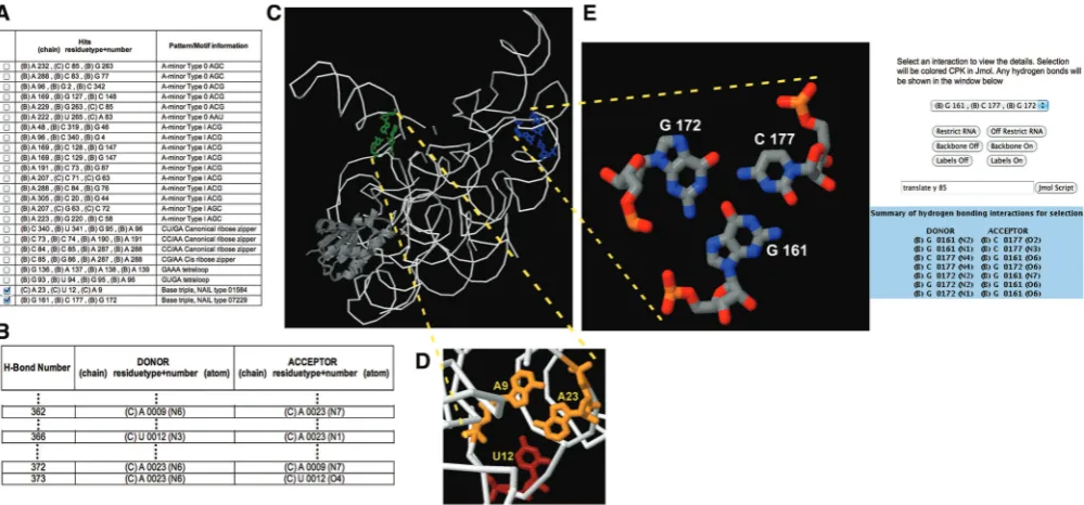
The NASSAM output lists the types of tertiary inter-action or base arrangement present in the query structure (Figure 1A). Each NASSAM run will, by default, also include calculations that identify the hydrogen bonding interactions between bases in the query RNA structures (Figure 1B). As many patterns and motifs in RNA struc-tures also include hydrogen bonding interactions in the W36 Nucleic Acids Research, 2012, Vol. 40, Web Server issue
at University of Sheffield on January 9, 2013
http://nar.oxfordjournals.org/
definition of the pattern, the generated list of hydrogen bond interactions can be used to identify whether the retrieved hits conform not only to the 3D arrangement of the bases but also to any additional parameters such as requirements for the presence of specific hydrogen bonds. In the case of the base triples, a NASSAM run is then filtered for cases where the base interactions will involve the formation of at least two hydrogen bonds per base. This filtering process will screen out numerous ‘loose’ opportunistic interactions involving triple-base ar-rangements (Tables 1 and 2) that are most likely function-ally insignificant or not highly conserved. Users have a further option of selecting hit patterns for visual examin-ation in a Jmol molecular visualizexamin-ation Java plug-in window (http://www.jmol.org/). Once the hits of interest have been selected, a Jmol window presenting the selected arrangements is opened (Figure 1C). Users can then browse through each base arrangement pattern using standard Jmol controls (Figure 1D) and select a specific interaction for further analysis. Any hydrogen bonding interactions that are present within the selected pattern is displayed in an auxiliary window (Figure 1E right panel).
NASSAM pattern database
The current NASSAM pattern database that is available for web searching includes patterns for both canonical and non-canonical base pairs (3,15), base triples (3,12,16), the A-minor motifs (17), kink-turn motifs (18), T-loops (19), ribose zippers (20) and tetraloops (21–23). The pseudo-atom vector representations for these interactions and motifs were mostly designed from the literature that
first presented their discoveries (as referenced previously) or via theoretical approaches (12,24). Many of the matrices representing the base pattern arrangements were ‘hand-crafted’ to achieve optimal retrieval and accuracy. Multiple vector combinations were tested and the representative vectors for a particular pattern was selected if it achieved optimal recall and precision when tested by searching against a dataset of PDB structures using the standalone NASSAM program. The NASSAM pattern database currently holds a total of 1041 pseudo-atom vector representations of base interaction or arrangement patterns, of which the majority are cur-rently base triple representations. The database will be updated to include other base motifs, or newly reported base motifs.
SEARCH EXAMPLES AND CASE STUDIES
We have recently used NASSAM to perform the annota-tion of base triples in RNA crystallographic structures in the PDB (12). This work revealed several yet unreported triple arrangements including a novel UAU stacked triples motif that was found to be highly conserved in available structures and sequences of the prokaryotic 23S ribosomal subunits (12). In this article, we discuss specific examples regarding the utility of NASSAM versus comparable programs.
[image:4.612.64.564.68.301.2]We had initially compared several programs namely: ARTS (6), WebFR3D (5), RNA FRABASE (7), FASTR3D (8) and R3D-BLAST (10) as a means of as-sessing the need for a web service such as NASSAM. Although these programs are intended, like NASSAM,
Figure 1. Snapshots of NASSAM output for the PDB structure of a bacterial ribonuclease P (PDB ID: 3Q1R): (A) A listing of the occurrences (hits) of base arrangement patterns or motifs; (B) excerpts from a list identifying hydrogen bonds between bases which in this extract is for an AUA triple; (C) Jmol window showing the triple hits selected in (A) relative to the whole structure; (D) Jmol window showing a magnification of the AUA triple selected in (A) where the hydrogen bonds formed by this interaction are presented in (B); (E) Jmol window showing the GCG triple selected in (A) with the hydrogen bonds present in the selection presented in an auxiliary Jmol window (right).
at University of Sheffield on January 9, 2013
http://nar.oxfordjournals.org/
to identify RNA 3D patterns, they differ significantly from NASSAM in terms of methodology, and/or input and output. Thus, the program RNA FRABASE (7) requires input in the form of sequences or secondary structures. The WebFR3D service (5) allows for either a symbolic search of up to 15 nucleotides where the user can specify an interaction matrix for the nucleotides, or a geometric search for structures that are already available in the database, but it does not appear to allow users to upload their own structures. Others such as ARTS (6) and R3D-BLAST (10) can basically identify motifs through structural similarities achieved via either 3D or 2D alignment-based methods. The intended operation of the CLICK web service (9) is to compare and align two 3D structures and not as a direct search engine to match and identify 3D motifs. NASSAM is therefore able to fill a
void in analytical capability for RNA 3D structures that is currently not served by these programs.
[image:5.612.43.552.85.237.2]The program MC-annotate (25) is, to our knowledge, the closest program to NASSAM in that it is able to perform an RNA structure annotation using a user-uploaded PDB file as input. NASSAM and MC-an-notate perform the annotations using very different approaches and as a result provide generally dissimilar structural analyses. However, as described later, these can in many respects be regarded as complementary to one another. The method used by MC-annotate has been reported by Gendron et al. (25). NASSAM provides three output types depending on the search options selected: (i) a specific search output based on the type of interaction or base arrangement the user is inter-ested in; or options of having all available patterns
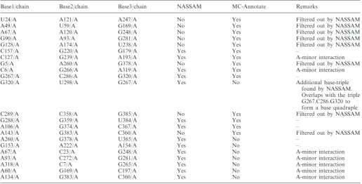
Table 2. Comparison of NASSAM and MC-annotate annotations for base triples in the structure 3G78
Base1/chain Base2/chain Base3/chain NASSAM MC-Annotate Remarks
U24/A A121/A A247/A No Yes Filtered out by NASSAM
A49/A U59/A G169/A No Yes Filtered out by NASSAM
A67/A A120/A G248/A No Yes Filtered out by NASSAM
G90/A A93/A G281/A No Yes Filtered out by NASSAM
G128/A A174/A U238/A No Yes Filtered out by NASSAM
C157/A G220/A G179/A Yes Yes –
C127/A G239/A A193/A Yes Yes A-minor interaction
G5/A A260/A G378/A No Yes Filtered out by NASSAM
C6/A G266/A A319/A Yes Yes A-minor interaction
G267/A C286/A G320/A Yes Yes –
G320/A U298/A G267/A Yes No Additional base-triple
found by NASSAM. Overlaps with the triple G267.C286.G320 to form a base quadruple
C289/A C358/A G385/A No Yes Filtered out by NASSAM
G288/A G359/A U384/A Yes Yes –
A106/A G374/A C367/A Yes Yes –
A143/A G383/A C360/A No Yes Filtered out by NASSAM
A260/A G378/A U365/A Yes No –
G153/A A222/A A154/A Yes No –
A67/A C23/A G248/A Yes No A-minor interaction
A93/A C272/A G281/A Yes No A-minor interaction
A318/A C7/A G265/A Yes No A-minor interaction
A60/A G169/A C197/A Yes No A-minor interaction
[image:5.612.42.559.282.544.2]A134/A G383/A C360/A Yes No A-minor interaction
Table 1. Comparison of NASSAM and MC-annotate annotations for base triples in the structure 3Q1R
Base1/chain Base2/chain Base3/chain NASSAM MC-Annotate Remarks
G4/B A96/B C340/B No Yes Filtered out by NASSAM
G46/B G48/B C319/B No Yes Filtered out by NASSAM
G56/B A276/B C325/B No Yes Filtered out by NASSAM
G76/B C84/B A288/B No Yes Filtered out by NASSAM
C73/A G87/B A191/B No Yes Filtered out by NASSAM
A116/B G123/B G153/B No Yes Filtered out by NASSAM
G161/B C177/B G172/B Yes Yes –
G107/B C209/B A206/B No Yes Filtered out by NASSAM
C231/B G263/B C85/B No Yes Filtered out by NASSAM
A23/C U12/C A9/C Yes Yes –
A112/B C66/C G19/C No Yes A non-planar interaction
filtered out by NASSAM
A158/B G115/B U154/B Yes No –
G22/C C13/C G56/C Yes No –
W38 Nucleic Acids Research, 2012, Vol. 40, Web Server issue
at University of Sheffield on January 9, 2013
http://nar.oxfordjournals.org/
annotated either (ii) inclusive or (iii) exclusive of base pairs.
Another advantage of using NASSAM is that it is able to annotate structures according to the nomenclature used in literature and has an up to date database for these tertiary motifs. As a result, NASSAM is able to annotate motifs such as the A-minor motifs, tetraloops, kink turns and ribose zippers. A NASSAM search using theHaloarcula marismortui23S rRNA subunit (PDB ID: 1FFK) as input can quickly provide a snapshot showing that there are at least 13 occurrences of the various tetraloops in the structure (Figure 2A). To demonstrate the utility of NASSAM, we compared the results of NASSAM searches against an MC-annotate annotation and here highlight the main differences in the outputs provided. We were unable to perform direct comparisons between MC-annotate and NASSAM on larger structures such as the rRNA subunits (e.g. 1FFK and 3U5D) as the MC-annotate server does not at present accept such large files for analysis, so in the examples below we focus on smaller RNA structures.
[image:6.612.112.510.321.697.2]The standard NASSAM search for base triples and MC-annotate annotations for the structures of a tRNA (PDB ID: 6TNA) and a bacterial ribonuclease P holoen-zyme in complex with tRNA (PDB ID: 3Q1R) were able to accurately identify the A23.U12.A9 (Figure 1) base triples in 6TNA as well as the A23.U12.A9 in the tRNA component and a G161.C177.G172 triple in 3Q1R (Table 1). NASSAM base triple searches were designed to filter out triples that were not in a planar arrangement and those that did not satisfy criteria of having at least two hydrogen bonds per base pairing. The remaining planar, doubly hydrogen-bonded triples are expected to be highly stable (12). The filtering mechanism therefore screens out less stable triple arrangements, which in large complexes such as the ribosomal subunits, are expected to be numerous but not necessarily of any interest to the user. However, users are given an option of turning off this screening mechanism to increase the search coverage. MC-annotate does not filter out its output for triples in the same way and therefore also includes base triple interactions that may have single
Figure 2. Snapshots of NASSAM output showing (A) the results of a GNRA tetraloop search for theH. marismortui23S rRNA structure (PDB ID: 1FFK) in the left panel with the selected GNRA tetraloop hits viewed relative to the whole structure in a Jmol window in the right panel; (B) the results listing the hits to A-minor motifs in the structure of a bacterial ribonuclease P (PDB ID: 3Q1R) in the left panel and a close up examination in Jmol of one selected hit (C85.A232.G263).
at University of Sheffield on January 9, 2013
http://nar.oxfordjournals.org/
hydrogen bonds connecting each base (Table 1). However, executing the NASSAM search at a distance tolerance of 60% revealed two more base triples that were not annotated by MC-annotate but which fitted the planarity and two hydrogen bond per base criteria (Table 1).
Another NASSAM versus MC-annotate comparison, using the ‘annotate all patterns except base pairs’ option, was performed for a group II intron structure from Oceanobacillus iheyensis (PDB ID: 3G78) (26,27). Both NASSAM and MC-annotate retrieved 14 base triples and A-minor motifs, but upon closer scrutiny, there were differences in the results retrieved. We found that MC-annotate yielded three triples that concurred with our filtering parameter and three A-minors, but the other eight hits did not fit the NASSAM filtering criteria (Table 2). On the other hand, the patterns retrieved by NASSAM consisted of six base triples, three of which were not retrieved by MC-annotate and eight A-minor motifs, five of which were not retrieved by MC-annotate (Table 2). Visual analysis of the triples annotated by NASSAM showed that one of the additional NASSAM triples (G267.U298.G320) overlaps with another G267.C286.G320 triple to form a base-quadruple inter-action (G267.C286.U298.G320). Both MC-annotate and NASSAM annotated the same set of T-loop and tetraloop structures (data not shown), but only NASSAM was able to annotate the two ribose zipper motifs (C272.U273.G92.A93 and C6.C7.A318.A319) presumably due to its use of an updated database of motifs reported in the literature. These results further illustrate the comple-mentarity of the NASSAM and MC-annotate programs that are a result of the different approaches used by the two programs to provide an annotation solution for RNA structures.
One caveat for the user is that, due to the nature of the base arrangements that the program looks for, and the use of higher search tolerances, a retrieved hit may not always be the desired target. As an example, one of the retrieved hits for an AGC A-minor Type 0 (A232.C85.G263) in 3Q1R when visually scrutinized does not fit the definition for that particular pattern despite the spatial arrangement matched by the search doing so (Figure 2B). In this particular example, the adenine is off plane and therefore not able to insert into the minor groove at the C285.G263 base pairing (Figure 2B). To aid visual examination and assessment of the hits in conforming to a particular defined arrangement, users have the option of selecting the hit(s) of interest and viewing them in the Jmol window.
SUMMARY
Perhaps, the most important capability of NASSAM is its ability to accept any existing complex RNA structure however large. This includes the ribosomal subunits from prokaryotes (such as PDB ID: 1FFK) (28) and eu-karyotes (such as PDB ID: 3U5D) (29). This thus enables NASSAM to be used as a standard first line annotation tool for tertiary motifs not only in complex structures such
as ribozymes but also in large complex assemblies such as the ribosomal subunits.
ACKNOWLEDGEMENTS
The authors thank Mohd Noor Mat Isa and Hafiza Aida Ahmad of the Malaysia Genome Institute for technical assistance with server operations. They gratefully acknow-ledge the Genome Computing Centre of the Malaysia Genome Institute for providing the computational infra-structure for the NASSAM server computations.
FUNDING
Ministry of Higher Education Malaysia FRGS [UKM-ST-06-FRGS0006-2009 to M.F.R.]; Universiti Kebangsaan Malaysia [UKM-GUP-KPB-08-33-132 to M.F.R.]; the Ministry of Higher Education, Malaysia (National Science Fellowship to S.D.A.) Universiti Sains Malaysia (Academic staff training fellowship to H.Y.H.). Funding for open access charge: Universiti Kebangsaan Malaysia [UKM-DLP-2012-018].
Conflict of interest statement. None declared.
REFERENCES
1. Berman,H.M., Westbrook,J., Feng,Z., Gilliland,G., Bhat,T.N., Weissig,H., Shindyalov,I.N. and Bourne,P.E. (2000) The Protein Data Bank.Nucleic Acids Res.,28, 235–242.
2. Nagaswamy,U., Larios-Sanz,M., Hury,J., Collins,S., Zhang,Z., Zhao,Q. and Fox,G.E. (2002) NCIR: a database of non-canonical interactions in known RNA structures.Nucleic Acids Res.,30, 395–397.
3. Tinoco,I.J. (1993) In: Gesteland,R.F. and Atkins,J.F. (eds),The RNA World. Cold Spring Harbor Laboratory Press, Cold Spring Harbor, NY, pp. 603–607.
4. Duarte,C.M. and Pyle,A.M. (1998) Stepping through an RNA structure: a novel approach to conformational analysis.J. Mol. Biol.,284, 1465–1478.
5. Petrov,A.I., Zirbel,C.L. and Leontis,N.B. (2011) WebFR3D—a server for finding, aligning and analyzing recurrent RNA 3D motifs.Nucleic Acids Res.,39, W50–W55.
6. Dror,O., Nussinov,R. and Wolfson,H. (2005) ARTS: alignment of RNA tertiary structures.Bioinformatics,21(Suppl. 2), ii47–ii53. 7. Popenda,M., Szachniuk,M., Blazewicz,M., Wasik,S., Burke,E.K.,
Blazewicz,J. and Adamiak,R.W. (2010) RNA FRABASE 2.0: an advanced web-accessible database with the capacity to search the three-dimensional fragments within RNA structures.
BMC Bioinformatics,11, 231.
8. Lai,C.E., Tsai,M.Y., Liu,Y.C., Wang,C.W., Chen,K.T. and Lu,C.L. (2009) FASTR3D: a fast and accurate search tool for similar RNA 3D structures.Nucleic Acids Res.,37, W287–W295. 9. Nguyen,M.N., Tan,K.P. and Madhusudhan,M.S. (2011)
CLICK—topology-independent comparison of biomolecular 3D structures.Nucleic Acids Res.,39, W24–W28.
10. Liu,Y.C., Yang,C.H., Chen,K.T., Wang,J.R., Cheng,M.L., Chung,J.C., Chiu,H.T. and Lu,C.L. (2011) R3D-BLAST: a search tool for similar RNA 3D substructures.Nucleic Acids Res.,39, W45–W49.
11. Harrison,A.M., South,D.R., Willett,P. and Artymiuk,P.J. (2003) Representation, searching and discovery of patterns of bases in complex RNA structures.J. Comput. Aided Mol. Des.,17, 537–549.
12. Firdaus-Raih,M., Harrison,A.M., Willett,P. and Artymiuk,P.J. (2011) Novel base triples in RNA structures revealed by graph theoretical searching methods.BMC Bioinformatics,12, S2. W40 Nucleic Acids Research, 2012, Vol. 40, Web Server issue
at University of Sheffield on January 9, 2013
http://nar.oxfordjournals.org/
13. Spriggs,R.V., Artymiuk,P.J. and Willett,P. (2003) Searching for patterns of amino acids in 3D protein structures.J. Chem. Inf. Comput. Sci.,43, 412–421.
14. Ullmann,J.R. (1976) An algorithm for subgraph isomorphism.
JACM,23, 31–42.
15. Xin,Y. and Olson,W.K. (2009) BPS: a database of RNA base-pair structures.Nucleic Acids Res.,37, D83–D88.
16. Abu Almakarem,A.S., Petrov,A.I., Stombaugh,J., Zirbel,C.L. and Leontis,N.B. (2012) Comprehensive survey and geometric classification of base triples in RNA structures.Nucleic Acids Res.,40, 1407–1423.
17. Nissen,P., Ippolito,J.A., Ban,N., Moore,P.B. and Steitz,T.A. (2001) RNA tertiary interactions in the large ribosomal subunit: the A-minor motif. Proc. Natl Acad. Sci. USA,98, 4899–4903.
18. Klein,D.J., Schmeing,T.M., Moore,P.B. and Steitz,T.A. (2001) The kink-turn: a new RNA secondary structure motif.EMBO J.,
20, 4214–4221.
19. Krasilnikov,A.S. and Mondragon,A. (2003) On the occurrence of the T-loop RNA folding motif in large RNA molecules.RNA,9, 640–643.
20. Tamura,M. and Holbrook,S.R. (2002) Sequence and structural conservation in RNA ribose zippers.J. Mol. Biol.,320, 455–474. 21. Correll,C.C. and Swinger,K. (2003) Common and distinctive
features of GNRA tetraloops based on a GUAA tetraloop structure at 1.4 A˚ resolution.RNA,9, 355–363.
22. Cheong,C., Varani,G. and Tinoco,I. Jr (1990) Solution structure of an unusually stable RNA hairpin, 50GGAC(UUCG)GUCC.
Nature,346, 680–682.
23. Jucker,F.M. and Pardi,A. (1995) Solution structure of the CUUG hairpin loop: a novel RNA tetraloop motif.Biochemistry,34, 14416–14427.
24. Walberer,B.J., Cheng,A.C. and Frankel,A.D. (2003) Structural diversity and isomorphism of hydrogen-bonded base interactions in nucleic acids.J. Mol. Biol.,327, 767–780.
25. Gendron,P., Lemieux,S. and Major,F. (2001) Quantitative analysis of nucleic acid three-dimensional structures.J. Mol. Biol.,308, 919–936.
26. Toor,N., Keating,K.S., Taylor,S.D. and Pyle,A.M. (2008) Crystal structure of a self-spliced group II intron.Science,320, 77–82. 27. Wang,J. (2010) Inclusion of weak high-resolution X-ray data for
improvement of a group II intron structure.Acta Crystallogr. D,
66, 988–1000.
28. Ban,N., Nissen,P., Hansen,J., Moore,P.B. and Steitz,T.A. (2000) The complete atomic structure of the large ribosomal subunit at 2.4 A˚ resolution.Science,289, 905–920.
29. Ben-Shem,A., Garreau de Loubresse,N., Melnikov,S., Jenner,L., Yusupova,G. and Yusupov,M. (2011) The structure of the eukaryotic ribosome at 3.0 A˚ resolution.Science,334, 1524–1529.
at University of Sheffield on January 9, 2013
http://nar.oxfordjournals.org/