CT = computed tomography; D2R = dopamine type 2 receptor; ER = estrogen receptor; FDG = 2-[18F]-2-fluoro-deoxyglucose; FESP = [18 F]-fluo-roethylspiperone; FIAU = 5-iodo-2′-fluoro-2′-deoxy-1-β-D-arabinofuranosyl-5-ioduracil; HSV1-tk= herpes simplex type 1 virus thymidine kinase; IRES = internal ribosomal entry sites; MRI = magnetic resonance imaging; PET = positron emission tomography; SPECT = single photon emission computed tomography.
Radionuclide imaging technologies
Two major technologies that employ radioactive isotopes (Supplementary Table 1) for imaging molecular events cur-rently exist: SPECT (carried out with a gamma camera),
employing isotopes in which a single high energy photon is directly emitted [1]; and PET, in which a positron is emitted from the radioactive isotope [2]. For positron imaging, positrons are emitted from nuclei of proton-rich isotopes,
Review
Recent advances in imaging endogenous or transferred gene
expression utilizing radionuclide technologies in living subjects:
applications to breast cancer
Frank Berger and Sanjiv Sam Gambhir
Department of Molecular and Medical Pharmacology, Crump Institute for Molecular Imaging, UCLA School of Medicine, Los Angeles, California, USA
Correspondence: Sanjiv Sam Gambhir, Crump Institute for Molecular Imaging, UCLA School of Medicine, Box 951770, Los Angeles, CA 90095-1770, USA. Tel: +1 310 206 1798; fax: +1 310 209 4655; e-mail: [email protected]
Abstract
A variety of imaging technologies is being investigated as tools for studying gene expression in living subjects. Two technologies that use radiolabeled isotopes are single photon emission computed tomography (SPECT) and positron emission tomography (PET). A relatively high sensitivity, a full quantitative tomographic capability, and the ability to extend small animal imaging assays directly into human applications characterize radionuclide approaches. Various radiolabeled probes (tracers) can be synthesized to target specific molecules present in breast cancer cells. These include antibodies or ligands to target cell surface receptors, substrates for intracellular enzymes, antisense oligodeoxynucleotide probes for targeting mRNA, probes for targeting intracellular receptors, and probes for genes transferred into the cell. We briefly discuss each of these imaging approaches and focus in detail on imaging reporter genes. In a PET reporter gene system for in vivoreporter gene imaging, the protein products of the reporter genes sequester positron emitting reporter probes. PET subsequently measures the PET reporter gene dependent sequestration of the PET reporter probe in living animals. We describe and review reporter gene approaches using the herpes simplex type 1 virus thymidine kinase and the dopamine type 2 receptor genes. Application of the reporter gene approach to animal models for breast cancer is discussed. Prospects for future applications of the transgene imaging technology in human gene therapy are also discussed. Both SPECT and PET provide unique opportunities to study animal models of breast cancer with direct application to human imaging. Continued development of new technology, probes and assays should help in the better understanding of basic breast cancer biology and in the improved management of breast cancer patients.
Keywords:animal models, imaging, microPET, positron emission tomography, single photon emission computed tomography
Received: 14 November 2000
Revisions requested: 22 November 2000 Revisions received: 23 November 2000 Accepted: 23 November 2000 Published: 11 December 2000
Breast Cancer Res2001, 3:28–35
This article may contain supplementary data which can only be found online at http://breast-cancer-research.com/content/3/1/028
commentary
review
reports
primary research
and eventually interact with electrons. Annihilation occurs, and the mass of the electron and positron is converted into two gamma rays that travel outward from the site of annihi-lation at ~180° to one another. Scintilannihi-lation crystals com-posed of different materials are used to capture the gamma rays for both SPECT and PET. The collection of many events allows reconstruction of the source of the emissions (Supplementary Figure 1). PET data can be corrected for attenuation, which occurs because some of the gamma rays do not traverse through all of the surrounding tissue. SPECT data is more difficult to correct for attenuation effects. PET is about 10-fold more sensitive than SPECT, primarily because the SPECT cameras use collimators that reject many of the counts from the source [3]. There are dedicated breast PET imaging systems under development [4] that may prove to be clinically important for breast and/or axillary imaging with improved resolution and/or sensitivity. We have recently developed microPET technol-ogy at UCLA, which allows high resolution (~2 mm in each axial direction) imaging of small animals such as mice [5] (Supplementary Figure 2).
Although the focus of this review is on radionuclide imaging technologies, it is worth briefly reviewing other imaging modalities and how they contrast to PET and SPECT. Magnetic resonance imaging (MRI) [6] and com-puted tomography (CT) [7] provide high resolution anatom-ical imaging that is not available through SPECT and PET. Development of new contrast agents for MRI is allowing the study of molecular events, but these approaches are still in the early developmental stage [8]. Magnetic reso-nance spectroscopy has also extensively been studied and is reviewed in detail elsewhere [9]. Optical approaches for imaging living small animals have been significantly aided by the use of green fluorescent protein [10] and firefly luciferase [11]. The use of highly sensitive charge-coupled device cameras has provided the ability to detect very low levels of light coming from deep within a living animal. This approach does not produce tomographic images, but pro-vides a rapid, low cost method to detect molecular events using an optical reporter gene.
The advantages of radionuclide technologies and, espe-cially, PET include high sensitivity (~10–11M), which
allows for detecting low levels of probes accumulated at a given site. Furthermore, because the isotopes available for PET can substitute for naturally occurring atoms in organic molecules, enormous versatility in the study of biochem-istry in vivo is possible. The radionuclide approaches do suffer from a lack of anatomical information that can be provided by the other technologies. Multiple efforts are currently underway to build combined MRI/PET and CT/PET systems [12]. We are performing microPET studies in our laboratories and are starting to use micro-CAT technology (Imtek Inc, Knoxville, TN, USA) [13] to obtain partially registered anatomical information. Clinical
scanners that combine CT and PET have also recently become available and should help to improve overall diag-nostic capability [14].
Radionuclide imaging probes
A key advantage of the radionuclide imaging technologies over other imaging approaches is the ability to label almost any chemical species with an isotope of choice. This has allowed the development of hundreds of radio-active imaging probes capable of imaging a variety of mol-ecular events [2,15].
A potentially useful way to discuss the general categories of probes is to start from targets at the cell membrane and to move to targets within the cell. Two major approaches are available for those genes that lead to expression of a protein on the cell surface. These involve antibodies or antibody fragments to target a specific protein, and spe-cific ligands to target receptors on cells.
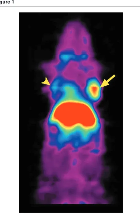
Antibody fragments are being engineered to achieve rapid targeting and rapid blood clearance [16]. A recent example from our laboratories of an antibody fragment approach tar-geting carcinoembryonic antigen expression in a mouse tumor xenograft model imaged in microPET is shown in Figure 1. The antibody fragment in this case is labeled with
64Cu, which has a half-life of approximately 12 h. It is likely
that engineered antibody fragments will provide methods to target many cell surface targets and should continue to be a general class of radiolabeled imaging probe.
Many possibilities for imaging molecular targets exist as one considers targets within a cell. Substrates that are trapped within the cell because they are metabolized by intracellular enzymes can be exploited for imaging pur-poses. A specific example of this approach is the widely used 2-[18F]-2-fluoro-deoxyglucose (FDG) [15,17], which
is actively transported into cells by a glucose membrane transporter. After hexokinase-mediated phosphorylation, phosphorylated FDG (FDG-6-PO4) cannot be further metabolized in the glucose metabolic pathway and is not able to leave the cell, leading to intracellular trapping of the radiolabeled compound [2,15] Breast carcinomas, like many cancers, show noticeable increased glucose metabo-lism, leading to a pronounced intracellular accumulation of FDG relative to surrounding tissues. FDG-PET can provide helpful information in the multimodality evaluation of breast lesions, therapy response and extent of metastatic disease. The inability to detect very small tumors (< 4 mm diameter) and the varying metabolic activity of the different tumor subtypes in breast cancer [18] cause some limitations.
[11C]-Methionine, a proliferation marker capable of detecting
In vivo imaging of intracellular receptors has also been reported. The growth of breast epithelial cells is an estro-gen-mediated process that depends on estrogen acting through an estrogen receptor (ER) and results in the induction of progestin receptor. The assay of the levels of these two receptors is important because it is consid-ered indicative of the responsiveness of a tumor to hor-monal agents [20]. The ER content of breast carcinomas is also known to be an important prognostic indicator. Successful research has focused mainly on imaging of estrogen receptors. [18F]-Fluoro-17β-estradiol has high
affinity for the ER, and an excellent correlation has been noted between the tumoral uptake of this estradiol on PET images and the tumor ER concentration measured
in vitro[21].
Another important approach that is still in its infancy is the use of small (15–20 bases) antisense oligodeoxynu-cleotides that target gene expression by specific hybridiza-tion to mRNA (Fig. 2). The basis for this approach is rooted in the use of antisense drugs as methods to inhibit transla-tion of mRNA [22]. The ability to image endogenous gene expression using a wide array of radiolabeled probes is quite a general and powerful approach. The rate-limiting step, however, is the ability to radiolabel a specific mole-cule and not to significantly alter its properties. It would be ideal if there could be a single universal probe that could image a wide variety of molecular events. The antibody fragment approach may satisfy this need for cell membrane targets but not intracellular targets. The antisense approach with mRNA targeting is a potential general approach, but it is currently limited and not likely to image genes with relatively low levels of transcription. If one is able to introduce an imaging or reporter gene into a group of cells, imaging gene expression with a universal probe becomes possible. Several reporter gene approaches for imaging transferred gene expression, including applica-tions to gene therapy models, are now discussed.
Principles of imaging reporter gene expression
[image:3.612.65.289.89.433.2]Molecular biologists have long used reporter genes to study promoter/regulatory elements involved in gene expression, inducible promoters to examine the transduc-tion of gene expression, and transgenes containing endogenous promoters fused to a reporter gene to study endogenous gene expression. This is a highly general
Figure 1
[image:3.612.318.555.92.281.2]MicroPET imaging of a mouse using a [64Cu]-labeled antibody fragment targeted against carcinoembryonic antigen (CEA) in a xenograft model. The mouse carried a C6 glioma xenograft on the left shoulder (negative control) and a LS174T xenograft expressing CEA on the right shoulder (arrows). The mouse was injected with 26 µCi 64Cu-anti-CEA minibody and imaged at 5 h, with the highest retention in the LS174T tumor (arrow) and liver, and lower retention in the control tumor (arrowhead). The liver signal occurs due to metabolism of the antibody fragment at that site.
Figure 2
approach because any promoter can be fused to a reporter gene, and detection of a reporter gene product is a way of ‘indirectly’ following the expression of the gene that the chosen promoter normally regulates. Once a reporter gene driven by a promoter of choice is introduced into the desired tissue, expression of the reporter gene can be monitored by several conventional methods like tissue biopsy, followed by immunohistochemistry. Conventional methods to detect reporter gene expression are, however, limited by their inability to determine ‘non-invasively’ the locations and magnitude of gene expression in living sub-jects over time. Approaches using green fluorescent protein [10] and firefly luciferase [11] as reporter genes allow localization of reporter gene expression in some living animals, but monitoring of the detailed location and magni-tude of reporter gene expression over time is difficult. Approaches using MRI are reviewed elsewhere [8].
Radionuclide imaging techniques offer the possibility of monitoring the detailed location, magnitude, and time varia-tion of reporter gene expression with high sensitivity for in vivouse in animals and humans [23]. A promoter of choice drives transcription of a reporter gene. The accumulation of the reporter probe is dependent directly on the expression of the reporter gene: the translation of the reporter gene mRNA leads to a protein product that can interact with a reporter probe. This interaction may be based on uptake and enzymatic conversion of the reporter probe via the reporter protein, leading to retention of the metabolite(s) or a receptor ligand based interaction (Fig. 3).
The choice of a promoter for driving reporter gene expres-sion depends on the intended application. Constitutive promoters can be used to produce continuous transcrip-tion of the reporter gene. Inducible promoters can be used to provide external control for varying the levels of tran-scription. To mimic the transcription of some endogenous gene, one can use the identical promoter of the endoge-nous gene to be imaged. This allows the use of reporter genes to indirectly monitor the expression of an endoge-nous gene. Two reporter gene approaches are discussed: the herpes simplex type 1 virus thymidine kinase (HSV1-tk) and the dopamine type 2 receptor (D2R). Other approaches have also been developed and are reviewed elsewhere [24,25].
The HSV1-tk gene (HSV1-TK is the corresponding enzyme) has its roots in a therapeutic approach as a ‘suicide gene’ [25,26]. HSV1-TK, like murine and human TK, phosphorylates thymidine. HSV1-TK, however, phos-phorylates acycloguanosines (eg acyclovir, ganciclovir, penciclovir) much more effectively than do mammalian TKs. Cellular enzymes then convert the acycloguanosine monophosphates to diphosphates and triphosphates that, if present in sufficient concentration, kill cells by incorpora-tion as chain-terminating derivatives or by direct inhibiincorpora-tion
of DNA polymerase. Because HSV1-TK converts acy-cloguanosine prodrugs to toxic compounds, this approach is used in cancer gene therapy protocols as a suicide gene approach [26]. The actual approach is to deliver the
HSV1-tk gene to tumor cells, then use acycloguanosines to kill the cells expressing HSV1-tk. The relaxed substrate specificity by the viral TK is critical in the approach of
HSV1-tk suicide gene therapy, but can also be exploited using the HSV1-tkgene in an imaging approach.
Two main categories of substrates, uracil nucleoside derivatives and acycloguanosine derivatives, have been investigated specifically as reporter probes for imaging
HSV1-tk reporter gene expression for SPECT and PET. These reporter probes are transported into cells, and trapped as a result of enzymatic phosphorylation by HSV1-TK. The choice of acycloguanosine derivatives (eg ganciclovir) as potential reporter probes was based on their ability to be radiolabeled with 18F, which has several
ideal characteristics as an isotope [27]. The reporter probe is only used in tracer doses (not in pharmacological doses as in suicide therapy), so there are no known prob-lems concerning cell toxicity.
8-[18F]-Fluoroganciclovir has been validated as a reporter
probe for HSV1-tk gene expression using a replication-deficient adenovirus, Ad-CMV-HSV1-tk, encoding HSV1-tk
commentary
review
reports
primary research
Figure 3
expressed under the control of the CMV promoter [28]. Other laboratories are also developing positron-labeled substrates to image the HSV1-tk PET reporter gene in living animals [29]. Radioactive 5-iodo-2′-fluoro-2′
-deoxy-1-β-D-arabinofuranosyl-5-ioduracil (FIAU) has been used to
image stable HSV1-tk gene expression in transplanted tumors and HSV1-tkgenes delivered in retroviral vectors to transplanted tumors. Using [124I]-labeled FIAU, data similar
to that observed with 8-[18F]-fluoroganciclovir and 8-[18
F]-fluoropenciclovir were obtained in imaging HSV1-tk
expression in tumors by PET. Because of its longer half-life (4 days), [124I]-FIAU may be a useful alternative to image HSV1-tk in some applications in which the background signal needs to be minimized. FIAU can additionally be labeled with single photon emitting iodine, allowing direct imaging with SPECT [30]. Further details of the HSV1-tk
reporter gene system and various reporter probes are
reviewed elsewhere [25]. We have also recently studied a mutant HSV1-sr39tkreporter gene with a better sensitivity for detecting lower levels of reporter gene expression [31]. Figure 4 presents an example of repetitive microPET imaging of a mutant HSV1-sr39tk reporter gene using a [18F]-labeled penciclovir molecule. An adenoviral vector is
used to deliver the HSV1-sr39tkreporter gene to the liver. The adenoviral vector targets the liver because of Cox-sackie adenoviral receptors present on hepatocytes.
[18F]-Fluoroethylspiperone (FESP) is a positron-labeled
analog of the dopamine antagonist spiperone. This tracer was developed to evaluate the level of D2R in vivo in animals and humans [32]. Endogenous high level D2R expression is limited primarily to the striatum. We have used the D2R gene as a reporter gene and FESP as a reporter probe for imaging gene expression [33]. There is no pharmacological effect of FESP in cells ectopically expressing D2R when FESP is used in small tracer con-centrations like in PET imaging. Imaging of both the
HSV1-tk and D2R reporter genes in animal models of tumor xenografts had been already successfully performed by our laboratories (Supplementary Figure 3) [34]. Further comparisons of the HSV1-tk and D2R reporter gene systems are provided elsewhere [25].
Potential applications of the reporter gene technologies developed for the study of breast cancer models are numerous. It should now be possible to mark any breast cancer cell line with a reporter gene for subsequent imaging of cell trafficking in living animals. Stable transfec-tion of the reporter gene into a given cell line is possible using plasmids carrying the reporter gene, and antibiotic selection markers or viral vectors with subsequent selec-tion of cell lines stably expressing the reporter gene. Once available, these cells can be directly implanted to study cell growth and trafficking with or without pharmacological intervention. Several models are now under study in which breast cancer metastases can be monitored non-invasively over time. Transgenic mice can also be developed in which a breast tissue specific promoter drives the reporter gene. This allows the study of spontaneous tumors as they develop and metastasize. The reporter genes also provide a unique way to optimize gene therapy through tracking the delivered genes, and are now discussed in detail.
Gene therapy applications
[image:5.612.56.299.102.383.2]Several gene therapeutic approaches to breast cancer treatment have been considered. A very important strategy is the attempt to correct or compensate specific genetic defects in breast cancer cells. Such efforts to compensate mutations include ablation of oncogenic products, restora-tion of receptor expression (eg estrogen receptor expres-sion), alteration of genes involved in the induction of apoptosis, or activation of tumor suppressor genes [35,36]. Mutations in the p53 gene are among the very
Figure 4
commentary
review
reports
primary research
common changes in breast cancer [37]. Wild type p53
functions as a tumor suppressor gene in breast cancer cells, as shown by the growth suppression of cells in vitro
when transfected with wild type expression vectors [38]. Retroviral delivery of wild type p53 into human breast cancer cells suppresses tumorigenicity in nude mice [38].
Hung et al [39] developed a phase I clinical trial of E1A
gene therapy targeting Her-2/neu overexpressing breast and ovarian cancer. HER2/neu overexpression in breast cancer is considered linked with lower response to chemotherapy and unfavorable prognosis. This group identified a viral transcriptional regulator, the adenovirus type 5 E1A, that can repress HER-2/neu overexpression. Expression of E1A in HER-2/neu overexpressing cancer cells resulted in downregulation of HER-2/neu in vitro, which reversed the malignant phenotype and restored sensitivity to chemotherapeutic agents. Preclinical in vivo
studies showed that the E1A gene was able to function therapeutically as a tumor suppressor gene in breast cancer xenograft models. In the phase I clinical trial, Hung
et al showed that the E1A gene can be delivered to cancer cells by a cationic liposomal delivery system. HER-2/neu was downregulated and the E1A gene induced apoptosis. A general drawback of the mutation compensa-tion approach is that transfeccompensa-tion of most of the tumor cells is needed to obtain a clinical effect, because intracel-lular responses are being modulated without effect on sur-rounding non-transfected cells.
Approaches such as molecular chemotherapy (shows a regional ‘bystander effect’) or immune therapy (has the potential to generate a systemic response) hold even more promise for becoming the breast cancer therapies of the future [35]. Molecular chemotherapy aims to increase the sensitivity of the tumor cells to chemotherapy or to find more specific ways of drug delivery to tumor cells [40]. The most studied system to accomplish cell killing uses the already discussed TK gene from the herpes simplex virus and gangiclovir as a prodrug.
Tumor specificity of molecular chemotherapy can be enhanced through the use of transductional or transcrip-tional targeting. Transductranscrip-tional targeting directs viral gene delivery vectors specifically to tumor cells, which express unique epitopes like erbB2 or the mucin protein core. Transcriptional targeting may be accomplished through the use of breast or tumor specific promoters such as Muc-1, HER-2, Myc and others [40].
Imaging modalities to verify the location, magnitude and duration of transgene expression are highly recommended in all these approaches, and will be very useful in attempts to optimize gene therapy protocols in general and time of prodrug application in particular.
A third strategy, genetic immunotherapy [35], aims to augment the specificity and magnitude of the immune response against tumor-associated antigens. These thera-pies include passive and active immunization, introduction of cytokines, and expression of T cell costimulatory mol-ecules [36]. An advantage of this approach is not having to introduce the therapeutic gene into all metastatic sites. The use of cytokines for immunotherapy has been limited by the toxicities associated with systemic administration of the proteins. Recombinant adenoviral vectors containing various cytokine genes have been used to deliver high local concentrations of the cytokines intratumorally. In a subcutaneous model of metastatic breast cancer in trans-genic mice, direct intratumoral injection of an adenoviral vector expressing IL-12 resulted in tumor regression in 75% of the treated animals [41]. Adenoviral-mediated gene delivery of IL-2 was recently evaluated in a phase I clinical trial in metastatic breast cancer and melanoma patients, showing the safety of this approach [42].
Given the magnitude of breast cancer as a clinical problem, however, few gene therapy clinical trials have until now been initiated. The exceptional heterogeneous molecular biology of this cancer, production of immuno-suppressive factors, and the widespread nature of the disease are three examples of the difficulties faced in breast cancer gene therapy [36]. Recent advances in understanding the molecular cell biology underlying breast cancer nevertheless revealed several potentially clinically useful gene therapy approaches for the near future.
Non-invasive imaging of transgene expression will be of great benefit in assessing organ tissue specificity as well as level and duration of transgene expression in vivo, and could therefore be very useful in validation of new delivery systems as well as in monitoring clinical gene therapy trials. Most therapeutic transgenes do not lend themselves to direct imaging of the transgene product. Most thera-peutic transgene products lack appropriate ligands or probes that can be radiolabeled and used to generate images that define the magnitude of transgene expression. It would also be very difficult to develop and validate new ligands and probes for each therapeutic transgene. Alter-natively, it is reasonable to develop and validate indirect imaging strategies using a reporter gene in combination with a therapeutic gene.
from this genetic difference, and the same promoter drives both genes. The expression of the reporter gene reflects, at the macroscopic tissue level, at least in some diagnos-tic situations, the expression of the therapeudiagnos-tic gene [44]. All three strategies are based on demonstrating a propor-tional and constant relationship in the coexpression of two transgenes over a wide range of expression levels. The advantage of these paradigms is that they can potentially be applied to any gene combination. Strict coexpression of two proteins in equimolar amounts can only be achieved by a fusion gene encoding both gene products. However, fusion proteins may not yield functional activity, may not localize in the appropriate subcellular compart-ment, or may induce immunogenic reactions; therefore, this approach is not ideal for in vivostudies. The propor-tional expression of two cis-linked genes is possible using an internal ribosomal entry sites (IRES) element within a single bicistronic transcription unit. The IRES element enables translation initiation within the bicistronic mRNA, thus permitting gene coexpression by cap-dependent translation of the first cistron and cap-independent, IRES-mediated translation of the second cistron. It will eventu-ally be important to assess whether IRES-based vectors are reliable indicators of transgene coexpression in differ-ent tissues, taking into account the half-life of each encoded protein. Whether the genes located upstream and downstream are equally expressed for a given appli-cation also has to be investigated. An example of using a bicistronic system to image gene expression in a tumor model is available elsewhere (Supplementary Figure 4) [43]. This work should help form the basis for extending this approach into gene therapy trials.
Future prospects
The future potential of radionuclide assays with SPECT and, particularly, PET imaging is quite promising. As small animal imaging technologies continue to spread to major research centers and the development of general probes expands, animal research should be significantly accelerated. The study of breast cancer cell trafficking and basic tumor biology in xenograft and transgenic models should be possi-ble. Optimization of gene therapy should also be possipossi-ble. The results from animal models should be rapidly translated into human applications because of a rapidly growing base of imaging scanners, which should lead to improved diagno-sis and management of breast cancer patients.
Acknowledgements
The authors would like to thank Drs H Herschman, M Phelps, JR Barrio, S Cherry, N Satyamurthy, T Toyokuni, L Wu and A Wu for their collabo-rative efforts in much of the work described in the paper. We would also like to thank the many undergraduate and graduate students as well as post-doctoral fellows who helped with some of the studies reviewed in this paper
This work is supported in part by US Army DAMD 17-98-1-8179, Department of Energy DE-FC03-87ER60615, NIH RO1 CA82214-01, and NIH CA86306; Frank Berger is supported by the Deutsche Forschungsgemeinschaft.
References
1. Budinger TF: Critical review of PET, SPECT and neuroreceptor studies in schizophrenia. J Neural Transm 1992, 36(suppl): 3–12.
2. Phelps ME: Inaugural article: positron emission tomography provides molecular imaging of biological processes. Proc Natl Acad Sci USA2000, 97:9226–9233.
3. Fleming JS, Alaamer AS: Influence of collimator characteristics on quantification in SPECT. J Nucl Med1996, 37:1832–1836. 4. Freifelder R, Karp JS: Dedicated PET scanners for breast
imaging. Phys Med Biol1997, 42:2463–2480.
5. Cherry SR, Shao Y, Silverman RW, Meadors K, Siegel S, Chatzi-ioannou A, Young JW, Jones WF, Moyers JC, Newport D, Boutef-nouchet A, Farquhar TH, Andreaco M, Paulus MJ, Binkley DM, Nutt R, Phelps ME: MicroPET: a high resolution PET scanner for imaging small animals. IEEE Trans Nucl Sci 1997, 44: 1161–1166.
6. Jackson EF, Ginsberg LE, Schomer DF, Leeds NE: A review of MRI pulse sequences and techniques in neuroimaging. Surg Neurol1997, 47:185–199.
7. Hopper KD, Singapuri K, Finkel A: Body CT and oncologic imaging. Radiology2000, 215:27–40.
8. Bogdanov A, Weissleder R: The development of in vivo imaging systems to study gene expression. Trends Biotechnol 1998, 16:5–10.
9. Rudin M, Beckmann N, Porszasz R, Reese T, Bochelen D, Sauter A: In vivo magnetic resonance methods in pharmaceutical research: current status and perspectives. NMR Biomed1999, 12:69–97.
10. Yang M, Baranov E, Moossa AR, Penman S, Hoffman RM: Visual-izing gene expression by whole-body fluorescence imaging. Proc Natl Acad Sci USA2000, 97:12278–12282.
11. Contag PR, Olomu IN, Stevenson DK, Contag CH: Biolumines-cent indicators in living mammals. Nat Med1998, 4:245–247. 12. Shao Y, Cherry SR, Farahani K, Slates R, Silverman RW, Meadors
K, Bowery A, Siegel S, Marsden PK, Garlick PB: Development of a PET detector system compatible with MRI/NMR systems. IEEE Trans Nucl Sci1997, 44:1167–1171.
13. Paulus MJ, Gleason SS, Kennel SJ, Hunsicker PR, Johnson DK: High resolution X-ray computed tomography: an emerging tool for small animal cancer research. Neoplasia2000, 2:62–70. 14. Beyer T, Townsend DW, Brun T, Kinahan PE, Charron M, Roddy R,
Jerin J, Young J, Byars L, Nutt R: A combined PET/CT scanner for clinical oncology. J Nucl Med2000, 41:1369–1379. 15. Phelps M: PET: the merging of biology and imaging into
mole-cular imaging. J Nucl Med2000, 41:661–681.
16. Wu AM: Designer genes: recombinant antibody fragments for biological imaging. Q J Nucl Med2000, 44:268–283.
17. Higashi K, Clavo AC, Wahl RL: Does FDG uptake measure the proliferative activity of human cancer cells? In vitro compari-son with DNA flow cytometry and tritiated thymidine uptake. J Nucl Med1993, 34:414–418.
18. Avril N, Rose CA, Schelling M, Dose J, Kuhn W, Bense S, Weber W, Ziegler S, Graeff H, Schwaiger M: Breast imaging with positron emission tomography and fluorine-18 fluo-rodeoxyglucose: use and limitations. J Clin Oncol 2000, 18:3495–3502.
19. Huovinen R, Leskinen-Kallio S, Nagren K, Lehikoinen P, Ruot-salainen U, Teras M: Carbon-11-methionine and PET in evalua-tion of treatment response of breast cancer. Br J Cancer 1993, 67:787–791.
20. Varagnolo L, Stokkel MP, Mazzi U, Pauwels EK: 18F-Labeled
radiopharmaceuticals for PET in oncology, excluding FDG. Nucl Med Biol2000, 27:103–112.
21. Mintun MA, Welch MJ, Siegel BA, Mathias CJ, Brodack JW, McGuire AH, Katzenellenbogen JA: Breast cancer: PET imaging of estrogen receptors. Radiology1988, 169:45–48.
22. Crooke ST, Lebleu B: Antisense Research and Applications. Ann Arbor: CRC Press, Inc., 1993:579.
23. Gambhir SS, Barrio JR, Herschman HR, Phelps ME: Imaging gene expression: principles and assays. J Nucl Cardiol1999, 6:219–233.
24. Rogers BE, Zinn KR, Buchsbaum DJ: Gene transfer strategies for improving radiolabeled peptide imaging and therapy. Q J Nucl Med2000, 44:208–223.
commentary
review
reports
primary research
Tjuvajev J, Blasberg R: Imaging transgene expression with radionuclide imaging technologies. Neoplasia2000, 2:118–138. 26. Moolten FL: Suicide genes for cancer therapy. Sci Med1997,
4:16–25.
27. Gambhir SS, Barrio JR, Herschman HR, Phelps ME: Assays for noninvasive imaging of reporter gene expression. Nucl Med Biol1999, 26:481–490.
28. Gambhir SS, Barrio J, Wu L, Iyer M, Namavari M, Satyamurthy N, Bauer E, Parrish C, MacLaren D, Borghei A, Berk A, Cherry S, Phelps ME, Herschman H: Imaging of adenoviral directed herpes simplex virus type 1 thymidine kinase gene expression in mice with ganciclovir. J Nucl Med1998, 39:2003–2011. 29. Tjuvajev JG, Joshi R, Lindsley L, Balatoni J, Finn R, Larson S,
Sadelain M, Blasberg R: Noninvasive imaging of HSV1-tk marker gene with FIAU for monitoring transfer and expres-sion of other therapeutic genes by multi-gene delivery vectors [abstract]. J Nucl Med1998, 39:130P.
30. Tjuvajev JG, Chen SH, Joshi A, Joshi R, Guo ZS, Balatoni J, Ballon D, Koutcher J, Finn R, Woo SL, Blasberg RG: Imaging adenovi-ral-mediated herpes virus thymidine kinase gene transfer expression in vivo. Cancer Res1999, 59:5186–5193.
31. Gambhir SS, Bauer E, Black ME, Liang Q, Kokoris MS, Barrio JR, Iyer M, Namavari M, Satyamurthy N, Green LA, Nguyen K, Cherry SR, Phelps ME, Herschman HR: A mutant herpes simplex virus type 1 thymidine kinase reporter gene shows improved sensi-tivity for imaging reporter gene expression with positron emission tomography. Proc Natl Acad Sci USA 2000, 97: 2785–2790.
32. Barrio JR, Huang SC, Phelps ME: Biological imaging and the molecular basis of dopaminergic diseases. Biochem Pharma-col1997, 54:341–348.
33. MacLaren DC, Gambhir SS, Satyamurthy N, Barrio JR, Sharfstein S, Toyokuni T, Wu L, Berk AJ, Cherry SR, Phelps ME, Herschman HR: Repetitive, non-invasive imaging of the dopamine D2
receptor as a reporter gene in living animals. Gene Ther1999, 6:785–791.
34. Iyer M, Bauer E, Barrio J, Iyer M, Namavari M, Satyamurthy N, Green LA, Nguyen K, Phelps ME, Herschman HR, Gambhir SS: 8-[18F]Fluoropenciclovir: an improved reporter probe for
imaging HSV1-tk reporter gene expression in vivo using positron emission tomography. J Nucl Med(in press). 35. Ruppert JMWM, Rosenfeld M, Grushcow J, Bilbao G, Curiel DT,
Strong TV: Gene therapy strategies for carcinoma of the breast. Breast Cancer Res Treat1997, 44:93–114.
36. Boxhorn HK, Eck SL: Gene therapy for breast cancer. Hematol Oncol Clin North Am1998, 12:665–675.
37. Phillips H: The role of the p53 tumour suppressor gene in human breast cancer. Clin Oncol (R Coll Radiol) 1999, 11: 148–155.
38. Wang NP, To H, Lee WH, Lee EY: Tumor suppressor activity of RB and p53genes in human breast carcinoma cells. Onco-gene1993, 8:279–288.
39. Hung MC, Hortobagyi GN, Ueno NT: Development of clinical trial of E1A gene therapy targeting HER-2/neu-overexpress-ing breast and ovarian cancer. Adv Exp Med Biol 2000, 465:171–180.
40. Patterson A, Harris AL: Molecular chemotherapy for breast cancer. Drugs Aging1999, 14:75–90.
41. Bramson JL, Hitt M, Addison CL, Muller WJ, Gauldie J, Graham FL: Direct intratumoral injection of an adenovirus expressing interleukin-12 induces regression and long-lasting immunity that is associated with highly localized expression of inter-leukin-12. Hum Gene Ther1996, 7:1995–2002.
42. Stewart AK, Lassam NJ, Quirt IC, Bailey DJ, Rotstein LE, Krajden M, Dessureault S, Gallinger S, Cappe D, Wan Y, Addison CL, Moen RC, Gauldie J, Graham FL: Adenovector-mediated gene delivery of interleukin-2 in metastatic breast cancer and melanoma: results of a phase 1 clinical trial. Gene Ther1999, 6:350–363.
43. Yu Y, Annala AJ, Barrio JR, Toyokuni T, Satyamurthy N, Namavari M, Cherry SR, Phelps ME, Herschman HR, Gambhir SS: Quan-tification of target gene expression by imaging reporter gene expression in living animals. Nat Med2000, 6:933–937. 44. Yaghoubi S, Liang Q, Barrio JR, Namavari M, Satyamurthy S,
Toyokuni T, Black M, Phelps ME, Herschman HR, Gambhir SS: Assessment of gene expression utilizing two adenoviral vectors and positron emission tomography [abstract]. FASEB
2000, 14:A568.
Supplementary Table 1
Radionuclides of interest for positron emission tomography (PET) and single photon emission computed tomography (SPECT) studies
Radionuclide Half-life Type
11C 20.3 min PET
13N 9.97 min PET
15O 122 s PET
18F 109.8 min PET
62Cu 9.74 min PET
64Cu 12.7 h PET
68Ga 68.1 min PET
76Br 16.1 h PET
124I 4.17 days PET
131I 8.0 days SPECT
123I 13.2 h SPECT
111In 2.8 days SPECT
67Ga 78.3 h SPECT
99mTc 6.0 h SPECT
201Tl 73.1 h SPECT
127Xe 36 days SPECT
Supplementary material
Supplementary Figure 1
Schematic illustrating single photon emission computed tomography (SPECT) and positron emission tomography (PET). (a)In SPECT, a single photon is produced as the isotope decays and this single photon must be detected through rotating detectors using a collimator. (b)In PET, annihilation eventually occurs with the positron and electron to produce two high energy (511 keV) gamma rays at ~180° that are detected using a circular ring of detectors.
Supplementary Figure 2
Photograph of the microPET scanner designed at UCLA, Los Angeles, CA, USA. The scanner is capable of imaging small animals such as mice in 30–60 min with a spatial resolution of ~2 mm in each axis (8 mm3). The scanner uses an 8 × 8 array of 2 × 2 × 10 mm3lutetium oxyorthosilicate crystals. The ring diameter is 17.1 cm. The transaxial and axial fields of view are 17.1 and 1.8 cm, respectively.
Supplementary Figure 3
Imaging of two reporter genes in the same mouse. A nude mouse carrying two tumors (left, dopamine 2 receptor [D2R]; right, herpes simplex virus type 1 thymidine kinase [HSV1-tk]) was imaged in a microPET system with tail-vein injection of ~250 µCi [18 F]-fluoroethylspiperone (FESP) and, 24 hours later, with ~250 µCi 8-[18F]-fluoropenciclovir (FPCV). Imaging began 1 hour after injection of each tracer. These results show the ability to image both the D2R and the HSV1-tkreporter genes in the same living mouse with microPET. The scale represents the accumulation of FESP and FPCV measured as percent injected dose per gram of tissue (%ID/g).
Supplementary Figure 4