Copyright © 2002, American Society for Microbiology. All Rights Reserved.
Regional Clustering of Shared Neutralization Determinants on Primary
Isolates of Clade C Human Immunodeficiency Virus Type 1
from South Africa
Renata Bures,
1Lynn Morris,
2Carolyn Williamson,
3Gita Ramjee,
4Mark Deers,
5Susan A. Fiscus,
6Salim Abdool-Karim,
4and David C. Montefiori
1*
Department of Surgery, Duke University Medical Center, Durham,1and Center for AIDS Research, Department of Microbiology and
Immunology, University of North Carolina at Chapel Hill, Chapel Hill,6North Carolina; National Institute for Virology,
Johannesburg,2Division of Medical Virology, University of Cape Town, Cape Town,3and Medical Research
Council, Durban,4South Africa; and Statistical Center for HIV/AIDS Research and Prevention,
Fred Hutchinson Cancer Research Center, Seattle, Washington 981095
Received 27 July 2001/Accepted 20 September 2001
Clade C is one of the most prevalent genetic subtypes of human immunodeficiency virus type 1 (HIV-1) in the world today and one of the least studied with respect to neutralizing antibodies. Most information on HIV-1 serology as it relates to neutralization is derived from clade B. Clade C primary isolates of HIV-1 from South Africa and Malawi were shown here to resemble clade B isolates in their resistance to inhibition by soluble CD4 and their sensitivity to neutralization by human monoclonal antibody immunoglobulin G1b12 and, to a lesser extent, 2F5. Unlike clade B isolates, however, all 16 clade C isolates examined resisted neutralization by 2G12. Infection with clade C HIV-1 in a cohort of female sex workers in South Africa generated antibodies that neutralized the autologous clade C isolate and T-cell-line-adapted (TCLA) strains of clade B. Neutralization of clade B TCLA strains was much more sensitive to the presence of autologous gp120 V3 loop peptides compared to the neutralization of clade C isolates in most cases. Thus, the native structure of gp120 on primary isolates of clade C will likely pose a challenge for neutralizing antibody induction by candidate HIV-1 vaccines much the same as it has for clade B. The autologous neutralizing antibody response following primary infection with clade C HIV-1 in South Africa matured slowly, requiring at least 4 to 5 months to become detectable. Once detectable, extensive cross-neutralization of heterologous clade C isolates from South Africa was observed, suggesting an unusual degree of shared neutralization determinants at a regional level. This high frequency of cross-neutralization differed significantly from the ability of South African clade C serum samples to neutralize clade B isolates but did not differ significantly from results of other combinations of clade B and C reagents tested in checkerboard assays. Notably, two clade C serum samples obtained after less than 2 years of infection neutralized a broad spectrum of clade B and C isolates. Other individual serum samples showed a significant clade preference in their neutralizing activity. Our results suggest that clades B and C are each comprised of multiple neutralization serotypes, some of which are more clade specific than others. The clustering of shared neutralization determinants on clade C primary HIV-1 isolates from South Africa suggests that neutralizing antibodies induced by vaccines will have less epitope diversity to overcome at a regional level.
An important goal in the development of an effective human immunodeficiency virus type 1 (HIV-1) vaccine is to overcome the extensive genetic heterogeneity of the virus. Nucleotide sequence comparisons have been used to define three groups of the virus known as group M (main), group O (outlier), and group N (non-M, non-O) (50, 69). Group M is further divided into 10 phylogenically related genetic subtypes (clades A, B, C, D, F1, F2, G, H, J, and K) that, together with a growing number of circulating intersubtype recombinant forms, com-prise the majority of HIV-1 variants in the world today. Clade C is emerging as most prevalent, being common in India (15, 16, 31, 41) and the southern African countries of Botswana, Zimbabwe, Malawi, Mozambique, and South Africa (7, 8, 25, 26, 60, 64, 79, 81). Clade B is dominant in North America and Western Europe and has been a major focus for vaccine
de-velopment (27). It is uncertain whether vaccines that are ulti-mately effective against clade B will be capable of targeting other genetic subtypes of the virus.
The uncertain relevance of genetic subtype to HIV-1 vac-cines is owed in part to a poor understanding of the immuno-type diversity of the virus as it relates both to cellular and humoral immunity. The fact that genetic subtypes tend to clus-ter geographically raises the possibility that distinct immuno-types of the virus have evolved along similar lines and, al-though a growing body of evidence suggests that this may not be true in a strict sense (4, 12, 29, 38, 56, 61, 82), additional studies seem warranted. For example, with respect to humoral immunity, the sporadic neutralizing activity of sera from HIV-1-infected individuals appears to be independent of genetic subtype (38, 56, 61, 82). That observation has led to a general notion that genetic subtype does not predict the neutralization serotype of the virus. An exception has been noted for clades B and E (E is now known as recombinant subtype A/E [32]), which appear to consist of different neutralization serotypes relative to each other. That conclusion was based on results of * Corresponding author. Mailing address: Department of Surgery,
Box 2926, Duke University Medical Center, Durham, NC 27710. Phone: (919) 684-5278. Fax: (919) 684-4288. E-mail: monte@duke .edu.
2233
on November 8, 2019 by guest
http://jvi.asm.org/
checkerboard assessments made with four serum samples and virus isolates from each clade (47) and when serum pools from both clades, selected for high neutralizing antibody titers, were tested with a larger panel of clade B and E isolates (45).
The concept of HIV-1 immunotypes may be particularly relevant to neutralizing antibodies. Neutralizing antibodies tar-get the surface gp120 and transmembrane gp41 envelope gly-coproteins of the virus (62, 65) and could be a valuable anti-viral immune response to generate with vaccines (44, 49, 52). These glycoproteins exist as a trimolecular complex of gp120-gp41 heterodimers (17, 24, 43, 83) and are essential for virus entry. Antibody-mediated neutralization of HIV-1 may take place either by blocking gp120 from binding its cellular recep-tor (CD4) or coreceprecep-tor (CCR5 and CXCR4) or by preventing gp41 from mediating fusion with the target cell membrane (2, 18, 21, 22, 28, 30, 39, 40, 74, 84). Both glycoproteins display an unusual degree of sequence variation that gives rise to complex epitopes. One manifestation of this variation during infection is the evolution of neutralization escape variants. For example, serum from individuals infected with clade B HIV-1 often fails to neutralize contemporaneous and later virus isolates but neutralizes earlier isolates from the respective individuals quite potently (1, 3, 6). Epitopes responsible for primary isolate neutralization by serum samples from HIV-1-infected individ-uals remain largely unknown.
Genetic variation also affects the higher-order structure of the native envelope glycoprotein complex, having a profound effect on antigenicity. For example, certain N-glycans and ter-tiary folds in the gp120 core can render primary HIV-1 isolates resistant to neutralization by soluble CD4 (sCD4) and many antibody specificities compared to T-cell-line-adapted (TCLA) strains of the virus (20, 68, 72, 85). This is very common for epitopes in the third variable cysteine-cysteine loop (V3 loop) of gp120 (5, 73, 77). Antibodies in sera from infected individ-uals and vaccinated volunteers may have potent neutralizing ac-tivity against the matched (autologous) HIV-1 variant but those antibodies have limited neutralizing activity against het-erologous variants (14, 34, 55, 58, 63). This is especially true in the case of vaccine-elicited antibodies (9, 48). Some excep-tions include a small number of human monoclonal antibodies (e.g., 2G12, immunoglobulin G1b12 [IgG1b12], and 2F5) and sera from a subset of HIV-1-infected long-term nonprogres-sors, in which cases a broad spectrum of HIV-1 isolates are neutralized (10).
Immunotype diversity could adversely impact the ability to develop a single HIV-1 vaccine that is broadly effective on a global scale. Alternatively, it may be possible to tailor vaccines to match the virus variant(s) circulating in specific regions targeted for vaccination. In either case, the ability to identify and predict the relevant immunotypes of the virus would has-ten vaccine efforts. With respect to neutralizing antibodies, very few virus isolates and serum samples belonging to clade C have been characterized, and in fact, very little is known about the neutralization properties of clade C isolates and serum samples from South Africa. The present studies were con-ducted to define the neutralization properties of these virologic and serologic reagents in preparation for future vaccine clinical trials in South Africa and other areas where clade C is com-mon. Our results identify important similarities and differences between clades B and C of HIV-1. Moreover, we provide
evidence that neutralization serotypes of HIV-1 may cluster geographically within a clade. The implications of these find-ings for vaccine development are discussed.
MATERIALS AND METHODS
Viruses, cells, and serum samples.Eight clade B and 17 clade C primary HIV-1 isolates were used in these studies. All clade B isolates possessed an R5 phenotype and were obtained during early seroconversion from subjects in the United States, Trinidad, and Italy (9). Nine clade C isolates (which begin with the prefix Du) were isolated in 1998 from female sex workers recruited from four truck stops along the major trucking route between Durban and Johannesburg, South Africa. These women were participating in a multicenter clinical trial of a potential vaginal microbicide (78). Details of the recruitment procedures are documented elsewhere (67). The women were screened monthly for sexually transmitted infections including HIV. HIV-1 seroconversion was assessed by enzyme-linked immunosorbent assay (ELISA) (Abbott, Chicago, Ill.) and con-firmed by using the Vironostika HIV Uniform II micro-ELISA 4 system (Om-nimed, Madison, Wis.). All HIV-1-positive tests were further confirmed by the Institute of Tropical Medicine, Antwerp, Belgium–-the central laboratory for the multicenter trial. At each visit, women were given pre- and posttest HIV-1 counseling and were provided with intensive condom counseling. Sexually trans-mitted diseases were treated according to the South African syndromic manage-ment guidelines. Virus was isolated within 4 months of initial seroconversion from five of these sex workers (Du123, 3 months; Du151, 1.5 months; Du156, 1 month; Du172, 1 month; Du422, 4 months) and at later time points from four additional sex workers (Du174, 19.5 months; Du179, 21 months; Du204, 12
months; Du368, 7.5 months). Sequence analysis ofgag,pol, andenvconfirmed
that all South African isolates were clade C and lacked intersubtype recombina-tion (Williamson et al., submitted for publicarecombina-tion).
Eight additional clade C isolates (which begin with the prefix S) were obtained from individuals attending the sexually transmitted disease clinic of the Lilongwe Central Hospital in Malawi (64). These isolates were shown to be clade C by serologic and sequence analysis of the V3 loop of gp120 (64). All clade C isolates from Africa had an R5 phenotype except for Du179, which was R5X4 and induced syncytium formation in MT-2 cells. Coreceptor usage of clade C isolates from South Africa was assessed in U87.CD4 cells transfected to express either CCR5 or CXCR4 as described (57). The coreceptor usage of the remaining isolates was reported previously (9, 64).
Viruses were isolated by peripheral blood mononuclear cell (PBMC) coculture as described (53, 57). All primary isolates were of a low passage number (three
passages or fewer) in PBMC exclusively. Stocks of the TCLA strains, HIV-1IIIB,
HIV-1MN, and HIV-1SF-2, were generated in H9 cells (54). All virus stocks were
made cell free by filtration (filter pore size, 0.45-m) and stored in aliquots at
⫺80°C until use. Two additional cell lines used in neutralization assays, MT-2
and CEMx174, have been described previously (35, 70). PBMC were maintained in RPMI 1640 containing 20% heat-inactivated fetal bovine serum and
supple-mented with gentamicin (50g/ml) and human interleukin-2 (4%). Growth
medium for the H9, MT-2, and CEMx174 cells consisted of RPMI 1640 con-taining 12% heat-inactivated fetal bovine serum supplemented with gentamicin
(50g/ml).
Serum samples presumed to be clade B were obtained from HIV-1-infected individuals attending the Duke University Medical Center Infectious Diseases
Clinic in Durham, N.C. The individuals had been infected with HIV-1 forⱖ2
years and donated serum between 1998 and 2000. A previous study assessed these serum samples for neutralizing activity against the eight clade B primary isolates used here (9). Additional serum samples were collected at multiple time points from each of the nine sex workers in South Africa. Due to personal preference, participation in a structured treatment-interruption protocol, or non-availability, no subjects were on antiretroviral therapy at the time of serum collection; this was done to avoid possible in vitro antiviral activity of the drugs that might be misinterpreted as neutralizing antibody activity at low serum dilutions. All sera were heat inactivated at 56°C for 45 min.
Monoclonal antibodies and sCD4.IgG1b12, 2G12, and 2F5 are human mono-clonal antibodies that bind conserved epitopes and cross-neutralize a variety of TCLA strains and primary isolates (11, 23, 36, 66, 75, 76). The epitope for IgG1b12 is located in the CD4-binding domain of gp120 and is sensitive to mutations in V2 and C3 (51). 2G12 recognizes an epitope in the C2-V4 region of gp120 that involves sites of N glycosylation (76). 2F5 recognizes a linear epitope in the ectodomain of gp41 having the amino acid sequence ELDKWA (59). sCD4 comprising the full-length extracellular domain of human CD4 and
on November 8, 2019 by guest
http://jvi.asm.org/
duced in Chinese hamster ovary cells was obtained from Progenics Pharmaceu-ticals, Inc. (Tarrytown, N.Y.).
DNA sequencing.The DNA sequence of the V3 loop of gp120 was determined by direct sequencing of amplified products generated by RT-PCR. PCR primers, with the reverse outer primer used as the reverse transcriptase primer in the
cDNA synthesis step, wereo-env, 6201 to 6227 and 9067 to 9095;i-env, 6815 to
6838 and 7322 to 7349 (numbered using the HIV-1 HXBr sequence, Los Alamos HIV sequence database). Amplified DNA fragments were purified using the QIAQUICK PCR Purification Kit (Qiagen, Valencia, Calif.). Sequencing was done using the Sanger dideoxyterminator strategy with fluorescence dyes at-tached to the dideoxynucleotides, and the sequence determination was made by
electrophoresis using an ABI 377 sequencer. A portion of thegag(939 bases),pol
(834 bases), and gp120-gp41 junction (340 bases) was also sequenced and shown to be clade C with no evidence of recombination (Williamson et al., submitted).
gp120-V3 peptides.Peptides corresponding to amino acid sequences in the V3 loop of clade C isolates were synthesized, purified, and analyzed by SynPep
Corporation (Dublin, Calif.). The HIV-1IIIBV3 peptide was purchased from
Sigma (Saint Louis, Mo.), whereas the HIV-1MNV3 peptide was purchased from
American BioTechnologies, Inc. (Cambridge, Mass.). All peptides were⬎90%
pure as judged by high-pressure liquid chromatography and mass spectrometry.
Neutralizing antibody assays.Neutralization of primary isolates was measured in PBMC by using a reduction in p24 Gag antigen synthesis as described previ-ously (9). Briefly, 500 50% tissue culture infective doses of virus were incubated with various dilutions of test samples (serum, monoclonal antibodies, and sCD4) in triplicate for 1 h at 37°C in 96-well U-bottom culture plates. PHA-PBMC were added and incubated for one day. The cells were then washed three times with
growth medium and resuspended in 200l of fresh growth medium. Culture
supernatants (25l) were collected twice daily thereafter and mixed with 225l
of 0.5% Triton X-100. The 25l of culture fluid removed each day was replaced
with an equal volume of fresh growth medium. Concentrations of p24 Gag antigen in Triton X-100 lysates were measured in an antigen capture ELISA as described by the supplier (DuPont/NEN Life Sciences, Boston, Mass.). Concen-trations of p24 in virus control wells (virus plus cells but no test serum) were determined for each harvest day. Concentrations in all remaining wells were determined for a harvest day that corresponded to a time when p24 production in virus control wells was in an early linear phase of increase that exceeded 3 ng/ml, which is when optimum sensitivity is achieved in this assay (87). The limit of detection in the p24 ELISA was 0.1 ng of p24/ml. Neutralization titers are given as the reciprocal of the minimum serum dilution (calculated prior to the addition of cells) that reduced p24 synthesis by 80% relative to a negative control serum sample from a healthy, HIV-1-negative individual.
Neutralization assay for TCLA strains were performed in either MT-2 cells
(HIV-1IIIBand HIV-1MN) or CEMx174 cells (HIV-1SF2) by using neutral red to
quantify the percentage of cells that survived virus-induced killing (54). Briefly, 500 50% tissue culture infective doses of virus were incubated with multiple dilutions of serum samples in triplicate for 1 h at 37°C in 96-well flat-bottom culture plates. Cells were added and the incubation continued until most but not all of the cells in virus control wells (cells plus virus but no serum sample) were involved in syncytium formation (usually 4 to 6 days). Cell viability was quantified by neutral red uptake as described (54). Neutralization titers are defined as the reciprocal serum dilution (before the addition of cells) at which 50% of cells were protected from virus-induced killing. A 50% reduction in cell killing cor-responds to an approximate 90% reduction in p24 Gag antigen synthesis in this assay (9). Each set of assays included a positive control serum that had been assayed multiple times and had a known average titer.
V3-specific neutralizing antibodies were assessed by incubating diluted serum samples (diluted with an equal volume of phosphate-buffered saline, pH 7.4) for
1 h at 37°C in the presence and absence of V3 peptide (50g/ml). Titers of
neutralizing antibodies were then determined in either the PBMC assay (in the
case of primary isolates) or the MT-2 cell assay (in the case of HIV-1IIIBand
HIV-1MN) as described above.
ELISA.V3 peptide-specific binding antibodies were assessed by ELISA in Nunc (Roskilde, Denmark) Immuno plates (MaxiSorb F96) using alkaline phos-phatase-conjugated, goat anti-monkey IgG as described (19). Plasma samples were assayed in duplicate at a 1:50 dilution, and values are given as the average absorbance at a wavelength of 405 nm.
Statistical analyses.Differences in positive response rates (i.e., positive neu-tralization at any serum dilution) between genetic subtypes of the virus for a given serum sample were tested for statistical significance by using the two-sided Fisher’s exact test. Differences in mean log neutralizing antibody titers between genetic subtypes of the virus for a given serum sample were tested by using the two-sided Wilcoxon rank sum test. For overall differences in either the positive response rates or mean log neutralizing antibody titers between genetic subtypes of the virus, pairwise results were pooled as a sample and tested for significance by using the nonparametric Wilcoxon sign rank test. All differences were
con-sidered significant ifPwasⱕ0.05. Analyses of overall differences included only
heterologous virus-serum pairs. This was done to eliminate bias, since only the Du samples contained autologous pairs.
RESULTS
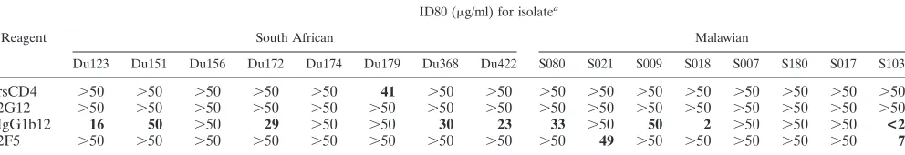
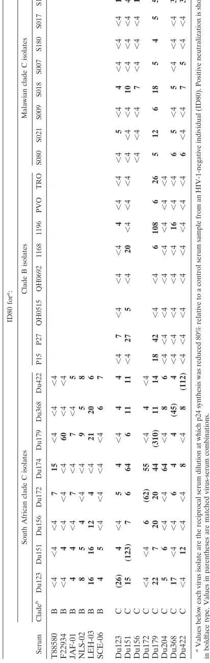
Neutralization of clade C primary HIV-1 isolates with sCD4 and monoclonal antibodies.Primary isolates from eight sub-jects in South Africa and eight subsub-jects in Malawi were char-acterized in neutralization assays with sCD4 and three broadly neutralizing human monoclonal antibodies. These clade C iso-lates all exhibited a high level of resistance to inhibition by sCD4 (Table 1). Of the three monoclonal antibodies tested, IgG1b12 was most effective, neutralizing five of eight isolates from South Africa and four of eight isolates from Malawi at
doses in the range of⬍1.8 to 50g/ml. Monoclonal antibody
2F5 neutralized two isolates from Malawi but none from South Africa. A third monoclonal antibody, 2G12, failed to neutralize all 16 clade C isolates tested. The broad resistance of clade C primary isolates to neutralization by 2G12 was in striking con-trast to the ability of this same preparation of 2G12 to neu-tralize six of eight clade B isolates tested previously (9). Con-trol assays performed with TCLA strains and primary isolates belonging to clade B confirmed no loss of activity of the sCD4 and monoclonal antibodies used in these experiments (data not shown).
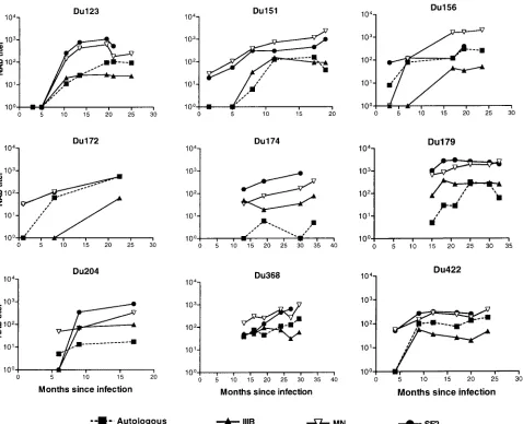
[image:3.587.45.541.85.170.2]Neutralizing antibody response following infection with clade C HIV-1. Serum samples collected at multiple time points from nine South African sex workers were assayed for neutralizing activity against the autologous virus isolate and three TCLA strains of clade B HIV-1. All subjects developed a neutralizing antibody response against their own virus (Fig. TABLE 1. Neutralization sensitivity of HIV-1 clade C isolates as measured with sCD4 and human monoclonal antibodies
Reagent
ID80 (g/ml) for isolatea
South African Malawian
Du123 Du151 Du156 Du172 Du174 Du179 Du368 Du422 S080 S021 S009 S018 S007 S180 S017 S103
rsCD4 ⬎50 ⬎50 ⬎50 ⬎50 ⬎50 41 ⬎50 ⬎50 ⬎50 ⬎50 ⬎50 ⬎50 ⬎50 ⬎50 ⬎50 ⬎50 2G12 ⬎50 ⬎50 ⬎50 ⬎50 ⬎50 ⬎50 ⬎50 ⬎50 ⬎50 ⬎50 ⬎50 ⬎50 ⬎50 ⬎50 ⬎50 ⬎50
IgG1b12 16 50 ⬎50 29 ⬎50 ⬎50 30 23 33 ⬎50 50 2 ⬎50 ⬎50 ⬎50 <2
2F5 ⬎50 ⬎50 ⬎50 ⬎50 ⬎50 ⬎50 ⬎50 ⬎50 ⬎50 49 ⬎50 ⬎50 ⬎50 ⬎50 ⬎50 7
aValues below each virus strain are the minimum concentration of sCD4 and monoclonal antibodies 2G12, IgG1b12, and 2F5 required to achieve an 80% reduction
in p24 synthesis in PBMC (ID80). Positive neutralization is shown in boldface type.
on November 8, 2019 by guest
http://jvi.asm.org/
1). The magnitude of this response varied between individuals
and was relatively potent in most cases (titers of⬎100 in seven
of nine cases). Although the timing of serum collection did not permit a refined assessment of the temporal response, it is accurate to say that autologous neutralizing antibodies were undetectable for at least the first 4 to 5 months of infection in three subjects (Du123, Du151, and Du422). Despite this delay, all three subjects eventually mounted a potent autologous neu-tralizing antibody response. Autologous neuneu-tralizing antibod-ies were low or undetectable at early time points and later rose in magnitude for subjects Du123, Du151, Du156, Du172, and Du422 as evidence that these five study subjects were in an early stage of seroconversion at the time of their enrollment. Autologous neutralizing antibodies in these five subjects were assessed with virus that was obtained shortly after infection. Autologous neutralizing antibodies in the remaining cases were measured with virus that was obtained either before (Du368) or after (Du174, Du179, and Du204) the time of first serum collection (an estimated 6 to 15 months from initial
ELISA positivity). Of these latter cases, virus from subject Du368 was neutralized potently by all serum samples collected 5.5 months later and thereafter. Virus isolated at month 21 from subject Du179 was neutralized weakly by serum samples obtained at month 15 and was neutralized potently by serum samples obtained at month 25 and thereafter. Virus from sub-ject Du174, isolated at month 19.5, was neutralized weakly or not at all by serum collected at months 13, 18, 30, and 34. A second study subject, Du204, had a similar weak autologous neutralizing antibody response.
TCLA strains belonging to clade B HIV-1 were highly sen-sitive to neutralization by the clade C serum samples. Overall neutralization-sensitivity with clade C serum samples was
HIV-1MN⬵HIV-1SF2⬎HIV-1IIIB(Fig. 1). Neutralizing antibodies
were sometimes detected with clade B TCLA strains before being detected with the autologous clade C isolate. Moreover, the potency of neutralization detected with TCLA strains usu-ally exceeded the potency detected with the autologous isolate. Two additional observations are worth noting. First, as men-FIG. 1. Neutralizing antibody response over time in infected individuals in South Africa. Neutralizing antibodies were measured in PBMC with the early autologous isolate and in either MT-2 or CEMx174 cells with the clade B TCLA strains IIIB (MT-2), MN (MT-2), and SF2 (CEMx174). Results that were negative (neutralization titers of⬍4 for autologous isolates and⬍20 for IIIB, MN, and SF2) were assigned a value of 1 for representation.
on November 8, 2019 by guest
http://jvi.asm.org/
[image:4.587.53.531.71.459.2]tioned above, sera from 5 subjects were capable of neutralizing the autologous isolate obtained prior to serum collection (Du123, 10.5 months; Du151, 8 months; Du156, 3 months; Du172, 8 months; Du422, 9 months). Autologous isolates in these cases were obtained 1 to 3 months after initial serocon-version and, as such, could closely resemble the transmitted variant that drove the initial antibody response, leading to antibodies that neutralized the early variant. Two subjects en-rolled later in infection (Du179 and Du368) had detectable autologous neutralizing antibodies that increased in magnitude over the observation period. Autologous isolates in these two cases were obtained 21 and 7.5 months after infection and therefore could be variants that escaped an earlier neutralizing antibody response. In this event, the neutralizing antibodies we detected might have arisen as a de novo antibody response to escape epitopes. Although earlier isolates were not available to test this possibility, a later isolate from subject Du179 (36.5
months) was sensitive to neutralization (titer⬎108) by serum
samples collected at months 25, 30, and 32.5, suggesting that the neutralization determinants were not evolving at a fast pace.
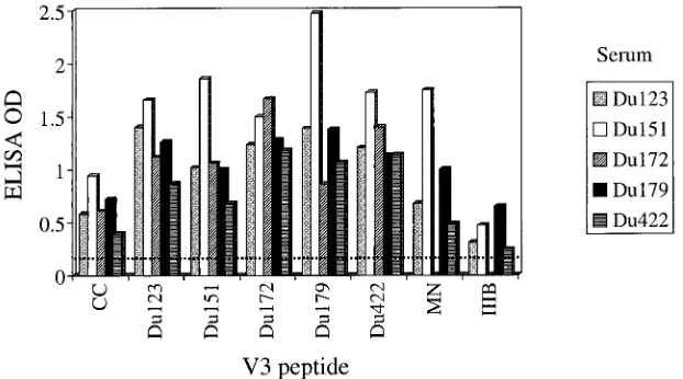
Antibody response to the V3 loop of gp120.V3-specific an-tibodies were assessed by ELISA and in competitive neutral-ization assays with peptides from five different clade C isolates
from South Africa (Du123, Du151, Du172, Du179, and Du422), the clade C consensus sequence (37), and the clade B
TCLA strains HIV-1MN and HIV-1IIIB. The amino acid
se-quence of each V3 peptide is shown in Table 2. Sera from South African subjects reacted strongly with the autologous peptide and exhibited broad cross-reactivity with heterologous clade C peptides (Fig. 2). Reactivity was somewhat diminished in the case of the consensus clade C peptide and more so in the
case of the IIIB peptide. Reactivity to the HIV-1MNpeptide
was variable, being relatively strong for subjects Du151 and Du179 and low to moderate for subjects Du123 and Du422.
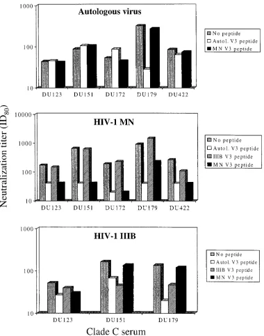
To determine whether these V3-specific antibodies had neu-tralizing activity, serum samples were preincubated with V3 peptides and then assayed for neutralizing activity against the autologous virus and two TCLA strains (Fig. 3). A major frac-tion of autologous neutralizing activity in serum Du179 was blocked by the matched Du179 V3 peptide, suggesting the presence of neutralizing antibodies directed against the V3
loop of this virus. As a control, the HIV-1MNpeptide had no
effect on the autologous neutralizing activity of this serum
sample. Matched V3 peptides and the HIV-1MN V3 peptide
had no effect on the autologous neutralizing activity of four
other clade C serum samples. By comparison, the HIV-1MN
-specific neutralizing activity of all five serum samples was
[image:5.587.41.554.82.182.2]dra-FIG. 2. ELISA reactivity to V3 peptides. Serum samples (1:50 dilution) were evaluated by ELISA for the presence of antibodies that could bind individual peptides derived from the V3 loop of the indicated virus strains. The amino acid sequence of each peptide is shown in Table 2. The dotted line represents the cutoff value for positive reactivity (twice the optical density [OD] of a control serum sample from a healthy, HIV-1-negative individual). The following serum samples were obtained after the estimated number of months of infection: Du123 (13.5 months), Du151 (11.3 months), Du172 (8 months), Du179 (25 months), Du422 (17 months). CC, clade C consensus sequence.
TABLE 2. Amino acid sequences of V3 peptides
HIV-1 isolate Amino acid sequencea
CC...T R P N N N T R K S I R I - - G P G Q T F Y A T G D I I G D I R Q
Du123 ...I R P N N N T R K S I R I - - G P G Q T F Y A T N D I I G D I R Q
Du151 ...T R P N N N T R K S I R I - - G P G Q T F Y A T D A I I G N I R E
Du172 ...T R P S N N T R K S V R I - - G P G Q T F F A T G D I I G D I R Q
Du179 ...T R P G N N T R K S I R I - - G P G Q A F Y - T N H I I G D I R Q
Du422 ...T R P N N N T R K S V R I - - G P G Q T F Y A T G A I I G D I R E
MN... N K R K R I H I - - G P G R A F Y T T K N I I G T I
IIIB ... N N N T R K S I R I Q R G P G R A F V T I G K – I G
aAmino acids that differ from the consensus clade C sequence (CC) are shown in boldface type
on November 8, 2019 by guest
http://jvi.asm.org/
[image:5.587.138.449.506.679.2]matically reduced in the presence of matched V3 peptides and
the HIV-1MNV3 peptide but not the HIV-1IIIBV3 peptide. In
addition, the HIV-1IIIB-specific neutralizing activity was
re-duced in the presence of matching V3 peptide and the
HIV-1IIIB V3 peptide but not the HIV-1MN V3 peptide. We note
that the reduction in HIV-1IIIB-specific neutralization was
mi-nor in one of three cases examined (Du123). Due to sample
availability and low neutralization titers against HIV-1IIIB, the
remaining two samples could not be tested in these
competi-tion assays with HIV-1IIIB.
Analysis of intra- and interclade neutralization.A checker-board analysis was performed with clade B and C virus isolates and serum samples. Clade C serum samples were selected from the South African sex workers at a time when neutralizing activity was detected with the autologous virus and TCLA strains. Clade B serum samples were obtained from individuals in North Carolina who had documented HIV-1 infection for
ⱖ2 years. These same clade B serum samples were assayed
[image:6.587.106.486.71.553.2]recently against the clade B viruses in our panel (9), and there-fore, those results were used in our comparative analyses. We FIG. 3. Ability of V3 peptides to out-compete the neutralizing activity of clade C serum samples from South Africa. Serum samples shown in Fig. 1 were evaluated for neutralizing activity in the presence and absence of gp120 V3 peptides (50g/ml) as described in Materials and Methods. Autologous (Autol.) V3 peptide refers to a peptide bearing the amino acid sequence of the virus from the subject who was the source of serum listed on thexaxis. The top panel shows autologous virus-serum combinations, the middle panel shows clade C serum samples assayed with the HIV-1MNvirus, and the bottom panel shows clade C serum samples assayed with the HIV-1IIIBvirus. ID80, 80% inhibitory dose.
on November 8, 2019 by guest
http://jvi.asm.org/
caution that a potential source of error in our checkerboard assaysisthelackofsequenceinformationtoconfirmthegeneticsub-type of virus from our presumed clade B serum donors from North Carolina. These subjects were presumed to be infected with clade B based on the high incidence of clade B in North America. We also note that our partial genetic and sero-logic characterization of the clade C isolates from Malawi does not entirely eliminate the possibility that these are recombi-nant viruses.
Cross-neutralization within and between genetic subtypes was readily apparent (Table 3). The clade C isolates appeared to be more sensitive to neutralization overall compared to clade B isolates. Specifically, the Du isolates were neutralized 70% of the time (excluding autologous virus-serum combina-tions) by Du serum samples, and 54% of the time by clade B serum samples. Moreover, the Malawi isolates were neutral-ized 48% of the time by Du serum samples. By comparison, clade B isolates were neutralized 25% of the time by Du serum samples (Table 3) and 27% of the time by clade B serum sam-ples (9). Overall trends were analyzed statistically based on pos-itive neutralization frequencies and mean neutralization titers. Only the positive response rates between Du and clade B iso-lates tested with Du serum samples were found to be statisti-cally significant (Table 4). We note that no significant differ-ences were identified based on mean neutralization titers (Table 4).
A number of clade-specific reactivities were observed within the overall analysis: (i) isolate Du174 was neutralized
prefer-entially by clade C sera (P⬍0.01 based on response rate), (ii)
serum Du151 exhibited a preferential neutralization of clade C
isolates from South Africa (P⫽0.01 based on response rate;
P⫽0.02 based on mean neutralization titer), (iii) serum
NLS-02 exhibited a preferential neutralization of clade C isolates
from South Africa (P⫽0.01 by both criteria), and (iv) serum
Du204 exhibited a preferential neutralization of clade C
iso-lated from South Africa (Pⱕ0.05 based on mean
neutraliza-tion titer). Notably, one serum sample from the Du cohort (Du179) neutralized 16 of 16 clade C isolates and six of eight clade B isolates (Table 3).
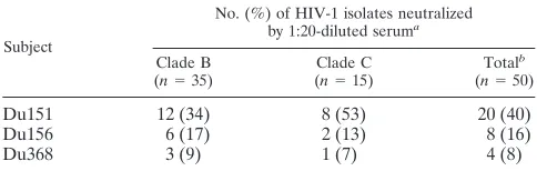
A broader assessment of cross-neutralizing activity with 50 heterologous primary HIV-1 isolates was performed on a small subset of serum samples for which an adequate supply of serum was available. These particular serum samples had po-tent neutralizing activity against the early autologous clade C
isolate (titer⬎100) and were able to neutralize TCLA strains
belonging to clade B. They were assayed at a 1:20 dilution with the 50 primary isolates to be selective for relatively potent heterologous neutralization specificities. The heterologous primary isolates consisted of the 8 clade B and 16 clade C isolates listed in Table 3 plus an additional 26 clade B isolates. The results are summarized in Table 5 and do not include autologous virus-serum combinations. Serum Du151 neutral-ized 40% of the isolates as evidence that this serum sample contains broadly cross-reactive neutralizing antibodies. Serum samples Du156 and Du368 were much less cross-reactive but nonetheless neutralized 7 to 17% of isolates. No clade-specific
preferences were observed with these two serum samples (P⫽
1.00).
DISCUSSION
The neutralization properties of primary isolates and serum samples from clade C HIV-1-infected individuals were charac-terized and compared to clade B. Our goal was to develop standard virologic reagents and to acquire information that would benefit vaccine design. A major focus was placed on South Africa because of the high rates of HIV-1 transmission and the lack of information on neutralizing antibodies in that country. A particular emphasis was placed on a group of fe-male sex workers located at multiple stops along a major truck-ing route between Durban and Johannesburg. These women were participating in HIV-1 prevention programs as part of a microbicide trial and were screened monthly for HIV-1 sero-conversion. Virus isolates and serum samples were available in some cases very soon after infection, which made it possible to examine the autologous and heterologous neutralizing activity of the earliest antibodies generated.
All but one clade C isolate possessed the R5 biologic phe-notype that is typical of transmitted variants. These isolates (eight from South Africa and eight from Malawi) resembled R5 clade B isolates in their sensitivity to neutralization by some but not all serologic reagents. Shared properties included a high level of resistance to inhibition by sCD4 and the frequent neutralization by monoclonal antibody IgG1b12. Resistance to sCD4 is a general property that distinguishes primary isolates from TCLA strains (20) and is thought to reflect the exposure of critical epitopes in and around the CD4 binding site of gp120 as affected by N-linked glycans and tertiary folds in the native gp120 molecule (85). It may be presumed that we pre-served the sCD4-resistant phenotype of these clade C isolates by passing them a minimum number of times in PBMC exclu-sively (71). Many sCD4-resistant variants of HIV-1 are difficult to neutralize with monoclonal antibodies and sera from in-fected individuals. The IgG1b12 epitope, which overlaps the CD4 binding domain of gp120 (51), is an exception in that neutralization by this monoclonal antibody is not predicted by sCD4 sensitivity. For example, the 89.6 and 89.6P variants of simian-human immunodeficiency virus are both moderately sensitive to inhibition by sCD4 yet exhibit a striking dichotomy in their sensitivity to neutralization by IgG1b12 (19). The abil-ity of IgG1b12 to neutralize many sCD4-resistant variants of clade C HIV-1 is additional evidence that this antibody spec-ificity would be highly beneficial for broadly effective vaccina-tion.
Another monoclonal antibody, 2F5, that is known to be effective against primary isolates was only able to neutralize 2 of 16 clade C isolates here. Since this low frequency was no different statistically from the three of eight clade B isolates neutralized by this same batch of 2F5 (9), the neutralizing activity of 2F5, like that of IgG1b12, does not appear to dis-tinguish clade B from clade C. A much different outcome occurred with the 2G12 monoclonal antibody in that it failed to neutralize all 16 clade C isolates from South Africa and Malawi
up to the highest dose tested (50g/ml). 2G12 has been shown
to neutralize primary HIV-1 isolates from multiple clades (9, 23, 46, 75, 76), but very high doses were needed to neutralize the small number of clade C isolates tested in those studies (75, 86). We conclude that 2G12 has very poor neutralizing activity against clade C isolates relative to other clades. It seems
on November 8, 2019 by guest
http://jvi.asm.org/
ly that the neutralizing specificity of 2G12 will have an impact in regions where clade C is prevalent, except in cases where it is synergistic with other neutralizing antibody specificities (86). Our analysis of sequential serum samples from a group of South African sex workers infected with clade C HIV-1 showed a delay in autologous neutralizing antibody production follow-ing primary infection of at least 4 to 5 months. That delay was similar to clade B infection (55, 63) and could be due to either early immunologic dysfunction, the poor immunogenicity of the envelope glycoproteins, or a combination of these factors. Following the delay, a potent autologous neutralizing antibody
response was detected in most cases (titers⬎100) as evidence
that the B-cell response to critical epitopes eventually matured. Despite potent neutralizing antibodies, one individual (Du151) never gained control of her viremia and progressed to AIDS rapidly. We do not know the status of this person’s HIV-1-specific cellular immune response, nor were we able to deter-mine whether a neutralization escape variant arose that might explain the rapid progression.
In addition to neutralizing the autologous isolate, clade C serum samples from South Africa neutralized TCLA strains of clade B HIV-1 with a level of potency similar to clade B serum samples. This is evidence that the antibody specificities for TCLA HIV-1 neutralization are conserved between clades B and C. In support of this notion, antibodies in clade C serum samples bound peptides corresponding to the V3 loop of clade B and C gp120, which is a major target for the neutralization of clade B TCLA strains (5, 73, 77). Although the amino acid sequence of the V3 loop of clades B and C can be quite different (37, 64), they appear to share antigenic features rec-ognized by monoclonal antibodies (33). This may explain why the neutralizing activity of clade C serum samples against clade B TCLA strains was sensitive to the presence of V3 loop peptides. The inability of V3 loop peptides to block autologous neutralization of clade C primary isolates in four of five cases examined, this despite the presence of antibodies to the autol-ogous V3 peptide, suggests that certain epitopes in the V3 loop of clade C primary isolates are poorly antigenic relative to TCLA strains. A similar phenomenon has been described for clade B (5, 73, 77). This dichotomy in antigenicity is an indi-cation that epitope exposure on the native envelope glycopro-tein complex of clade C primary isolates will pose a challenge for neutralizing antibody induction by candidate HIV-1 vac-cines much the same as it has for clade B (52, 85).
Primary isolates of clade B and C HIV-1 were further com-pared in checkerboard neutralization assays that differed from those in previous reports in several ways: (i) a large number of clade C isolates and serum samples were included, (ii) virus isolates were obtained early in infection, and (iii) the clade C serum samples corresponded to an early stage of neutralizing antibody production. In general, the earliest neutralizing anti-bodies to be detected in an HIV-1-infected individual have a limited range of specificity (14, 55). It was therefore unexpect-ed to find 70% of heterologous clade C virus-serum combina-tions testing positive from South Africa. This high frequency is unusual and, with the exception of sera from some long-term nonprogressors (13, 63), has not be documented previously.
The extensive cross-neutralization within the South African serum samples and virus isolates may be an indication of shared neutralization determinants. This would be
encourag-TABLE 3. Checkerboard analysis of clade B and C neutralization ID80 for a: South African clade C isolates Clade B isolates Malawian clade C isolates Serum Clade b Du123 Du151 Du156 Du172 Du174 Du179 Du368 Du422 P15 P27 QH0515 QH0692 1168 1196 PVO TRO S080 S021 S009 S018 S007 S180 S017 S103 T88580 B ⬍ 4 ⬍ 4 ⬍ 4 71 5 ⬍ 4 ⬍ 4 ⬍ 4 F22934 B ⬍ 4 4 ⬍ 4 ⬍ 4 ⬍ 4 60 ⬍ 4 ⬍ 4 JAF-01 B 44 ⬍ 4 7 ⬍ 4 7 ⬍ 4 5 NLS-02 B 85 4 ⬍ 4 ⬍ 4 95 8 LEH-03 B 16 16 12 4 ⬍ 4 21 20 6 SCE-06 B 45 ⬍ 4 ⬍ 4 ⬍ 4 ⬍ 4 67 Du123 C (26) 4 ⬍ 4 54 ⬍ 4 44 ⬍ 4 7 ⬍ 4 ⬍ 4 ⬍ 4 4 ⬍ 4 ⬍ 4 ⬍ 4 5 ⬍ 4 4 ⬍ 4 ⬍ 4 ⬍ 4 12 Du151 C 15 (123) 7 6 64 6 11 11 ⬍ 4 27 5 ⬍ 4 20 ⬍ 4 ⬍ 4 ⬍ 4 ⬍ 4 ⬍ 4 ⬍ 4 10 ⬍ 4 ⬍ 4 ⬍ 4 46 Du156 C ⬍ 4 ⬍ 4 ⬍ 4 7 ⬍ 4 ⬍ 4 ⬍ 4 17 Du172 C ⬍ 4 ⬍ 4 6 (62) 55 ⬍ 4 4 ⬍ 4 Du179 C 22 7 20 20 44 (310) 11 14 18 42 ⬍ 4 ⬍ 4 6 108 6 26 5 12 6 18 5 4 5 55 Du204 C 56 ⬍ 4 ⬍ 4 64 ⬍ 4 86 ⬍ 4 ⬍ 4 ⬍ 4 ⬍ 4 ⬍ 4 ⬍ 4 ⬍ 4 ⬍ 4 Du368 C 17 ⬍ 4 ⬍ 4 6 4 4 (45) 4 ⬍ 4 ⬍ 4 ⬍ 4 ⬍ 4 ⬍ 4 16 ⬍ 4 ⬍ 4 65 ⬍ 4 5 ⬍ 4 ⬍ 4 ⬍ 4 37 Du422 C ⬍ 4 12 ⬍ 4 ⬍ 4 8 ⬍ 4 8 (112) ⬍ 4 ⬍ 4 ⬍ 4 ⬍ 4 ⬍ 4 ⬍ 4 ⬍ 4 ⬍ 4 6 ⬍ 4 ⬍ 4 75 ⬍ 4 ⬍ 4 39 aValues below each virus isolate are the reciprocal serum dilution at which p24 synthesis was reduced 80% relative to a control serum sample from an HIV-1-negative individual (ID80). Positive neutralization is shown in boldface type. Values in parentheses are matched virus-serum combinations. bClade B serum samples were from HIV-1-infected individuals in North Carolina. Clade C serum samples were from HIV-1-infected subjects in South Afric a. These serum samples were obtained after the estimated number of months of infection: Du123 (13.5 months), Du151 (11.3 months), Du172 (8 months), Du179 (25 months), Du204 (9 months), Du368 (19 months), Du4 22 (12 months).
on November 8, 2019 by guest
http://jvi.asm.org/
[image:8.587.52.258.82.732.2]ing for vaccine efforts, as it suggests that neutralizing antibod-ies will have less epitope diversity to overcome at a regional level. The broad implications of this concept depend in part on the geographic range of the shared determinant that defines the corresponding neutralization serotypes. Less epitope diver-sity would be expected if multiple subjects in our cohort were infected by the same individual. Shared determinants might also arise as a consequence of serial transmission of selected isolates in these sex workers. Early evidence in South Africa suggested multiple introductions of the virus into the country, with the diversity observed being greater than one would ex-pect from founder-type effects such as seen in Thailand (8, 79). Similarly, within this sex worker cohort, the analysis of the
relationships betweengag,pol, andenvsequences showed
rel-atively high diversity, and little phylogenetic clustering of se-quences was detected (Williamson et al., submitted). Single-donor and serial transmission of selected isolates are therefore not obvious explanations for our results. However, full-length sequencing does show reliable clustering of two of the isolates (Du151 and Du422), suggesting that at least for these two isolates there was a common source in the not-too-distant past (80). Moreover, we cannot be certain that these female sex workers were not infected multiple times to give rise to a cross-reactive polyvalent neutralizing antibody response. Along this same line, it has been reported that initial virus populations are more heterogeneous in women than in men immediately fol-lowing transmission (42). Additional studies will be needed to
clarify the events that gave rise to the cross-neutralizing anti-body responses in the South African subjects and how those events might impact vaccine development.
The 70% positive neutralization rate within the set of South African clade C viruses and serum samples differed signifi-cantly only from the overall ability of these same serum sam-ples to neutralize clade B isolates. This case of clade-specific neutralization did not extend to other combinations in our checkerboard assays with clade B and C reagents, suggesting that clades B and C are comprised in part of overlapping neutralization serotypes. Evidence that the overlap was only partial comes from a number of individual cases of clade-specific neutralization. One example is a clade C isolate (Du174) that was neutralized preferentially by clade C serum samples. In addition, two South African clade C serum samples neutralized South African clade C isolates in a specific manner. Taken together with the selective neutralizing activity of 2G12, these examples reflect subtle associations between genetic sub-type and neutralization serosub-type that have gone unnoticed in previous studies, owing in part to the complexity of the neu-tralization determinants on the virus and the limited number of clade C reagents used in the past. One unexpected finding was the preferential neutralization of clade C isolates by a pre-sumed clade B serum sample. Unfortunately, a lack of se-quence information on the virus from this individual makes it possible that infection was with a non-clade B virus. Nonethe-less, this is an additional case of a serum that recognized a neutralization serotype found in clade C that was rarely de-tected in clade B. Our combined results are an indication that within each genetic subtype are multiple neutralization sero-types, some of which are more clade-specific than others.
During the course of our studies we discovered two individ-uals in South Africa (Du151 and Du179) whose serum after less than 2 years of infection neutralized a large number of clade B and C primary isolates. This cross-neutralizing activity was not unlike that of sera from some HIV-1-infected long-term nonprogressors (13, 63) and appears to be relatively unique for recent seroconverters. The cross-reactive neutraliz-ing activity could be due to host factors, viral factors, or a combination of the two. It also serves to confirm that clades B and C consist in part of overlapping neutralization serotypes. The viral envelope glycoproteins from these two individuals are interesting candidates for vaccine development.
[image:9.587.44.283.85.165.2]It seems possible that some neutralization serotypes track more with geography than with genetic subtype, as was sug-gested by our observations in a cohort of sex workers in South Africa infected with clade C HIV-1. If generally true, targeted immunogens might prove beneficial for vaccines in this and other parts of the world. The selection of immunogens for such tailored vaccines will require extensive surveys of virus isolates and serum samples. Alternatively, new approaches for cross-reactive neutralizing antibody induction are under investiga-tion that may make tailoring efforts unnecessary (52). With few exceptions (e.g., 2G12 epitope), an immunogen that generates antibodies capable of neutralizing many clade B isolates may be expected to neutralize many clade C isolates and vice versa. Until such immunogens become available, efforts to induce neutralizing antibodies with candidate HIV-1 vaccines might benefit the most by including envelope glycoproteins from one or more strains of the virus that predominate in the specific TABLE 4. Statistical analysis of checkerboard results
Seruma Virusesa
Pderived fromb:
Positive
response rate NAb titerMean
Du Du vs B 0.03 0.31
B Du vs B 0.13 0.13
Du Mw vs B 0.19 0.50
Du Du vs Mw 0.25 0.13
aDu, clade C from South Africa; B, clade B; Mw, clade C from Malawi.
bOverall differences in either the positive neutralization response rate or
mean log neutralizing antibody (NAb) titers between genetic subtypes of the virus were tested for significance by using the nonparametric Wilcoxon sign rank
test. Differences were considered significant ifPwasⱕ0.05. To eliminate bias,
results of autologous serum-virus pairs in the Du samples were excluded from analysis, since this was the only group where autologous pairs were present.
TABLE 5. Cross-neutralizing activity of serum samples from three subjects infected with clade C HIV-1
Subject
No. (%) of HIV-1 isolates neutralized
by 1:20-diluted seruma
Clade B
(n⫽35) (Clade Cn⫽15) Total
b
(n⫽50)
Du151 12 (34) 8 (53) 20 (40)
Du156 6 (17) 2 (13) 8 (16)
Du368 3 (9) 1 (7) 4 (8)
aSerum samples from subjects Du151 (17.3 months), Du156 (17 months), and
Du368 (27 months) were assayed at a 1:20 dilution with 35 clade B and 15 heterologous clade C primary HIV-1 isolates. Neutralization was considered
positive if p24 synthesis was reducedⱖ80% relative to a negative control (serum
from a healthy, HIV-1-negative individual). Values do not include autologous virus-serum combinations.
bThis is the fraction and percent of isolates neutralized for the 50 isolates
total.
on November 8, 2019 by guest
http://jvi.asm.org/
[image:9.587.42.284.585.662.2]regions targeted for vaccination. We add as a cautionary note that it is not clear at this time what level of neutralizing anti-body induction and clinical benefit will be achieved with those envelope glycoproteins.
ACKNOWLEDGMENTS
We thank Mary Phoswa and Melissa Kerkau for virus isolation and Tonie Cilliers for virus phenotyping. We also thank Myron S. Cohen for providing access to samples from Malawi, Dennis Burton for IgG1b12, and Herman Katinger and John Mascola for 2G12 and 2F5. This work was supported by the U.S. National Institutes of Health (grants AI46705, P30-HD37260, and DK49381), the South African AIDS Vaccine Initiative, the Poliomyelitis Foundation, and the Na-tional Research Foundation of South Africa.
REFERENCES
1.Albert, J., B. Abrahamsson, K. Nagy, E. Aurelius, H. Gaines, G. Nystrom, and E. M. Fenyö.1990. Rapid development of isolate-specific neutralizing antibodies after primary HIV-1 infection and consequent emergence of virus
variants which resist neutralization by autologous sera. AIDS4:107–112.
2.Alkhatib, G., C. Combadiere, C. C. Broder, Y. Feng, P. E. Kennedy, P. M. Murphy, and E. A. Berger.1996. CC CKR5: a RANTES, MIP-1␣, MIP-1
receptor as a fusion cofactor for macrophage-tropic HIV-1. Science272:
1955–1958.
3.Arendrup, M., C. Nielsen, J.-E. S. Hanson, C. Pedersen, L. Mathiesen, and J. O. Nielsen.1992. Autologous HIV-1 neutralizing antibodies: emergence of neutralization-resistant escape virus and subsequent development of escape
virus neutralizing antibodies. J. Acquir. Immune Defic. Syndr.5:303–307.
4.Betts, M. R., J. Krowka, C. Santamaria, K. Balsamo, F. Gao, G. Mulundu, C. Luo, N. N’Gandu, H. Sheppard, B. H. Hahn, S. Allen, and J. A. Frelinger.
1997. Cross-clade human immunodeficiency virus (HIV)-specific cytotoxic
T-lymphocyte responses in HIV-infected Zambians. J. Virol.71:8908–8911.
5.Bou-Habib, D. C., G. Roderiquez, T. Oravecz, P. W. Berman, P. Lusso, and M. A. Norcross.1994. Cryptic nature of envelope V3 region epitopes pro-tects primary monocytotropic human immunodeficiency virus type 1 from
antibody neutralization. J. Virol.68:6006–6013.
6.Bradney, A. P., S. Scheer, J. M. Crawford, S. P. Buchbinder, and D. C. Montefiori.1999. Neutralization escape in human immunodeficiency virus
type 1-infected long-term nonprogressors. J. Infect. Dis.179:1264–1267.
7.Bredell, H., G. Hunt, B. Morgan, C. T. Tiemessen, D. J. Martin, and L. Morris.2000. Identification of HIV-1 inter-subtype recombinants in South
Africa usingenvandgagheteroduplex mobility assays. AIDS Res. Hum.
Retrovir.16:493–497.
8.Bredell, H., C. Williamson, P. Sonnenberg, D. J. Martin, and L. Morris.
1998. Genetic characterization of HIV type 1 from migrant workers in three
South African gold mines. AIDS Res. Hum. Retrovir.14:677–684.
9.Bures, R., A. Gaitan, T. Zhu, C. Graziosi, K. McGrath, J. Tartaglia, P. Caudrelier, R. E. L. Habib, M. Klein, A. Lazzarin, D. Stablein, M. Deers, L. Corey, M. L. Greenberg, D. H. Schwartz, and D. C. Montefiori.2000. Im-munization with recombinant canarypox vectors expressing membrane-an-chored gp120 followed by gp160 protein boosting fails to generate antibodies that neutralize R5 primary isolates of human immunodeficiency virus type 1.
AIDS Res. Hum. Retrovir.16:2019–2035.
10.Burton, D. R., and D. C. Montefiori.1997. The antibody response in HIV-1
infection. AIDS11(Suppl. A):S87–S98.
11.Burton, D. R., J. Pyati, R. Koduri, S. J. Sharp, G. B. Thornton, P. W. H. I. Parren, L. S. W. Sawyer, R. M. Hendry, N. Dunlop, P. L. Nara, M. Lamac-chia, E. Garratty, E. R. Stiehm, Y. J. Bryson, Y. Cao, J. P. Moore, D. D. Ho, and C. F. Barbas III.1994. Efficient neutralization of primary isolates of
HIV-1 by a recombinant human monoclonal antibody. Science266:1024–
1027.
12.Cao, H., P. Kanki, J.-L. Sankalé, A. Dieng-Sarr, G. P. Mazzara, S. A. Kalams, B. Korber, S. Mboup, and B. D. Walker.1997. Cytotoxic T-lym-phocyte cross-reactivity among different human immunodeficiency virus type
1 clades: implications for vaccine development. J. Virol.71:8615–8623.
13.Cao, Y., L. Qin, L. Zhang, J. Safrit, and D. D. Ho.1995. Virologic and immunologic characterization of long-term survivors of human
immunode-ficiency virus type 1 infection. N. Engl. J. Med.332:201–208.
14.Carotenuto, P., D. Looij, L. Keldermans, F. de Wolf, and J. Goudsmit.1998.
Neutralizing antibodies are positively associated with CD4⫹T-cell counts
and T-cell function in long-term AIDS-free infection. AIDS12:1591–1600.
15.Cassol, S., B. G. Weniger, P. G. Babu, M. O. Salminen, X. Zheng, M. T. Htoon, A. Delaney, M. O’Shaughnessy, and C.-Y. Ou.1996. Detection of
HIV type 1envubtypes A, B, C, and E in Asia using dried blood spots: a new
surveillance tool for molecular epidemiology. AIDS Res. Hum. Retrovir.
12:1435–1441.
16.Cecilia, D., S. S. Kulkarni, S. P. Tripathy, R. R. Gangakhedkar, R. S. Paranjape, and D. A. Gadkari.2000. Absence of coreceptor switch with
disease progression in human immunodeficiency virus infections in India.
Virology271:253–258.
17.Chan, D. C., D. Fass, J. M. Berger, and P. S. Kim.1997. Core structure of
gp41 from the HIV envelope glycoprotein. Cell89:263–273.
18.Choe, H., M. Farzan, Y. Sun, N. Sullivan, B. Rollins, P. D. Ponath, L. Wu, C. R. Mackay, G. LaRosa, W. Newman, N. Gerard, C. Gerard, and J. Sodroski.1996. The -chemokine receptors CCR3 and CCR5 facilitate
infection by primary HIV-1 isolates. Cell85:1135–1148.
19.Crawford, J. M., P. L. Earl, B. Moss, K. A. Reimann, M. S. Wyand, K. H. Manson, M. Bilska, J. T. Zhou, C. D. Pauza, P. W. H. I. Parren, D. R. Burton, J. G. Sodroski, N. L. Letvin, and D. C. Montefiori.1999. Charac-terization of primary isolate-like variants of simian-human
immunodefi-ciency virus. J. Virol.73:10199–10207.
20.Daar, E. S., X. L. Li, T. Moudgil, and D. D. Ho.1990. High concentrations of recombinant soluble CD4 are required to neutralize primary human
immunodeficiency virus type 1 isolates. Proc. Natl. Acad. Sci. USA87:6574–
6578.
21.Deng, H., R. Liu, W. Ellmeier, S. Choe, D. Unutmaz, M. Burkhart, P. Di Marzio, S. Marmon, R. E. Sutton, C. M. Hill, C. B. Davis, S. C. Peiper, T. J. Schall, D. R. Littman, and N. R. Landau.1996. Identification of a major
co-receptor for primary isolates of HIV-1. Nature381:661–666.
22.Dragic, T., V. Litwin, G. P. Allaway, S. R. Martin, Y. Huang, K. A. Na-gashima, C. Cayanan, P. J. Maddon, R. A. Koup, J. P. Moore, and W. A. Paxton.1996. HIV-1 entry into CD4⫹cells is mediated by the chemokine
receptor CC-CKR-5. Nature381:667–673.
23.D’Souza, M. P., D. Livnat, J. A. Bradac, and S. H. Bridges.1997. Evaluation of monoclonal antibodies to human immunodeficiency virus type 1 primary isolates by neutralization assays: performance criteria for selecting candidate
antibodies for clinical trials. J. Infect. Dis.175:1056–1062.
24.Earl, P. L., R. W. Doms, and B. Moss.1990. Oligomeric structure of the human immunodeficiency virus type 1 envelope glycoprotein. Proc. Natl.
Acad. Sci. USA87:648–652.
25.Engelbrecht, S., I. Koulinska, T. L. Smith, J. Barreto, and E. J. van Rens-burg.1998. Variation in HIV type 1 V3 regionenvsequences from
Mozam-bique. AIDS Res. Hum. Retrovir.14:803–805.
26.Engelbrecht, S., T. L. Smith, P. Kasper, E. Faatz, M. Zeier, D. Moodley, C. G. Clay, and E. J. van Rensburg.1999. HIV type 1 V3 domain serotyping and genotyping in Gauteng, Mpumalanga, KwaZulu-Natal, and Western
Cape Provinces of South Africa. AIDS Res. Hum. Retrovir.15:325–328.
27.Esparza, J., and N. Bhamarapravati.2000. Accelerating the development and future availability of HIV-1 vaccines: why, when, where and how? Lancet
355:2061–2066.
28.Feng, Y., C. C. Broder, P. E. Kennedy, and E. A. Berger.1996. HIV-1 entry cofactor: functional cDNA cloning of a seven-transmembrane, G
protein-coupled receptor. Science272:872–877.
29.Ferrari, G., W. Humphrey, M. J. McElrath, J.-L. Excler, A.-M. Duliege, M. L. Clements, L. C. Corey, Dani P. Bolognesi, and K. J. Weinhold.1997. Clade B-based HIV-1 vaccines elicit cross-clade cytotoxic T lymphocyte
reactivities in uninfected volunteers. Proc. Natl. Acad. Sci. USA94:1396–
1401.
30.Freed, E. O., E. L. Delwart, G. L. Buchschacher, Jr., and A. T. Panganiban.
1992. A mutation in the human immunodeficiency virus type 1 transmem-brane glycoprotein gp41 dominantly interferes with fusion and infectivity.
Proc. Natl. Acad. Sci. USA89:70–74.
31.Gadkari, D. A., D. Moore, H. W. Sheppard, S. S. Kulkarni, S. M. Mehendale, and R. C. Bollinger.1998. Transmission of genetically diverse strains of
HIV-1 in Puna, India. Indian J. Med. Res.107:1–9.
32.Gao, F., D. L. Robertson, S. G. Morrison, H. Hui, S. Craig, J. Decker, P. N. Fultz, M. Girard, G. M. Shaw, B. H. Hahn, and P. M. Sharp.1996. The heterosexual human immunodeficiency virus type 1 epidemic in Thailand is caused by an intersubtype (A/E) recombinant of African origin. J. Virol.
70:7013–7029.
33.Gorny, M. K., T. C. VanCott, C. Hioe, Z. R. Israel, N. L. Michael, A. J. Conley, C. Williams, J. A. Kessler, I. I., P. Chigurupati, S. Burda, and S. Zolla-Pazner.1997. Human monoclonal antibodies to the V3 loop of HIV-1
with intra- and interclade cross-reactivity. J. Immunol.159:5114–5122.
34.Graham, B. S.1994. Serologic responses to candidate AIDS vaccines. AIDS
Res. Hum. Retrovir.10:S145–S148.
35.Harada, S., Y. Koyanagi, and N. Yamamoto.1985. Infection of HTLV-III/ LAV in HTLV-I-carrying cells MT-2 and MT-4 and application in a plaque
assay. Science229:563–566.
36.Kessler, J. A., II, P. M. McKenna, E. A. Emini, C. P. Chan, M. D. Patel, S. K. Gupta, G. E. Mark III, C. F. Barbas III, D. R. Burton, and A. J. Conley.1997. The recombinant human monoclonal antibody IgG1b12 neutralizes diverse human immunodeficiency virus type 1 primary isolates. AIDS Res. Hum.
Retrovir.13:575–582.
37.Korber, B., B. Hahn, B. Foley, J. W. Mellors, T. Leitner, G. Meyers, F. McCutchen, and C. L. Kuiken.1997. Human Retroviruses and AIDS 1997: a compilation and analysis of nucleic acid and amino acid sequences. The-oretical Biology and Biophysics Group, Los Alamos National Laboratory, Los Alamos, N.Mex.
38.Kostrikis, L. G., Y. Cao, H. Ngai, J. P. Moore, and D. D. Ho.1996.