R E S E A R C H
Open Access
Recombinant production of self-assembling
β
-structured peptides using SUMO as a fusion
partner
Abhinav Prakash
1, Stephen J Parsons
1, Stuart Kyle
1,2and Michael J McPherson
1,2*Abstract
Background:Self-assembling peptides that form nanostructured hydrogels are important biomaterials for tissue engineering scaffolds. The P11-family of peptides includes, P11-4 (QQRFEWEFEQQ) and the complementary peptides P11-13 (EQEFEWEFEQE) and P11-14 (QQOrnFOrnWOrnFOrnQQ). These form self-supporting hydrogels under
physiological conditions (pH 7.4, 140 mM NaCl) either alone (P11-4) or when mixed (P11-13 and P11-14). We report a SUMO-peptide expression strategy suitable for allowing release of native sequence peptide by SUMO protease cleavage.
Results:We have expressed SUMO-peptide fusion proteins from pET vectors by using autoinduction methods. Immobilised metal affinity chromatography was used to purify the fusion protein, followed by SUMO protease cleavage in water to release the peptides, which were recovered by reverse phase HPLC. The peptide samples were analysed by electrospray mass spectrometry and self-assembly was followed by circular dichroism and transmission electron microscopy.
Conclusions:The fusion proteins were produced in high yields and theβ-structured peptides were efficiently released by SUMO protease resulting in peptides with no additional amino acid residues and with recoveries of 46% to 99%. The peptides behaved essentially the same as chemically synthesised and previously characterised recombinant peptides in self-assembly and biophysical assays.
Keywords:Self-assembly, Peptide, Hydrogel, Recombinant expression, Scaffold
Background
Rationally designed self-assembling peptides have recently attracted widespread attention for the development of novel biomaterials in applications such as tissue engineer-ing scaffolds [1-5] and dental enamel remineralisation [6]. The P11-family of peptides comprises over 20 different
pep-tides designed by Aggeli and colleagues to self-assemble intoβ-sheet structures under various physicochemical con-ditions to form isotropic hydrogels at peptide concentra-tions of 10–30 mg/mL [7-9]. These peptides have varying overall charge, hydrophobicity, and polar amino acids resulting in a difference in properties such as solvent affin-ity, dissolution rate and rigidity of gels.
Chemically synthesised peptide P11-4 (CH3CO-QQRFE
WEFEQQ-NH2) is a pH responsive self assembling peptide
which formsβ-sheets and nematic gels at a concentration of 12.6 mM in water on a pH trigger [7,8] or under physio-logical conditions in cell culture medium (DMEM) at pH 7.4 [10]. Peptides P11-13 (CH3CO-EQEFEWEFEQE-NH2)
and P11-14 (CH3CO-QQOFOWOFOQQ-NH2) are
com-plementary assembling peptides that will not self-assemble independently but when combined they self-assemble with each other to form a hydrogel [11].
Recently there have been reports of recombinant pro-duction of self-assembling peptides although there can be issues of low production levels, cell toxicity and deg-radation by proteases [12-14]. For short peptides <50 amino acids these issues are normally addressed by expressing the peptide as part of a larger‘fusion partner protein’ to optimise intracellular stability and facilitate affinity isolation during purification. However, there
* Correspondence:[email protected]
1Institute of Molecular and Cellular Biology, Faculty of Biological Sciences,
University of Leeds, Leeds LS2 9JT, UK
2Astbury Centre for Structural Molecular Biology Faculty of Biological
Sciences University of Leeds, Leeds LS2 9JT, UK
remains the challenge of separating the peptide from its fusion partner.
Previous work on recombinant production of the β -structured P11 self-assembling peptides has focused upon
a ketosteroid isomerase fusion partner to produce peptides P11-4 [10,12] and P11-2 [15]. These studies reported a
maximal yield of 4.6 g fusion protein/L culture for P11-4
[10] by autoinduction [16]. However, the fusion proteins accumulate as insoluble inclusion bodies and peptides are released by denaturation followed by chemical cleavage of a methionine residue with cyanogen bromide [10,12] or a cysteine residue with 1-cyano-4-dimethylaminopyridinium [15]. The use of chemicals for cleavage can lead to pro-blems of disposal on scale-up and results in a non-native peptide sequence. Thioredoxin has also been used as a fu-sion partner with enzymatic cleavage to release fused pep-tides, in this case with tobacco etch virus protease [17]. However, this leaves either a Gly or Ser as the N-terminal residue and so usually results in a non-native N-terminus.
In the present study, we have used SUMO (small ubiqui-tin-related modifier) protein as a fusion partner for various self-assembling P11-peptides to address the question, can
high yields of self assembling P11-family peptide with native
termini be produced from a soluble fusion protein system? SUMO fusion technology has been widely used for difficult to express peptides and proteins [18] and due to the nature of cleavage by the SUMO protease, which recognises a structural rather than sequence feature, allows the produc-tion of proteins or peptides with any N-terminal residue [19,20]. The approach uses theS. cerevisiaeSUMO as a fu-sion partner to allow soluble expresfu-sion of fufu-sion proteins which can be easily purified using an affinity purification tag. The tertiary structure of SUMO, rather than a se-quence motif, is recognised and cleaved by SUMO prote-ase which cleaves after two Gly residues at the C-terminus of SUMO thus releasing the associated protein or peptide with a native N-terminus. SUMO has been successfully used for production of vesicle forming peptides [21] and another self-assembling peptide EAK16[22].
We have developed a SUMO-peptide expression strat-egy suitable for producing soluble fusion proteins and have recovered three different P11-family peptides, P11-4
and the complementary peptides P11-13 and P11-14 (K). In
the latter case the ornithine in the chemically prepared peptide is replaced by lysine residues. We have charac-terised these recombinant peptides and show that they be-have essentially the same as previously characterised chemically synthesised and recombinant peptides.
Results
Cloning and expression strategy
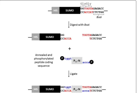
The pET SUMOadapt vector was kindly provided by Bosse-Doenecke [23]. This modified vector carries an inser-tion of a multiple cloning site with aBsaI site positioned
conveniently to allow the cloning of a coding DNA se-quence in-frame with the Gly-Gly motif at the C-terminal cleavage site of SUMO protease [23]. However, the Invitro-gen parent vector is based on pBR322 which has a low to moderate copy number. As our objective was to enhance the level of protein production we subcloned the SUMO adapt region into a high copy number pET-28 vector. A PCR reaction was performed using the primers CpRd_NcoI_F and T7 reverse, and the product was cloned into pET28c using restriction enzymes NcoI and BamHI. This generated the expression vector pET28_SUMOadapt. Peptide coding sequences, containing a translation termin-ation codon to ensure a native C-terminal end, were cloned into theBsaI site (Figure 1).
A S. cerevisiae SUMO protease gene codon-optimised for E. coli expression was synthesised by Genscript and was sub-cloned into the expression vector pET11a.
Expression of SUMO_P11-N and SUMO protease by
autoinduction
The term P11-N is used to represent any of the P11
fam-ily of peptides. The pET28_SUMOadapt was tested for SUMO protein production to select the optimal growth medium and induction time. Auto-induction trials indi-cated production of soluble protein using E. coli BL21 Star (DE3). Terrific broth (TB) and 8ZY media supple-mented with 6% (v/v) 50 X 5052 [16] were tested and TB with 5052 was found to result in a higher cell culture density and level of SUMO production over the growth period tested. The maximum OD600was 45
correspond-ing to a fusion protein level of 1.5 g/L. A harvest time of 64 hours was selected for maximal soluble protein production.
Optimal yield of soluble SUMO protease was also achieved under these conditions after 64 hours culture.
Extraction and purification of SUMO_P11-N and SUMO
Protease
clear that the properties of the peptide influence the SDS-PAGE migration characteristics of the SUMO-peptide fusion proteins. SUMO-P11-4 and SUMO-P11-13
migrate in a similar manner and upon SUMO protease cleavage the SUMO protein migrates further within the
gel. By contrast the positively charged peptide causes the SUMO-P11-14(K) to migrate more rapidly than the P11-4
or P11-13 fusion proteins. However, following SUMO
protease cleavage of P11-14(K) the SUMO shows an
[image:3.595.58.545.89.417.2]ap-parent decrease in migration rate to a position
Figure 2SDS-PAGE gels showing the cleavage of SUMO_P11-N with SUMO protease. A)Uncleaved and SUMO protease cleaved SUMO_P11-4 in either buffer (lanes 1 and 2) or water (lanes 3 and 4).B)SUMO_P11-13 uncleaved (lane 1) and SUMO protease cleaved in water
(lane 2) andC)SUMO_P11-14 (K) uncleaved (lane 1) and SUMO protease cleaved in water (lane 2). Lanes 3 and 4 show overloaded samples of
[image:3.595.56.540.535.685.2]Lanes 1 and 2 respectively to allow visualisation the released P11-14 (K) peptide (lane 4). Lane 5 shows SUMO protease
corresponding to the cleaved SUMO proteins from the P11-4 and P11-13 fusion samples.
Reverse phase HPLC (RP-HPLC)
Following peptide cleavage it was necessary to separate the peptide from other reaction components by RP-HPLC. The cleavage reaction mixture was adjusted to pH 9.0 with NH4OH and incubated overnight before filtering (0.22μm)
prior to injection onto a C18 column. The elution profile was monitored at 220 and 280 nm and Figure 3 shows chro-matograms of typical separations. The fractions were col-lected using the 220 nm absorbance setting rather than 214 nm due to a limitation with the fraction collector. The
fractions corresponding to distinct peaks were collected sep-arately and analysed by mass spectrometry. The peptides were in the major peaks eluting between 4 and 8 minutes and were pooled and lyophilised. The difference in retention time for P11-14 and associated SUMO compared with the
P11-4 and P11-13 samples is most likely due to the
alterna-tive buffer system used for this posialterna-tively charged peptide.
Mass spectrometry characterisation
[image:4.595.59.532.264.673.2]To confirm the identity of the fusion proteins and peptides electrospray mass spectrometry was performed. Fusion pro-teins were dialysed overnight against 50 mM ammonium bicarbonate (pH 8.0) using Spectra/POR 6 dialysis 1 kDa
Figure 3Average absorbance traces of cleaved SUMO_P11-N when purified using RP-HPLC on a C18 column.Absorbance measurements were made at 280 nm and 220 nm with the fraction collector programmed to collect peaks at 220 nm. The positions of the peaks subsequently identified to contain the peptide and SUMO protein are indicated by arrows.A)P11-4 purification with the sharp peak at 6–7 minutes
corresponding to P11-4 and the broader peak between 11 and 20 minutes corresponding to SUMO.B)P11-13 purification.C)P11-14 (K)
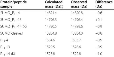
cut off membrane (Spectrum laboratories). Peptide samples were lyophilised. The theoretical mass of the fusion proteins were calculated by Expasy Protoparam for protein samples lacking the N-terminal methionine and the masses mea-sured by mass spectrometry are shown in Table 1. The masses were in excellent agreement confirming the iden-tities of the fusion proteins and the peptides.
Electrospray MS-MS sequencing confirmed the iden-tity of nine of the eleven amino acids in each of the peptides.
Peptide quantification
UV spectroscopy was used to quantify the peptides based on their theoretical extinction coefficients. SUMO protein has 118 amino acids excluding the N-terminal methionine, while the peptides comprise 11 amino acids, representing between 10.3 and 10.7% of the mass of the fusion proteins. In an experiment to compare the yields of each peptide, a series of parallel purifications were performed and the results are shown in Table 2. These show that the peptides were well purified, with yields of 99.6% for P11-13, 84% for P11-4 and 46.1% for P
achieved. The reason for the lower yield for P11-14 may
be due to the different buffer system or the positively charged nature of the peptide. The yield of 46% is good but further work is required to determine the underlying reason for the lower recovery and to try to optimise re-covery of this peptide.
Formation of peptide hydrogels
The purified peptides were dissolved in 140 mM NaCl to a final concentration of 10 mg/mL. The pH of the P11-4
solution was adjusted to ca. 2.0 to trigger formation of a self supporting gel. P11-13 and P11-14 were also prepared
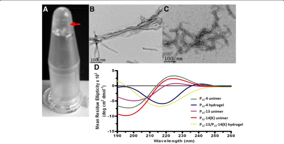
to a final concentration of 10 mg/mL at ca. pH 7 and were mixed in equal volumes to form a self supporting gel. Figure 4A shows the hydrogel state for P11-13/P11-14
when combined.
Characterisation of peptide fibril formation
Aggeli and colleagues reported the morphological struc-ture of chemically synthesised P11-4 fibrils by
transmis-sion electron microscopy (TEM) [7,8]. Self supporting gels for P11-4 and P11-13/P11-14 were diluted to 100μM
in distilled water at pH 2.0 or 7.4 respectively. Fibril morphologies were observed by TEM using uranyl acet-ate negative staining. Figure 4B and C show the inter-twining of fibrils creating fibres for P11-4 and P11-13/
P11-14(K) hydrogels, respectively. Both long and short
fibre structures were observed as isolated structures as well as entangled masses. These are similar to structures that have been previously observed with other samples of these peptides [7,8,10,12].
The secondary structure of the peptide samples was examined as a function of pH using circular dichroism. Peptide hydrogels were diluted to 100 μM solutions. Monomeric forms were also prepared as controls. The results as shown in Figure 4D confirm the random coil conformation of the monomeric forms of P11-4, P11-13
and P11-14(K), and theβ-sheet conformations for the P11
-4 and P11-13/P11-14(K) hydrogels.
Discussion
Our previous work led to the development of E. coli ex-pression systems capable of producing large amounts of short self-assembling peptides of up to 370 mg/L culture [10,12]. These were produced in the form of inclusion bod-ies with recovery of the peptides from their fusion partner by urea solubilisation and chemical cleavage. We were interested to explore the extent to which we could express the P11-family ofβ-structured peptides in a soluble format
[image:5.595.56.291.125.242.2]through the use of soluble peptide fusions. We chose to test the SUMO system as it enhances the solubility of
Table 1 Comparison of calculated and mass spectrometry determined molecular masses of fusion protein and peptide samples
Protein/peptide sample
Calculated mass (Da){
Observed mass (Da)
Difference (Da)
SUMO_P11-4 14821.4 14820.8 −0.6
SUMO_P11-13 14796.3 14796.4 +0.1
SUMO_P11-14 (K) 14790.5 14789.6 −0.9
SUMO cleaved 13284.8 13284.0 −0.8
P11-4 1554.6 1553.7 −0.9
P11-13 1529.5 1528.6 −0.9
P11-14 (K) 1523.8 1522.8 −1.0
[image:5.595.59.539.668.733.2]{The calculated molecular mass is of SUMO protein lacking the N-terminal methionine residue.
Table 2 Yields of purified SUMO fusion proteins and peptides
Peptide Fusion protein yield (mg/L)
Peptide as percentage mass of fusion protein (%)
Theoretical yield of peptide (mg/L)
Actual yield of peptide purified (mg/L)
Percent theoretical yield (%)
P11-4 400 10.49 42.0 35.3 84.0
P11-13 500 10.34 51.7 51.5 99.6
proteins and has been effective for recombinant produc-tion of difficult proteins with good yields and efficient puri-fication [19,24-27]. Moreover, SUMO protease is highly efficient and does not alter the target protein sequence thereby producing native protein of high quality [28]. To enhance the level of protein and peptide production we required high levels of SUMO expression and therefore subcloned the SUMO adapt region from pET SUMO adapt [23] into a pET28c vector. Constructs were trans-formed into E. coli strain BL21 (DE3) Star and TB-5052 media was used for expression of protein. SUMO-peptide fusions were recovered at 0.37 to 0.5 g/L with HPLC purifi-cation from the SUMO protease cleavage reaction resulting in yields of soluble P11-peptides of between 18 and 35 mg/
L demonstrating that we have achieved good levels of re-covery of purified peptides. It is likely this can be increased significantly as the maximum yield of SUMO protein achieved was 1.5 g/L compared to 637 mg/mL reported by Li et al., [28] using a similar expression strategy, while for EAK16 peptide production 250 mg/L of fusion protein was reported by Satakarni et al., [22] by IPTG induction.
Cleavage of fusion protein by SUMO protease was highly efficient in both cleavage buffer and in water. The possibility of performing cleavage reactions in water fur-ther demonstrates the efficiency of the system allowing largely salt free solutions containing peptides to be obtained thus reducing difficulties in downstream removal
of salts prior to subsequent steps. It is interesting that the SUMO protease works so efficiently in the presence of a low concentration of reducing agent required for cysteine protease function. The dithiothreitol (DTT) concentration in the protease stock solution is 0.5 mM so upon dilution by up to 10,000-fold in water for the cleavage reaction the final concentration would be 50 nM.
We have previously characterised both chemically and recombinantly produced P11- peptides [7,8,10-12]. In
this study TEM showed that rP11-4 formed fibrils with
lengths and widths similar to those reported for cP11-4
[7,8,10,12] and to our previously purified rP11-4 and
rP11-4(hsl) [10,12] and cP11-13/cP11-14 similar to those
previously observed [11]. Circular dichroism analysis suggested that the predominant secondary structure of rP11-4 and P11-13/P11-14 hydrogels at physiological pH
(pH 7.4) was antiparallel β-sheet as has been observed previously [7,8,10-12]. Upon adjusting rP11-4 to pH 9, a
conformational transition to random coil occurred while solutions of rP11-13 and rP11-14 alone also displayed
random coil conformation. We have previously demon-strated that these P11- peptides are cytocompatible with
human primary fibroblasts [10,11].
Conclusions
[image:6.595.58.537.90.333.2]We have demonstrated the use of a SUMO fusion approach for efficient production and purification of Figure 4Hydrogel analysis of P11-13/P11-14. A)Hydrogel formed upon equimolar mixing of P11-13 and P11-14(K) indicated by arrow. B and C)
Transmission electron microscopy (TEM) images of self-assembledB)P11-4 at pH 2 andC)P11-13/P11-14(K). D)Circular dichroism analysis of P11-4,
P11-13 and P11-14(K) unimers, and P11-4 and P11-13/P11-14(K) hydrogels at pH 7.4. Random coil conformation is observed for peptide unimers
β-structured recombinant self assembling peptides with native N- and C-termini. We have demonstrated that SUMO protease displays efficient cleavage of the fusion protein in water with a very low concentration of reducing agent (50 nM) . The efficiency of SUMO protease cleavage in water can be exploited to produce essentially salt and ion-free solutions of various proteins/peptides for subse-quent processing. The purified recombinant peptides be-have similarly to chemically synthesized versions. Whilst recombinant production has the potential to produce large quantities of self-assembling peptides for tissue en-gineering and industrial applications, the greatest benefits are likely to derive from the soluble expression of such peptides as fusions to a range of functional peptide and protein domains to impart tailored biological functions within self-assembled peptidic biomaterials.
Methods
Bacterial strains, plasmids and cell culture media
E. coli strain XL1 Blue (Stratagene, La Jolla, CA.) was used for routine cloning while BL21 Star (DE3) Star (Invitrogen, Carlsbad, CA) was used for expression. The expression vectors, pET 28c+ and pET 11a+ (Novagen) were used to express the SUMO fusion construct and SUMO protease respectively. pET SUMOadapt was a gift from Dr E. Bosse-Doenecke [23]. A codon optimised SUMO protease gene was synthesised by Genscript and delivered in a pUC 57 plasmid.
The growth media Luria Burtani broth (LB), 8ZY and LB agar were prepared according to [29]. Terrific Broth (TB) was purchased from HiMedia. Auto-induction media were prepared according to the methods of Stu-dier [16] and were supplemented by 6% (v/v) 50 X 5052 supplements. 5052 supplement corresponds to 0.5% (v/ v) glycerol, 0.05% (w/v) glucose and 2% (w/v) lactose. Antibiotics carbenicillin and kanamycin were added to media to final concentrations of 100 μg/mL and 50μg/ mL respectively.
Generation of recombinant E. coli strains
Generation of pET28_SUMO fusion construct
The SUMO protein coding region was PCR amplified from pET SUMOadapt [23] with an NcoI site at the 5’ end and a BamHI site at the 3’end to allow cloning into pET28c . A 50 μL PCR reaction containing 0.5 ng of template DNA (pET SUMOadapt), 15 pmoles each of CpRd_NcoI_F (GAGATATACCATGGGC) and T7 ter-minator primer (TAGTTATTGCTCAGCGGTGG), 0.2 mM dNTPs, 1mM MgSO4, 1 X KOD buffer, 1 U KOD Hot Start Polymerase (Novagen) was subjected to 1 mi-nute at 95°C, 36 cycles of 30 seconds at 95°C, 30 seconds at 55°C and 1 minute extension at 72°C followed by a final extension for 10 minutes at 72°C. PCR products were analysed on a 1.5% agarose gel and the correct
product was purified using a QIAquick PCR clean up kit (Qiagen cat. # 28104). This DNA fragment was digested with NcoI and BamHI and purified as before. A 15 μL reaction containing 1.2 μg of the pET28c vector was digested with 20 units of NcoI and BamHI followed by dephosphorylation with 10 units of Antarctic phosphat-ase (NEB cat. #M0289L) and the vector backbone was gel extracted (QIAquick Gel Extraction kit cat. #28704). A 10μL ligation reaction was set up containing 50 ng of pET28c vector backbone, 100 ng of SUMO insert, 10 units of T4 DNA ligase and 1 X T4 DNA ligase buffer.
Generation of rP11-N coding regions
Complementary oligonucleotides encoding self-assembling peptides P11-4 (QQRFEWEFEQQ), P11-13
(EQEFEWE-FEQE) and P11-14(K) (QQKFKWKFKQQ) were designed
to have a stop codon (bold) and 5’TGGT or ACCA single strand overhangs (underlined);
P11-4 F 5’-tggtcagcagcgctttgaatgggaatttgaacagcagtaa-3’
P11-4 R 5’-accattactgctgttcaaattcccattcaaagcgctgctg-3’
P11-13 F 5’-tggtgaacaggaatttgaatgggaatttgaacaggaataa-3’
P11-13 R 5’-accattattcctgttcaaattcccattcaaattcctgttc-3’
P11-14 (K) F 5’-tggtcagcagaaatttaaatggaaatttaaacagcagtaa-3’
P11-14 (K) R 5’-accattactgctgtttaaatttccatttaaatttctgctg-3’
The SUMOadapt region was designed by Bosse-Doenecke [23] to exploit the BsaI restriction site for cloning. In this case the ends created are ACCA allowing peptide insertion immediately adjacent to the C-terminal Gly-Gly motif (Figure 1).
Oligonucleotides were dissolved to a concentration of 100 pmol/μl in distilled water. A 15μL phosphoryl-ation reaction was set up at 37°C for 30 min containing 600 pmol of oligonucleotide, 5 U of T4 polynucleotide kinase (NEB cat. #M0236L) and 1 X T4 DNA ligase buf-fer. The reaction mixture was heat inactivated at 65°C for 20 minutes before 400 pmol of phosphorylated forward and reverse oligonucleotides were mixed with 20 μl of 10 X annealing buffer (400mM Tris HCl pH 8.0, 100mM MgCl2and 500 mM NaCl) in a final
vol-ume of 200 μl. The mixture was heated at 99°C for 10 minutes and allowed to cool down slowly to 50°C to allow the complementary sequences to anneal.
Generation of fusion proteins
by colony PCR using Go-Taq Polymerase (Promega) and T7 forward and reverse primers to identify recombinants which were then sequenced to confirm their integrity.
Generation of SUMO protease expression construct A His-SUMO protease [30] codon optimised DNA se-quence was synthesised by Genscript in a pUC57 vector. This was subcloned into pET 11a digested withNdeI and
BamHI and dephosphorylated. Colony PCR was used to screen transformants and the coding region was verified by DNA sequencing.
Fusion protein expression
Protein expression studies were performed using the auto-induction protocol developed by Studier [16]. A single col-ony was used to inoculate 2 mL of TB media containing antibiotic with growth at 37°C for 6 hours at 250 rpm. A 250μl aliquot of the culture was used to inoculate 400 mL of TB-5052 auto-induction media in a 2L flask and incu-bated at 25°C at 250 rpm. For time course experiments, 1 mL samples were collected at time intervals up to 64 h. Cells were harvested at 64 h post inoculation by centrifu-gation at 6000 rpm for 20 min using fixed angle rotor (Sorvall SLA1500).
Preparation of soluble proteins fromE. colicultures Cell pellets were re-suspended in extraction buffer (50 mM NaH2PO4, 300 mM NaCl, 20 mM imidazole, pH 8.0)
supplemented with protease inhibitor cocktail (Complete EDTA free protease inhibitors) (1 tablet/40 mL) and 20 Units of Omnicleave endonuclease. The cell suspension was then subjected to two cycles of mechanical disruption at 30,000 psi using a cell disruptor (Constant Cell Disrup-tion Systems model 2 PLUS). Following cell lysis, the sol-uble fraction was isolated by centrifugation at 13,000 x g
for 45 minutes to pellet the insoluble fraction. The super-natant (soluble phase) was removed, filtered through a 0.22μm filter (Sartorius) and stored at−80°C.
SUMO_peptide purification by immobilised IMAC
The Novagen ‘Batch Purification of 6X His-tagged pro-teins from E. coli under native conditions’ protocol was followed. The soluble fractions were filtered with a 0.45μm filter (Sartorius) and loaded onto a pre-equilibrated Ni-NTA column. Columns were then washed with extraction buffer containing 50 mM then 100 mM imidazole and pro-teins were eluted in elution buffer (50 mM NaH2PO4, 300
mM NaCl, 250 mM imidazole, pH 8.0). Proteins were con-centrated using centrifugal filters (Amicon Ultra, Millipore) and buffer exchanged in to either PBS or dH2O prior to
SUMO protease cleavage. This exchange was achieved by repeated dilution and centrifugation with the required buf-fer to remove trace salts.
SDS-PAGE analysis
Protein samples were mixed in SDS loading buffer (12% SDS, 6% mercaptoethanol, 30% Glycerol, 0.05% bromo-phenol blue in 1M Tris–HCl (pH 6.8) in a 4:1 (v/v) ratio of sample to loading buffer and heated at 95°C for 5 minutes. Samples were analysed using 4–12% NuPAGE Novex Bis-tris precast gels (Invitrogen, UK) or home-made 12% SDS-PAGE gels at 150 V for 1 hour. Protein bands were visualized using Coomassie Blue G-250 (BDH Chemicals) or Simply BlueTMSafe stain (Invitro-gen, UK).
Protein concentration determination
Protein concentrations were determined either by Brad-ford assay (Bio-Rad cat. 500–0201) or UV spectroscopy. Theoretical extinction coefficients were calculated using ProtParam (http://ca.expasy.org/cgi-bin/protparam) to es-timate the concentration according to Beer-Lambert Law. The extinction coefficients for SUMO was 1490 M-1cm-1, P11-4 was 5500 M-1 cm-1 while SUMO protease was
30035 M-1cm-1.
SUMO protease expression, purification and storage SUMO protease was expressed by autoinduction in E. coli BL21 Star (DE3) and harvested after 64 hours. Cell pellets were resuspended in binding buffer (50 mM NaPi, 300 mM NaCl, 20 mM imidazole, pH 8.0) supple-mented with Omnicleave endonuclease. SUMO protease was purified by IMAC and immediately following elu-tion, DTT was added to a concentration of 1 mM and the purified SUMO protease was dialysed against phos-phate buffered saline (PBS; 50 mM NaPi, 300 mM NaCl, pH 8.0) with 1 mM DTT. This was concentrated and quantified using a Bradford assay as 5 mg/mL. Finally glycerol was added to 50% (v/v) and the SUMO protease was stored at−80°C until required.
Cleavage of SUMO_peptide fusion protein with recombinant SUMO protease
SUMO protease was used in a 1:1000 molar ratio for 2 hours or 1:10,000 molar ratio overnight for cleavage of SUMO fusion protein. The reaction was carried out in phosphate buffered saline (PBS) buffer (pH 8.0) in pres-ence of 1mM DTT or in water without DTT and incu-bated at 37°C. SDS-PAGE analysis was performed to verify the cleavage.
Reversed phase high performance liquid chromatography (RP HPLC)
and P11-13 were purified using Buffer A (5%
nitrile/95% water, pH 9.0) and Buffer B (95% aceto-nitrile/5% water, pH 9.0) which were adjusted to pH 9.0 using ammonium hydroxide (NH4OH) to maintain the
peptides’unimeric conformation. Peptide P11-14 (K) was
purified with NH4OH, 0.1% and 0.06% trifluoroacetic
acid (TFA) added to Buffer A and Buffer B respectively. All buffers were filtered through a 0.2μm filter prior to use. Samples were filtered using 0.45 μm filter prior to loading. The semiprep column was initially equilibrated with Buffer A at a flow rate of 2.0 mL/min and a blank run was performed. Following sample injection at t = 0, a gradient elution was performed from 0–90% Buffer B over 20 minutes. The elution fractions were analysed using absorbance measurement at 280 nm and 220 nm. The fraction collector was programmed to collect peaks from 220 nm absorbance. The process was repeated for multiple injections to purify maximum amount of pep-tide and peppep-tide containing peaks were collected, pooled and lyophilized before being submitted for mass spec-trometry as a powder.
Mass spectrometry
Protein samples were dialysed against excess 50 mM ammonium bicarbonate (pH 8.0) or dH2O overnight and
peptide samples were prepared by dissolving HPLC puri-fied lyophilised powder in 20μL of methanol containing 1 μL of 100% formic acid. Samples were submitted to the Mass Spectrometry Facility, Astbury Centre, Univer-sity of Leeds and analysed by Dr. James Ault on a Synapt HDMS (Waters UK Ltd.) mass spectrometer. Peptide samples were subsequently sequenced by tandem mass spectrometry (MS-MS).
Formation of peptide hydrogels
Lyophilised peptide samples were dissolved in 140 mM NaCl solution at pH 7.4 to a concentration of 10 mg/mL. To unimerise the peptides, the pH was adjusted using 1 M NaOH and 1 M HCl (high pH for negative P11-4 and
P11-13 and low pH for P11-14 (K)). Self-assembly was
induced in P11-4 samples by reducing the pH below pH
7.0. For P11-13 and P11-14 (K) complementary-assembly
was achieved by mixing equimolar volumes.
Transmission electron microscopy
Self-assembled peptide gels of P11-4 and of P11-13/
P11-14 (K) were formed at 6.3 mM as described above.
Following overnight incubation at room temperature, these were diluted with double distilled water to 100
μM. The morphology of the resulting fibrils was visua-lized by TEM using uranyl acetate negative staining. Glow-discharged, carbon coated 400 hexagonal mesh copper grids were activated by UV light for 20 minutes. Grids were covered with 20 μl of peptide solution and
allowed to adsorb for 1 minute. Excess sample was drained with filter paper and grids were negatively stained using 10μl of 4% uranyl acetate solution (w/v in water) for 20 seconds. Excess solution was removed using filter paper and grids were allowed to air dry be-fore TEM analysis. Images were obtained using a Jeol 1200 EX TEM operating at 80 kV. Peptide hydrogels were analysed in duplicate.
Circular dichroism of recombinant peptides
Peptide gels were diluted to 100μM in water as described above. Samples were loaded in quartz cuvettes (Hellma) with a path length of 1 mm. Mean residual ellipticity read-ings were taken at far UV region of spectrum (190 nm to 260 nm) using a Jasco J-750 spectropolarimeter. Each spectrum was the average of 8 scans with a step resolution of 0.5 nm, scan speed 50 nm.min-1, response time of 1 second and a sensitivity of 50 m° at 20°C. Blank readings were taken for all samples and subtracted from the pep-tide samples results
Abbreviations
DTT: Dithiothreitol; IMAC: Immobilised metal affinity chromatography; Ni-NTA: Nickel-nitriloacetic acid; Orn: Ornithine; RP-HPLC: Reverse phase HPLC; SDS-PAGE: Sodium dodecyl sulphate-polyacrylamide gel electrophoresis; SUMO: Small ubiquitin-related modifier; TB: Terrific broth; TEM: Transmission electron microscopy.
Competing interest
The authors declare that there is no competing interest involved.
Authors’contribution
AP and SJP carried out the experiments. SK advised and analysed CD and TEM experiments. MJM conceived, designed and co-ordinated the study and was involved in data analysis. All authors were involved in the production and review of the manuscript and have read and approved the final manuscript.
Acknowledgements
SJP was funded by a BBSRC-CASE studentship with Dow Chemicals. SK was supported by a Wellcome Trust studentship. This work was part supported through WELMEC, a Centre of Excellence in Medical Engineering funded by the Wellcome Trust and EPSRC, under grant number WT 088908/Z/09/Z. We thank Dr Eva Bosse-Doenecke, Institut für Biochemie und Biotechnologie, Martin-Luther Universität Halle-Wittenberg for providing the plasmid pETSUMOadapt, Denise Ashcroft for DNA sequencing, James Ault for mass spectrometry, Iain Manfield for advice and assistance with RP-HPLC and Mike Ward for TEM analysis.
Received: 6 March 2012 Accepted: 8 June 2012 Published: 3 July 2012
References
1. Webber MJ, Tongers J, Newcomb CJ, Marquardt KT, Bauersachs J, Losordo DW, Stupp SI:Supramolecular nanostructures that mimic VEGF as a strategy for ischemic tissue repair.Proc Natl Acad Sci USA2011,
108:13438–13443.
2. Cho H, Balaji S, Sheikh AQ, Hurley JR, Tian YF, Collier JH, Crombleholme TM, Narmoneva DA:Regulation of endothelial cell activation and
angiogenesis by injectable peptide nanofibers.Acta Biomater2012,
8:154–164.
3. Gelain F, Unsworth LD, Zhang S:Slow and sustained release of active cytokines from self-assembling peptide scaffolds.J Control Release2010,
4. Bell CJ, Carrick LM, Katta J, Jin Z, Ingham E, Aggeli A, Boden N, Waigh TA, Fisher J:Self-assembling peptides as injectable lubricants for osteoarthritis.J Biomed Mater Res A2006,78:236–246.
5. Firth A, Aggeli A, Burke JL, Yang XB, Kirkham J:Biomimetic self-assembling peptides as injectable scaffolds for hard tissue engineering.
Nanomedicine2006,1:189–199.
6. Kirkham J, Firth A, Vernals D, Boden N, Robinson C, Shore RC, Brookes SJ, Aggeli A:Self-assembling peptide scaffolds promote enamel remineralization.J Dent Res2007,86:426–430.
7. Aggeli A, Bell M, Boden N, Keen JN, Knowles PF, McLeish TCB, Pitkeathly M, Radford SE:Responsive gels formed by the spontaneous self-assembly of peptides into polymeric beta-sheet tapes.Nature1997,386:259–262. 8. Aggeli A, Bell M, Boden N, Carrick LM, Strong AE:Self-assembling peptide
polyelectrolyte beta-sheet complexes form nematic hydrogels.Angew Chemie Int Ed Engl2003,42:5603–5606.
9. Aggeli A, Nyrkova IA, Bell M, Harding R, Carrick L, McLeish TCB, Semenov AN, Boden N:Hierarchical self-assembly of chiral rod-like molecules as a model for peptide beta-sheet tapes, ribbons, fibrils, and fibers.Proc Natl Acad Sci USA2001,98:11857–11862.
10. Kyle S, Aggeli A, Ingham E, McPherson MJ:Recombinant self-assembling peptides as biomaterials for tissue engineering.Biomaterials2010,
31:9395–9405.
11. Kyle S, McPherson MJ, Aggeli A, Ingham E:Rational molecular design of complementary self-assembling peptide hydrogels.Adv Healthcare Mater, in press.
12. Riley JM, Aggeli A, Koopmans RJ, McPherson MJ:Bioproduction and characterization of a pH responsive self-assembling peptide.Biotechnol Bioeng2009,103:241–251.
13. Kyle S, Aggeli A, Ingham E, McPherson MJ:Production of self-assembling biomaterials for tissue engineering.Trends Biotechnol2009,27:423–433. 14. McPherson MJ, James K, Kyle S, Parsons S, Riley J:Recombinant production
of self-assembling peptides. InEngineering aspects of self-organising materials. Volume 35. Koopmans RJ: Elsevier; 2009:80–117.
15. Hartmann BM, Kaar W, Falconer RJ, Zeng B, Middelberg APJ:Expression and purification of a nanostructure-forming peptide.J Biotechnol2008,
135:85–91.
16. Studier FW:Protein production by auto-induction in high-density shaking cultures.Protein Expres Purif2005,41:207–234.
17. Hartmann BM, Kaar W, Yoo IK, Lua LH, Falconer RJ, Middelberg AJP:The chromatography-free release, isolation and purification of recombinant peptide for fibril self-assembly.Biotechnol Bioeng2009,104:973–985. 18. Satakarni M, Curtis R:Production of recombinant peptides as fusions with
SUMO.Protein Expres Purif2011,78:113–119.
19. Malakhov M, Mattern M, Malakhova O, Drinker M, Weeks S, Butt T:SUMO fusions and SUMO-specific protease for efficient expression and purification of proteins.J Struct Funct Genomics2004,5:75–86. 20. Lee CD, Sun HC, Hu SM, Chiu CF, Homhuan A, Liang SM, Leng CH, Wang
TF:An improved SUMO fusion protein system for effective production of native proteins.Protein Sci2008,17:1241–1248.
21. van Hell AJ, Costa C, Flesch FM, Sutter M, Jiskoot W, Crommelin DJA, Hennink WE, Mastrobattista E:Self-assembly of recombinant amphiphilic oligopeptides into vesicles.Biomacromolecules2007,8:2753–2761. 22. Satakarni M, Koutinas AA, Webb C, Curtis R:Enrichment of fermentation
media and optimization of expression conditions for the production of EAK16 peptide as fusions with SUMO.Biotechnol Bioeng2009,102:725–735. 23. Bosse-Doenecke E, Weininger U, Gopalswamy M, Balbach J, Knudsen SM,
Rudolph R:High yield production of recombinant native and modified peptides exemplified by ligands for G-protein coupled receptors.Protein Expres Purif2008,58:114–121.
24. Butt TR, Edavettal SC, Hall JP, Mattern MR:SUMO fusion technology for difficult-to-express proteins.Protein Expres Purif2005,43:1–9.
25. Marblestone JG, Edavettal SC, Lim Y, Lim P, Zuo X, Butt TR:Comparison of SUMO fusion technology with traditional gene fusion systems: Enhanced expression and solubility with SUMO.Protein Sci2006,15:182–189. 26. Zuo X, Li S, Hall J, Mattern M, Tran H, Shoo J, Tan R, Weiss SR, Butt TR:
Enhanced expression and purification of membrane proteins by SUMO fusion inEscherichia coli.J Struct Funct Genomics2005,6:103–111. 27. Guzzo CM, Yang DCH:Systematic analysis of fusion and affinity tags
using human aspartyl-tRNA synthetase expressed inE. coli.Protein Expres Purif2007,54:166–175.
28. Li J, Zhang J, Zhang Z, Ma H, Zhang J, Zhang S:Production of bioactive human beta-defensin-4 inEscherichia coliusing SUMO fusion partner. Protein J2010,29:314–319.
29. Sambrook J:Fritsch EF, Maniatis T:Molecular cloning: A laboratory manual. New York: Cold Spring Harbor Laboratory Press, Cold Spring Harbor; 1989. 30. Assenberg R, Delmas O, Graham SC, Verma A, Berrow N, Stuart DI, Owens RJ, Bourhy H, Grimes JM:Expression, purification and crystallization of a lyssavirus matrix (M) protein.Acta Cryst2008,F64:258–262.
doi:10.1186/1475-2859-11-92
Cite this article as:Prakashet al.Recombinant production of self-assemblingβ-structured peptides using SUMO as a fusion partner.
Microbial Cell Factories201211:92.
Submit your next manuscript to BioMed Central and take full advantage of:
• Convenient online submission
• Thorough peer review
• No space constraints or color figure charges
• Immediate publication on acceptance
• Inclusion in PubMed, CAS, Scopus and Google Scholar
• Research which is freely available for redistribution