JOURNAL OFVIROLOGY, July 2011, p. 7353–7362 Vol. 85, No. 14 0022-538X/11/$12.00 doi:10.1128/JVI.00141-11
Copyright © 2011, American Society for Microbiology. All Rights Reserved.
Sprouty 2 Binds ESCRT-II Factor Eap20 and Facilitates
HIV-1 Gag Release
䌤
G. N. Medina,
1L. S. Ehrlich,
1M. H. Chen,
1M. B. Khan,
2M. D. Powell,
2and C. A. Carter
1*
Department of Molecular Genetics & Microbiology, Stony Brook University, Stony Brook, New York 11794-5222,1andDepartment of Microbiology, Biochemistry & Immunology, Morehouse School of Medicine, Atlanta, Georgia 303102
Received 20 January 2011/Accepted 22 April 2011
The four ESCRT (endocyticsortingcomplexesrequired fortransport) complexes (ESCRT-0, -I, -II, and -III) normally operate sequentially in the trafficking of cellular cargo. HIV-1 Gag trafficking and release as virus-like particles (VLPs) require the participation of ESCRTs; however, its use of ESCRTs is selective and nonsequential. Specifically, Gag trafficking to release sites on the plasma membrane does not require ESCRT-0 or -II. It is known that a bypass of ESCRT-0 is achieved by the direct linkage of the ESCRT-I component, Tsg101, to the primary L domain motif (PTAP) in Gag and that bypass of ESCRT-II is achieved by the linkage of Gag to ESCRT-III through the adaptor protein Alix. However, the mechanism by which Gag suppresses the interaction of bound ESCRT-I with ESCRT-II is unknown. Here we show (i) that VLP release requires the steady-state level of Sprouty 2 (Spry2) in COS-1 cells, (ii) that Spry2 binds the ESCRT-II component Eap20, (iii) that binding Eap20 permits Spry2 to disrupt ESCRT-I interaction with ESCRT-II, and (iv) that coex-pression of Gag with a Spry2 fragment that binds Eap20 increases VLP release. Spry2 also facilitated release of P7L-Gag (i.e., release in the absence of Tsg101 binding). In this case, rescue required the secondary L domain (YPXnL) in HIV-1 Gag that binds Alix and the region in Spry2 that binds Eap20. The results identify
Spry2 as a novel cellular factor that facilitates release driven by the primary and secondary HIV-1 Gag L domains.
HIV-1 utilizes the host cell endosomal trafficking pathway for release from infected cells (16). This pathway, which is used by the cell to deliver cargo at the cell surface to degradative compartments in the cell interior, to traffic biosynthetic mate-rials synthesized in the cell interior to the plasma membrane, and to recycle components between the two locations, requires the sequential participation of four ESCRT complexes (ESCRT-0, -I, -II, and -III) and an AAA ATPase (reviewed in reference 21). ESCRT-0 and ESCRT-I recognize and bind cargo. ESCRT-II interaction with Rab7-interacting lysosomal protein (RILP) (38, 47) links the machinery to the dynein motor that promotes interior-directed movement of cargo to-ward perinuclear multivesicular bodies (MVBs) (37). ESCRT-III action on MVBs leads to the formation of cargo-laden intraluminal vesicles that eventually mature into or fuse with lysosomes for cargo degradation (49). We and others previ-ously showed that trafficking of HIV-1 Gag (the structural precursor polyprotein responsible for viral assembly and re-lease) does not require ESCRT-0 or -II (26, 35, 36). Given that the ESCRT-II function would oppose Gag trafficking toward the plasma membrane, the mechanism by which Gag hijacks Tsg101 and ESCRT-I must include a means of circumventing ESCRT-II interaction with ESCRT-I. Here we show that Sprouty 2 (Spry2), a cellular protein previously shown to dis-rupt ESCRT-0 interaction with ESCRT-I by competing with Tsg101 (in ESCRT-I) for binding to Hrs (in ESCRT-0) (24), is
involved in this process. Spry2 has not been linked previously to HIV replication.
The Spry2 gene is a member of a family of conserved genes with homologues inDrosophila,Xenopus, mice, zebrafish, and humans. These genes are best known as modulators of recep-tor tyrosine kinase endocytic trafficking and signaling (re-viewed in references 9, 18, and 19). Spry2 and the ESCRT adaptor protein, Alix, have been reported to cooperatively interfere with downregulation of the epidermal growth factor receptor (EGFR) (40, 41). The fact that Spry2 alters the traf-ficking of cellular proteins by interacting with ESCRT factors and adaptors prompted us to determine whether it also plays a role in viral trafficking and release.
We chose to initially investigate the role of Spry2 in HIV-1 Gag assembly and release in COS-1 cells, as transfection of cells like COS-1, HeLa, and 293T provide an opportunity to examine the effects of Spry2 depletion and overexpression in model cell systems in which assembly and release driven by the so-called primary and secondary late (L) domains in Gag (i.e., PTAP and LYPXnL, respectively) have been well
character-ized (reviewed in references 2, 5, 14, 15, and 34). We found that efficient Gag release from COS-1 cells required the steady-state level of endogenous Spry2. Spry2 recognized ES-CRT-II component Eap20 and interfered with ESCRT-I inter-action with ESCRT-II. A fragment of the Spry2 protein con-taining the region that binds Eap20 promoted virus-like particle (VLP) release when coexpressed with Gag. Spry2 also facilitated release of a Gag mutant with a disrupted Tsg101 binding site (P7L-Gag). The Alix binding site in P7L-Gag and the Eap20-binding site in Spry2 were required for the rescue. We conclude that Spry2 facilitates both primary and secondary L domain-driven HIV-1 Gag release.
* Corresponding author. Mailing address: Life Sciences Bldg., Rm. 248, Department of Molecular Genetics & Microbiology, Stony Brook University, Stony Brook, NY 11794-5222. Phone: (631) 632-8801. Fax: (631) 632-9797. E-mail: [email protected].
䌤Published ahead of print on 4 May 2011.
7353
on November 7, 2019 by guest
http://jvi.asm.org/
MATERIALS AND METHODS
Plasmids and reagents.Plasmid pCMV-HIV-gagencoding HIV-1 Gag C-ter-minally tagged with green fluorescent protein (GFP) has been described previ-ously (32). Site-directed mutation of the Gag PTAP motif to generate the construct P7L-Gag-GFP encoding a Gag with a nonfunctional domain, LTAP, was constructed by site-directed mutagenesis (Qiagen). A similar strategy was used to make Y36S-Gag GFP and P7L/Y36S-Gag-GFP constructs. Plasmids encoding the previously described (24) full-length human Spry2 N-terminally tagged with hemagglutinin (HA) (pCGN-Spry2) and all mutants were generous gifts of D. Bar-Sagi (NYU, NY). Sprouty2 was detected by using a polyclonal antibody (Sigma-Aldrich) that recognizes an antigenic site in the C-terminal domain (CTD) of Spry2. HA-tagged proteins were detected by using a mono-clonal antibody directed at the tag (Sigma-Aldrich). C-terminally FLAG-tagged human Eap20 or Myc-tagged Vps28 and Eap45 constructs were generous gifts from W. Sundquist (University of Utah). FLAG-tagged and Myc-tagged proteins were detected using monoclonal or polyclonal antibodies raised against the tags (Sigma-Aldrich). Gag-related proteins tagged with GFP were detected with a monoclonal antibody against GFP (Clontech Laboratories) or a polyclonal an-tibody against the native form of the capsid protein (10). Fusion protein expres-sion in the yeast two-hybrid assay was detected with monoclonal antibodies
against the activation and DNA binding domains (Clontech). Actin levels were detected by using a monoclonal antibody against actin (Sigma-Aldrich).
Yeast two-hybrid assays.Proteins of interest were tested for interactions using
the Matchmaker GAL4 yeast two-hybrid system (Clontech) withSaccharomyces
cerevisiaestrain AH109 as instructed by the manufacturer. Inserts were cloned
into vectors using the 5⬘EcoRI and 3⬘SalI cloning sites.
Cell culture and transfection.COS-1 cells were cultured in Dulbecco’s mod-ified Eagle medium (DMEM) supplemented with fetal bovine serum (5%) and antibiotics (1%) to 60% confluence at 37°C. The cells were transfected using Fugene6 reagent (Roche) for DNA or Lipofectamine for small interfering RNA (siRNA) (Invitrogen). At 48 h posttransfection, the culture medium was sepa-rated and cells were scraped in lysis buffer (50 mM Tris [pH 7.4], 137 mM NaCl,
1.5 mM MgCl2, 1 mM EDTA, 1% Triton X-100, 10% glycerol) with a rubber
policeman and collected by centrifugation. Virus particles were passed through a 0.45-um-pore-size filter and isolated by ultracentrifugation through a cushion of 20% sucrose at 36,000 rpm for 90 min at 4°C using a Beckman SW41 rotor.
[image:2.585.110.474.70.408.2]Immunoprecipitation and immunoblotting.Where indicated, proteins were isolated by immunoprecipitation. Briefly, cells were lysed in buffer containing protease and phosphatase inhibitors. Cell lysates were passed through 26-guage (G) three-eighths-inch needles four times and clarified by centrifugation at
FIG. 1. Depletion of the endogenous pool of Spry2 inhibits HIV-1 Gag release. (A) Qualitative analysis of Spry2 mRNA expression. COS-1 cells were treated with siRNA against Spry2 or against a nonspecific target (C) 24 h prior to transfection with DNA encoding Gag-GFP. A total of 48 h post-DNA transfection, total RNA was harvested, and cDNAs were generated. The blot shows Spry2 mRNA expression following treatment of COS-1 cells with control or targeted siRNA. (B) Analysis of Spry2 depletion by qPCR. cDNA levels were quantified by qPCR analysis using primers specific to Spry2. The data were normalized to the housekeeping geneh60Slevels. The results represent the average of three independent experiments. (C) Analysis of Spry2 depletion by Western analysis. Cell lysates prepared as described in Materials and Methods were centrifuged to remove particulate material and then analyzed by Western blotting for exogenous Spry2 protein expression using antibody directed to the Spry2 C-terminal region. (D) Effect of Spry2 depletion on VLP release from cells. Gag proteins in cell lysates and VLPs in media from samples treated with control (lanes 1 to 3) or targeted siRNA (lanes 4 to 6) were isolated as described in Materials and Methods and analyzed by SDS-PAGE and Western blotting. (E) Semiquantitative analysis of VLP release. The panel shows the ratio of the Gag signal in VLP isolated from the media to the sum of the Gag signal in VLPs plus the cell lysate.
on November 7, 2019 by guest
http://jvi.asm.org/
10,000⫻gfor 15 min at 4°C. The supernatants were incubated with primary antibodies for 1 h at 4°C followed by incubation with protein A-Sepharose beads (Pierce), prewashed with the lysis buffer. The mixture was incubated further at 4°C in a rotating device for 60 min. Following isolation by centrifugation, the beads were washed, suspended in SDS-PAGE loading buffer, and heated at 95°C for 5 min. Proteins were separated in 10 to 12.5% SDS polyacrylamide gels and identified by Western blotting. Proteins were visualized using an infrared-based imaging system (Odyssey; LI-COR Biosciences). The secondary antibodies used to detect protein expression were Alexa Fluor 680 goat anti-mouse IgG (Molec-ular Probes) and IRDyeTM800-conjugated affinity-purified goat anti-rabbit IgG (Rockland).
RNA interference.siRNA targeting human Spry2, designed using the ON-TARGETplus siRNA database on Dharmacon’s website, was purchased from Dharmacon and transfected as described above.
Quantitative RT-PCR (qRT-PCR).RNA was prepared from COS-1 cells that were transfected with siRNA against human Spry2 or a nontargeting control siRNA and DNA encoding HIV-1 Gag, using the Qiagen RNeasy kit according
to the manufacturer’s protocol. Approximately 5g of RNA was reverse
tran-scribed using oligo(dT) and Super Script III reverse transcriptase (Invitrogen). Quantitative PCR (qPCR) analysis was performed on a LightCycler (Roche) utilizing the cDNA templates and different primer pairs. The reaction consisted
of FastStart DNA Master SYBR green I (Roche), 2.5 mM MgCl2, and 25M
each primer. PCR conditions were as follows: 95°C for 10 min; 45 amplification cycles of 95°C for 5 s, 55°C for 5 s; 72°C for 10 s; and a final incubation at 82°C to avoid primer dimerization prior to quantification. All primers were designed in regions flanking introns to exclude data alteration by possible DNA
contam-ination.Spry2primers, designed by using Primer3 software and generated by
Operon Biotechnologies (Huntsville, AL), were theSpry2forward primer (5⬘-C
TAAGCCTGCTGGAGTGACC-3⬘), with a melting point (Tm) of 64.5°C, and
theSpry2reverse primer (5⬘-GTGTTTCGGATGGCTCTGAT-3⬘), with aTmof
60.4°C). The P1 subunit of the human 60S ribosomal gene was utilized as an internal control housekeeping gene in the qPCRs. The primers generated were
as follows: h60S forward primer (5⬘-ATCTACTCGGCCCTCATTCTGC-3⬘),
with aTmof 58.7°C, and the h60S reverse primer (5⬘GGTCCACCGGCCCCT
ACATT-3⬘), with aTmof 62.1°C.
Fluorescence microscopy.COS-1 cells were grown to 40% confluence on large square coverslips (22 by 22 mm) in six-well plates in DMEM supplemented with fetal bovine serum and antibiotics as previously described (32). At 24 h post-transfection, the cells were washed twice with PBS and fixed in 3.7% formalde-hyde in PBS for 20 min. Samples were then washed three times with PBS for 5 min each wash, permeabilized with 0.1% Triton X-100 for 4 min, and washed three times again with PBS. After blocking for 10 min in PBS containing 1% bovine serum albumin (BSA), the cells were incubated with primary antibody for 1 h at 37°C, rinsed three times with PBS, and then incubated with the secondary antibody conjugated with the fluorophore Texas Red for 30 min. The nuclear stain Hoeschst (Molecular Probes) was added in the last 10 min. Cells were mounted using Vectashield hard set medium (Vector Laboratories). Confocal images were captured with an inverted fluorescent/differential interference con-trast (DIC) Zeiss Axiovert 200 M microscope equipped with an AxioCam HRm
camera (Zeiss) and mercury arc lamp light source using a 63⫻Plan-Apochromat
(numerical aperture [NA] of 1.40) oil objective and operated using Axiovision, version 4.5, software. Figures show images captured at the center z plane unless otherwise noted. Where appropriate, protein colocalization was assessed in 10 to 15 cells by determination of Pearson coefficients of correlation (11) using Image J software and regarded as significant when values of 0.6 or higher (equivalent to a 95% level of confidence for that number of cells) were observed.
RESULTS
HIV-1 Gag release from COS-1 cells requires the steady-state level of endogenous Spry2.Targeted siRNA or a nontar-geting control siRNA was added to cultures of COS-1 cells 24 h prior to transfection with DNA encoding HIV-1 Gag. The cells were harvested at 48 h post-gag transfection. qRT-PCR and Western analysis were used to monitor Spry2 mRNA deple-tion. As shown in Fig. 1A, treatment with 20 to 80 nM of the targeted siRNA (Fig. 1A, lanes 4 to 6), but not the nontarget-ing control siRNA (Fig. 1A, lanes 1 to 3), depleted the pool of Spry2 mRNA to an undetectable level. Although some off-target effect was detected at the highest dose of siRNA tested,
[image:3.585.332.506.71.354.2]comparison with 60S rRNA, used as an internal standard, indicated that the predominant effect was Spry2 specific (Fig. 1B, lanes 1 to 6). The steady-state level of the endogenous Spry2 protein was below the level of detection by Western analysis. We therefore assessed the effect of the targeted siRNA on adventitious expression of HA-tagged Spry2 pro-tein. As shown in Fig. 1C, treatment with increasing amounts of the targeted siRNA resulted in a dose-dependent reduction in HA-Spry2 protein accumulation in the cytoplasm (Fig. 1C, lanes 5 to 7) compared to the absence of inhibition observed for cells transfected with the nontargeting control siRNA (Fig. 1C, lanes 2 to 4). Moreover, the HA-Spry2 level in cells treated with the control siRNA was comparable to the Spry2 level in the mock-treated sample (Fig. 1C, lane 1), indicating that the control and targeted siRNAs behaved appropriately. Spry2 depletion correlated with a dose-dependent inhibition of VLP production (Fig. 1D, top lanes 4 to 6) compared to the control (Fig. 1D, top lanes 1 to 3), while the levels of Gag or actin in the cytoplasm were comparable to the levels detected in the control cells (Fig. 1D, bottom lanes 1 to 6). A quantitative analysis indicated that the efficiency of VLP release was re-duced in a dose-dependent manner⬃2- to 4-fold (Fig. 1E).
FIG. 2. HA-Spry2 coimmunoprecipitates with Eap20-FLAG. (A) Immunoprecipitation reactions. DNA encoding Myc-tagged Eap45 (lane 1), wild-type (WT) tagged Spry2 (lane 2), or HA-Spry2 ⌬36-66 (lane 3) was coexpressed with FLAG-tagged Eap20. Eap20-FLAG was immunoprecipitated (IP) from the lysates with an anti (␣)-Myc (lane 1) or anti-HA antiserum (lanes 2 and 3) and de-tected by Western blotting (WB) using anti-FLAG antibody. (B) Total cell lysates showing expression of input proteins. Note that total cell lysates show Spry2 as a doublet rather than the single band detected in Fig. 1. Lane 4 in panels A and B tests for cross-reactive bands.
VOL. 85, 2011 Spry2 FACILITATES HIV-1 Gag RELEASE 7355
on November 7, 2019 by guest
http://jvi.asm.org/
Thus, the steady-state level of Spry2 was required for efficient release of VLPs.
Spry2 associates with ESCRT-II component Eap20. (i) Co-immunoprecipitation.Sprouty proteins are devoid of any rec-ognizable protein interaction domain, and clues as to how they function have been derived mainly by screening for interacting partners. Previous studies demonstrated that HA-tagged Spry2 is biologically active and interacts with the ESCRT-0 compo-nent, Hrs (24). Since HIV-1 Gag requires Spry2 (Fig. 1) but not Hrs (36) (data not shown) or ESCRT-II factors (26, 35), we investigated the possibility that Spry2 facilitated selective usage of ESCRT factors through its ability to sequester components of the ESCRT complexes. To test this, we determined whether HA-Spry2 recognized a FLAG-tagged form of the ESCRT-II factor Eap20 in coimmunoprecipitation experiments. Eap20 was used for these experiments because a soluble pool of the protein that can be recruited by components in ESCRT-II and
-III is known to exist (23, 51). In a control experiment, we confirmed that Eap20-FLAG was precipitated by antibody di-rected at the Myc tag on Eap45, an ESCRT-II factor with which Eap20 is known to interact (26) (Fig. 2A, lane 1). As shown in Fig. 2A, lane 2, Eap20-FLAG also was precipitated with HA-Spry2 by anti-HA antibody. Eap20 was detected spe-cifically, as no cross-reactive proteins were detected (Fig. 2A, lane 4). Deletion of the most highly conserved region in the N-terminal domain (NTD) of the Spry2 protein, residues 36 to 66, prevented the interaction (Fig. 2A, lane 3). Examination of cell lysates indicated that the proteins used for each set of immunoprecipitation reactions were expressed in the samples (Fig. 2B).
(ii) Confocal microscopy.The results of the coimmunopre-cipitation experiments were supported by using confocal mi-croscopy to examine cells coexpressing the proteins (Fig. 3). Consistent with previous studies (51), Eap20-FLAG exhibited
FIG. 3. HA-Spry2 colocalizes with Eap20. COS-1 cells were transfected with DNA encoding Eap20-FLAG (green) either alone (N), with WT HA-Spry2 (A to D; red); with Spry2⌬aa36-66 (E to G; red), or with Spry2-Y55F (H to M, red). (C1 and D1) Enlargements of the regions indicated in panels C and D, respectively. The cells were fixed and stained with primary antibodies against the tags and fluorescein isothiocyanate (FITC) or Texas Red fluorescent secondary antibodies to detect protein expression. (O) A full-lane immunoblot of the HA-Spry2 proteins with the HA antibody used to detect HA-Spry2 in confocal samples. M, marker. Pearson’s values for colocalizing pairs were 0.75 (panel C), 0.73 (panel D), 0.5 (panel G), and 0.70 (panel M).
on November 7, 2019 by guest
http://jvi.asm.org/
a somewhat dispersed punctate and diffuse pattern when ex-pressed alone (Fig. 3N). Eap20-FLAG reproducibly colocal-ized with HA-Spry2 in cells in which the proteins were coex-pressed (Fig. 3A to D) (n ⫽ 3 independent experiments). Significant Pearson coefficients of correlation (see legend to Fig. 3), determined as described in Materials and Methods, indicated that the colocalization was not random. In⬃75% of the cells, the colocalized proteins were detected in the cell interior in a perinuclear punctate pattern (Fig. 3C; Fig. 1 3 C1 shows an enlargement of the boxed region in Fig. 3C). In the remaining 25%, the colocalized proteins were detected on the plasma membrane (Fig. 3D; Fig. 1 3 D1 shows an enlargement of the boxed region in Fig. 3D). Colocalization was completely ablated by deletion of the residues (amino acids [aa] 36 to 66) in the Spry2 NTD that blocked Spry2-Eap20 coimmunopre-cipitation (Fig. 3E to G). Interestingly, mutation of Y55 within this region to F, which prevents phosphorylation of the residue, eliminated colocalization in the cell interior (Fig. 3H to J), but colocalization at the plasma membrane was still detected in 25% of the cells that coexpressed the proteins (Fig. 3K to M). This indicated that phosphorylation of Y55 was important for colocalization in the cell interior but not at the plasma
mem-brane and suggested that the interactions were not identical. Fig. 3O shows a full-lane immunoblot of the HA-Spry2 pro-teins with the HA antibody and provides evidence that the immunofluorescence is depicting only the HA-Spry2 doublet. The results indicate that Spry2 associates with ESCRT-II com-ponent Eap20 and suggests that the manner of interaction is phosphorylation and location dependent.
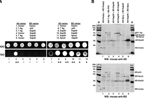
[image:5.585.45.543.71.407.2](iii) Yeast two-hybrid assay.We tested for direct interaction of Spry2 with Eap20 in the yeast two-hybrid assay (Fig. 4). Briefly, in this assay, proteins of interest that are translationally fused to the DNA-binding or the activation domain (BD or AD, respectively) of the yeast transcriptional activator, Gal 4, will reconstitute Gal 4 activity if they interact and thereby promote expression of a reporter gene (52). As shown in Fig. 4A, yeast transformants with the AD-and-BD chimeric protein pairs tested grew on the double-dropout selection medium (DDO; Trp and Leu), indicating that the plasmid pairs were expressed. When tested on the triple-dropout medium (TDO; Trp, Leu, and His), which tests for protein-protein interaction, the negative controls (empty vector pairings or T Ag with Eap20 or -45) gave no growth signal (Fig. 4A, lanes 1 and 3 to 7). The positive controls (i.e., T Ag paired with p53 [25]) and
FIG. 4. Spry2 interacts with Eap20 in the yeast two-hybrid assay. (A) Growth assay. Eap20, Eap45, and Spry2 fusions (lanes 9 to 12) or control constructs (lanes 1 to 8) were coexpressed as indicated and tested for cotransformation of the fusion pairs on DDO medium (top) or for positive yeast two-hybrid interactions on TDO medium (bottom). Images were captured by digital camera. The brightness of the samples reflects relative cell growth in 24 h. (B) Fusion protein expression. Fusion protein pair expression was determined by sequential probings in Western analysis using monoclonal antibodies against the activation domain (first probing; AD) and then the DNA binding-domain (second probing; BD). Filled circles mark the migration position of the AD binding and BD binding domains alone (lane 1) or fusion proteins pairs (lanes 2 to 6).
VOL. 85, 2011 Spry2 FACILITATES HIV-1 Gag RELEASE 7357
on November 7, 2019 by guest
http://jvi.asm.org/
Eap20 paired with Eap45 (Fig. 4A, lanes 2 and 8 to 10) inter-acted, as expected. Coexpression of Eap20 and Spry2 (Fig. 4A, lanes 11 and 12) produced a signal that was weaker than that detected for Eap20 and Eap45 but reproducibly stronger than that generated by the negative controls. Fusion protein pair expression was confirmed by sequential probings in Western analysis using monoclonal antibodies against the AD and BD (Fig. 4B). Although the Eap20-Eap45 pairs grew comparably on TDO medium irrespective of the fusion partner, the expres-sion of both proteins was lower in the AD-Eap20 and BD-Eap45 pairing than in the AD-BD-Eap45 and BD-Eap20 pairing (compare Fig. 4B, lanes 3 and 4). Similarly, although compa-rable growth on TDO medium was detected for the Eap20-Spry2 pairs irrespective of the fusion partner, expression of AD-Spry2 and BD-Eap20 (Fig. 4, lane 5) was weaker than that of AD-Eap20 and BD-Spry2 (Fig. 4, lane 6). Apparently, a certain threshold level of protein expression is sufficient to report interaction by the TDO medium growth test. The results support the conclusion that Spry2 binds directly to Eap20.
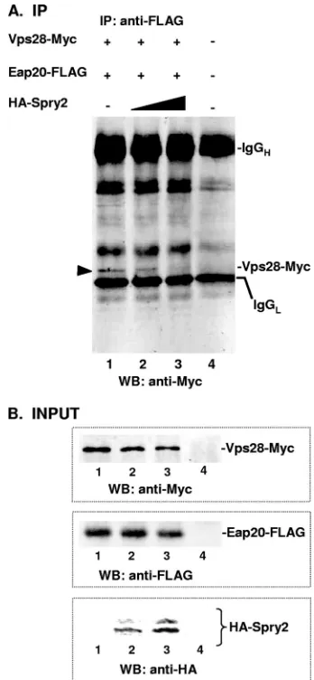
Spry2 disrupts ESCRT-I interaction with ESCRT-II. Struc-tural and genetic studies show that regions of Eap45, Eap30, and Eap20 in ESCRT-II form a composite surface that is required for interaction with Vps28 and Tsg101 in ESCRT-I (17, 22, 26). As the results above indicated that Spry2 bound Eap20, we investi-gated the possibility that it might disrupt ESCRT-I interaction with ESCRT-II. Consistent with the notion that Eap20 and Vps28 participate in the ESCRT-I–ESCRT-II interaction, we found that a Myc-tagged form of Vps28 was coimmunoprecipitated by an antibody targeted to the FLAG tag on Eap20 (Fig. 5, lane 1). As shown in Fig. 5, lanes 2 and 3, the expression of HA-Spry2 at 1:1 and 2:1 ratios to Eap20 resulted in a dose-dependent reduc-tion in pull-down of Vps28, indicating reducreduc-tion of the Vps28-Eap20 interaction. Vps28-Myc was detected specifi-cally, as no cross-reactive protein was detected in this region of the gel (Fig. 5, lane 4). The results suggest that Spry2 interferes with ESCRT-I–ESCRT-II interactions.
Spry2-NTD associates with Eap20 and promotes Gag re-lease.If the interaction of Spry2 with Eap20 is linked to Spry2-mediated facilitation of Gag release (Fig. 1), then coexpression of Spry2 with Gag should sequester the Eap20 protein and thereby promote VLP release. Contrary to this expectation, we observed no positive effect of overexpressing the full-length Spry2 protein on Gag release (Fig. 6A). However, consistent with the observation that aa 36 to 66 in the Spry2 NTD play an important role in Eap20 recognition (Fig. 2), we found that expression of the Spry2 NTD alone (aa 1 to 172) had a positive effect on Gag release (Fig. 6B). This effect was not observed upon overexpression of the CTD (aa 175 to 315) (Fig. 6C). Coexpression of Gag and the Spry2 NTD increased VLP re-lease 4- to 5-fold in 4/4 independent experiments compared to 0/4 when Gag was coexpressed with the Spry2 CTD (Fig. 6D). This correlated with the ability of the NTD fragment, but not that of the CTD, to coimmunoprecipitate with Eap20-FLAG (Fig. 6E, lane 4, arrow). For unknown reasons, the antibody against the HA tag on the Spry2 NTD did not coprecipitate Eap20-FLAG even though (i) the full-length HA-tagged Spry2 protein recognized Eap20-FLAG (Fig. 2, lane 2) and (ii) Eap20-FLAG coprecipitated the Spry2 NTD. The increase in VLP release correlated with colocalization of Eap20 and Spry2 NTD on the plasma membrane (Fig. 6, panels F to H). In
contrast, Eap20 and the Spry2 CTD did not colocalize (Fig. 6I to K). Thus, coimmunoprecipitation with Eap20, colocalization with Eap20 at the plasma membrane, and enhancement of Gag release all map to the NTD of Spry. We conclude that Spry2 facilitates Gag release by sequestering the ESCRT-II Eap20 protein, thereby preventing its interaction with ESCRT-I.
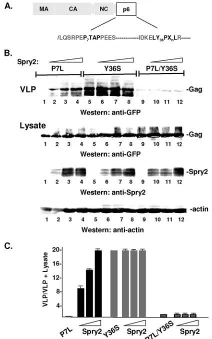
[image:6.585.332.506.69.445.2]Spry2 overexpression rescues release of P7L-Gag.Previous studies have demonstrated that proteins that function with ESCRT machinery, such as Alix and members of the Nedd4 family of ubiquitin ligases, can rescue release of Gag when primary and secondary L domain functions are impaired (re-viewed in reference 34; 1, 6, 42, 45, 48). We therefore investi-gated the effect of adventitious HA-Spry2 expression on the release of P7L-Gag. Figure 7A shows the location of the dis-ruptive mutations, P7L and Y36S, within the primary (P7TAP) and secondary (Y36PLXnL) L domain sequence in HIV-1 Gag.
FIG. 5. HA-Spry2 interferes with ESCRT-I–ESCRT-II interaction. (A) Immunoprecipitation reactions. COS-1 cells were transfected with DNA encoding Eap20-FLAG (0.5g) and Vps28-Myc (0.5g) in the absence (lane 1) or presence of 0.5g and 1.0g of DNA encoding HA-Spry2 (lanes 2 and 3, respectively). Immunoprecipitation and Western blotting were carried out with anti-FLAG and anti-Myc an-tibodies as indicated. (B) Total cell lysates showing expression of input proteins. Lane 4 in panels A and B tests for cross-reactive bands.
on November 7, 2019 by guest
http://jvi.asm.org/
As expected based on previous studies (20, 30, 32), release of P7L-Gag was highly defective (Fig. 7B, lane 1). However, as shown in Fig, 7B, lanes 2 to 4, HA-Spry2 expression rescued release in a dose-dependent manner. A quantitative analysis indicated that P7L-Gag release was stimulated 10- to 20-fold compared to release of P7L-Gag alone (Fig. 7C). We deter-mined whether Spry2-mediated rescue utilized the secondary L domain in HIV-1 Gag by mutating a critical residue, Y36, to Ser. This mutation was previously shown to disrupt secondary L domain function (12, 28, 44). As shown in Fig. 7B, lanes 5 to 8, the mutation had no detectable effect on WT Gag release and HA-Spry2 expression did not stimulate or inhibit release. This is as expected, since the primary L domain is intact. However, as shown in Fig. 7B, lanes 9 to 12, and Fig. 7C, mutation of Y36 to Ser in the P7L-Gag mutant (P7L/Y36S) abolished the ability of Spry2 to induce rescue.
Examination of Spry2 revealed that the same region required for Eap20 binding was required for rescue of P7L-Gag. As noted
above, expression of the full-length Spry2 protein rescued P7L-Gag release (Fig. 8B, lanes 1 to 4). Indeed, WT HA-Spry2 re-stored the release efficiency of P7L-Gag to the level of the WT Gag protein (compare Fig. 8B, lanes 1 to 4, to Fig. 8A, lanes 1 and 2, and Fig. 8C). The HA-Spry2 NTD fragment also dose-depend-ently rescued P7L-Gag release, although not to the level observed for the WT Spry2 protein (Fig. 8B, lanes 5 to 7). Deletion of aa 36 to 66 reduced the ability of the overexpressed Spry2 protein to rescue P7L release but did not prevent rescue (Fig. 8B, lanes 8 to 10, and C). We conclude that the conserved region in the Spry2 NTD that determines the interaction with Eap20 contributes to a P7L-Gag rescue pathway that is mediated through the secondary L domain in Gag.
DISCUSSION
Spry2 belongs to a family of proteins that modulate signaling of receptor tyrosine kinases [RTKs) (reviewed in references 9,
FIG. 6. Spry2 NTD binds Eap20 and promotes Gag release. (A to C) Effect of Spry2, Spry2 NTD, and Spry2 CTD on VLP release from cells. Proteins in cell lysates and VLPs in media from samples cotransfected with DNA-encoding Gag-GFP (3g) and HA-Spry2 (A), HA-Spry2 NTD (B), or HA-Spry2 CTD (C) (0.5, 1.0, 1.5, and 3.0g) were isolated as described in Materials and Methods and analyzed by SDS-PAGE and Western blotting. (D) Semiquantitative analysis of VLP release determined as described in the legend to Fig. 1. (E) Immunoprecipitation reactions. Cell lysates prepared from cells cotransfected with DNA encoding Eap20-FLAG and HA-tagged Spry2 NTD or CTD were tested for ability to coimmunoprecipitate using the indicated antibodies (preimmune serum [P]: lanes 1, 3, 5, and 7; anti-HA: lanes 2 and 6; or anti-FLAG: lanes 4 and 8). Eap20 and Spry2 were detected by Western blotting using rabbit (Rb) anti-FLAG or mouse (M) anti-HA antibodies, respectively. The arrow in panel E denotes Spry2 NTD that coimmunoprecipitated with Eap20. Confocal microscopy of cells coexpressing Eap20-FLAG with the Spry2 NTD (F to H) or CTD (I to K). Pearson’s values for the colocalizing pairs were 0.84 (panel H) and 0.59 (panel K).
VOL. 85, 2011 Spry2 FACILITATES HIV-1 Gag RELEASE 7359
on November 7, 2019 by guest
http://jvi.asm.org/
18, and 19). Spry2 is known to disrupt the downregulation of EGFR by interfering with the Hrs-Tsg101 interaction and thereby disrupting ESCRT-mediated trafficking from early to late endosomes (24). Our studies identify Spry2 as a novel Eap20-interacting factor that may coordinate the release func-tions directed by the primary and secondary L domains in HIV-1 Gag. Moreover, since our results indicate that HIV-1 Gag requires steady-state levels of Spry2 for efficient release from COS-1 cells, this Spry2 function is important, at least in this model cell environment. We and others previously dem-onstrated (i) that HIV-1 Gag trafficking does not require the ESCRT-II complex (26, 35) and (ii) that ESCRT-II factors cannot substitute for Tsg101 function in the rescue of HIV-1 Gag-ESCRT chimeric proteins (35). Our studies provide a basis for understanding how the Tsg101 bound to Gag might achieve linkage of ESCRT-I to -III while suppressing interac-tions with ESCRT-II; we hypothesize that Spry2 participates in
viral budding by binding the ESCRT-II component Eap20 and preventing ESCRT-I interaction with ESCRT-II. Presumably, such interactions would be nonproductive, as Gag bound to Tsg101 might be delivered to late endosomes/multivesicular bodies by ESCRT-II, whose Eap45 factor contains a phospha-tidylinositol 3,5-bisphosphate [PI(3,5)P2]-binding domain that
targets the complex to this compartment upon activation by ESCRT-I (reviewed in reference 21). Since, as noted above, Spry2 also can interfere with recruitment of ESCRT-I by ESCRT-0 (24), both Spry2 functions (i.e., disruption of ESCRT-I interaction with ESCRT-0 and -II) might be ex-ploited by Gag. We showed here that depletion of Spry2 in-hibits wild-type (WT) Gag release and that Spry2 overexpres-sion promoted release of Gag in the absence of Tsg101 binding. Release under the latter condition is known to be mediated through binding of the cellular factor Alix (3, 7, 29, 42, 43, 46). Supporting this, mutation of a residue in the Alix-binding site in the p6 region of P7L-Gag ablated the Spry2-mediated boost to release (Fig. 8). Interestingly, the Alix bind-ing site in the nucleocapsid domain, NC, was not sufficient to provide the Alix-binding function required for the Spry2-me-diated boost, indicating that, for Spry2, the p6- and NC-Alix-binding sites are not equivalent.
If the sequestration of Eap20 is solely responsible for the promotion of both WT and P7L-Gag release, one might expect that Eap 20 depletion would, in the former case, enhance virus release and, in the latter case, boost Alix function for P7L-Gag release. The fact that Eap20 knock-down does not promote WT Gag release (26, 35) suggests that the situation is more complex. For example, Eap20 depletion would reduce its recruitment by Chmp 6 and thereby impair overall ESCRT-III function. Such disruption of overall ESCRT-III function would negatively impact HIV release and possibly offset any detectable effects of Eap20 depletion. The situation could be similar for P7L-Gag, whose release is promoted through Alix bridging to Chmp 4 in ESCRT-III. It should be noted that although the Spry2-NTD invariably promoted WT VLP release (n ⫽ 4), full-length Spry2 variably inhibited (n⫽4/14 independent trials) or had no effect on VLP release (n ⫽ 10/14 independent transfections). The reason for this inconsistency is unclear. When inhibition was observed, it mapped to a determinant in the CTD that binds PI(4,5)P2, a phospholipid shown to be required for Gag membrane targeting and budding (reviewed in reference 34). These observations indicate that Spry2 could play more than one role in Gag assembly. Given that direct recruitment of ESCRT-I is bypassed with P7L-Gag, the release advantage contributed by the WT Spry2 protein may not nec-essarily be interference with the ESCRT-I and ESCRT-II in-teraction. Rather, another Spry2 function may be unmasked when Tsg101 is not bound to Gag.
Our results suggest that Spry2, through its NTD, can direct Eap20 to the plasma membrane (Fig. 3D and 6F). An important caveat is that our localization studies involve overexpression of an ESCRT protein, and it is well known that this can lead to aberrant localization patterns and for-mation of structures and compartments that are not ob-served at physiological expression levels. It is therefore pos-sible that the colocalization patterns that we observe are artifacts of Eap20 (or Spry2) overexpression.
Notwithstand-FIG. 7. HA-Spry2 rescues P7L-Gag. (A) Schematic drawing of Gag showing location of disruptive mutations in primary and secondary L domains (shown in bold). (B) COS-1 cells were transfected with a fixed amount of DNA encoding the indicated Gag mutant (3.0g; P7L: lanes 1 to 4; Y36S: lanes 5 to 8; P7L/Y36S: lanes 9 to 12) and increasing amounts of DNA encoding HA-Spry2 (0g: lanes 1, 5, and 9; 3.0g: lanes 2, 6, and 10; 6.0g: lanes 3, 7, and 11; 12g: lanes 4, 8, and 12). VLPs in the media (top) or proteins in cell lysates (bottom) were isolated as described in Materials and Methods and analyzed by SDS-PAGE and Western blotting. (C) Semiquantitative analysis of VLP release.
on November 7, 2019 by guest
http://jvi.asm.org/
[image:8.585.56.268.69.408.2]ing, in this regard, Spry2 appears to function like the phys-iological Eap20 binding partners Eap45 and Chmp6, which direct Eap20 to endosomal membranes during MVB protein sorting. Moreover, finding that Spry2 is associated with Eap20 at the plasma membrane is consistent with our pro-posal that Spry2 limits the Eap20–ESCRT-II interaction with ESCRT-I at the Gag budding sites on the plasma mem-brane. These considerations provide some confidence that the membrane association is not specious.
A recent report suggests that Spry2 and the envelope (Env) protein of Jaagsiekte sheep retrovirus (JSRV) have a func-tional relationship through a shared signaling network (4). JSRV is a betaretrovirus capable of transforming target cellsin vitroandin vivo. The JSRV Env induces expression of Spry2, and Spry2 overexpression interferes with Env-mediated trans-formation. Residue Y55 in the Spry2 NTD is critical for the interference. Spry2 is known to associate with a wide range of signaling molecules like Nedd4, c-Cbl, human seven in
absen-tia homolog 2 (SIAH2), protein phosphatase 2A (PP2A), and the adaptor protein CrkL through aa 36 to 66 in the NTD (8; reviewed in reference 9). Here also, the key residue within the region is Y55, whose phosphorylation state regulates Spry2 activation and the affinity of binding (13, 27, 31, 33, 39, 50). Our studies indicate that residues 36 to 66 are also critical for Spry2 interaction with Eap20 as indicated by immunoprecipi-tation assays and confocal microscopy (Fig. 2 and 3, respec-tively).
As noted above, Kim et al. (24) previously demonstrated that Spry2 can disrupt the interaction of ESCRT-0 with ESCRT-I. Our studies provide further evidence that Spry2 interferes with ESCRT-mediated trafficking through the dem-onstration that the protein can disrupt the interaction of ESCRT-I with ESCRT-II. In addition, our results indicate that Spry2 can facilitate release whether driven by the primary or the secondary HIV-1 Gag L domain. In future studies, it will be of interest to determine whether the sequestration of
FIG. 8. HA-Spry2 residues aa 36 to 66 contribute to rescue of P7L-Gag. (A) Comparison of WT (lane 1) and P7L (lane 2) Gag VLP production. (B) COS-1 cells were transfected with DNA-encoding P7L-Gag (3.0g, lane 1) and increasing amounts of DNA encoding HA-Spry2 (3.0g: lanes 2 to 4; 6.0g: lanes 5 to 7; 12g: lanes 8 to 10). VLPs in the media (top) or proteins in total cell lysates (bottom) were isolated as described in Materials and Methods and analyzed by SDS-PAGE and Western blotting. (C) Semiquantitative analysis of VLP release.
VOL. 85, 2011 Spry2 FACILITATES HIV-1 Gag RELEASE 7361
on November 7, 2019 by guest
http://jvi.asm.org/
ESCRT-II factors is solely responsible for the requirement for Spry2 in HIV-1 release or if additional functions of the Spry2 protein also contribute to productive Gag trafficking, assembly, and budding.
ACKNOWLEDGMENTS
We thank H. J. Kim, L. J. Zhong, and D. Bar-Sagi for reagents and for helpful discussions in the initial stages of this study. We also thank J. Keller for technical assistance with qPCR experiments and E. Ivanova for preparation of L domain mutants.
G.N.M. was supported by W. Burghardt Turner Predoctoral Fellow-ships (NSF-HRD-funded SUNY AGEP, grant no. 35583). This study was supported by NIH R01 award AI068463 (to C.A.C.).
REFERENCES
1.Calistri, A., et al.2009. Role of the feline immunodeficiency virus L-domain in the presence or absence of Gag processing: involvement of ubiquitin and
Nedd4-2s ligase in viral egress. J. Cell. Physiol.218:175–182.
2.Carter, C. A., and L. S. Ehrlich.2008. Cell biology of HIV-1 infection of
macrophages. Annu. Rev. Microbiol.62:425–443.
3.Chen, C., O. Vincent, J. Jin, O. A. Weisz, and R. C. Montelaro.2005. Functions of early (AP-2) and late (AIP1/ALIX) endocytic proteins in
equine infectious anemia virus budding. J. Biol. Chem.280:40474.
4.Chitra, E., et al.2010. Functional interaction between Env oncogene from Jaagsiekte sheep retrovirus and tumor suppressor Sprouty2. Retrovirology
7:62.
5.Chu, H., J. J. Wang, and P. Spearman.2009. Human immunodeficiency virus type-1 gag and host vesicular trafficking pathways. Curr. Top. Microbiol.
Immunol.339:67–84.
6.Chung, H. Y., et al.2008. NEDD4L overexpression rescues the release and infectivity of human immunodeficiency virus type 1 constructs lacking PTAP
and YPXL late domains. J. Virol.82:4884–4897.
7.Dussupt, V., et al.2009. The nucleocapsid region of HIV-1 Gag cooperates with the PTAP and LYPXnL late domains to recruit the cellular machinery
necessary for viral budding. PLoS Pathog.5:e1000339.
8.Edwin, F., K. Anderson, and T. B. Patel.2010. HECT domain-containing E3 ubiquitin ligase Nedd4 interacts with and ubiquitinates Sprouty2. J. Biol.
Chem.285:255–264.
9.Edwin, F., K. Anderson, C. Ying, and T. B. Patel.2009. Intermolecular interactions of Sprouty proteins and their implications in development and
disease. Mol. Pharmacol.76:679–691.
10.Ehrlich, L., H. G. Krausslich, E. Wimmer, and C. A. Carter.1990. Expres-sion in Escherichia coli and purification of human immunodeficiency virus
type 1 capsid protein (p24). AIDS Res. Hum. Retroviruses6:1169–1175.
11.Fernandes, F., et al.2011. Phosphoinositides direct equine infectious anemia
virus gag trafficking and release. Traffic12:438–451.
12.Fisher, R. D., et al.2007. Structural and biochemical studies of ALIX/AIP1
and its role in retrovirus budding. Cell128:841–852.
13.Fong, C. W., et al.2003. Tyrosine phosphorylation of Sprouty2 enhances its
interaction with c-Cbl and is crucial for its function. J. Biol. Chem.278:
33456–33464.
14.Fujii, K., J. H. Hurley, and E. O. Freed.2007. Beyond Tsg101: the role of
Alix in ‘ESCRTing’ HIV-1. Nat. Rev. Microbiol.5:912–916.
15.Ganser-Pornillos, B. K., M. Yeager, and W. I. Sundquist.2008. The
struc-tural biology of HIV assembly. Curr. Opin. Struct. Biol.18:203–217.
16.Garrus, J. E., et al.2001. Tsg101 and the vacuolar protein sorting pathway
are essential for HIV-b1 budding. Cell107:55–65.
17.Gill, D. J., et al.2007. Structural insight into the ESCRT-I/-II link and its role
in MVB trafficking. EMBO J.26:600–612.
18.Guy, G. R., et al.2003. Sprouty: how does the branch manager work? J. Cell
Sci.116:3061–3068.
19.Guy, G. R., R. A. Jackson, P. Yusoff, and S. Y. Chow.2009. Sprouty proteins:
modified modulators, matchmakers or missing links? J. Endocrinol.203:191–
202.
20.Huang, M., J. M. Orenstein, M. A. Martin, and E. O. Freed.1995. p6Gag is required for particle production from full-length human immunodeficiency
virus type 1 molecular clones expressing protease. J. Virol.69:6810–6818.
21.Hurley, J. H.2010. The ESCRT complexes. Crit. Rev. Biochem. Mol. Biol.
45:463–487.
22.Im, Y. J., and J. H. Hurley.2008. Integrated structural model and membrane
targeting mechanism of the human ESCRT-II complex. Dev. Cell14:902–
913.
23.Im, Y. J., T. Wollert, E. Boura, and J. H. Hurley. 2009. Structure and function of the ESCRT-II-III interface in multivesicular body biogenesis.
Dev. Cell17:234–243.
24.Kim, H. J., L. J. Taylor, and D. Bar-Sagi.2007. Spatial regulation of EGFR
signaling by Sprouty2. Curr. Biol.17:455–461.
25.Lane, D. P., and L. V. Crawford.1979. T antigen is bound to a host protein
in SV40-transformed cells. Nature278:261–263.
26.Langelier, C., et al.2006. Human ESCRT-II complex and its role in human
immunodeficiency virus type 1 release. J. Virol.80:9465–9480.
27.Lao, D. H., et al.2007. Direct binding of PP2A to Sprouty2 and phosphor-ylation changes are a prerequisite for ERK inhibition downstream of
fibro-blast growth factor receptor stimulation. J. Biol. Chem.282:9117–9126.
28.Lazert, C., N. Chazal, L. Briant, D. Gerlier, and J. C. Cortay.2008. Refined study of the interaction between HIV-1 p6 late domain and ALIX.
Retro-virology5:39.
29.Martin-Serrano, J., A. Yarovoy, D. Perez-Caballero, and P. D. Bieniasz.
2003. Divergent retroviral late-budding domains recruit vacuolar protein sorting factors by using alternative adaptor proteins. Proc. Natl. Acad. Sci.
U. S. A.100:12414–12419.
30.Martin-Serrano, J., T. Zang, and P. D. Bieniasz.2001. HIV-1 and Ebola virus encode small peptide motifs that recruit Tsg101 to sites of particle
assembly to facilitate egress. Nat. Med.7:1313.
31.Mason, J. M., et al.2004. Tyrosine phosphorylation of Sprouty proteins regulates their ability to inhibit growth factor signaling: a dual feedback loop.
Mol. Biol. Cell15:2176–2188.
32.Medina, G., et al.2008. Tsg101 can replace Nedd4 function in ASV Gag
release but not membrane targeting. Virology377:30–38.
33.Nadeau, R. J., J. L. Toher, X. Yang, D. Kovalenko, and R. Friesel.2007. Regulation of Sprouty2 stability by mammalian Seven-in-Absentia homolog
2. J. Cell. Biochem.100:151–160.
34.Pincetic, A., and J. Leis.2009. The mechanism of budding of retroviruses
from cell membranes. Adv. Virol.2009:6239691–6239699.
35.Pincetic, A., G. Medina, C. Carter, and J. Leis.2008. Avian sarcoma virus and human immunodeficiency virus, type 1 use different subsets of ESCRT
proteins to facilitate the budding process. J. Biol. Chem.283:29822–29830.
36.Pornillos, O., et al.2003. HIV Gag mimics the Tsg101-recruiting activity of
the human Hrs protein. J. Cell Biol.162:425–434.
37.Progida, C., et al.2007. RILP is required for the proper morphology and
function of late endosomes. J. Cell Sci.120:3729–3737.
38.Progida, C., M. R. Spinosa, A. de Luca, and C. Bucci.2006. RILP interacts with the VPS22 component of the ESCRT-II complex. Biochem. Biophys.
Res. Commun.347:1074–1079.
39.Satoh, T., S. Torii, K. Nakayama, and E. Nishida.2010. CrkL is a novel
target of Sprouty2 in fibroblast growth factor signaling. Genes Cells15:161–
168.
40.Schmidt, M. H., I. Dikic, and O. Bogler.2005. Src phosphorylation of Alix/ AIP1 modulates its interaction with binding partners and antagonizes its
activities. J. Biol. Chem.280:3414–3425.
41.Schmidt, M. H., et al.2004. Alix/AIP1 antagonizes epidermal growth factor receptor downregulation by the Cbl-SETA/CIN85 complex. Mol. Cell. Biol.
24:8981–8993.
42.Sette, P., J. A. Jadwin, V. Dussupt, N. F. Bello, and F. Bouamr.2010. The ESCRT-associated protein Alix recruits the ubiquitin ligase Nedd4-1 to facilitate HIV-1 release through the LYPXnL L domain motif. J. Virol.
84:8181–8192.
43.Strack, B., A. Calistri, S. Craig, E. Popova, and H. G. Go¨ttlinger.2003. AIP1/ALIX is a binding partner for HIV-1 p6 and EIAV p9 functioning in
virus budding. Cell114:689–699.
44.Usami, Y., S. Popov, and H. G. Gottlinger.2007. Potent rescue of human immunodeficiency virus type 1 late domain mutants by ALIX/AIP1 depends
on its CHMP4 binding site. J. Virol.81:6614–6622.
45.Usami, Y., S. Popov, E. Popova, and H. G. Go¨ttlinger.2008. Efficient and specific rescue of human immunodeficiency virus type 1 budding defects by
a Nedd4-like ubiquitin ligase. J. Virol.82:4898–4907.
46.von Schwedler, U. K., et al.2003. The protein network of HIV budding. Cell
114:701–713.
47.Wang, T., and W. Hong.2006. RILP interacts with VPS22 and VPS36 of ESCRT-II and regulates their membrane recruitment. Biochem. Biophys.
Res. Commun.350:413–423.
48.Weiss, E. R., et al. 2010. Rescue of HIV-1 release by targeting widely divergent NEDD4-type ubiquitin ligases and isolated catalytic HECT
do-mains to Gag. PLoS Pathog.6:e1001107.
49.Wollert, T., C. Wunder, J. Lippincott-Schwartz, and J. H. Hurley.2009.
Membrane scission by the ESCRT-III complex. Nature458:172–177.
50.Wong, E. S., J. Lim, B. C. Low, Q. Chen, and G. R. Guy.2001. Evidence for
direct interaction between Sprouty and Cbl. J. Biol. Chem.276:5866–5875.
51.Yorikawa, C., et al. 2005. Human CHMP6, a myristoylated ESCRT-III protein, interacts directly with an ESCRT-II component EAP20 and
regu-lates endosomal cargo sorting. Biochem. J.387:17–26.
52.Young, K. H.1998. Yeast two-hybrid: so many interactions, (in) so little time.
Biol. Reprod.58:302–311.