Bacteriophage-Encoded Chaperonin
Lidia P. Kurochkina,aPavel I. Semenyuk,bVictor N. Orlov,bJohan Robben,cNina N. Sykilinda,aand Vadim V. Mesyanzhinova
Shemyakin-Ovchinnikov Institute of Bioorganic Chemistry, Russian Academy of Sciences, Moscow, Russiaa; Belozersky Research Institute of Physico-Chemical Biology, Lomonosov Moscow State University, Moscow, Russiab; and Division of Biochemistry, Molecular and Structural Biology, Katholieke Universiteit Leuven, Leuven, Belgiumc
Chaperonins promote protein folding
in vivo
and are ubiquitously found in bacteria, archaea, and eukaryotes. The first viral
chaperonin GroEL ortholog, gene product 146 (gp146), whose gene was earlier identified in the genome of bacteriophage EL, has
been shown to be synthesized during phage propagation in
Pseudomonas aeruginosa
cells. The recombinant gp146 has been
ex-pressed in
Escherichia coli
and characterized by different physicochemical methods for the first time. Using serum against the
recombinant protein, gp146’s native substrate, the phage endolysin gp188, has been immunoprecipitated from the lysate of
EL-infected bacteria and identified by mass spectrometry.
In vitro
experiments have shown that gp146 has a protective effect against
endolysin thermal inactivation and aggregation, providing evidence of its chaperonin function. The phage chaperonin has been
found to have the architecture and some properties similar to those of GroEL but not to require cochaperonin for its functional
activity.
C
haperonins are known to promote the correct folding of
newly synthesized polypeptides and to prevent aggregation of
proteins denatured under stress. Chaperonins are large cylindrical
oligomers consisting of two rings arranged back to back. Each ring
contains a central cavity, where unfolded proteins may be
encap-sulated and undergo productive folding in an ATP-dependent
manner (
19
,
22
,
23
).
Based on protein sequence and structural features,
chaper-onins fall into two groups. Group I chaperchaper-onins (including heat
shock proteins such as hsp60) are found in bacteria,
endosymbi-otic organelles (mitochondria and chloroplasts), and subsets of
archaea (
17
,
22
). Group II chaperonins are found in archaea and
the eukaryotic cytosol (
4
,
14
).
Chaperonin GroEL from
Escherichia coli
, which carries out its
function with a cofactor, GroES, is the most studied of group I
chaperonins (
4
,
14
,
22
,
45
). GroEL consists of 14 identical 57-kDa
subunits arranged in two heptameric rings (
6
,
41
). GroES 10-kDa
subunits also form a heptameric ring that acts as a detachable lid of
the central cavity when GroEL forms a complex with GroES (
37
,
44
). Group II chaperonins act independently of the detachable
cofactor. The helical protrusion at the tip of the apical domain
substitutes for the cofactor as a built-in lid of the central cavity
(
29
). Group II chaperonins are almost always more complicated
than group I chaperonins in the subunit composition of their
complexes, which are hetero-oligomers rather than
homo-oli-gomers (
4
,
14
).
Some bacteriophages (
, T4, RB49) are known to use host
GroEL for folding their capsid proteins during morphogenesis.
While phage
uses both host GroEL and GroES, the GroES
co-chaperonin function in T4 and RB49 is performed instead by
phage-encoded orthologs gp31 and CocO, respectively (
2
,
21
,
42
).
As revealed by structural analysis and later by cryoelectron
mi-croscopy, the gp31 cap may create a somewhat larger folding cage
with GroEL than with the GroES cap to accommodate the
rela-tively large 56-kDa T4 major capsid protein (
3
,
12
,
27
).
The first chaperonin GroEL ortholog gene has been identified
in the genome of
Pseudomonas aeruginosa
bacteriophage EL
(211,215 bp, 201 open reading frames [ORFs]) (
24
), related to the
giant phiKZ-like
Myoviridae
(
11
). The putative GroEL-like
chap-eronin protein (558 amino acids [aa]; GenBank accession number
5176674
) encoded by gene 146 has only about 25% amino acid
sequence identity with
E. coli
GroEL (547 aa) and
P. aeruginosa
GroEL (548 aa), while these bacterial chaperonins show 80%
mu-tual sequence identity. Nevertheless, conservation of most of the
residues involved in ATP/ADP and Mg
2⫹binding (
5
,
9
,
44
)
sug-gests that gp146 belongs to this ubiquitous family of proteins. The
two other GroEL orthologs encoded by phage genomes were
pre-dicted (
30
,
13
), but none of them have been experimentally
stud-ied yet.
In the present work, chaperonin activity of a
bacteriophage-encoded protein is demonstrated for the first time by functional
and physicochemical characterization of the putative chaperonin,
gp146, encoded by the genome of
P. aeruginosa
bacteriophage EL.
MATERIALS AND METHODS
Cloning, expression, purification, and antibody preparation of gp146.
The gene 146 was amplified from EL genomic DNA using the forward primer 5=-GTACGCATATGTCTCAAACGCTACTG-3=and the reverse primer 5=-TCCTTTAGGATCCCCTTACCGCACCTT-3=, which con-tained recognition sequences for NdeI and BamHI (underlined), respec-tively. The amplicon was cloned into the pET-22b(⫹) vector (Novagene, United States) and expressed inE. coliBL21(DE3) as previously described (40). The transformants were cultured in 600 ml of 2⫻tryptone-yeast extract (TY) medium containing 200g/ml of ampicillin at 37°C. When theA600reached 0.7, the recombinant bacteria were induced with 1 mM
isopropyl--D-thiogalactopyranoside (IPTG) and incubated for 3 h at 25°C. The cells were harvested by centrifugation at 2,500⫻gfor 10 min (Megafuge 2.0 R; Heraeus Instruments, Germany).
To purify gp146, the cell pellet was resuspended in 6 ml of 50 mM
Received18 April 2012Accepted3 July 2012
Published ahead of print11 July 2012
Address correspondence to Lidia P. Kurochkina, [email protected].
Copyright © 2012, American Society for Microbiology. All Rights Reserved.
doi:10.1128/JVI.00940-12
on November 7, 2019 by guest
http://jvi.asm.org/
washed with 10 volumes of buffer, the protein was eluted by a linear gradient from 100 to 500 mM NaCl in 50 mM Tris-HCl (pH 7.5) and analyzed by SDS–10% PAGE (31). Fractions containing pure protein were concentrated using an Amicon Ultra-15 centrifugal filter device (molec-ular weight cutoff [MWCO], 100,000; Millipore, United States). The gp146 concentration was determined spectrophotometrically at 280 nm using a theoretical absorption coefficient of 35,870 M⫺1cm⫺1.
To produce antibodies, female BALB/c mice (14 to 17 g) were immu-nized intraperitoneally with 0.2 ml of a suspension containing 50 to 100 g of pure gp146 in 150 mM NaCl and an equal volume of complete Freund’s adjuvant. Two booster injections were given with the same pro-tein preparation and incomplete Freund’s adjuvant at two- to three-week intervals. Serum was recovered 7 days after the third immunization. As determined by enzyme-linked immunosorbent assay (ELISA), the titer of antibody was 1:250,000.
Western blot analysis.After separation by SDS-PAGE, proteins were transferred by electroelution from the gel onto a nitrocellulose membrane (Bio-Rad) in electroblotting buffer (0.1 M Tris, 0.1 M boric acid, 0.01 M EDTA) at 200 mA for 60 min. Antigen was detected with antiserum fol-lowed by rabbit anti-mouse horseradish peroxidase (HRP)-conjugated antibodies (1:6,000; Sigma, United States). Antibodies were diluted in phosphate-buffered saline (PBS) supplemented with 3% milk and 0.05% Tween 20, and incubations were for 1 h at room temperature with gentle rocking. Blots were developed with 0.02% 3=,3=-diaminobenzidine (Sigma) in hydrogen peroxide solution.
Preparation of lysate from the EL-infected cells.TheP. aeruginosa
PAO1 cells were grown in 150 ml of 2⫻TY medium at 37°C to a density of 2⫻108cells/ml, infected by bacteriophage EL at a multiplicity of infection
of 5, and incubated for 40 min at 37°C. The cells were harvested by cen-trifugation at 2,500⫻gfor 10 min. The pellet was resuspended in 50 mM Tris-acetate (pH 8) and 5 mM EDTA and sonicated. NaCl and phenyl-methylsulfonyl fluoride (PMSF) were added to final concentrations of 140 mM and 1 mM, respectively. The lysate preparation was centrifuged at 12,000⫻gfor 10 min.
Immunoprecipitation of gp146 from the lysate.Serum against gp146 was incubated with insoluble protein A (Sigma) in 50 mM Tris-acetate (pH 8) for 2 h at 4°C with gentle rocking. Protein A with attached anti-bodies was centrifuged at 4,000⫻gfor 5 min and subsequently washed in solution I (50 mM Tris-acetate [pH 8], 5 mM EDTA, 150 mM NaCl, 0.5% sodium deoxycholate, 1% Triton X-100), solution II (50 mM Tris-acetate [pH 8], 5 mM EDTA, 500 mM NaCl, 1% Triton X-100), solution III (50 mM acetate [pH 8], 5 mM EDTA, 82 mM KCl), and 50 mM Tris-acetate (pH 8) and 5 mM EDTA.
To precipitate phage chaperonin, protein A with attached antibodies against gp146 was incubated with the lysate of the EL-infected bacterial cells (45 min postinfection) at 4°C for 2.5 h. Nonbound proteins were removed by washing with solutions I to III. The pellet of protein A with bound immune complex was supplemented with SDS-sample loading buffer, incubated at 98°C for 5 min, and analyzed by SDS–10% PAGE. A sample prepared in parallel from the lysate of noninfectedP. aeruginosa
PAO1 cells was used as a control.
Coimmunoprecipitation of gp146-substrate complexes and sub-strate identification.Coimmunoprecipitation of chaperonin-substrate complexes was performed as described above, except that specifically bound proteins were eluted from the protein A with 50 mM Tris-acetate
protein sequencing system (PE Applied Biosystems, United States). Phe-nylthiohydantoin derivatives of the amino acids were identified by a 120A PTH analyzer (PE Applied Biosystems).
Cloning, expression and purification of gp188.The gene 188 was amplified from EL genomic DNA using the forward primer 5=-TAAATA CATATGAACTTCCGGACGAAG-3=and the reverse primer 5=-GGAAA ACTCGAGGAATCAATACGAAATAACGTG-3=(NdeI and XhoI recog-nition sites are underlined). The PCR product was cloned into the pET-28b(⫹) vector (Novagene) and expressed inE. colicells at 25°C. The recombinant protein containing a 6⫻His affinity tag at the N terminus (gp188*) was purified on His-Select nickel affinity gel (Sigma), dialyzed against 50 mM Tris-HCl (pH 7.5), and concentrated using an Amicon Ultra-15 centrifugal filter device (MWCO, 30,000). Protein concentration was determined spectrophotometrically at 280 nm using a theoretical ab-sorption coefficient of 58,455 M⫺1cm⫺1.
Endolysin activity assay.The muralytic activity of endolysin (gp188*) was determined as previously described (7). TheP. aeruginosaPAO1 cells with the outer membrane permeabilized by chloroform treatment were used as a substrate. A 30-l sample of protein solution was mixed with 270 l cells, and optical density was measured at 490 nm for 30 min using a Wallac 1420 microbiology reader (PerkinElmer, United States). The ac-tivity was determined from the decrease in absorbance as a function of time using a standardized calculation method. Negative controls (buffer without enzyme) were subtracted from sample measurements.
Analytical ultracentrifugation.Sedimentation-velocity experiments were carried out at 22°C using a model E analytical ultracentrifuge (Beck-man Instruments, United States) equipped with photoelectric scanning absorption optical system, with spinning rates of 20,000 rpm for gp146 and 48,000 rpm for gp188* and scanning at 280 nm. Protein concentra-tion was 1 mg/ml. Data analysis was carried out using the SEDFIT pro-gram (39).
Hydrodynamic diameter determination.The hydrodynamic diame-ters of proteins were determined using dynamic light scattering (DLS). All experiments were carried out at 25°C on a ZetaSizer NanoZS instrument (Malvern) with a laser wavelength of 633 nm. Data were analyzed using Dispersion Technology software, version 5.10.
Estimated protein density was calculated on the assumption of spher-ical particles from the equation ⫽Mw/4NarH3, whereMwis molecular
weight,Nais Avogadro’s number, andrHis the hydrodynamic radius.
Electron microscopy.Purified protein was loaded on a Pioloform-coated copper grid. The specimen was then contrasted with 1% uranyl acetate and observed in a Jeol (Japan) JEM 1400 electron microscope.
DSC.The thermal denaturation of proteins was investigated by differ-ential scanning calorimetry (DSC) using a DASM-4 microcalorimeter (Biopribor, Russia) with 0.47-ml capillary platinum cells. All of the mea-surements were carried out at a heating rate of 1°C min⫺1in the
temper-ature range from 5 to 90°C and at a constant pressure of 2 atm. Curves were corrected for time response as earlier described (35). The second heating was used as the instrument baseline because of irreversible dena-turation found for all samples. The chemical baseline was calculated and subtracted using Origin 1.16 software (MicroCal, Inc., United States). The standard reaction buffer used in the experiments was 50 mM Tris-HCl (pH 7.5), 10 mM MgCl2, and 100 mM KCl. The concentration of gp146
was 0.58M. Protein concentration and molar excess heat capacity were calculated on a tetradecameric complex with a molecular mass of 863.8
on November 7, 2019 by guest
http://jvi.asm.org/
kDa. To investigate the influence of nucleotide binding on gp146 struc-ture, ATP or ADP (Sigma) was added to the protein solution to a final concentration of 1 mM.
Fluorescence measurements.Fluorescence spectra of protein solu-tions were measured in a 2-ml quartz cell at various temperatures with a FluoroMax-3 spectrofluorometer (Horiba Jobin Yvon, France) using an excitation wavelength of 280 nm. The samples were loaded in the cell and heated to 75°C at the average rate of 2°C/min. The emission spectra were recorded at 300 to 400 nm at the corresponding temperatures. Experimental curves representing the temperature dependence of wavelength of the emis-sion maximum (max) were fitted using a Boltzmann sigmoid curve.
CD spectroscopy.Circular dichroism (CD) measurements were per-formed using a Chiroscan CD spectrometer (Applied Photophysics, United Kingdom) in a 0.5-mm cell. Spectra were recorded in the range of 200 to 260 nm and were baseline corrected by subtracting the buffer spec-trum. Each point was measured for 1 s. The observed value was converted to mean residue ellipticity.
Isothermal titration calorimetry (ITC).Nucleotide binding was in-vestigated on a VP-ITC instrument (MicroCal Ltd., United States) with a 1.4-ml cell. ATP and ADP concentrations were measured spectrophoto-metrically using a UV-1601 instrument (Shimadzu Scientific Instruments Inc., Japan) at 259 nm with an extinction coefficient of 15,400 M⫺1cm⫺1.
All experiments were carried out at 10°C in 50 mM Tris-HCl (pH 7.5), 10 mM MgCl2, and 100 mM KCl. Titration experiments were performed by
successive 10-l injections of 40M ATP or 60M ADP solution into gp146 (0.58M for the tetradecamer), and the interval between injections was 5 min. All samples were degassed before the experiment. Binding isotherms were corrected by subtracting the ligand dilution isotherms, determined by titrating ATP and ADP solutions into buffer. Data analysis was carried out using MicroCal Origin 7.0 software with the “one set of sites” model. Resulting coefficients of determination were calculated from
the equation,R2⫽1⫺
兺
i共Expi兲2兺
i共Expi⫺mean共Exp兲兲2, where Exp and Fit are
experimental and fitted ordinate arrays, respectively, and exceeded 0.99.
gp188* thermal inactivation.The gp188* samples were incubated at 0.1M in 50 mM Tris-HCl (pH 7.5), 10 mM MgCl2, and 100 mM KCl in the
absence or presence of 0.2M gp146 (for the tetradecamer) and 3 mM ATP at 37 and 50°C. At various times, aliquots were withdrawn and chilled on ice and the muralytic activity was determined at 23°C as described above.
gp188* aggregation assay.The effect of gp146 on gp188* aggregation was investigated in 50 mM Tris-HCl (pH 7.5), 10 mM MgCl2, and 100
mM KCl at 45°C using DLS. The gp188* samples at a constant
concentra-tion of 3M were incubated with different concentrations of gp146 in the absence or presence of 1.2 mM ATP for a few hours, and the hydrody-namic diameters of particles were continuously measured. The molar ra-tio of gp188* to gp146 was calculated for the monomer and tetradecam-ers, respectively.
RESULTS
Expression and purification of gp146.
A plasmid construct for
ex-pression of gene 146 in
E. coli
cells was designed. The bacterial
expression of gp146 (61.7 kDa) was carried out at a lowered
tem-perature (25°C) to increase the yield of soluble protein.
Low-tem-perature induction is known to result in a significant
improve-ment in solubility by slowing down the rate of protein expression
and allowing time for folding (
43
). The recombinant protein was
precipitated from the cell lysate by adding ammonium sulfate,
followed by purification on a Q-Sepharose column (
Fig. 1
), and
used for antibody preparation. It has been shown by Western blot
analysis that the polyclonal antibodies obtained specifically bind
to the recombinant gp146 but recognize neither bacterial nor EL
structural proteins (not shown).
Analysis of gp146 expression
in vivo
.
According to the
struc-tural proteome analysis of bacteriophage EL, gp146 is not part of
the mature phage particle (
33
). However, it has been found during
phage EL propagation. To investigate gp146 expression
in vivo
, the
proteins from EL-infected bacterial cells at different infection
stages were resolved by SDS-PAGE followed by Western blot
anal-ysis. An apparent band (about 60 kDa) likely corresponding to
gp146 was detected from 15 min postinfection onwards with
se-rum against the recombinant protein (not shown).
In vivo
-ex-pressed native gp146 was immunoprecipitated from the clarified
lysate of the EL-infected bacterial cells (45 min postinfection)
us-ing antibodies against the recombinant protein attached to
insol-uble protein A. A similar procedure was performed
simultane-ously with an equivalent amount of lysate from noninfected cells.
The immunoprecipitated complexes were supplemented by
SDS-sample loading buffer and heated. Upon this treatment, the
asso-ciated proteins were released from protein A into solution and
separated by SDS-PAGE (
Fig. 2
). An additional band with an
elec-trophoretic mobility coinciding with that of the recombinant
gp146 was found in the protein pattern from the EL-infected cells
(
Fig. 2A
, lane 3) compared to a control. No additional bands,
FIG 1SDS–10% PAGE of expressed gp146 (lane 1) and gp146 purified by precipitation with 30% ammonium sulfate (lane 2) and subsequent chroma-tography on Q-Sepharose (lane 3). Lane M, protein marker.
FIG 2Immunoprecipitation of native gp146 expressedin vivofrom the lysate of EL-infectedP. aeruginosacells (45 min postinfection) using serum attached to protein A. Proteins were resolved by SDS–10% PAGE and visualized in the gel by staining with Coomassie brilliant blue (A) and serum against gp146 after being transferred from the gel onto a nitrocellulose membrane (B). Shown are protein patterns of serum (lanes 1) and immunoprecipitated complexes from the lysate of noninfected (lanes 2) and EL-infected (lanes 3) bacterial cells. Lane M, protein marker; lanes c, recombinant gp146.
on November 7, 2019 by guest
http://jvi.asm.org/
[image:3.585.301.542.63.180.2] [image:3.585.92.238.66.244.2]except for the bands typical of the heavy and light chains of
anti-bodies (
Fig. 2A
, lane 1), were observed in the precipitate from
noninfected cells (
Fig. 2A
, lane 2). This band was clearly revealed
by immunoblotting with serum against the recombinant gp146
(
Fig. 2B
). The identity of the immunoprecipitated protein to
gp146 was confirmed by N-terminal amino acid sequencing. Six
amino acid residues were determined, and all of them were
com-pletely identical to the gp146 primary structure. Thus, gp146 is
really synthesized
in vivo
during bacteriophage EL propagation in
P. aeruginosa
cells. Further investigations of the recombinant
pro-tein were performed. To test whether gp146 can function as a
chaperonin, it was necessary to find its native substrate.
Isolation and identification of substrate for gp146.
Antibod-ies against gp146 attached to protein A were further used for
co-immunoprecipitation of the putative chaperonin along with the
substrate proteins associated with it from the lysate of EL-infected
bacteria. To rule out nonspecific protein binding to antigen, the
immune complexes were thoroughly washed with buffers
con-taining detergents and NaCl (see Materials and Methods). To
en-sure ATP-dependent release of substrate proteins from gp146, the
precipitated complexes were incubated with ATP- and Mg
2⫹-containing buffer at an elevated temperature. Eluted proteins were
separated by SDS-PAGE. The gel lane was cut into slices and
sub-jected to in-gel trypsin digestion. Peptides were extracted and
sep-arated by liquid chromatography followed by MS analysis. One of
the phage EL proteins, gp188 (292 aa, 32.5 kDa), was identified. A
recombinant protein with a 6
⫻
His affinity tag at the N terminus
(gp188*, 34.3 kDa) was expressed in
E. coli
cells in a soluble,
en-zymatically active form and purified by metal chelate
chromatogra-phy for further study. Using serum against the recombinant gp188*,
gp188 expression was observed during phage EL multiplication in
bacterial cells, but 45 min later than that of gp146 (not shown).
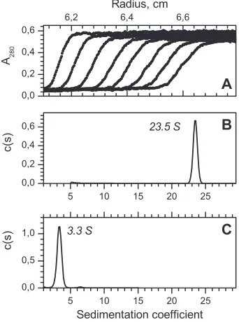
Architecture of gp146 and gp188*.
The structural
characteris-tics and homogeneity of the purified recombinant gp146 and
gp188* were investigated using analytical ultracentrifugation and
DLS. One-component raw sedimentation profiles were observed
for both proteins (
Fig. 3A
). The sedimentation coefficients
equaled 23.5
⫾
0.3 S and 3.3
⫾
0.3 S for gp146 and gp188*,
re-spectively (
Fig. 3B
and
C
). The fitting of the frictional coefficient
for gp146 yielded a value of 1.39, suggesting a nonspherical form
of the protein. Based on the calculated values of the sedimentation
and frictional coefficients, the protein’s molecular mass was
esti-mated. Its value (872 kDa) most likely corresponds to a
tetra-decamer, considering that the calculated molecular mass of the
gp146 monomer is 61.7 kDa. The estimated molecular mass of
gp188* (35 kDa) is close to that of a protein monomer. According
to DLS data, the hydrodynamic diameters of gp146 and gp188* are
about 13.2 and 5.6 nm, respectively (not shown). Estimation of
the gp146 density was 1.191 g/ml, which is lower than the mean
density of a typical protein with the same molecular weight (
18
),
suggesting the presence of an inner cavity in the complex. An
electron microscopy assay confirmed that the phage chaperonin is
composed of double-stacked heptameric rings with a central
cav-ity (
Fig. 4
). Thus, the architecture of the recombinant gp146 is
close to that of GroEL from
E. coli
(
6
).
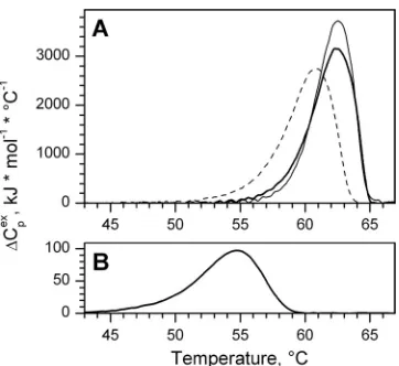
Thermal denaturation of gp146 and gp188*.
Thermal
dena-turation of the recombinant proteins was analyzed by DSC. The
DSC profile of the putative chaperonin showed a cooperative
en-dothermic transition with a maximum at 62.1°C and a
calorimet-ric enthalpy change of about 12,600 kJ/mol for the complex (
Fig.
5A
;
Table 1
). The calorimetric transition was found to be
irrevers-ible (after heating to 90°C and a subsequent cooling to 5°C, no
transition was observed during the second scan) and
concentra-tion independent (2 to 16
M for a protein subunit), which
indi-cates an absence of dissociation stages in melting.
Since chaperonins assist the folding of polypeptide chains in an
ATP-dependent manner, we examined the effect of ATP and ADP
binding on thermal denaturation of gp146 (
Fig. 5A
;
Table 1
).
Ad-dition of ADP increased cooperativeness and enthalpy of the
gp146 denaturation, which suggests chaperonin stabilization. In
contrast, ATP binding results in complex destabilization, which is
indicated by a slight decrease of the temperature of the heat
capac-ity maximum (
T
m). This result closely correlates with the effect of
low ATP concentrations on GroEL thermal denaturation (
20
).
FIG 3Sedimentation data of recombinant gp146 (1.2M) and gp188* (14.6 M) at 22°C. (A) Raw data for gp146; every third curve is shown. (B and C) Fitting of sedimentation coefficients for gp146 and gp188*, respectively.
FIG 4Electron microscopic images of the purified recombinant gp146 nega-tively stained with 1% uranyl acetate. The inset demonstrates the 7-fold sym-metry of the chaperonin complex.
on November 7, 2019 by guest
http://jvi.asm.org/
[image:4.585.331.511.65.230.2] [image:4.585.78.249.68.298.2]Thermal denaturation of gp188* is also irreversible and takes
place at temperatures lower than those for gp146 (
Fig. 5B
;
Table 1
).
Fluorescence and CD spectroscopies.
Fluorescence and CD
spectroscopies were used to detect changes in the tertiary and
secondary structures of proteins, respectively, at different
temper-atures. Red shifts in the wavelength of the emission maximum
(
max) without appreciable changes in fluorescence intensity were
observed for both proteins when the temperature was increased
from 25 to 75°C.
Figure 6A
shows the temperature dependence of
maxof the emission spectra obtained by exciting gp146 and
gp188* solutions at 280 nm. The far-UV CD spectra of proteins
have two minima near 208 and 220 nm (
Fig. 6B
, inset), which is
characteristic of proteins with a significant amount of
␣
-helical
content. When temperature was increased, the ellipticity at 208
nm was decreased dramatically with no change in the peak
posi-tion for either protein (
Fig. 6B
). All experimental curves
indicat-ing changes in the tertiary and secondary structures of proteins
upon their heating have an abrupt sigmoid form, which means a
cooperative protein transition from its native structure to a
disor-dered one. However, the destruction of the secondary structures of
gp146 and gp188* was observed at temperatures higher than that at
which the tertiary structure was destroyed (
Fig. 6
). When gp188* was
heated to 50°C, half of its tertiary structure was lost, whereas its
sec-ondary structure did not change. Both methods have shown that
gp188* undergoes structural changes at lower temperatures than
gp146. The recombinant gp146 remains intact at temperatures up to
52°C. These results coincide with those obtained by DSC.
Isothermal titration calorimetry of phage chaperonin.
To
study ATP and ADP binding to the recombinant gp146 more
ac-curately, nucleotide titration was carried out using ITC. Typical
raw traces of the calorimetric titration of ADP and ATP to pure
gp146 are shown in
Fig. 7A
and
C
, respectively. The downward
peak represents the heat release at each injection of the nucleotide
solution. The binding isotherm of the nucleotide was obtained by
integrating each peak and subtracting the dilution heat. Such
iso-therms for ADP and ATP are shown in
Fig. 7B
and
D
, respectively.
The recombinant gp146 has been shown to bind both ATP and
ADP. The observed binding isotherms were fitted to the “one set
of sites” model. In this model, the putative chaperonin has several
identical nucleotide binding sites, which are independent of each
other and have a uniform binding constant,
K
a, and enthalpy
change,
⌬
H
. The parameter values fitting this model best are listed
in
Table 2
. As displayed in
Fig. 7B
and
D
, the theoretical curves
(solid lines) show a good match with the experimental data
(squares) for ADP and ATP, respectively. Our results indicate that
nucleotide binding to gp146 could be well interpreted by the
non-cooperative model, while ATP is known to induce non-cooperative
conformational changes in GroEL (
28
). The ADP binding
con-stant and enthalpy are a little higher than those for ATP. Observed
stoichiometry of nucleotide binding to the recombinant gp146 is
FIG 5DSC profiles of recombinant gp146 (A) and gp188* (B) in 50 mM Tris-HCl (pH 7.5), 10 mM MgCl2, and 100 mM KCl showing the dependence
[image:5.585.74.255.64.230.2]of the heat capacity on temperature for gp146 (0.58M) and gp188* (7.3M) in the absence of nucleotides (thick solid lines) and in the presence of ATP (A, dashed line) and ADP (A, thin solid line).
TABLE 1Thermodynamic parameters of gp146 thermal denaturation: effect of nucleotide binding
Sample ⌬Hcal(kJ/mol) Tm(°C)
gp188* 608⫾30 54.8⫾0.1
gp146 12,619⫾939 62.1⫾0.5
gp146/ATP 12,737⫾517 60.6⫾0.3
gp146/ADP 14,036⫾380 62.2⫾0.3
FIG 6Temperature-induced changes in the tertiary (A) and secondary (B) structures of gp188* (thick line, filled circles) and gp146 (thin line, open cir-cles) in 50 mM Tris-HCl (pH 7.5), 10 mM MgCl2, and 100 mM KCl observed
by fluorescence and CD spectroscopies. (A) Temperature dependence of wavelength of the emission maximum (max) of the fluorescent spectra of
gp146 (0.12M) and gp188* (1.4M). (Inset) Fluorescence emission spectra of gp146 (thin line) and gp188* (thick line) at 25°C by excitation at 280 nm. (B) Temperature dependence of the mean residue ellipticity at 208 nm of the far-UV CD spectra of gp146 (0.12M) and gp188* (2.9M). (Inset) Far-UV CD spectra of gp188* at 25, 55, 58, and 73°C and gp146 at 25, 60, 63, and 75°C (the ellipticity decreases with the rising temperature).
on November 7, 2019 by guest
http://jvi.asm.org/
[image:5.585.317.524.65.366.2] [image:5.585.40.288.665.725.2]close to 7 for both ADP and ATP (
Fig. 7B
and
D
;
Table 2
). In
addition, the data for the competitive titration indicated that the
binding sites are universal and able to bind both ATP and ADP
(not shown). Considering the binding stoichiometry, two
hepta-meric rings may differ in their abilities for nucleotide binding.
Another explanation points to sample heterogeneity, suggesting
that various structural conformations of the chaperonin are
avail-able for nucleotide binding.
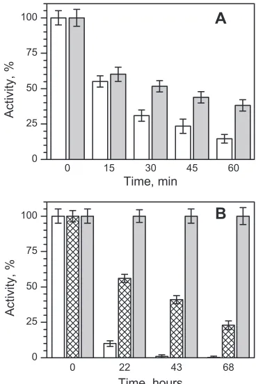
Effect of gp146 on gp188* thermal inactivation
in vitro
.
Chaperonins are thought to play a cellular role not only in
assist-ing the foldassist-ing of nascent proteins but also in protectassist-ing mature
proteins undergoing stress. It is known that chaperonin GroEL
can stabilize enzymes against thermal inactivation (
36
,
38
). To
find out whether gp146 can carry out a similar role, we
investi-gated the effect of gp146 on heat inactivation of its substrate,
gp188*,
in vitro
. According to DSC profiles (
Fig. 5
) and
fluores-cence and CD data (
Fig. 6
), gp188* denaturation starts at
approx-imately 47°C, while gp146 seems to remain native at temperatures
up to 52°C, even in the presence of ATP. Therefore, gp188*
inac-tivation was examined under physiological conditions at 50°C.
Aliquots were removed at appropriate intervals and assayed for
residual muralytic activity. Inactivation of the free gp188*
fol-lowed first-order kinetics with a half-time of about 16 min (not
shown). As shown in
Fig. 8A
, the loss of the gp188* enzymatic
activity during the incubation was retarded by gp146 in the
[image:6.585.124.463.66.322.2]pres-FIG 7ITC profiles of the binding of ADP (A and B) and ATP (C and D) to recombinant gp146. (A and C) Raw data for successive injections of ADP (ATP) into gp146; (B and D) integrations of each injection (squares) and the best fit by the “one set of sites” model (solid lines) versus molar ratio of ADP (ATP) to gp146.
TABLE 2Best-fit parameters obtained by the “one set of sites” model
Nucleotide Stoichiometry Ka(M⫺1) ⌬H(kJ/mol)
⌬S
(J · mol⫺1· °C⫺1)
ATP 7.33⫾0.03 5.48⫾0.24 ⫺36.17⫾0.23 1.7 ADP 8.76⫾0.03 7.76⫾0.36 ⫺42.50⫾0.22 ⫺17.6
FIG 8Activity of gp188* (0.1M for monomer) during thermal inactivation in 50 mM Tris-HCl (pH 7.5), 10 mM MgCl2, and 100 mM KCl at 50°C (A) and
37°C (B). Shown are free gp188* (white bars) and gp188* in the presence of gp146 (0.2M for the tetradecamer) with 3 mM ATP (gray bars) and without ATP (hatched bars). The graph shows the results of three independent thermal inactivation assays, with error bars indicating the standard deviations.
on November 7, 2019 by guest
http://jvi.asm.org/
[image:6.585.328.512.386.662.2] [image:6.585.38.287.679.723.2]ence of ATP. The enzymatic activity still remaining 60 min later
was more than twofold higher in the presence of gp146 than in its
absence. It was found that the free gp188* was also inactivated at
37°C, while in the presence of gp146 it remained intact for a long
time (
Fig. 8B
). It should be noted that gp188* thermal inactivation
is also slowed down by gp146 in the absence of ATP, but to a lesser
extent than in its presence (
Fig. 8B
).
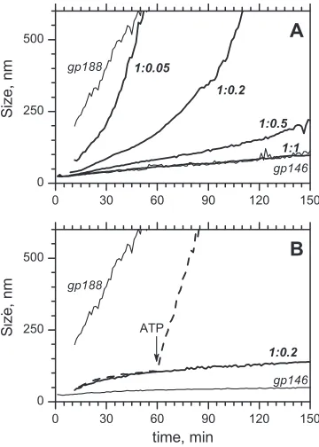
Effect of gp146 on gp188* aggregation
in vitro
.
Heat
inacti-vation of gp188* was found to be accompanied by irreversible
aggregation. The effect of gp146 on the thermal aggregation of
gp188* under physiological conditions was studied using DLS,
which allows sizing of the protein aggregates. To observe the
for-mation of aggregates, the gp188* concentration was increased to 3
M in comparison with the concentration used in the thermal
inactivation assay.
Figure 9
shows DLS curves of the gp188*
ag-gregation at 45°C. The free gp188* agag-gregation to large-sized
par-ticles takes less than 30 min, irrespective of the presence or absence
of ATP. It was found that gp146 had the capacity to suppress
denatured enzyme aggregation both in the presence and absence
of ATP. The effect of gp146 on the aggregation of gp188* in the
presence of ATP is shown in
Fig. 9A
. The degree of suppression of
the aggregation correlates with the chaperonin content in the
so-lution. This is indicated by the shifting of the curves from the
reference to the right when the molar ratio of gp146 to substrate
increases. At an equimolar ratio, gp146 was found to completely
suppress gp188* aggregation. Indeed, the related curve has no
difference to that for free gp146 (
Fig. 9A
). The replacement of
gp146 by bovine serum albumin (BSA) did not result in a
protec-tive effect (not shown).
A similar influence of gp146 on the gp188* aggregation was
observed under the same conditions in the absence of ATP (
Fig.
9B
). However, in this case the effect seems to be caused by the
formation of highly stable long-lived binary complexes between
gp146 and the thermally unfolded substrate. Bound to gp146,
gp188* cannot undergo unspecific aggregation. Control
experi-ments with BSA at molar excess do not show a comparable
sup-pression of gp188* aggregation under these conditions (not
shown), indicating a specific effect of gp146 similar to that earlier
observed for chaperonin GroEL (
10
,
25
). The complex of
ther-mally inactivated substrate and gp146 can be easily dissociated by
adding ATP. ATP-dependent dissociation results in the release of
gp188*, which denatures and aggregates rapidly, as indicated by
the increase in large protein particles (
Fig. 9B
).
DISCUSSION
In this study, we have characterized one of the GroEL orthologs
pre-dicted from phage genomes (
13
,
24
,
30
) and demonstrated its
chap-eronin activity for the first time. The phage chapchap-eronin was shown to
be synthesized during bacteriophage EL propagation in
P. aeruginosa
cells. The native protein was isolated by immunoprecipitation from
EL-infected bacterial cells using serum against recombinant gp146
and identified by N-terminal amino acid sequencing.
Physicochem-ical characterization of the phage chaperonin by a number of
differ-ent techniques was carried out on the recombinant gp146 produced
by
E. coli
cells. It was found that the recombinant gp146
self-assem-bled into a large oligomeric complex with the architecture typical of
chaperonins. Similar to GroEL from
E. coli
, the phage chaperonin is a
homotetradecamer composed of two stacked seven-member rings,
each with a central cavity.
A native phage substrate of gp146, gp188, was isolated and
identified. Characterization of recombinant proteins by different
methods revealed that gp188* was less thermostable than gp146.
The recombinant gp188* underwent inactivation and aggregation
in vitro
under physiological conditions at elevated temperatures
below 50°C, while gp146 still remained native under these
condi-tions. It was demonstrated that gp146 can protect the enzymatic
activity of gp188* and suppress the irreversible aggregation of the
thermally unfolded substrate molecules
in vitro
. These results
pro-vided evidence of the gp146 chaperonin function.
Whether the phage chaperonin plays a role in the EL life cycle is
still unclear. We suppose that gp146 somehow affects lysis, because its
substrate, gp188, appears to be an endolysin (
8
). Endolysins are
known to be phage-encoded enzymes produced during the late phase
of gene expression in the lytic cycle to degrade peptidoglycan, the
main constituent of the bacterial cell wall, thereby enabling progeny
virions to be liberated (
34
,
46
). We confirmed that gp188 synthesis
actually started during the late stage of the virus reproduction cycle,
much later than gp146 synthesis. It is known that under physiological
conditions many proteins undergo continuous denaturation in the
cell, resulting in the loss of their biological activity (
15
). Considering
this fact, we suppose that, during EL propagation in bacterial cells, the
phage chaperonin could perform the same protective role in relation
to its substrate as it does
in vitro
and thereby increase the half-life of
endolysin, which otherwise might be inactivated and might
aggre-gate. Further application of genetic approaches is required to prove
the correctness of this hypothesis.
The phage chaperonin has shown to function in both
ATP-dependent and ATP-inATP-dependent manners. The first one seems to
be similar to the chaperonin functional ATPase cycle, which
re-FIG 9Effect of gp146 on the aggregation of gp188* (3M) at 45°C in the presence of 1.2 mM ATP (A) and in the absence of ATP (B). Thin lines mean DLS trends of free proteins. Thick lines correspond to their mixture at various molar ratios (the number near each of the curves indicates a molar ratio of monomeric enzyme to tetradecameric gp146). The dashed line (B) corre-sponds to the gp188*-gp146 mixture at a molar ratio of 1:0.2; the arrow shows the time when ATP was added.
on November 7, 2019 by guest
http://jvi.asm.org/
[image:7.585.73.253.66.318.2]phage chaperonin are probably similar to those of bacterial
GroEL. However, unlike GroEL, the phage chaperonin does not
require a cochaperonin for its activity. Indeed, we have shown by
in vitro
experiments using purified recombinant proteins (gp146
and gp188*) that the phage chaperonin is able to function without
any additional protein cofactors. It should be noted that no phage
or host cochaperonin was found to coimmunoprecipitate
to-gether with the substrate protein from the EL-infected bacteria.
Therefore, it is possible to assume that, like group II chaperonins,
the phage chaperonin appears to have a helical protrusion, which
can play a role equivalent to GroES, sealing off the central cavity
from the outside. According to our preliminary observations of
the gp146 shape in solution by small-angle X-ray scattering
(SAXS), the chaperonin complex can adopt either an open or
closed conformation that appears to be regulated by the position
of the helical protrusion (
1
). Further structural and biochemical
investigations are required to reveal the detailed mechanism of its
functioning, which can be distinct from the mechanisms of the
other known chaperonins. Further analysis of its functional and
structural characteristics may provide important insights into the
nature of the phage chaperonin, which is probably a
representa-tive of a new group of chaperonins.
ACKNOWLEDGMENTS
This work was supported by grant 11-04-00935 from the Russian Fund for Basic Research.
We are grateful to V. N. Krylov for bacteriophage EL and Y. F. Leonova for N sequencing. We thank N. N. Magretova and P. V. Kalmykov for sedimentation analysis and I. I. Kireev for help with the electron micro-scope.
REFERENCES
1.Amarantov SV, Naletova IN, Kurochkina LP.2011. Simulation of the shape of chaperonins using the small-angle X-ray scattering curves and torus form factor. J. Exp. Theor. Phys. (Mosc.)113:322–338.
2.Ang D, et al.2001. Pseudo-T-even bacteriophage RB49 encodes CocO, a cochaperonin for GroEL, which can substitute forEscherichia coli’s GroES and bacteriophage T4’s Gp31. J. Biol. Chem.276:8720 – 8726.
3.Bakkes PJ, Faber BW, van Heerikhuizen H, van der Vies SM.2005. The T4-encoded cochaperonin, gp31, has unique properties that explain its requirement for the folding of the T4 major capsid protein. Proc. Natl. Acad. Sci. U. S. A.102:8144 – 8149.
4.Bigotti MG, Clarke AR.2008. Chaperonins: the hunt for the group II mechanism. Arch. Biochem. Biophys.474:331–339.
5.Boisvert DC, Wang J, Otwinowski Z, Horwich AL, Singler PB.1996. The 2.4 Å crystal structure of the bacterial chaperonin GroEL complexed with ATP gamma S. Nat. Struct. Biol.3:170 –177.
6.Braig K, et al.1994. The crystal structure of the bacterial chaperonin GroEL at 2.8 Å. Nature371:578 –586.
7.Briers Y, Lavigne R, Volckaert G, Hertveldt K.2007. A standardized approach for accurate quantification of murein hydrolase activity in high-throughput assays. J. Biochem. Biophys. Methods70:531–533. 8.Briers Y, et al.2007. Muralytic activity and modular structure of the
endolysins ofPseudomonas aeruginosabacteriophagesKZ and EL. Mol. Microbiol.65:1334 –1344.
OBP and comparative genome analysis of the diverseKZ-related phages. J. Virol.86:1844 –1852.
14. Cowan NJ, Lewis SA. 2001. Type II chaperonins, prefoldin, and the tubulin-specific chaperones. Adv. Protein Chem.59:73–104.
15. Dill KA, Shortle D. 1991. Denatured states of proteins. Annu. Rev. Biochem.60:795– 825.
16. Dumont D, Noben Raus J-PJ, Stinissen P, Robben J.2004. Proteomic analysis of cerebrospinal fluid from multiple sclerosis patients. Proteom-ics4:2117–2124.
17. Fenton WA, Horwich AL.2003. Chaperonin-mediated protein folding: fate of substrate polypeptide. Q. Rev. Biophys.36:229 –256.
18. Fischer H, Polikarpov I, Craievich AF.2004. Average protein density is a molecular-weight-dependent function. Protein Sci.13:2825–2828. 19. Frydman J.2001. Folding of newly translated proteins in vivo: the role of
molecular chaperones. Annu. Rev. Biochem.70:603– 647.
20. Galan A, et al.1999. ATP hydrolysis induces an intermediate conforma-tional state in GroEL. Eur. J. Biochem.259:347–355.
21. Georgopoulos CP, Hendrix RW, Casjens SR, Kaiser AD.1973. Host participation in bacteriophage lambda head assembly. J. Mol. Biol.76:45– 50.
22. Hartl FU, Hayer-Hartl M.2002. Molecular chaperones in the cytosol: from nascent chain to folded protein. Science295:1852–1858.
23. Hartl FU, Hayer-Hartl M.2009. Converging concepts of protein folding
in vitroandin vivo.Nat. Struct. Mol. Biol.16:574 –581.
24. Hertveldt K, et al.2005. Genome comparison ofPseudomonas aeruginosa
large phages. J. Mol. Biol.354:536 –545.
25. Holl-Neugebauer B, Rudolph R.1991. Reconstitution of a heat shock effect in vitro: influence of GroE on the thermal aggregation of␣ -gluco-sidase from yeast. Biochemistry30:11609 –11614.
26. Horwich AL, Apetri AC, Fenton WA.2009. The GroEL/GroES cis cavity as a passive anti-aggregation device. FEBS Lett.583:2654 –2662. 27. Hunt JF, van der Vies SM, Henry L, Deisenhofer J.1997. Structural
adaptations of the of the specialized bacteriophage T4 cochapereronin gp31expand the size of the Anfinsen cage. Cell.90:361–371.
28. Inobe T, Makio T, Takasu-Ishikawa E, Terada TP, Kuwajima K.2001. Nucleotide binding to the chaperonin GroEL: non-cooperative binding of ATP analogs and ADP, and cooperative effect of ATP. Biochim. Biophys. Acta1545:160 –173.
29. Kanzaki T, et al.2008. Sequential action of ATP-dependent subunit conformational change and interaction between helical protrusions in the closure of the built-in lid of group II chaperonins. J. Biol. Chem.283: 34773–34784.
30. Kiljunen S, et al.2005. Yersiniophage phiR1-37 is a tailed bacteriophage having a 270 kb DNA genome with thymidine replaced by deoxyuridine. Microbiology151:4093– 4102.
31. Laemmli UK.1970. Cleavage of structural proteins during the assembly of the head of bacteriophage T4. Nature227:680 – 685.
32. Lavigne R, Noben et al.2006. The structural proteome ofPseudomonas aeruginosabacteriophageKMV. Microbiology152:529 –534.
33. Lecoutere E, et al.2009. Identification and comparative analysis of the structural proteomes of phiKZ and EL, two giant Pseudomonas aerugi-nosa bacteriophages. Proteomics9:3215–3219.
34. Loessner MJ.2005. Bacteriophage endolysins— current state of research and applications. Curr. Opin. Microbiol.8:480 – 487.
35. Lopez Mayorga O, Freire E.1987. Dynamic analysis of differential scan-ning calorimetry data. Biophys. Chem.27:87–96.
36. Martin J, Horwich AL, Hartl F-U.1992. Prevention of protein denatur-ation under heat stress by the chaperonin Hsp60. Science258:995–998. 37. Mayhew M, et al.1996. Protein folding in the central cavity of the
GroEL-GroES chaperonin complex. Nature379:420 – 426.
38. Mendoza JA, Wilson M, Joves F, Ackermann E.1996. Thermostabilization of enzymes by the chaperonin GroEL. Biotechnol. Tech.10:535–540.
on November 7, 2019 by guest
http://jvi.asm.org/
39. Schuck P.2000. Size-distribution analysis of macromolecules by sedi-mentation velocity ultracentrifugation and Lamm equation modeling. Biophys. J.78:1606 –1619.
40. Studier FW, Rosenberg AH, Dunn JJ, Dubendorff JW.1990. Use of T7 RNA polymerase to direct expression of cloned genes. Methods Enzymol.
185:60 – 89.
41. Thirumalai D, Lorimer GH.2001. Chaperonin-mediated protein fold-ing. Annu. Rev. Biophys. Biomol. Struct.30:245–269.
42. van der Vies SM, Gatenby AA, Georgopoulos C.1994. Bacteriophage T4 encodes a co-chaperonin that can substitute for Escherichia coli GroES in protein folding. Nature368:654 – 656.
43. Vera A, Gonzalez-Montalban N, Aris A, Villaverde A.2007. The con-formational quality of insoluble recombinant proteins is enhanced at low growth temperatures. Biotechnol. Bioeng.96:1101–1106.
44. Xu ZH, Horwich AL, Sigler PB.1997. The crystal structure of the asymmetric GroEL-GroES-(ADP)7chaperonin complex. Nature388:
741–750.
45. Young JC, Agashe VR, Siegers K, Hartl FU.2004. Pathways of chaper-one-mediated protein folding in the cytosol. Nat. Rev. Mol. Cell Biol.
5:781–791.
46. Young R, Wang I-N, Roof WD.2000. Phages will out: strategies of host cell lysis. Trends Microbiol.8:120 –128.
on November 7, 2019 by guest
http://jvi.asm.org/