Ames Laboratory Publications
Ames Laboratory
10-21-2002
Structural studies of Fe0.81Ga0.19 by reciprocal
space mapping
D. Viehland
Virginia Polytechnic Institute and State University
JieFang Li
Virginia Polytechnic Institute and State University
Thomas A. Lograsso
Iowa State University, [email protected]
A. R. Ross
Iowa State UniversityManfred Wuttig
University of Maryland, College Park
Follow this and additional works at:
http://lib.dr.iastate.edu/ameslab_pubs
Part of the
Condensed Matter Physics Commons
, and the
Metallurgy Commons
The complete bibliographic information for this item can be found at
http://lib.dr.iastate.edu/
ameslab_pubs/127
. For information on how to cite this item, please visit
http://lib.dr.iastate.edu/
howtocite.html
.
Structural studies of Fe0.81Ga0.19 by reciprocal space mapping
Abstract
Reciprocal lattice mapping has been performed on Fe
0.81Ga
0.19crystals by ω–ω/2θ, Ψ–ϕ, and ω–ϕ scans. A
strong elongation of the
〈
001
〉
cpeak was found along the
〈
110
〉
cdirection. ω scans revealed short lateral
correlation lengths ξ along
〈
110
〉
cand strong diffuse scattering along the
〈
001
〉
c. Multiple domains with
monoclinic symmetry (angle
∼
190°) were observed by Ψ–ϕ and ω–ϕ scans on the (001)
cface, and were also
tilted with respect to each other. The results show an average cubic structure with orthorhombic structural
modulations, and two structural domain states that result in a limiting monoclinic symmetry.
Keywords
iron alloys, gallium alloys, crystal structure, X-ray scattering
Disciplines
Condensed Matter Physics | Metallurgy
Comments
The following article can be found in
Applied Physics Letters
81 (2002): 3185 and may be found at
http://dx.doi.org/10.1063/1.1516231
.
Rights
Copyright 2002 American Institute of Physics. This article may be downloaded for personal use only. Any
other use requires prior permission of the author and the American Institute of Physics.
Structural studies of Fe
0.81Ga
0.19by reciprocal space mapping
D. Viehlanda)and J. F. Li
Department of Materials Science and Engineering, Virginia Tech, Blacksburg, Virginia 24061
T. A. Lograsso and A. Ross
Metals and Ceramic Sciences, Ames Laboratory, Ames, Iowa 50011
Manfred Wuttig
Department of Materials and Nuclear Engineering, University of Maryland, College Park, Maryland 20742
共Received 24 June 2002; accepted 29 August 2002兲
Reciprocal lattice mapping has been performed on Fe0.81Ga0.19crystals by–/2,⌿–, and–
scans. A strong elongation of the
具
001典
c peak was found along the具
110典
c direction. scans revealed short lateral correlation lengths along具
110典
c and strong diffuse scattering along the具
001典
c. Multiple domains with monoclinic symmetry共angle⬃190°兲were observed by ⌿–and–scans on the (001)cface, and were also tilted with respect to each other. The results show an average cubic structure with orthorhombic structural modulations, and two structural domain states that result in a limiting monoclinic symmetry. © 2002 American Institute of Physics.
关DOI: 10.1063/1.1516231兴
The magnetostrictive strain (3/2)100 of Fe is well
known to be small, on the order of 30 ppm. Introduction of Ga into crystalline solution with Fe results in pronounced increases in (3/2)100,1–3as long as the BCC phase remains stable for the Fe1⫺xGax solution,2 even though Ga is non-magnetic. For gallium concentrations x⬎0.15, this crystal-line solution is not stable in the annealed condition.4 How-ever, rapid quenching of specimens is known to extend the limit of stability up to x⫽0.19.2 The highest magnetostric-tion reported to date for any material is found for Fe0.81Ga0.19, with (3/2)100⬃400 ppm. Even though the
magnetic moment is being diluted, an enhancement of the magnetostriction occurs.
The magnetostriction is strongly peaked near x⫽0.19.3 At higher and lower Ga contents 共i.e., ⌬x⬃⫾0.02), (3/2)100 is decreased by ⬃50%. The anomalous magneto-striction cannot be due to a conventional mechanism. It has been conjectured that short-range ordering of Ga atoms oc-curs along the 关100兴 in the␣-Fe structure.2,5 Wuttig et al.5 have developed a model to explain this unique magnetostric-tion based upon Ga pairs. Accordingly, the magnetostricmagnetostric-tion would increase quadratically as a function of Ga concentra-tion and decrease at higher Ga contents due to the formaconcentra-tion of DO3 or B2 ordered states.2
Previous elastic constant measurements have shown a significant decrease in Young’s modulus with increasing Ga,5,6indicating that the lattice is softening as the magneto-striction is increasing. However, a structural understanding of this decrease, and hence the increase of the magnetostric-tion, remains lacking. Therefore, the purpose of this study was to investigate Fe0.81Ga0.19crystals using reciprocal space
mapping and texture analysis. The results show an average cubic structure, which has orthorhombic structural modula-tions, and two structural domain states that result in a limit-ing monoclinic symmetry.
Fe0.81Ga0.19crystals were grown by a Bridgman method,
as previously described.3The crystals were homogenized at 900 °C and rapidly quenched to stabilize the BCC ␣-Fe structure.2,3 The crystal was oriented along the
具
001典
c and was cut to dimensions 10⫻10⫻2 mm3. A Philips MPD high resolution diffractometer equipped with an open Eulerian cradle was used for structural investigations. The具
001典
cface was investigated by–/2,⌿–, and–scans.Angular–/2scans are done by measuring numerous coupled/2Bragg scans for a range of incident anglesas starting values by cradle rocking. The angle⌿is the angular position of the sample surface normal in the direction per-pendicular to the diffraction plane, whereasis the rotation angle perpendicular to the plane of diffraction. A⌿–scan performs a series of coupled rotations about the two axes perpendicular to /2, while maintaining a constant 2.7 This gives the integration volume of reciprocal space on a plane perpendicular to the plane of diffraction. Rotation of this perpendicular plane yields the three-dimensional spatial distribution of the diffraction intensity 共or texture兲 at con-stant Bragg conditions. A – scan utilizes a series of curves taken at different positions, at constant 2. The integration volume of reciprocal space appears as thin rods perpendicular to the diffraction plane, as the instrument broadening is quite large in this direction.5From these data, detailed information concerning the average crystal structure, domain texture, lateral coherency, and mosaicity can be ob-tained.
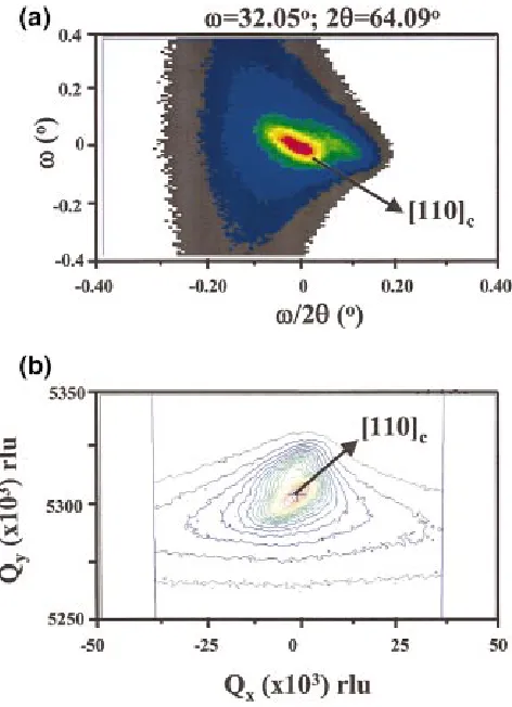
Figure 1共a兲 shows a –/2 scan. A single diffraction peak was found along /2, and splitting was not observed along . Thus, the structure is clearly cubic, with a⫽b⫽c ⫽2.906 Å and ␣⫽⫽␥⫽90°. This is consistent with the BCC ␣-Fe structure. However, the peak was strongly elon-gated along the
具
110典
c, shifting significantly in with changes in /2. The diffraction intensity along the具
110典
c elongation was very strong and could only be determined by rocking the sample.Reciprocal lattice plots were calculated, as shown in Fig.
a兲Electronic mail: [email protected]
APPLIED PHYSICS LETTERS VOLUME 81, NUMBER 17 21 OCTOBER 2002
3185
1共b兲. In this figure, the peak was elongated along both Qx and Qy. The
具
110典
corientation of the elongation is marked in the figure. The具
110典
c directionality demonstrates the presence of pseudo-orthorhombic symmetry. The broadness of this peak demonstrates that the pseudo-orthorhombic symmetry has significant structural irregularity. The line broadening along the direction yields the lateral correlation length . The Scherrer equation8 ⫽/关2 FWHMcos(2/2)兴 can be used to determine , where FWHM is the full width at half maximum, is the wavelength of the Cu K␣ radiation 共1.5406 Å兲and共2/2兲 is the peak position of the principle peak occurring at 64.094°. The FWHM was ⬃0.15° (2.62⫻10⫺3 rad), thus ⬍300 Å.In addition, Fig. 1共a兲 exhibits strong diffuse scattering along
具
001典
c, as can be seen from the dark colored contour regions. The diffuse scattering along this direction was much more broad in than that along the具
110典
c. The results demonstrate strong structural irregularity along both具
001典
c and具
110典
c; however, that along the具
001典
c did not possess any significant lateral correlation. This is consistent with the reports of short-range chemical ordering along the具
001典
cin Fe0.81Ga0.19.2,5 It appears that Fe0.81Ga0.19 is structurallyfrustrated, containing elements of both the BCC ␣-Fe and DO3 or B2 structures. In this structurally inhomogeneous
state, anomalous magnetostriction may exist due to a large elastic compliance of elastic microtwins under magnetic field, during magnetization rotation.
Figure 2共a兲 shows a– scan. This data demonstrates
two preferred orientations that are rotated in by 190°, and are tilted in by ⬃2.5° with respect to each other. This rotation produces monoclinic symmetry, as is noticeably greater than 180°. The results unambiguously demonstrate the presence of two domain states whose c-axes are slightly tilted away from the 关001兴c. This is directly revealed from the symmetry in the diffraction intensity of the – scan. The average crystal symmetry remains cubic consistent with the bcc␣-Fe structure. However the domain-averaged limit-ing symmetry is monoclinic. Neumann’s law will then re-quire that the property tensor matrices have monoclinic 共point group兲symmetry.9
The resolution of our instrument in the -direction is significantly higher than that in the -direction. Thus, the
–scan will offer better resolution when mapping of the spatial distribution of domains. Figure 2共b兲 shows an– scans taken along the (001)c. A single thin rod can be seen that has nonuniform intensity. This demonstrates that the do-main state is spatially nonuniform. The relative intensity of the rod can be seen to vary as a function of . At some
-positions, the intensity was much stronger than at others. These results are consistent with a stacking of micro-twins, resulting in a domain-averaged monoclinic limiting symmetry.
[image:4.612.59.295.52.378.2]The results of this investigation for Fe0.81Ga0.19 crystals can be summarized as 共i兲 the average crystal structure is cubic, with a domain-averaged monoclinic limiting symme-try;共ii兲structural nonuniformity exists along both the
具
110典
c and具
001典
c; and 共iii兲 orthorhombic structural modulations exist with a short lateral correlation length.FIG. 1. 共Color兲X-ray diffraction data for a 具001典c oriented Fe0.81Ga0.19 single crystal, which had been rapidly quenched.共a兲⫺/2scan, and共b兲 reciprocal lattice map.
FIG. 2.共Color兲X-ray diffraction data taken at constant Bragg conditions for a具001典coriented Fe0.81Ga0.19single crystal.共a兲-scan.共b兲-scan. 3186 Appl. Phys. Lett., Vol. 81, No. 17, 21 October 2002 Viehlandet al.
[image:4.612.325.556.52.370.2]The research was supported by the Office of Naval Re-search under grants N000140210340, N000140210126, and MURI N000140110761. One of the authors 共T. A. L.兲 ac-knowledges the support of the Office of Basic Energy Sci-ences, Materials Sciences Division, of the U.S. Department of Energy under Contract No. W-7405-ENG-82.
1
A. E. Clark, J. B. Restorff, M. Wun-Fogle, T. A. Lograsso, and D. L. Schlagel, IEEE Trans. Magn. 36, 3238 共2000兲; A. E. Clark, M. Wun-Fogle, J. B. Restorff, T. A. Lograsso, A. R. Ross, and D. Schlagel,
Pro-ceedings of the Actuator 2000 Conference, Bremen, Germany, June 2000. 2
A. E. Clark, M. Wun-Fogle, J. B. Restorff, T. Lograsso, J. R. Cullen, IEEE Trans. Magn. 37, 2678共2001兲.
3A. E. Clark, M. Wun-Fogle, J. B. Restorff, T. Lograsso, Proceedings of the
Fourth Pacific Rim International Conference on Advanced Materials and Processing (PRICM4), edited by S. Hanada, Z. Zhong, S. Nam, and R.
Wright,共The Japan Institute of Metals ..., 2001兲, p. 1711. 4
See for example: Binary Alloy Phase Diagrams, 2nd ed., edited by T. B. Massalski共ASM International, Materials Park, OH, 1990兲.
5M. Wuttig, L. Dai, and J. Cullen, Appl. Phys. Lett. 80, 1135共2002兲. 6Liyang Dai, M. S. Thesis, University of Maryland, 2002.
7
Xin Wang, Ph.D. Dissertation No. 557, Linkoping University, 1998. 8B. D. Cullity, Elements of X-ray Diffraction共Addison-Wesley, Reading,
MA, 1978兲. 9
J. F. Nye, Physical Properties of Crystals: Their Representation by
Ten-sors and Matrices共Oxford University Press, Oxford, 1985兲.
3187 Appl. Phys. Lett., Vol. 81, No. 17, 21 October 2002 Viehlandet al.