Copyright © 1997, American Society for Microbiology
Complete Sequence and Genomic Analysis of Murine
Gammaherpesvirus 68
HERBERT W. VIRGIN IV,
1PHILIP LATREILLE,
2PAMELA WAMSLEY,
2KYMBERLIE HALLSWORTH,
2KAREN E. WECK,
1ALBERT J. DAL CANTO,
1ANDSAMUEL H. SPECK
1*
Department of Pathology and Center for Immunology
1and Genome Sequencing Center, Department
of Genetics,
2Washington University School of Medicine, St. Louis, Missouri 63110
Received 19 February 1997/Accepted 16 April 1997
Murine gammaherpesvirus 68 (
g
HV68) infects mice, thus providing a tractable small-animal model for
analysis of the acute and chronic pathogenesis of gammaherpesviruses. To facilitate molecular analysis of
g
HV68 pathogenesis, we have sequenced the
g
HV68 genome. The genome contains 118,237 bp of unique
sequence flanked by multiple copies of a 1,213-bp terminal repeat. The GC content of the unique portion of the
genome is 46%, while the GC content of the terminal repeat is 78%. The unique portion of the genome is
estimated to encode at least 80 genes and is largely colinear with the genomes of Kaposi’s sarcoma herpesvirus
(KSHV; also known as human herpesvirus 8), herpesvirus saimiri (HVS), and Epstein-Barr virus (EBV). We
detected 63 open reading frames (ORFs) homologous to HVS and KSHV ORFs and used the HVS/KSHV
numbering system to designate these ORFs.
g
HV68 shares with HVS and KSHV ORFs homologous to a
complement regulatory protein (ORF 4), a D-type cyclin (ORF 72), and a G-protein-coupled receptor with close
homology to the interleukin-8 receptor (ORF 74). One ORF (K3) was identified in
g
HV68 as homologous to
both ORFs K3 and K5 of KSHV and contains a domain found in a bovine herpesvirus 4 major immediate-early
protein. We also detected 16 methionine-initiated ORFs predicted to encode proteins at least 100 amino acids
in length that are unique to
g
HV68 (ORFs M1 to 14). ORF M1 has striking homology to poxvirus serpins, while
ORF M11 encodes a potential homolog of Bcl-2-like molecules encoded by other gammaherpesviruses (gene 16
of HVS and KSHV and the BHRF1 gene of EBV). In addition, clustered at the left end of the unique region are
eight sequences with significant homology to bacterial tRNAs. The unique region of the genome contains two
internal repeats: a 40-bp repeat located between bp 26778 and 28191 in the genome and a 100-bp repeat located
between bp 98981 and 101170. Analysis of the
g
HV68, HVS, EBV, and KSHV genomes demonstrated that each
of these viruses have large colinear gene blocks interspersed by regions containing virus-specific ORFs.
Interestingly, genes associated with EBV cell tropism, latency, and transformation are all contained within
these regions encoding virus-specific genes. This finding suggests that pathogenesis-associated genes of
gam-maherpesviruses, including
g
HV68, may be contained in similarly positioned genome regions. The availability
of the
g
HV68 genomic sequence will facilitate analysis of critical issues in gammaherpesvirus biology via
integration of molecular and pathogenetic studies in a small-animal model.
Gammaherpesviruses are characterized biologically by their
association with tumors in immunosuppressed hosts. The
pro-totypic gamma-2 herpesvirus, herpesvirus saimiri (HVS),
causes lymphomas in some primates and rabbits and can
trans-form T lymphocytes (32, 37, 46, 50). Epstein-Barr virus (EBV)
is associated with lymphomas and nasopharyngeal carcinoma
in humans (39, 60), and Kaposi’s sarcoma herpesvirus (KSHV)
is associated with Kaposi’s sarcoma, body cavity-based
lympho-mas, and Castleman’s disease in humans (12, 15, 20, 34, 40, 49,
63). Because EBV and KSHV have been found to be species
specific, the only effective animal models for analysis of
gam-maherpesvirus pathogenesis have been HVS infection of
pri-mates or rabbits and EBV infection of marmosets. To help
elucidate the pathogenesis of acute and chronic
gammaherpes-virus infection, a mouse model of gammaherpesgammaherpes-virus infection
has recently been developed.
Murine gammaherpesvirus 68 (
g
HV68; also referred to as
MHV68) is a natural pathogen of wild murid rodents (7),
capable of infecting both outbred and inbred mice (8, 48, 58,
67). Viral genome structure and limited sequence analysis have
indicated that
g
HV68 is related to primate
gammaherpesvi-ruses (24, 25, 55, 64, 65).
g
HV68 infects multiple organs of
inbred mice and can establish a latent or persistent infection in
the spleen (8, 58, 67, 68, 76). Both CD4 and CD8 T cells are
important for control of
g
HV68 infection (10, 26, 76), with
persistent rather than latent infection seen in CD8-deficient
mice (76) and progressive infection seen in CD4-deficient mice
(10). One study implicated B lymphocytes as a potential
res-ervoir of persistent and/or latent virus in infected mouse
spleens (68). In addition, a B-lymphoma cell line chronically
infected with
g
HV68 has been isolated from an infected mouse
(73). However, the issue of the cellular reservoir for latent
virus remains unclear since subsequent analyses has
demon-strated efficient establishment of latency in mice lacking
ma-ture B cells (76) and persistence of
g
HV68 DNA in lungs of
B-cell-deficient mice (74), raising the possibility that there are
multiple or alternate sites of
g
HV68 latency. In one study, a
significant portion of mice infected with
g
HV68 developed
lymphoproliferative disorders, and treatment with cyclosporin
A increased the frequency of lymphoproliferative disease (66).
These initial studies strongly suggest that
g
HV68 is biologically
similar to primate gammaherpesviruses. To establish a
molec-ular basis for analysis of
g
HV68 pathogenesis, we have
se-quenced the
g
HV68 genome.
* Corresponding author. Mailing address: Department of Pathology,
Box 8118, Washington University School of Medicine, 660 S. Euclid
Ave., St. Louis, MO 63110. Phone: (314) 0367. Fax: (314)
362-4096. E-mail: [email protected].
5894
on November 9, 2019 by guest
http://jvi.asm.org/
MATERIALS AND METHODS
Virus isolation and assay.gHV68 was kindly provided by Peter Doherty (St. Jude’s Hospital, Memphis, Tenn.). We clonedgHV68 by limiting dilution twice and designated the isolate that we sequenced thegHV68 WUMS (for Washing-ton University School of Medicine) clone. This strain has been submitted to the American type culture collection. NIH 3T12 and mouse embryonic fibroblasts (MEFs) were maintained in Dulbecco’s modified Eagle medium supplemented with 10% fetal calf serum, penicillin (100 U/ml), streptomycin (100 mg/ml), and 2 mML-glutamine. MEFs were obtained as previously described (57). Plaque assays were performed on NIH 3T12 cells as previously described (76).
Preparation of viral DNA.Virus stocks for preparation of DNA were grown (multiplicity of infection of 1.0) in mouse NIH 3T12 fibroblasts. Virus was harvested from the culture supernatants 4 to 6 days postinfection (three T225 tissue culture flasks of NIH 3T12 fibroblasts were infected for DNA prepara-tion). Cellular debris was removed from the culture supernatant by centrifuga-tion at 1,0003g for 20 min. Virus was pelleted by centrifugation at 14,0003g
for 2 h at 4°C. The virus pellet was gently washed three times with 5 ml of cold phosphate-buffered saline and then resuspended in 1 ml of DNase I digestion buffer (50 mM Tris [pH 7.5], 10 mM MgCl2, 50mg of bovine serum albumin per ml) and adjusted to a final volume of 2 ml with DNase I digestion buffer. DNase I (Worthington) was added to a final concentration of 1.4mg/ml and allowed to digest for 30 min at 37°C. The virus suspension was then layered onto a 20% sucrose cushion (20% sucrose, 20 mM Tris [pH 7.5], 150 mM NaCl, 1 mM EDTA) in TLS55 tubes (Beckman), and virus was pelleted at 111,0003g for 1 h
at room temperature in a TLS55 swinging-bucket rotor (Beckman). The viral pellet was resuspended in 3 ml of 10 mM Tris (pH 8.0)–1 mM EDTA, and 3 ml of 23lysis buffer (40 mM Tris [pH 7.5], 200 mM NaCl, 20 mM EDTA, 2% Sarkosyl, 0.5% sodium dodecyl sulfate) was added. Proteinase K was added to a final concentration of 333mg/ml, and the reaction mixture was incubated at 37°C overnight. The reaction mixture was then gently extracted with an equal volume phenol-chloroform (1:1, vol/vol) by rocking for 30 min, followed by centrifuga-tion at 3,0003g for 10 min. The aqueous phase was reextracted with an equal
volume of chloroform by rocking for 30 min and centrifuged to separate the phases. The aqueous phase was recovered, and viral DNA was precipitated by addition of sodium acetate to a final concentration of 0.3 M and 2 volumes of ethanol. DNA was analyzed by pulsed-field gel electrophoresis and shown to migrate as a single large band.
To ensure that the DNA isolated contained all sequences required forgHV68 replication, we transfected the full-length DNA into NIH 3T12 fibroblast cells by the calcium phosphate-dimethyl sulfoxide shock method (11). In two indepen-dent experiments, transfection of the purified viral DNA generated infectious gHV68 (not shown). As a control, we showed that no infectious virus was present in the viral DNA preparation by the absence of viral cytopathic effect in MEF cultures to which viral DNA was added (without transfection). The MEF assay used is 10-fold more sensitive for detecting infectiousgHV68 than the plaque assay on NIH 3T12 fibroblasts (76).
DNA sequence generation.Purified viral DNA was sheared by sonication; ends were blunted with mung bean nuclease, size fractionated by gel electrophoresis, purified, ligated into M13 vector restricted with SmaI, and phosphatase treated as described elsewhere (77). Single-stranded DNA from M13 subclones was prepared by using the modified high-throughput ThermoMAX procedure (45). The single-stranded DNA was sequenced by using a cycling protocol (30) with fluorescent dye-labeled ET primers (35) and Thermosequenase (Amersham Inc., Arlington Heights, Ill.). Reaction products were analyzed on ABI 373A (stretch modified) and 377 automated sequencers (77). DNA sequence data was pro-cessed by using the OTTO script (33a). Base calling and sequence assembly were performed by using PHRED and CONSED (32a) followed by XGAP (19). The program FINISH (45a) was used to analyze the initial assembly in order to generate a list of specific reads necessary to obtain complementary strand cov-erage, close gaps, and resolve ambiguities. All final reads necessary to complete the genome sequence were performed and entered into the database (77). The entire sequence was represented by coverage either on both strands or with orthologous chemistries to resolve any ambiguities. The sequence data for the two tandem repeats were derived as discussed elsewhere in this report. Sequence assemblies were verified by constructing EcoRI, HindIII, and BamHI maps of the assembled sequence and comparing them with the published maps of thegHV68 genome (25).
To obtain sequences within the 100-bp internal repeat, the HindIII (95,678 bp)-EcoRI (102,216 bp) fragment of the genome containing the repeat was cloned into BlueScript KS1(Stratagene). This clone was digested with NheI (cuts at 101,676 bp in the viral genome) and PstI (cuts within the polylinker of BlueScript). Nested deletions were generated by using an exonuclease III, S1 nuclease deletion kit (Erase-a-Base kit; Promega) according to the manufactur-er’s protocol. Appropriate size deletions were sequenced as described above in the presence of 7.5% dimethyl sulfoxide to help resolve sequence compressions.
DNA sequence analysis and analysis of putative gene products.Repeated sequences were identified within the finished sequences, using TANDEM and INVERTED (22a). Initial analysis of the coding content of the genome was performed by using ACEDB (22b). For analysis of individual open reading frames (ORFs), the genome was downloaded into VECTOR NTI version 4.0 Deluxe (Informax Inc., Gaithersburg, Md.). ORFs were translated in VECTOR
NTI and downloaded to the National Center for Biotechnology Information (NCBI) BLAST server for analysis by BLASTP. Regions of thegHV68 nucle-otide sequence were downloaded for analysis by BLASTN and BLASTX. Unless otherwise specified, default settings (including filters) were used for BLAST searches. Alignments betweengHV68 ORFs and genes in other viruses, or host proteins, were analyzed by using the DNASTAR suite of programs (DNASTAR Inc., Madison Wis.). Alignments were performed with MegAlign, using the PAM250 residue weight table. Identities were determined from the PAM250 MegAlign alignments and are reported as the percentage of amino acids in a gene identical to thegHV68 sequence divided by the number of amino acids in thegHV68 ORF. Phylogenetic analyses were performed with MegAlign and the PAM250 weight table. Analysis of potential tRNA genes present in thegHV68 genome was carried out by using the tRNAscan-SE program (43a), which uses a modified, optimized version of tRNAscan version 1.3 (28), coupled with a new implementation of a multistep weight matrix algorithm for identification of eukaryotic tRNA promoter regions (54) as well as the RNA covariance analysis package Cove version 2.4.2 (23).
Assignment of ORFs and nomenclature.We initially scanned the viral genome for methionine-initiated ORFs encoding putative proteins at least 100 amino acids in length. Each of these ORFs was searched versus the nonredundant NCBI database, using the BLASTP algorithm and default settings, between January 1 and January 30, 1997. Viral and/or host homologs of allgHV68 ORFs with significant scores were downloaded for alignment and analysis. Subse-quently, a list of genes in HVS that were not found in thegHV68 sequence by using the initial criteria was compiled. The presence of these genes was reeval-uated by searching regions of thegHV68 genome (assuming the HVS gene order) and analyzing all methionine-initiated ORFs encoding proteins at least 50 to 60 amino acids in length, using BLASTP. In addition, these regions of the gHV68 genome were screened by using BLASTX to assess the presence of short ORFs with homology to HVS genes not identified by the previous searches. These approaches were successful in identifying several homologs of HVS ORFs that were missed on the initial search due to their short length. ORFs with homology to previously described gammaherpesvirus genes and ORFs were named for their HVS and KSHV homologs. After assignment of ORFs with known viral homologs, we examined the remaining 100 amino acid ORFs, using the following criteria. We evaluated whether the nucleotide sequences encoding these ORFs overlapped by more than 30% the nucleotide sequences encoding ORFs with homology to HVS, KSHV, and/or EBV genes. If ORFs overlapped known ORFs by less than 30%, we concluded that they might represent gHV68-specific ORFs and designated them M ORFs (for murine gHV68 putative ORFs). ORFs that overlapped more extensively with ORFs encoding homologs of known gammaherpesvirus proteins are not reported. This method of analysis does not address the possibility of gene products smaller than 100 amino acids in length or those that arise from splicing small ORFs and thus is most likely a conservative estimate of the number of genes encoded bygHV68.
GenBank accession number.The complete sequence of the unique region of thegHV68 genome has been deposited in the NCBI database. The accession number is GAMMAHV U97553.
RESULTS AND DISCUSSION
Primary structure of the
g
HV68 genome.
Sequences from
2,336 random M13 bacteriophage recombinants (and
addi-tional clones described below) were assembled into a
contigu-ous sequence (contig) representing the entire unique region of
the
g
HV68 genome linked to a copy of the 1,213-bp terminal
repeat at the right end of the unique sequence (Fig. 1; total
length of the contig is 119,450 bp). The linear organization of
the genome was deduced from the previous characterization of
the
g
HV68 genome (25), which demonstrated the presence of
multiple 1.2-kb direct repeats (terminal repeats) flanking the
unique region of the genome. Based on the orientation of the
HVS and KSHV genomes, the first base pair of unique
se-quence on the left end of the genome adjacent to the terminal
repeat was assigned nucleotide position 1. The length of the
unique region of the genome was determined from the
se-quence to be 118,237 bp, in close agreement with the 118 kb
predicted by physical mapping of the
g
HV68 genome (25). The
previously deduced BamHI, HindIII, and EcoRI restriction
endonuclease maps of the genome matched closely those
pre-dicted from the nucleotide sequence (Fig. 1A and reference
25). Exceptions were several small fragments not previously
observed: (i) a 209-bp HindIII fragment (HindIII a) between
the HindIII H and Y fragments, (ii) a 280-bp BamHI fragment
(BamHI U) between the BamHI K and A2 fragments, and (iii)
on November 9, 2019 by guest
http://jvi.asm.org/
a 53-bp EcoRI fragment (EcoRI U) between the EcoRI NI*
and B fragments (Fig. 1A).
The unique region of the genome is ca. 46% G
1
C, while the
terminal repeat is 77.6% G
1
C. This is comparable to the
53.5% G
1
C content of the unique region and 84.5% G
1
C
content of the terminal repeats of KSHV (61). Similarly, the
G
1
C content of the unique region of equine herpesvirus 2
(EHV2) is 58% (70). However, the G
1
C contents of the
unique region of both
g
HV68 and KSHV are significantly
higher than the 34.5% G
1
C content of the unique region of
HVS (3). Similar to both
g
HV68 and KSHV, the terminal
repeat of HVS is very G
1
C rich (70.8%) (3).
Analysis of putative ORFs in
g
HV68.
Computer sequence
analysis predicted the presence of 80 ORFs in the
g
HV68
genome (Fig. 1 and Table 1). The basis for assigning ORFs was
(i) ATG-initiated ORFs predicted to encode a minimum of
100 amino acids (exceptions were made in cases where a
known or predicted gene product is encoded by another
gam-maherpesvirus), (ii) significant sequence homology to other
known viral or cellular genes, and/or (iii) less than a 30%
overlap with ORFs defined by the first two criteria. These
predicted ORFs are shown relative to the
g
HV68 map in Fig.
1. Table 1 summarizes their locations in the genome, predicted
polypeptide sizes, and percent sequence identities to KSHV,
HVS, and EBV gene products. Putative gene products with
homology to HVS gene products were assigned the HVS/
KSHV gene number (i.e., ORF 4 corresponds to HVS gene 4),
while
g
HV68 ORF products which are not obvious homologs
of other gammaherpesvirus genes were designated M ORFs
(e.g., M1 [Fig. 1 and Table 1]). It is likely that this ORF
analysis reflects a conservative estimate of the genes encoded
by
g
HV68 since numerous small ORFs which did not meet
these criteria are not represented in Table 1 and Fig. 1.
Fur-thermore, this analysis would not detect spliced genes
contain-ing either short (
,
300 bp) or non-ATG-initiated ORFs.
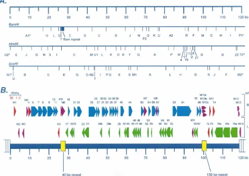
[image:3.612.60.556.76.427.2]Of the 80 ORFs identified in the
g
HV68 genome, 63 are
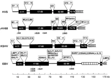
FIG. 1. Organization and ORF analysis of thegHV68 genome. (A) Predicted BamHI, EcoRI, and HindIII restriction endonuclease cleavage maps of thegHV68 genome based on the complete nucleic acid sequence. With the exception of several small fragments (BamHI U, HindIII a, and EcoRI U), the predicted maps closely match the published analysis of Efstathiou et al. (25). Asterisks indicate fragments at the ends of the genome map which have an undefined size due to the presence of a variable number of terminal repeats. (B) ORFs and tRNA-like genes present in thegHV68 genome. ThegHV68 genome is composed of a 118,237-bp unique region (shown as the thick blue line) flanked by multiple copies of a 1,213-bp terminal repeat (depicted as open rectangles at the ends of the genome). In addition, there are two internal repeats within the unique region, which are shown in yellow. The criteria for selecting candidate ORFs are described in the text. Those ORFs with substantial homology to genes present in the HVS and KSHV genomes were assigned the corresponding HVS/KSHV gene number. Rightward ORFs of HVS homologs are depicted in blue, while leftward ORFs of HVS homologs are depicted in green (with the exception of those which are homologs of known cellular genes). gHV68 ORFs with no obvious homology to any other gammaherpesvirus genes were designated M ORFs (M1 to M14).gHV68 ORFs which are homologs of known cellular genes or of genes in viruses other than HVS, KSHV, and EBV are depicted in red. Those M ORFs which do not exhibit significant homology to any known cellular or viral gene products are indicated in purple. Also shown clustered near the left end of the unique region are eight potential tRNA-like genes (see Table 2 for details).on November 9, 2019 by guest
http://jvi.asm.org/
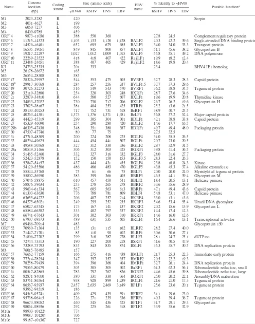
TABLE 1. Putative
g
HV68 gene products and homologs present in other gammaherpesviruses
Name Genomelocation (bp)
Coding strand
Size (amino acids) EBV name
% Identity togHV68
Possible functiona
gHV68 KSHV HVS EBV KSHV HVS EBV
M1 2023–3282 R 420 Serpin
M2 4031–4627 L 199 M3 6060–7277 L 406 M4 8409–9785 R 459
ORF 4 9873–11036 R 388 550 360 27.8 24.5 Complement regulatory protein ORF 6 11215–14523 R 1,103 1,133 1,128 1,128 BALF2 40.3 42.2 39.6 Single-stranded DNA binding protein ORF 7 14526–16481 R 652 695 679 685 BALF3 34.0 34.0 33.3 Transport protein
ORF 8 16505–19051 R 849 845 808 857 BALF4 51.1 45.6 38.2 Glycoprotein B ORF 9 19217–22297 R 1,027 1,012 1,009 1,015 BALF5 55.8 55.1 49.2 DNA polymerase ORF 10 22269–23522 R 418 418 407 422 RajiLF1 19.9 18.2 12.4
ORF 11 23488–24651 R 388 407 405 429 RajiLF2 18.6 19.8 20.4
K3 24733–25335 L 201 333 23.4 BHV4 IE1 homolog
M5 26178–26672 R 165 M6 26554–28308 R 585
ORF 17 28326–29957 L 544 553 475 605 BVRF3 32.7 28.3 28.3 Capsid protein ORF 18b 29917–30768 R 284 257 256 247 BVLF1.5 37.7 37.3 29.6
ORF 19 30726–32273 L 516 549 543 570 BVRF1 36.2 38.8 34.5 Tegument protein ORF 20 32119–32880 L 254 320 303 248 BXRF1 28.7 27.6 34.6
ORF 21 32879–34810 R 644 580 527 607 BXLF1 19.6 19.9 20.8 Thymidine kinase ORF 22 34833–37022 R 730 730 717 706 BXLF2 26.7 26.2 19.6 Glycoprotein H ORF 23 37025–38167 L 381 404 253 425 BTRF1 25.2 13.6 21.5
ORF 24 38103–40253 L 717 752 731 618 BcRF1 38.9 40.7 25.5
ORF 25 40263–44381 R 1,373 1,376 1,371 1,381 BcLF1 56.8 57.2 52.4 Major capsid protein ORF 26 44423–45319 R 299 305 304 301 BDLF1 42.1 38.8 33.8 Capsid protein ORF 27 45329–46090 R 254 290 280 420 BDLF2 14.6 17.7 16.5
ORF 29b 46395–47438 L 348 351 380 387 BDRF1 49.7 49.4 48.0 Packaging protein ORF 30 47507–47746 R 80 77 75 27.5 32.5
ORF 31 47710–48309 R 200 224 208 225 BDLF4 31.0 35.5 26.5 ORF 32 48294–49625 R 444 454 441 507 BGLF1 20.7 23.0 20.5 ORF 33 49588–50568 R 327 312 330 336 BGLF2 29.7 32.9 31.5
ORF 29a 50549–51466 L 306 312 303 325 BGRF1 39.8 41.4 36.5 Packaging protein ORF 34 51465–52460 R 332 327 316 332 BGLF3 28.0 31.6 27.7
ORF 35 52423–52878 R 152 150 150 153 BGLF3.5 28.3 22.4 26.3 ORF 36 52847–54157 R 437 444 431 455 BGLF4 23.8 18.8 24.3 Kinase
ORF 37 54129–55586 R 486 486 483 470 BGLF5 43.8 45.3 37.4 Alkaline exonuclease ORF 38 55544–55768 R 75 61 66 75 BBLF1 20.0 20.0 24.0 Myristylated tegument protein ORF 39 55802–56950 L 383 399 366 405 BBRF3 46.5 44.1 39.4 Glycoprotein M
ORF 40 57046–58875 R 610 457 450 541 BBLF2 18.4 20.5 13.1 Helicase-primase ORF 42 58876–59634 L 253 278 265 278 BBRF2 33.6 35.6 28.9
ORF 43 59634–61334 L 567 605 563 613 BBRF1 47.1 49.4 43.6 Capsid protein ORF 44 61303–63630 R 776 788 781 809 BBLF4 54.8 53.1 47.0 Helicase-primase ORF 45 63655–64272 L 206 407 257 214 BKRF4 33.0 13.1 13.6
ORF 46 64275–65021 L 249 255 252 255 BKRF3 54.6 53.4 55.4 Uracil DNA glycosylase ORF 47 65027–65545 L 173 167 141 137 BKRF2 20.2 15.6 13.9 Glycoprotein L ORF 48 65584–66582 L 333 402 797 537 BKRF2 14.4 17.4 12.3
ORF 49 66741–67643 L 301 302 303 310 BRRF1 14.6 16.0 12.6
ORF 50 67907–69373 R 489 631 535 605 BRLF1 16.4 26.6 13.1 Transcriptional activator
M7 69466–70914 R 483 Glycoprotein 150
ORF 52 70960–71364 L 135 131 115 162 BLRF2 28.2 27.4 40.0 ORF 53 71447–71701 L 85 110 90 102 BLRF1 30.6 30.6 27.1
ORF 54 71806–72702 R 299 318 287 278 BLLF3 28.1 24.7 25.1 dUTPase ORF 55 72744–73313 L 190 227 200 218 BSRF1 41.6 40.5 47.9
ORF 56 73289–75793 R 835 843 835 874 BSLF1 35.3 35.7 30.5 DNA replication protein M8 76015–76485 R 157
ORF 57 76662–77159 R 166 275 416 438 BMLF1 21.7 25.3 22.3 Immediate-early protein ORF 58 77214–78254 L 347 357 357 357 BMRF2 20.5 22.2 19.3
ORF 59 78258–79439 L 394 396 368 404 BMRF1 32.7 26.1 24.4 DNA replication protein ORF 60 79565–80479 L 305 305 305 302 BaRF1 61.3 62.3 56.1 Ribonucleotide reductase, small ORF 61 80517–82865 L 783 792 767 826 BORF2 44.6 45.6 39.8 Ribonucleotide reductase, large ORF 62 82871–84010 L 380 331 330 364 BORF1 25.0 20.2 22.1 Assembly/DNA maturation ORF 63 83751–86564 R 938 928 899 1,239 BOLF1 21.6 21.0 17.3 Tegument protein ORF 64 86567–93937 R 2,457 2,635 2,469 3,149 BPLF1 25.6 23.6 20.1 Tegument protein M9 93962–94519 L 186
ORF 66 94515–95741 L 409 429 435 591 BFRF2 31.1 29.6 23.0
ORF 67 95738–96415 L 226 271 235 336 BFRF1 40.3 39.4 36.7 Tegument protein ORF 68 96673–98052 R 460 545 436 525 BFLF1 31.7 29.1 29.3 Glycoprotein ORF 69 98061–98936 R 292 225 261 318 BFLF2 33.9 35.6 32.9
M10a 98903–101224 R 774 M10b 99087–101204 R 706 M10c 99187–101367 R 727
Continued on following page
on November 9, 2019 by guest
http://jvi.asm.org/
homologs of HVS genes, all of which are also present in the
KSHV genome and many of which are present in the EBV
genome (Table 1). One ORF, K3, appears to be present in the
KSHV and
g
HV68 genomes but not in the HVS or EBV
genome. The overall identity of
g
HV68 ORFs to their
ho-mologs in the other gammaherpesviruses indicates that
g
HV68
is more closely related to HVS and KSHV than to EBV (Table
1). However, at the level of individual ORFs, there is
consid-erable variability in the extent of homology, and there are
several ORFs for which the putative
g
HV68 ORF product is
most closely related to the EBV gene product (see below). It
should be noted that we assigned
g
HV68 genes as homologs of
other gammaherpesvirus genes only when BLASTP analyses
identified regions of homology (see Materials and Methods).
Thus, for example, the
g
HV68 M9 ORF may be the homolog
of HVS and KSHV ORF 65 and/or EBV BFRF3, but none of
these gene products were sufficiently homologous to the
puta-tive
g
HV68 M9 gene product to score in the BLASTP search.
There was generally good size conservation between
g
HV68
ORFs and those in KSHV, HVS, and EBV (Table 1). One
exception is ORF 57 of
g
HV68, which is significantly shorter
than the KSHV, HVS, and EBV homologs. On further
analy-sis, we found that ORF 57 is open upstream of the ATG at
position 76662, which was used to calculate the size of the
putative ORF 57 protein (Table 1). A BLASTP search of the
ORF upstream of position 76662 revealed significant
homol-ogy to HVS gene 57. In addition, alignment of this upstream
region demonstrated significant homology with portions of the
HVS and KSHV gene 57 proteins and the EBV BMLF1 gene.
This finding suggests that
g
HV68 ORF 57 may be a spliced
gene, with the ATG-initiated ORF in Table 1 present as the
C-terminal portion of a longer ORF 57 product.
It is also notable that there are three copies of ORF 75,
which encodes the enzyme N-formylglycinamide ribotide
amidotransferase. Interestingly, HVS also contains an
addi-tional copy of this gene (ORF 3). However, the other
charac-terized gammaherpesviruses appear to encode only a single
copy of ORF 75. The significance of multiple copies of this
gene in
g
HV68 and HVS in the biology of these viruses is not
readily apparent.
In general, the
g
HV68 ORFs are very closely spaced on the
genome (Fig. 1 and Table 1). The exception to this is near the
left end of the unique region, where the presence of ORFs
which meet the criteria described above is relatively sparse. As
discussed below, this may be notable since among the
gamma-herpesviruses, this region is very divergent and, in the cases of
EBV and HVS, is known to encode transformation-associated
gene products (6, 21, 32, 37, 39, 47, 50). At the left end of the
g
HV68 genome, five short (40- to 104-bp) sequences with
significant homology to multiple (at least three different)
bac-terial tRNAs were detected by a BLASTN search of bp 1 to
10000 of the genome (Fig. 1). Additional sequences with some
homology to a single bacterial tRNA were also found in this
region. BLASTN searches of the remainder of the genome did
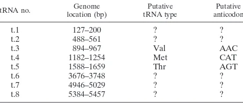
not reveal any additional tRNA-like sequences. The first
10,000 bp of the genome was further evaluated by using the
tRNAscan-SE program (see Materials and Methods), which
predicted the presence of eight tRNA-like genes (Table 2).
The role of these putative tRNA-like molecules in the
g
HV68
life cycle is not clear, and it is possible that they do not function
as tRNAs since five of the eight tRNA-like genes lack clear
anticodons (Table 2). Interestingly, the putative tRNAs are
encoded in a region positionally homologous to the region
encoding the HVS U-RNAs (2, 3, 31, 50).
[image:5.612.317.555.626.727.2]Three of the
g
HV68-specific ORFs (M12, M13, and M14)
are partially or entirely encoded within the terminal repeat
(below). The significance of this remains to be determined,
since the high G
1
C content of the terminal repeats may
pre-dict the presence of ORFs which are not actually transcribed.
Similarly, three overlapping ORFs (M10a, M10b, and M10c)
which are largely encoded within the 100-bp repeat were
de-tected. These share a common repetitive sequence encoded by
the 100-bp repeat but are predicted to contain unique carboxy
termini. In addition, M10a is predicted to have a unique
ami-no-terminal sequence (compared to ORFs M10b and M10c)
since this ORF initiates 78 bp to the left of the 100-bp repeat.
ORF M6 spans the 40-bp repeat and is predicted to have a
repetitive amino acid sequence.
TABLE 1—Continued
Name Genomelocation (bp)
Coding strand
Size (amino acids) EBV name
% Identity togHV68
Possible functiona
gHV68 KSHV HVS EBV KSHV HVS EBV
ORF 72 102426–103181 L 252 257 254 25.0 21.0 Cyclin D homolog M11 103418–103930 R 171 175 160 191 BHRF1 11.7 11.7 24.8 Bcl-2 homolog (gene 16?) ORF 73 103927–104868 L 314 1,162 407 24.2 7.3 Immediate-early protein ORF 74 105057–106067 R 337 342 321 23.1 22.8 GCR (IL-8 receptor homolog?) ORF 75c 106070–109999 L 1,310 1,296 1,299 1,318 BNRF1 24.7 23.4 22.4 Tegument protein/FGARATc
ORF 75b 110077–113901 L 1,275 1,296 1,299 1,318 BNRF1 22.6 22.5 22.3 Tegument protein/FGARAT ORF 75a 114032–117904 L 1,291 1,296 1,299 1,319 BNRF1 22.7 22.5 22.2 Tegument protein/FGARAT M12 117992–118681 R 212
M13 118149–118784 R 230 M14 118808–119125 L 106
aFunctions of individual viral genes were based on information summarized in references 3, 6, 61, and 70.
bThere is significant homology betweengHV68, KSHV, and HVS ORF 18 sequences and two overlapping reading frames in the EBV genome. The putative
BVLF1.5 gene joins these ORFs based on elimination of a hypothetical frameshift as previously suggested (3).
cFGARAT, N-formylglycinamide ribotide amidotransferase.
TABLE 2. Putative tRNA-like genes encoded by
g
HV68
tRNA no. location (bp)Genome tRNA typePutative anticodonPutative
t.1
127–200
?
?
t.2
488–561
?
?
t.3
894–967
Val
AAC
t.4
1182–1254
Met
CAT
t.5
1588–1659
Thr
AGT
t.6
3676–3748
?
?
t.7
4946–5029
?
?
t.8
5384–5457
?
?
on November 9, 2019 by guest
http://jvi.asm.org/
Analysis of repeats in the
g
HV68 genome.
In addition to the
terminal repeat, the
g
HV68 genome contains two internal
re-peats (Fig. 2), a 40-bp repeat located between bp 26778 and
28191 in the genome and a 100-bp repeat located between bp
98981 and 101170 in the genome. Both repeats are very G
1
C
rich; the 100-bp repeat is 86% G
1
C, while the 40-bp repeat is
83% G
1
C.
(i) The 100-bp repeat.
The region of the 100-bp repeat (Fig.
2A) was constructed by combining the following data: (i)
se-quences of multiple M13 clones spanning the left and right
junctions between the 100-bp repeats and unique sequence, (ii)
sequencing a series of exonuclease III-generated nested
dele-tions spanning the 100-bp repeat region (see Materials and
Methods), and (iii) estimation of the size of the repeat region
(cloned from genomic DNA) by restriction endonuclease
di-gestion. These multiple pieces of data were required because
the highly repetitive nature and size of the region prevented us
from obtaining unambiguous sequence from M13 clones alone.
This was due to our inability to unambiguously anchor
se-quences initiating within the 100-bp repeat in the contig. The
100-bp repeat is predicted to span from bp 98981 to 101170
and to contain 21 complete copies of the 100-mer repeat shown
in Fig. 2A and a 90-bp partial repeat at the right end of the
100-bp repeat region.
(ii) The 40-bp repeat.
The region of the 40-bp repeat was
constructed by using the following data: (i) sequences of
mul-tiple M13 clones spanning the left and right borders between
the 40-bp repeats and unique sequence and (ii) estimation of
the size of the repeat region (derived from genomic DNA) by
restriction endonuclease digestion analyses. We failed to stably
clone the 40-bp repeat region; numerous clones were
recov-ered, all of which had spontaneously deleted portions of the
repeat. M13 clones spanning the left junction between unique
sequence and the repeat region entered into a series of 40-bp
repeats each containing a BamHI site (type A repeats [Fig.
2B]). The first A repeat at the left-hand end is a partial repeat
containing the last 27 bp of the type A repeat. M13 clones
spanning the right junction between unique sequence and the
repeat region spanned several repeats that were similar in
structure to the A repeats (B, C, D, and E repeats [Fig. 2B])
prior to entering A repeats. The rightmost two repeats (type E
repeats) are 41-mers that lack the internal BamHI site. There
is also a slightly longer 51-bp variant (type D repeat) which
contains the internal BamHI site and two other 41-mer
vari-ants (type B and C repeats) which also contain the internal
BamHI site. We concluded that the majority of the repeat is
composed of an estimated 27 copies of the 40-mer A repeat
(Fig. 2B) based on the fact that M13 clones from both right and
left junctions between unique sequence and repeat sequence
end with 40-bp repeat A sequences. It is possible that other
sequences that could not be detected by our analysis are
present centrally within the 40-bp repeat sequence. It is
per-haps notable that all variant repeats which were detected map
to the right end of the repeat, suggesting that maintaining the
integrity of the central and left-hand portions of this repeat
region may be functionally important.
(iii) The terminal repeat.
The terminal repeat was
se-quenced multiple times from M13 clones spanning the 5
9
and
3
9
junctions between unique sequence and the terminal
re-peats. A single copy of the 1,213-bp terminal repeat is attached
to the right end of the
g
HV68 unique sequence submitted to
GenBank. It should be noted that the beginning of the
con-sensus terminal repeat unit was defined by the border between
the terminal repeat and nucleotide 1 of the genome. This
assignment, based on the sequences of M13 clones spanning
the right-hand junction between unique sequence and the
ter-minal repeat, results in a 26-bp portion of the terter-minal repeat
at the right-hand end of the unique region of the
g
HV68
genome before the first intact terminal repeat. As discussed
above, the terminal repeat encodes a putative polypeptide
(M14) which would be initiated from one of two potential
methionine initiation codons present in this very G
1
C rich
sequence, thus generating an ORF which is represented many
times in the intact viral genome. In addition, two ORFs (M12
and M13) begin outside the terminal repeat but extend into the
terminal repeat. We also evaluated the left end of the genome
attached to the terminal repeat but found no ORFs meeting
our criteria (see above). The significance of these putative
ORFs that include G
1
C-rich terminal repeat sequences is not
known.
Comparison of genome organization among
gammaherpes-viruses.
The genomes of herpesviruses tend to contain blocks
of conserved genes interspersed with blocks of genes specific to
a viral family or a specific virus (17). In agreement with the
close structural homology between the
g
HV68 genome and the
genomes of KSHV, HVS, and EBV, phylogenetic analyses of
the predicated DNA polymerase and major capsid proteins
supported the assignment of
g
HV68 as a gammaherpesvirus
(data not shown) (17, 38). To further assess the relationship
between
g
HV68 and primate gammaherpesviruses, we
com-pared the gene organizations of HVS, KSHV,
g
HV68, and
EBV genomes. This analysis revealed a strong conservation of
specific blocks of genes, separated by genes that appear to be
largely virus specific (Fig. 3). None of the EBV
latency-asso-ciated genes (i.e., the EBNA or LMP genes) appears to be
con-served among the gammaherpesviruses. In addition, the
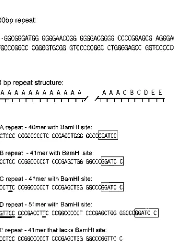
trans-FIG. 2. Sequence and structure of the internal repeats present in thegHV68genome. (A) Sequence of the 100-bp repeat (only the top strand is shown). Analysis of the 100-bp repeat region indicates that the repeat is made up of 21 intact copies of the repeat and a partial copy of the repeat at the right-hand end which is lacking the last 10 bp of the repeat. (B) Organization and sequence of the 40-bp repeat. The repeat is estimated to contain 27 complete copies of the A repeat linked to several degenerate forms of the repeat located at the right-hand end of the repeat (type B to E repeats), as illustrated. The sequences of each type of repeat is shown below the schematic diagram of the repeat structure. Residues which differ from the type A repeat are underlined, and the presence of a BamHI
site is indicated by the open rectangle.
on November 9, 2019 by guest
http://jvi.asm.org/
[image:6.612.91.265.65.307.2]formation-associated HVS gene products STP and TIP do not
appear to have homologs in the other gammaherpesviruses.
This observation raises the possibility that many of the
latency-and transformation-associated genes in
g
HV68 and KSHV are
encoded by virus-specific genes. In addition, it suggests that
while functions required for lytic replication of these viruses
have generally been well conserved, strategies for latency have
diverged significantly. This may reflect both cell-type- and
species-specific constraints. Alternatively, it is possible that the ability of
gammaherpesviruses to establish a latent infection has
inde-pendently evolved in each of these viruses. The latter
possibil-ity seems unlikely given that the abilpossibil-ity to establish a latent
infection is a hallmark of all characterized herpesviruses.
Homologs of cellular or viral genes potentially involved in
pathogenesis, latency, or tumor induction.
It is notable that
g
HV68 contains homologs of several viral and host genes that
may be important for gammaherpesvirus pathogenesis (Fig. 4).
Similar to HVS and KSHV,
g
HV68 contains an ORF (ORF 4)
with significant homology to multiple complement regulatory
proteins (3, 42, 61). The presence of four regions within
g
HV68 ORF 4 with homology to complement control protein
repeats or short consensus repeats results in significant
BLASTP alignments with many complement regulatory
pro-teins (42). Notably,
g
HV68 ORF 4 aligns particularly well over
the entire length of the predicted protein with murine
decay-accelerating factor (DAF) and human membrane cofactor
pro-tein (MCP) (Fig. 4A). Both DAF and MCP regulate C3
con-vertase (although by different mechanisms), suggesting that
regulation of C3 convertase is a potential action of ORF 4 of
g
HV68. This hypothesis is consistent with studies of the HVS
gene 4 product, which inhibits C3-mediated lysis (29).
ORF 72 of
g
HV68 is predicted to encode a D-type cyclin.
The fact that KSHV, HVS, and
g
HV68 encode cyclin
ho-mologs (13, 16, 36, 41, 51), combined with the fact that EBV
regulates cellular cyclin D expression (62), strongly argues for
a conserved role of this gene product in gammaherpesvirus
biology. An alignment of the
g
HV68 ORF 72 with mouse
cyclin D1, KSHV v-cyclin, and HVS v-cyclin is shown in Fig.
4B. The proteins all share conserved residues (e.g., the
g
HV68
TWM sequence at position 62 in the alignment) within the
cyclin box. A mutagenesis study of this region of the HVS
v-cyclin showed that several residues conserved between
[image:7.612.124.494.68.330.2]g
HV68 ORF 72 and the HVS v-cyclin are involved in binding
to cyclin-dependent kinase 6 (cdk6) (36). In addition, the
FIG. 3. Comparison of genome organization among gammaherpesviruses. The structures of the indicated viruses were compared to the HVS genome structure. The conserved blocks of genes are indicated in the shaded rectangles, and the numbers correspond to the HVS gene numbers. Genes missing from particular blocks are indicated below each block (specific genes which are missing are indicated with aD), while genes inserted within a particular block of genes are indicated above each block. The terminal repeats located at the ends of the gammaherpesvirus genomes are shown only at the right-hand end of the respective genomes. The major internal repeat of EBV is shown near the right-hand end of the genome as a series of connected open rectangles. ORFs designated with an S are largely unique to HVS; ORFs designated with an M are largely unique togHV68; ORFs designated with a K are largely unique to KSHV.FIG. 4. Alignments of fivegHV68 ORFs with viral or host homologs. All alignments were performed with MegAlign (DNASTAR) and are presented without reediting after the initial alignment was performed. Residues shown in green are chemically related, while red indicates the presence of identical residues among some of the sequences (at some positions both red and green highlighted residues are shown, which indicates that all of the colored residues are chemically related and some of them are identical). Blue denotes those residues which are identical in all the aligned protein sequences. For each of the alignments described below, the number in parentheses is the GenBank or SWISS-PROT accession number (GB or SP acc.). All HVS, KSHV, and EBV sequences were derived from the original publications (3, 6, 61). (A) Alignment of mouse DAF (GB acc. L41366), human MCP (SP acc. P15529), andgHV68 ORF 4. (B) Alignment of murine cyclin D1 (GB acc. M64403), gHV68 ORF 72, KSHV gene 72, and HVS gene 72. (C) Alignment of mouse bcl-2 (GB acc. M16506),gHV68 ORF M11, and EBV BHRF1. (D) Alignment ofgHV68 ORF M1, rabbitpox SPI-1 (SP acc. P42928), and cowpox crmA (GB acc. M14217). (E) Alignment of the mouse IL-8 receptor (SP acc. P35343),gHV68 ORF 74, KSHV gene 74, and HVS gene 74.
on November 9, 2019 by guest
http://jvi.asm.org/
on November 9, 2019 by guest
http://jvi.asm.org/
KSHV v-cyclin associates with cdk6 and to a lesser extent with
cdk4 (41). These data suggest that the
g
HV68 v-cyclin may also
interact with specific cyclin-dependent kinases. Interestingly,
we have not found a clear homolog of the LxCxE motif found
to be important for retinoblastoma protein binding to cyclin D
(22, 27). Notably, this motif is also lacking in the HVS and
KSHV v-cyclins, raising the possibility that the v-cyclins do not
interact with the retinoblastoma protein. Alternatively, it is
possible that additional exons are spliced to the viral ORF 72
to form a protein containing the LxCxE motif (or other
se-quences). The fact that KSHV and HVS v-cyclins are
ex-pressed in Kaposi’s sarcoma tissues (13) and transformed cell
lines (36), respectively, raises the possibility that the
g
HV68
v-cyclin is involved in oncogenesis or latency. However, a role
in lytic infection has not been ruled out by studies to date of
the HVS and KSHV v-cyclins.
g
HV68 ORF M11 has weak overall homology to bcl-2 family
members and encodes a protein with 24% amino acid identity
with the EBV BHRF1 gene product (Fig. 4C). The latter has
been shown to inhibit apoptosis in several systems (33, 69).
Similarly, the KSHV bcl-2 homolog (gene 16) inhibits
apopto-sis (14). We designated this ORF M11 rather than calling it
either ORF 16 (as a homolog of the HVS and KSHV gene 16)
or BHRF1, since the level of homology is weak enough that the
proteins could have distinct functions (Fig. 4C). It is clear from
a survey of the known viral Bcl-2 homologs that there is only
limited sequence conservation between these proteins and the
cellular counterparts. Thus, it is perhaps not surprising that the
viral bcl-2 genes may have also diverged significantly from each
other. The greatest local homology between Bcl-2 family
mem-bers and the
g
HV68 M11 ORF product is within the box 1
motif involved in dimerization of Bcl-2 family members (data
not shown). This raises the possibility that
g
HV68 M11
func-tions by interacting with itself or host Bcl-2 family members,
although this type of association may not be essential for the
KSHV gene 16 product to inhibit apoptosis (14).
It is notable that the
g
HV68 M11 ORF (potential bcl-2
homolog) is located in the same position in the genome as the
EBV bcl-2 homolog (BHRF1 gene). Thus, EBV and
g
HV68
have a common arrangement of their bcl-2 homologs. Notably,
while the KSHV bcl-2 homolog (gene 16) is located in the same
place as the HVS gene 16, the KSHV bcl-2 homolog is adjacent
to an HVS gene 70 homolog (Fig. 3). This finding suggests that
in an ancestral gammaherpesvirus, the bcl-2 homolog was
lo-cated near the right-hand end of the unique region (near gene
70). Rearrangement of this ancestral genome could have
moved the bcl-2 homolog to its current location in the HVS
and KSHV genomes. In the case of KSHV, this speculative
rearrangement included gene 70. This argues for placement of
the ancestral bcl-2 homolog in the same position as the
g
HV68
M11 and EBV BHRF1 genes.
g
HV68 also contains an ORF (M1) which is predicted to
encode a protein homologous to several poxvirus serpins (Fig.
4D). This ORF product is the first reported serpin homolog
encoded by a herpesvirus. The fact that similar ORFs have not
been found in HVS, KSHV, and EBV suggests that this ORF
may provide a virus- or host-specific function unique to
g
HV68.
Significant homology was found between
g
HV68 M1 and the
SPI-1 genes of rabbitpox, cowpox, vaccinia, and variola viruses
(Fig. 4D) (4). Interestingly, the poxvirus SPI-1 gene can
regu-late host cell range by altering apoptosis (9). There is also
significant homology between the
g
HV68 M1 ORF and crmA
(Fig. 4D) (59). This raises the possibility that the
g
HV68 serpin
may alter the function of interleukin 1 (IL-1)-converting
en-zyme-like proteases. It is notable that poxvirus serpins have
been implicated in a broad range of processes in addition to
regulating apoptosis, including regulation of inflammatory
re-sponses and altering arachidonate (4, 9, 18, 44, 52, 53, 56, 59,
72). The specific role of viral serpins may depend on the
en-zyme(s) targeted (43) and the host pathogenesis model
evalu-ated (71).
g
HV68 contains an ORF (ORF 74) homologous to genes
encoding G-protein-coupled receptors (GCRs). Similar to
gene 74 in both HVS and KSHV, the
g
HV68 ORF 74 is most
homologous to the IL-8 receptor (Fig. 4E) (1, 3, 5, 51, 61).
Structure analysis demonstrates seven potential
transmem-brane domains (not shown). The HVS ORF 74 product has
been shown to be a functional IL-8 receptor in transfection
experiments (1). This finding suggests the possibility that host
cytokines signaling through the viral GCR could alter the virus
life cycle. In contrast, the KSHV gene 74 product is a
consti-tutively active GCR that can bind both
a
and
b
chemokines
and can provide a signal for cell growth (5). As the KSHV gene
has been demonstrated to be expressed in tumors (13), it is
possible that these receptors play a role in tumor induction or
maintenance of tumor growth. However, a role in latency or
lytic infection is also possible.
g
HV68 contains an ORF (K3) homologous to ORFs K3 and
K5 of KSHV as well as the bovine herpesvirus 4 major
imme-diate-early (BHV4 IE1) transcript (61, 75). The homology
be-tween KSHV K3, KSHV K5,
g
HV68 K3, and BHV4 IE1 is
local and is marked by complete conservation of a motif
CWIC-(10/11x)-CxCxxxxxxxHxxCxxxWxxxSxxxxCxxCxxxY.
In-terestingly, this motif is also present in a hypothetical swinepox
virus protein (C7; SWISS-PROT accession no. P32225) and is
well conserved in the amino-terminal portion of the
Caeno-rhabditis elegans F58E6.1 protein (EMBL accession no. Z70754).
This is an additional example (together with the serpin
ho-molog described above) of conservation between poxvirus and
herpesvirus genes. The importance of this motif is not known,
but it may represent a part of a zinc finger motif. Conservation
of this motif across multiple viruses and C. elegans suggests an
important role in the function of these proteins.
Summary.
The complete nucleotide sequence of the
g
HV68
genome demonstrates that this murine herpesvirus is a
gam-maherpesvirus with similarities to HVS, KSHV, and EBV. The
presence of cyclin, GCR, serpin, Bcl-2, and DAF/MCP
ho-mologs provides an opportunity to evaluate the roles of the
genes encoding these proteins in viral pathogenesis. In
addi-tion, the identification of a number of apparently virus-specific
ORFs in the
g
HV68 genome offers the opportunity to assess
their contribution to
g
HV68 pathogenesis, latency, and tumor
induction in a tractable small-animal model.
ACKNOWLEDGMENTS
This work was partially supported by grants to H.W.V. from the NIH
(AI39615), the Mallinckrodt Foundation, and the Council for Tobacco
Research and by American Cancer Society Junior Faculty Research
Award JFRA-525. K.E.W. was supported by NIH grant K08
AI01279-01AI. S.H.S. was supported by NIH grants CA43143, CA52004, and
CA58524.
We thank Dave O’Brien and Alicia Gibson for their work producing
the
g
HV68 sequence.
REFERENCES
1. Ahuja, S. K., and P. M. Murphy. 1993. Molecular piracy of mammalian interleukin-8 receptor type B by herpesvirus saimiri. J. Biol. Chem. 268: 20691–20694.
2. Albrecht, J.-C., and B. Fleckenstein. 1992. Nucleotide sequence of HSUR6 and HSUR7, two small RNAs of herpesvirus saimiri. Nucleic Acids Res.
20:1810.
3. Albrecht, J.-C., J. Nicholas, D. Biller, K. R. Cameron, B. Biesinger, C.
Newman, S. Wittmann, M. A. Craxton, H. Coleman, B. Fleckenstein, and R. W. Honess.1992. Primary structure of the herpesvirus saimiri genome. J. Virol. 66:5047–5058.
on November 9, 2019 by guest
http://jvi.asm.org/
4. Ali, A. N., P. C. Turner, M. A. Brooks, and R. W. Moyer. 1994. The SPI-1 gene of rabbitpox virus determines host range and is required for hemorragic pock formation. Virology 202:305–314.
5. Arvanitakis, L., E. Geras-Raaka, A. Varma, M. C. Gershengorn, and E.
Cesarman.1997. Human herpesvirus KSHV encodes a constitutively active G-protein-coupled receptor linked to cell proliferation. Nature 385:347–350. 6. Baer, R., A. T. Bankier, M. D. Biggin, P. L. Deininger, P. J. Farrell, T. J.
Gibson, G. Hatfull, G. S. Hudson, S. C. Satchwell, C. Seguin, P. S. Tuffnell, and B. G. Barrell.1984. DNA sequence and expression of the B95-8 Epstein-Barr virus genome. Nature 310:207–209.
7. Blaskovic, D., M. Stancekova, J. Svobodova, and J. Mistrikova. 1980. Isola-tion of five strains of herpesviruses from two species of free living small rodents. Acta Virol. 24:468.
8. Blaskovic, D., D. Stanekova, and J. Rajcani. 1984. Experimental pathogen-esis of murine herpesvirus in newborn mice. Acta Virol. 28:225–231. 9. Brooks, M. A., A. N. Ali, P. C. Turner, and R. W. Moyer. 1995. A rabbitpox
virus serpin gene controls host range by inhibiting apoptosis in restrictive cells. J. Virol. 69:7688–7698.
10. Cardin, R. D., J. W. Brooks, S. R. Sarawar, and P. C. Doherty. 1996. Progressive loss of CD81T cell-mediated control of a gamma-herpesvirus in the absence of CD41T cells. J. Exp. Med. 184:863–871.
11. Cavanaugh, V. J., R. M. Stenberg, T. L. Staley, H. W. Virgin, M. R.
Mac-Donald, S. Paetzold, H. E. Farrell, W. D. Rawlinson, and A. E. Campbell.
1996. Murine cytomegalovirus with a deletion spanning HindIII-J and -I displays altered cell and tissue tropism. J. Virol. 70:1365–1374.
12. Cesarman, E., Y. Chang, P. S. Moore, J. W. Said, and D. M. Knowles. 1995. Kaposi’s sarcoma-associated herpesvirus-like DNA sequences in AIDS-re-lated body-cavity-based lymphomas. N. Engl. J. Med. 332:1186–1191. 13. Cesarman, E., R. G. Nador, F. Bai, R. A. Bohenzky, J. J. Russo, P. S. Moore,
Y. Chang, and D. M. Knowles.1996. Kaposi’s sarcoma-associated herpesvi-rus contains G protein-coupled receptor and cyclin D homologs which are expressed in Kaposi’s sarcoma and malignant lymphoma. J. Virol. 70:8218– 8223.
14. Chang, E. H.-Y., J. Nicholas, D. S. Bellows, G. S. Hayward, H.-G. Guo, M. S.
Reitz, and J. M. Hardwick.1997. A bcl-2 homolog encoded by Kaposi’s sarcoma-associated virus, human herpesvirus 8, inhibits apoptosis but does not heterodimerize with Bax or Bak. Proc. Natl. Acad. Sci. USA 94:690–694. 15. Chang, Y., E. Cesarman, M. S. Pessin, F. Lee, J. Culpepper, D. M. Knowles,
and P. S. Moore.1994. Identification of herpesvirus-like DNA sequences in AIDS-associated Kaposi’s sarcoma. Science 266:1865–1869.
16. Chang, Y., P. S. Moore, S. J. Talbot, C. H. Boshoff, T. Zarkowska, D.
Godden-Kent, H. Paterson, R. A. Weiss, and S. Mittnacht.1996. Cyclin encoded by KS herpesvirus. Nature 382:410.
17. Chee, M. S., S. B. Bankier, C. M. Bohni, R. C. Brown, T. Horsnell, C. A.
Hitchinson, T. Kouzarides, J. A. Martignetti, E. Preddie, S. C. Satchwell, P. Tomlinson, K. M. Weston, and B. G. Barrell.1990. Analysis of the protein-coding content of the sequence of human cytomegalovirus strain AD169. Curr. Top. Microbiol. Immunol. 154:125–169.
18. Chua, T. P., C. E. Smith, R. W. Reith, and J. D. Williamson. 1990. Inflam-matory responses and the generation of chemoattractant activity in cowpox virus-infected tissues. Immunology 69:202–208.
19. Dear, S., and R. Staden. 1991. A sequencing assembly and editing program for efficient management of large projects. Nucleic Acids Res. 19:3907–3911. 20. Decker, L. L., P. Shankar, G. Khan, R. B. Freeman, B. J. Dezube, J.
Lieber-man, and D. A. Thorley-Lawson.1996. The Kaposi’s sarcoma-associated herpesvirus (KSHV) is present as an intact latent genome in KS tissue but replicates in the peripheral blood mononuclear cells of KS patients. J. Exp. Med. 184:283–288.
21. Desrosiers, R. C., A. Bakker, J. Kamine, L. A. Falk, R. D. Hunt, and N. W.
King.1985. A region of the herpesvirus saimiri genome required for onco-genicity. Science 228:184–187.
22. Dowdy, S. F., P. W. Hinds, K. Louie, S. I. Reed, A. Arnold, and R. A.
Weinberg.1993. Physical interaction of the retinoblastoma protein with human D cyclins. Cell 73:499–511.
22a.Durbin, R. (MRC Laboratory of Molecular Biology, Cambridge, England). Unpublished data.
22b.Durbin, R. (MRC Laboratory of Molecular Biology, Cambridge, England)
and J. Thierry-Mieg (Centre de Recherche en Biochimie Macromoleculaire, CNRS, Montpellier, France).Unpublished data.
23. Eddy, S. R., and R. Durbin. 1994. RNA sequence analysis using covariance models. Nucleic Acids Res. 22:2079–2088.
24. Efstathiou, S., Y. M. Ho, S. Hall, C. J. Styles, S. D. Scott, and U. A. Gompels. 1990. Murine herpesvirus 68 is genetically related to the gammaherpesvi-ruses Epstein-Barr virus and herpesvirus saimiri. J. Gen. Virol. 71:1365– 1372.
25. Efstathiou, S., Y. M. Ho, and A. C. Minson. 1990. Cloning and molecular characterization of the murine herpesvirus 68 genome. J. Gen. Virol. 71: 1355–1364.
26. Ehtisham, S., N. P. Sunil-Chandra, and A. A. Nash. 1993. Pathogenesis of murine gammaherpesvirus infection in mice deficient in CD4 and CD8 T cells. J. Virol. 67:5247–5252.
27. Ewen, M. E., H. K. sluss, C. J. Sherr, H. Matsushime, J.-Y. Kato, and D. M.
Livingston.1993. Functional interactions of the retinoblastoma protein with mammalian D-type cyclins. Cell 73:487–497.
28. Fichant, G. A., and C. Burks. 1991. Identifying potential tRNA genes in genomic DNA sequences. J. Mol. Biol. 220:659–671.
29. Fodor, W. L., S. A. Rollins, S. Bianco-Caron, R. P. Rother, E. R. Guilmette,
W. V. Burton, J.-C. Albrecht, B. Fleckenstein, and S. P. Squinto.1995. The complement control protein homolog of herpesvirus saimiri regulates serum complement by inhibiting C3 convertase activity. J. Virol. 69:3889–3892. 30. Fulton, L. L., and R. K. Wilson. 1994. Variations on cycle sequencing.
BioTechniques 17:298–301.
31. Geck, P., S. A. Whitaker, M. M. Medveczky, T. J. Last, and P. G. Medveczky. 1994. Small RNA expression from the oncogenic region of a highly onco-genic strain of herpesvirus saimiri. Virus Genes 8:25–34.
32. Geck, P., S. A. Whitaker, M. M. Medveczky, and P. G. Medveczky. 1990. Expression of collagen like sequences by a tumor virus, herpesvirus saimiri. J. Virol. 64:3509–3515.
32a.Green, P. (University of Washington, Seattle). Unpublished data. 33. Henderson, S., D. Huen, M. Rowe, C. Dawson, G. Johnson, and A.
Rickin-son.1993. Epstein-Barr virus-coded BHRF1 protein, a viral homologue of Bcl-2, protects human B cells from programmed cell death. Proc. Natl. Acad. Sci. USA 90:8479–8483.
33a.Hillier, L. (The Genome Sequencing Center, Department of Genetics,
Wash-ington University School of Medicine).Unpublished data.
34. Huang, Y. Q., J. J. Li, M. H. Kaplan, B. Poiesz, E. Katabira, W. C. Zhang,
D. Feiner, and A. E. Friedman-Kien.1995. Human herpesvirus-like nucleic acid in various forms of Kaposi’s sarcoma. Lancet 345:759–761.
35. Ju, J., C. Ruan, C. W. Fuller, A. N. Glazer, and R. A. Mathies. 1995. Fluorescence energy transfer dye-labelled primers for DNA sequencing and analysis. Proc. Natl. Acad. Sci. USA 92:4347–4351.
36. Jung, J. U., M. Stager, and R. C. Desrosiers. 1994. Virus-encoded cyclin. Mol. Cell. Biol. 14:7235–7244.
37. Jung, J. U., J. J. Trimble, N. W. King, B. Biesinger, B. W. Fleckenstein, and
R. C. Desrosiers.1991. Identification of transforming genes of subgroup A and C strains of herpesvirus saimiri. Proc. Natl. Acad. Sci. USA 88:7051– 7055.
38. Karlin, S., E. S. Mocarski, and G. A. Schachtel. 1994. Molecular evolution of herpesviruses: genomic and protein sequence comparisons. J. Virol. 68: 1886–1902.
39. Kieff, E. 1996. Epstein-Barr virus and its replication, p. 2343–2396. In B. Fields, D. Knipe, and P. Howley (ed.), Fields’ virology. Lippincott-Raven, Philadelphia, Pa.
40. Li, J. J., Y. Q. Huang, C. J. Cockerell, and A. E. Friedman-Kien. 1996. Localization of human herpes-like virus type 8 in vascular endothelial cells and perivascular spindle-shaped cells of Kaposi’s sarcoma lesions by in situ hybridization. Am. J. Pathol. 148:1741–1748.
41. Li, M., H. Lee, D.-W. Yoon, J.-C. Albrecht, B. Fleckenstein, F. Neipel, and
J. U. Jung.1997. Kaposi’s sarcoma-associated herpesvirus encodes a func-tional cyclin. J. Virol. 71:1984–1991.
42. Liszewski, M. K., T. C. Farries, D. M. Lublin, I. Rooney, and J. P. Atkinson. 1996. Control of the complement system. Adv. Immunol. 61:201–283. 43. Lomas, D. A., D. L. Evans, C. Upton, G. McFadden, and R. W. Carrell. 1992.
Inhibition of plasmin, urokinase, tissue plasminogen activator, and CIs by a myxoma virus serine proteinase inhibitor. J. Biol. Chem. 268:516–521. 43a.Lowe, T., and S. R. Eddy (The Genome Sequencing Center, Department of
Genetics, Washington University School of Medicine).Unpublished data. 44. Macen, J. L., C. Upton, N. Nation, and G. McFadden. 1993. SERP-I, a serine
proteinase inhibitor encoded by myxoma virus, is a secreted glycoprotein that interferes with inflammation. Virology 195:348–363.
45. Mardis, E. R. 1994. High-throughput detergent extraction of M13 clones for fluorescent DNA sequencing. Nucleic Acids Res. 22:2173–2175.
45a.Marth, G., and L. Hillier (The Genome Sequencing Center, Department of
Genetics, Washington University School of Medicine).unpublished 46. Medveczky, M., P. Geck, J. L. Sullivan, D. Serbousek, J. Y. Djeu, and P. G.
Medveczky.1993. IL-2 independent growth and cytotoxicity of herpesvirus saimiri-infected human CD8 cells and involvement of two open reading frame sequences of the virus. Virology 196:402–412.
47. Medveczky, M. M., P. Geck, R. Vassallo, and P. G. Medveczky. 1993. Ex-pression of the collagen-like putative oncoprotein of herpesvirus saimiri in transformed T cells. Virus Genes 7:349–365.
48. Mistrikova, J., and D. Blaskovic. 1985. Ecology of the murine alphherpes-virus and its isolation from lungs of rodents in cell culture. Acta Virol.
29:312–317.
49. Moore, P. S., and Y. Chang. 1995. Detection of herpesvirus-like DNA se-quences in Kaposi’s sarcoma in patients with and without HIV infection. N. Engl. J. Med. 332:1181–1185.
50. Murthy, S. C. S., J. J. Trimble, and R. C. Desrosiers. 1989. Deletion mutants of herpesvirus saimiri define an open reading frame necessary for transfor-mation. J. Virol. 63:3307–3314.
51. Nicholas, J., K. R. Cameron, and R. W. Honess. 1992. Herpesvirus saimiri encodes homologs of G protein-coupled receptors and cyclins. Nature 355: 362–365.
52. Palumbo, G. J., W. C. Glasgow, and R. M. L. Buller. 1997. Poxvirus-induced
on November 9, 2019 by guest
http://jvi.asm.org/
alteration of arachidonate metabolism. Proc. Natl. Acad. Sci. USA 90:2020– 2024.
53. Palumbo, G. J., D. J. Pickup, T. N. Fredrickson, J. McIntyre, and R. M.
Buller.1989. Inhibition of inflammatory response is mediated by a 38-kDa protein of cowpoxvirus. Virology 172:262–273.
54. Pavesi, A., F. Conterio, A. Bolchi, G. Dieci, and S. Ottonello. 1994. Identi-fication of new eukaryotic tRNA genes in genomic DNA databases by using a multistep weight matrix analysis of transcription control regions. Nucleic Acids Res. 22:1247–1256.
55. Pepper, S. D., J. P. Stewart, J. R. Arrand, and M. Mackett. 1996. Murine gammaherpesvirus-68 encodes homologues of thymidine kinase and glyco-protein H: sequence, expression, and characterization of pyrimidine kinase activity. Virology 219:475–479.
56. Pickup, D. J., B. S. Ink, W. Hu, C. A. Ray, and W. K. Joklik. 1986. Hemor-rhage in lesions caused by cowpox virus is induced by a viral protein that is related to plasma protein inhibitors of serine proteases. Proc. Natl. Acad. Sci. USA 83:7698–7702.
57. Pollock, J. L., and H. W. Virgin. 1995. Latency, without persistence, of murine cytomegalovirus in spleen and kidney. J. Virol. 69:1762–1768. 58. Rajcani, J., D. Blaskovic, J. Svobodova, F. Ciampor, D. Huckova, and D.
Stanekova.1985. Pathogenesis of acute and persistent murine herpesvirus infection in mice. Acta Virol. 29:51–60.
59. Ray, C. A., R. A. Black, S. R. Kronheim, T. A. Greenstreet, P. R. Sleath, G. S.
Salvesen, and D. J. Pickup.1992. Viral inhibition of inflammation: cowpox virus encodes an inhibitor of the interleukin-1-beta converting enzyme. Cell
69:597–604.
60. Rickinson, A. B., and E. Kieff. 1996. Epstein-Barr virus, p. 2397–2446. In B. N. Fields, D. M. Knipe, and P. M. Howley (ed.), Virology. Lippincott-Raven, Philadelphia, Pa.
61. Russo, J. J., R. A. Bohenzky, M.-C. Chien, J. Chen, M. Yan, D. Maddalena,
J. P. Parry, D. Peruzzi, I. S. Edelman, Y. Chang, and P. S. Moore.1996. Nucleotide sequence of the Kaposi sarcoma-associated herpesvirus (HHV8). Proc. Natl. Acad. Sci. USA 93:14862–14867.
62. Sinclair, A. J., I. Palmero, A. Holder, G. Peters, and P. J. Farrell. 1995. Expression of cyclin D2 in Epstein-Barr virus positive Burkitt’s lymphoma cell lines is related to methylation status of the gene. J. Virol. 69:1292–1295. 63. Soulier, J., L. Grollet, E. Oksenhendler, P. Cacoub, D. Cazals-Hatem, P.
Babinet, M.-F. d’Agay, J.-P. Clauvel, M. Raphael, L. Degos, and F. Signaux.
1995. Kaposi’s sarcoma-associated herpesvirus-like DNA sequences in mul-ticentric Castleman’s disease. Blood 86:1276–1280.
64. Stewart, J. P., N. J. Janjua, S. D. Pepper, G. Bennion, M. Mackett, T. Allen,
A. A. Nash, and J. R. Arrand.1996. Identification and characterization of
murine gammaherpesvirus 68 gp150: a virion membrane glycoprotein. J. Vi-rol. 70:3528–3535.
65. Stewart, J. P., N. J. Janjua, N. P. Sunil-Chandra, A. A. Nash, and J. R.
Arrand.1994. Characterization of murine gammaherpesvirus 68 glycopro-tein B (gB) homolog: similarity to Epsglycopro-tein-Barr virus gB (gp110). J. Virol.
68:6496–6504.
66. Sunil-Chandra, N. P., J. Arno, J. Fazakerley, and A. A. Nash. 1994. Lym-phoproliferative disease in mice infected with murine gammaherpesvirus 68. Am. J. Pathol. 145:818–826.
67. Sunil-Chandra, N. P., S. Efstathiou, J. Arno, and A. A. Nash. 1992. Viro-logical and pathoViro-logical features of mice infected with murine gammaher-pesvirus 68. J. Gen. Virol. 73:2347–2356.
68. Sunil-Chandra, N. P., S. Efstathiou, and A. A. Nash. 1992. Murine gamma-herpesvirus 68 establishes a latent infection in mouse B lymphocytes in vivo. J. Gen. Virol. 73:3275–3279.
69. Tarodi, B., T. Subramanian, and G. Chinnadurai. 1994. Epstein-Barr virus BHRF1 protein protects against cell death induced by DNA-damaging agents and heterologous viral infection. Virol. 201:404–407.
70. Telford, E. A. R., M. S. Watson, H. C. Aird, J. Perry, and A. J. Davison. 1995. The DNA sequence of equine herpesvirus 2. J. Mol. Biol. 249:520–528. 71. Thompson, J. P., P. C. Turner, A. N. Ali, B. C. Crenshaw, and R. W. Moyer.
1993. The effects of serpin gene mutations on the distinctive pathobiology of cowpox and rabbitpox virus following intranasal inoculation of Balb/c mice. Virology 197:328–338.
72. Upton, C., J. L. Macen, D. S. Wishart, and G. McFadden. 1990. Myxoma virus and malignant rabbit fibroma virus encode a serpin-like protein impor-tant for virus virulence. Virology 179:618–631.
73. Usherwood, E. J., J. P. Stewart, and A. A. Nash. 1996. Characterization of tumor cell lines derived from murine gammaherpesvirus-68-infected mice. J. Virol. 70:6516–6518.
74. Usherwood, E. J., J. P. Stewart, K. Robertson, D. J. Allen, and A. A. Nash. 1996. Absence of splenic latency in murine gammaherpesvirus 68-infected B cell-deficient mice. J. Gen. Virol. 77:2819–2825.
75. Van Santen, V. L. 1991. Characterization of the bovine herpesvirus 4 major immediate-early transcript. J. Virol. 65:5211–5224.
76. Weck, K. E., M. L. Barkon, L. I. Yoo, S. H. Speck, and H. W. Virgin. 1996. Mature B cells are required for acute splenic infection, but not for estab-lishment of latency, by murine gammaherpesvirus 68. J. Virol. 70:6775–6780. 77. Wilson, R. K., and E. R. Mardis. Shotgun sequencing. In M. Cozza (ed.), Genomics: a laboratory manual, in press. Cold Spring Harbor Laboratory
Press, Cold Spring Harbor, N.Y.