Controlling the Burst Eect of a Drug by
Introducing Starch in the Structure of Magnetic
Polyurethane Microspheres Containing
Superparamagnetic Iron Oxide Nanoparticles
M. Mahmoudi
1;and S. Laurent
2Abstract. The aim of this research is to prepare superparamagnetic polyurethane microspheres using SPIONs. Theophylline was used as a drug and the various stoichiometric ratios of isocyanate/polyol were studied to assess their application as a targeting delivery system. Magnetic polyurethane microspheres containing superparamagnetic iron oxide nanoparticles loaded with theophylline were prepared by the water in oil in water emulsion technique. The ratio of hexamethylenediisocyanate and polyol ("-polycaprolactone and starch) was modied and the release of theophylline was determined for a period of 20 days. These microspheres were characterized by TEM, SEM, FTIR and magnetometry. The microscopy images show the morphological shape of the superparamagnetic microspheres with an average size of 5 m. The FTIR and the magnetometry conrmed the presence of superparamagnetic nanoparticles in the microspheres. The release of theophylline was studied and dosed by UV spectrophotometer. With the use of PCL with a low molecular weight or in the presence of starch in the structure of polyurethane, the burst eect of drug is decreased. Finally, a SEM study showed an important degradation of the microspheres after the release process. The use of starch as a polyol caused signicant improvement in burst eect of the superparamagnetic polyurethane microspheres.
Keywords: Superparamagnetic iron oxide nanoparticles; Polyurethane; Magnetic microspheres; Theo-phylline.
INTRODUCTION
Polyurethane, which is a biodegradable polymer, has been employed extensively in various biomedical appli-cations, such as tissue engineering (e.g. skin, cardiovas-cular and bone), ureteral stents, meniscal reconstruc-tion and drug delivery [1-9]. A distinguishing feature of polyurethane is its great biocompatibility without causing tissue damage or inducing a considerable in-ammatory response [10,11]. Furthermore, the decom-position products of polyurethane, which are obtained due to its biodegradability, have been well recognized
1. National Cell Bank, Pasteur Institute of Iran, Tehran, P.O. Box 11365-8639, Iran.
2. Department of General, Organic, and Biomedical Chem-istry, NMR and Molecular Imaging Laboratory, University of Mons, B-7000 Mons, Belgium.
*. Corresponding author. E-mail: [email protected] Received 26 May 2010; received in revised form 6 July 2010; accepted 16 August 2010
to be not only non-cytotoxic, but also a supporter for cell growth [12,13]. Biodegradable microparticles have been intensively investigated as controlled release systems for drugs and biomolecules [14]. A major advantage of microparticles and microspheres is the possibility of controlled drug release rate, which is obtained by their degradation rate, as well as the fact that the bioactive molecules are protected from an unfavorable environment [15,16]. In this regard, the drugs/biomolecules should be enclosed within a polyurethane shell via a facile microencapsulation method.
Several groups have investigated polyurethane mi-crospheres containing various drugs [17,18] (e.g. theo-phylline [19] which is used for the treatment of asthma and chronic bronchitis [20]). Raenia et al. [6] probed the preparation and characterization of polyurethane microspheres containing theophylline.
Nowadays, multi-functional microspheres with both biocompatible and superparamagnetic properties
have received tremendous attention, due to their ex-cellent ability for targeting delivery [21]. The most general method for induction of magnetic properties to the polymeric microspheres is trapping the mag-netic nanoparticles in the polymer structure through a polymerization route [22]. Due to their superparam-agnetic properties, excellent biocompatibility, control-lable shape and size, the most promising candidates are superparamagnetic iron oxide nanoparticles (SPI-ONs) [23-33].
To our knowledge, magnetic nanoparticles have not been used yet in polyurethane microspheres to increase their ability for targeting purposes; therefore, the purpose of this research is to prepare superpara-magnetic polyurethane microspheres using SPIONs. Theophylline was used as a drug and the various stoichiometric ratios of isocyanate/polyol were stud-ied to assess their application as a targeting deliv-ery system. Additionally, the eect of starch (a polysaccharide with alpha-D-glucopyranose links) as a polyol was probed to explore any improvement in burst eect of superparamagnetic polyurethane micro-spheres.
MATERIALS
Chemicals for SPIONs
Polyvinyl alcohol (MW = 30; 000 40; 000 g/mol) was
purchased from Fluka (USA). Iron chloride and sodium hydroxide (NaOH) of analytical grades were supplied by Merck Inc. (Germany) and used without further purication.
Chemicals for Microspheres
Polycaprolactone (PCL; molecular weights of 530 and 2000 g/mol) and polyvinyl pyrrolidone (PVP; molecular weight of 24 000 g/mol) which were used as a polyester polyol and suspension stabilizer, re-spectively, were obtained from Aldrich (Germany). The isocyanate (Hexamethylenediisocyanate (HMDI)), the chain extending agent (Butanediol (BD)), the polyurethane solvent (acetone) and further polyol (starch) were all acquired from Merck (Germany). Theophylline was obtained from Pharma Chemie Com-pany (Iran).
METHODS
Synthesis of Monodisperse SPIONs
The SPIONs were prepared according to the optimal co-precipitation parameters as reported elsewhere [28]. Typically, solutions were prepared using deionized (DI) water after 30 minutes bubbling with neutral gas (i.e. argon) for de-oxygenation. The iron salts were
dissolved in DI water containing 0.5 M HCl where the mole fraction of Fe2+to Fe3+was adjusted to 1:2. The
black color magnetite nanoparticles were achieved by dropwise addition of an iron salt solution to a 1.6 M NaOH aqueous solution under an inert atmosphere and vigorous stirring rate for homogenization (with stirring rate of 9000 rpm). The reactions for the formation of magnetite nanoparticles are [28]:
Fe3++ 3OH ! Fe(OH)
3; (1)
Fe(OH)3! FeOOH + H2O; (2)
Fe2++ 2OH ! Fe(OH)
2; (3)
2FeOOH + Fe(OH)2! Fe3O4+ 2H2O: (4)
In order to control mass transfer which may allow particles to combine and build larger polycrystalline particles turbulent ow was created by placing the reaction ask in an ultrasonic bath (the reactor is illustrated in Scheme 1) [28]. After 30 min, the obtained SPIONs were collected via a strong magnet, and washed with DI water several times. Consequently, a PVA solution with a suitable polymer concentration (polymer/iron mass ratio of 2) [32] was added by syringe as a stabilizer, and the reaction mixture was stirred at a constant temperature of 35C for an
additional 30 min. The nanoparticles were collected by magnetic eld and washed several times with DI water; the obtained ferrouid was kept at 4C for
future use.
Preparation of Magnetic Microspheres
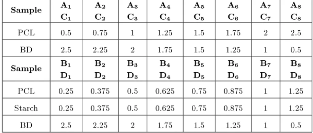
Microspheres were achieved via the well-known water in oil in water emulsion technique. Various molar stoichiometries of HMDI, PCL (molecular weight of 530 g/mol), starch and BD were employed, as shown in Table 1.
As seen in Table 1, group A contains PCL only as polyol and group B contains the same amount of
Table 1. Molar equivalents of BD and PCL (MW = 530 g/mol or MW = 2000 g/mol) for samples A or C and that of BD,
starch and PCL (MW = 530 g/mol or MW = 2000 g/mol) for samples B or D.
Sample A1 C1 A2 C2 A3 C3 A4 C4 A5 C5 A6 C6 A7 C7 A8 C8
PCL 0.5 0.75 1 1.25 1.5 1.75 2 2.5
BD 2.5 2.25 2 1.75 1.5 1.25 1 0.5
Sample B1 D1 B2 D2 B3 D3 B4 D4 B5 D5 B6 D6 B7 D7 B8 D8
PCL 0.25 0.375 0.5 0.625 0.75 0.875 1 1.25
Starch 0.25 0.375 0.5 0.625 0.75 0.875 1 1.25
BD 2.5 2.25 2 1.75 1.5 1.25 1 0.5
both polyols (PCL and starch). By repeating the same molar amount of PCL as in groups A and B, but with molecular weight of 2000 g/mol, groups C and D were achieved, respectively. PCL was dried under vacuum at 60C for 9 hours, dissolved in acetone and mixed
with theophylline in a 1 litre glass reactor. HMDI was dissolved in acetone and added dropwise to the reactor. After 1 hour, the SPIONs (iron concentration of 15 mg/ml) were added to the reactor. It is worth noting that the volume of the ferrouid was 10 times lower than the mixture quantity. Consequently, the chain extender was added to the reactor contents and the reaction mixture was continued for an additional three hours. The total amount of incorporated theophylline in each sample was 5% (w/w) of the glass reactor contents. The mixture was homogenized at 1200 rpm under neutral atmosphere (argon gas) in oil bath and the temperature was xed at 60C. In order to prepare
the water phase, 2 g of the surfactant (PVP) was dissolved in 200 ml of DI water and transferred to a new reactor with vigorous homogenization rate (10 800 rpm). The obtained pre-polymer (30 cm3) was
added dropwise to the 1% (w/v) solution of PVP in DI water to obtain microspheres. The superparam-agnetic microspheres were trapped from the solution using magnetic columns (MACS; A Mini MACS r Separation Unit, Miltenyi Biotec Inc, Germany) and washed several times with DI water in order to remove the solvent residuals, which have toxic eects, together with the suspension stabilizer and the free theophylline. Upon removing the magnetic column from the strong magnetic elds, the microspheres were collected and freeze-dried for two days at 60C and kept in a
desiccator with silica gel.
Characterizations
The morphologies of the nano- and micro-particles were characterized by Transmission Electron Microscopy (TEM;ZEISS, EM-10C, Germany) and Scanning
Elec-tron Microscopy (SEM;Philips, XL30), operating at 100 kV and 20 kV, respectively. Polyurethane for-mations were conrmed by Fourier-transform infrared spectra (FTIR) of the samples with KBr pellets using an ABB Bomem MB-100 FTIR spectrophotometer. Phase characterization was accomplished using the X-ray diraction (XRD;Siemens, D5000, Germany) technique with Cu K radiation. The magnetization of the samples in a variable magnetic eld was measured using a vibrating sample magnetometer (VSM) with a sensitivity of 10 3 emu and a maximum magnetic
eld of 8 kOe. A UV spectrophotometer (Milton Roy Spectronic 601) was used for dening the drug concentration in solution.
In Vitro Drug Loading and Release
The loading of theophylline was measured by UV spectrophotometer (Milton Roy Spectronic 601) af-ter solubilisation of the magnetic microsphere par-ticles in acetonitrile. The insoluble SPIONs from the theophylline-containing solutions were removed by centrifugation at 14000 rpm. The quantity of drug in the supernatant was analyzed using a UV spectrophotometer at wavelength of 274 nm. Typi-cally, one gram of superparamagnetic microspheres was dissolved in 10 ml buer solution and the release of drug was tracked using the UV spectrophotometer. In this regard, the calibration curve was determined at dierent theophylline concentrations (0.1-1 mg/ml) in phosphate buer saline (PBS; a buer solution which is commonly used in biological research) at pH=7.4. Finally, in vitro theophylline release was studied for all groups for a period of 20 days.
RESULTS AND DISCUSSION
Transmission electron microscopy (TEM) of the ppared superparamagnetic iron oxide nanoparticles re-vealed the formation of spherically-shaped SPIONs
Figure 1. TEM image, XRD pattern and VSM curve of synthesized SPIONs.
with an average size of 4 nm and a narrow size distribution (Figure 1). Furthermore, the XRD spectra match well with magnetite (Fe3O4, reference JCPDS
No. 82-1533), indicating that the samples have a cubic crystal system [33]. In addition, the sample was analyzed by VSM and showed negligible remanence and coercivity in the hysteresis loops, which conrmed the superparamagnetic behavior, with a magnetic satura-tion of 50 emu/g (Figure 1).
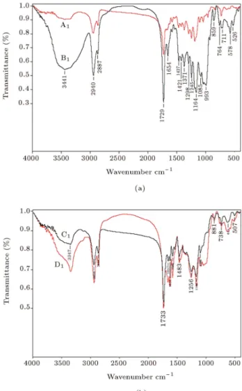
Figure 2 shows the FTIR spectrum of each group candidate (i.e. A1, B1, C1 and D1) conrming the
for-mation of a polyurethane structure given the presence of carbonyl (C=O) and (N-H) peaks at about 1733 and 3340 cm 1, respectively, and the absence of an
isocyanate (NCO) peak at around 2270 cm 1. Thus,
there is almost no residue of the monomer remaining in the polymer and pure and compatible polyurethanes are formed in the reactions. Furthermore, the FTIR spectra conrm the existence of SPIONs that exhibit a band in the low-frequency region (1000-500 cm 1) due
to the iron oxide skeleton [28]; more specically, the characteristic band of Fe-O at 572 cm 1 shows that
the microspheres consist of Fe3O4.
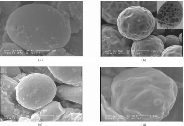
The results of the Scanning Electron Microscopy (SEM) are presented in Figure 3. The SEM images show the morphology of synthesized superparamag-netic microspheres containing theophylline as a round spherical shape (average size of 5 m) and that there is little dierence between samples with various PCL molecular weights. More specically, the average size distribution of those superparamagnetic microspheres which were synthesized by PCL with lower molecular weight is smaller. The reason is that the lower molecular weight could decline the viscosity which may
Figure 2. FTIR spectra of (a) A1 (PCL
(MW = 530 g/mol)) and B1 (PCL (MW = 530 g/mol) and
starch), and (b) C1 (PCL (MW = 2000 g/mol)) and D1
(PCL (MW = 2000 g/mol) and starch).
increase the stirring speed and consequently results in smaller microspheres [34]. Moreover, groups B and D, which contain both starch and PCL, have lower average particle sizes than groups A and C due to the attribution of the starch eect in declining the viscosity of the solution during the formation of microspheres [34]. However, addition of starch to the samples caused surface roughness due to the granular shape of starch particles which were trapped in the polymeric network [35].
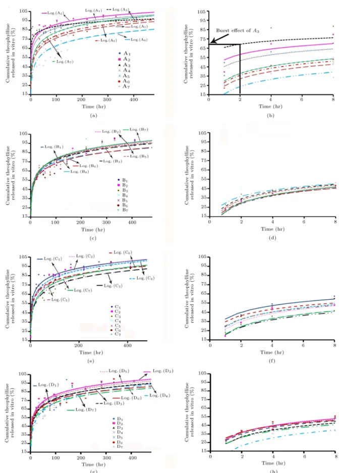
The VSM curves of the superparamagnetic micro-spheres are presented in Figure 4. Figure 5 shows the result of calibration curve which has been determined at dierent theophylline concentrations. Figure 6 shows the release graphs of all samples. According to these graphs, all groups have a burst eect of between 15-65 percent during their rst 4 hours of the release process. As can be seen, by decreasing the molecular weight of PCL, the burst eect is decreased for groups
Figure 3. SEM micrographs of superparamagnetic polyurethane microspheres for (a) A1 (PCL (MW = 530 g/mol)), (b)
B2 (PCL (MW = 530 g/mol) and starch), (c) C3(PCL (MW = 2000 g/mol)) and (d) D4 (PCL (MW = 2000 g/mol) and
starch). Inset at the top right is the TEM image of a microsphere.
Figure 4. Magnetization curves for samples (a) A1 (PCL
(MW = 530 g/mol)), (b) B1 (PCL (MW = 530 g/mol) and
starch), (c) C1 (PCL (MW = 2000 g/mol)) and (d) D1
(PCL (MW = 2000 g/mol) and starch).
A and C, from 65 to 30%. It is worth noting that by decreasing the PCL molecular weight, the eect of polyol to the chain extender ration was minor and the scatter release proles for samples in the same group were not observed (compare Figures 6a and 6c). On the other hand, by employing starch in the structure of the polyurethane texture, the burst eect showed a considerable decline from 65% (for groups A and C) to
Figure 5. The calibration curve for theophylline
concentrations determination according to its absorbance.
20% (for groups B and D). In addition, the eect of polyol to the chain extender ration was negligible and did not change the release prole regardless of PCL's molecular weight.
High molecular weight starch chains together with their granular shape in lateral specimens may form entanglements with the polyurethane polymer, there-fore causing the signicant decline in the burst eect.
Figure 6. In vitro drug release pattern of the samples in (a) group A (PCL (MW = 530 g/mol)), (c) group B (PCL
(MW = 530 g/mol) and starch), (e) group C (PCL (MW = 2000 g/mol)), and (g) group D (PCL (MW = 2000 g/mol) and
starch). (b), (d), (f) and (h) patterns are the release prole in the rst 8 hours of (a), (c), (e) and (g), respectively. Plain lines represent the logarithmic tting.
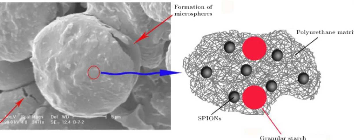
Figure 7. SEM image of the microspheres objects and the possible schematic of their matrix, showing the linkage between various ingredients. Diagram is not to scale in representing the proportions of the dierent objects.
Figure 8. SEM images of microspheres of sample B1for (a) before and (b) after the drug release process; (c) SEM image
of (b) with higher magnication, showing the existence of granular starch after degradation of microspheres.
The predetermined starch barrier may also contribute to a lesser extent in the formation of polyurethane chains as a polyol. More specically, PCL has a much lower molecular weight in comparison with starch, and therefore can oer a higher diusion rate in comparison to starch for reaction with HMDI and BD. Figure 7 schematically illustrate the existence of starch in the
polyurethane matrix; in this case, the diusion rate of the drug from the polymeric matrix to the buer solution would be decreased.
After the release study, the superparamagnetic microspheres were investigated by SEM (Figure 8). The results conrm that the most degradation of microspheres occurs after 20 days of interaction with
a PBS environment. Probing SEM images of the degraded microspheres with higher magnications (e.g. Figure 8c) proved the existence of the granular starch, conrming our claim in the dispersion of starch in the polymeric matrix.
CONCLUSION
Magnetic polyurethane microspheres loaded with theo-phylline were successfully prepared by the water in oil in water emulsion technique. Various molar stoichiome-tries of HMDI, PCL (MW = 530 or 2000 g/mol), starch
and BD were used. Polyurethane formation was con-rmed by IR, and the morphology of the microparticles was characterized by TEM and SEM. The release of theophylline was studied during a period of 20 days and dosed by UV. With the use of PCL with a low molecular weight or in the presence of starch in the structure of polyurethane, the burst eect of the drug is decreased. A SEM study showed an important degradation of the microspheres after the release process. Thus, this kind of new superparamagnetic microspheres is very promising, since it oers the opportunity to control the drug release and also the biodegradable character of the particles.
REFERENCES
1. Li, B., Davidson, J.M. and Guelcher, S.A. \The eect of the local delivery of platelet-derived growth factor from reactive two-component polyurethane scaolds on the healing in rat skin excisional wounds", Bioma-terials, 30(20), pp. 3486-3494 (2009).
2. Zhang, J.-Y., Beckman, E.J., Hu, J., Yang, G.-G., Agarwal, S. and Hollinger, J.O. \Synthesis, biodegrad-ability, and biocompatibility of lysine diisocyanate-glucose polymers", Tissue Eng., 8(5), pp. 771-785 (2002).
3. Santerre, J.P., Woodhouse, K., Laroche, G. and Labow, R.S. \Understanding the biodegradation of polyurethanes: From classical implants to tissue en-gineering materials", Biomaterials, 26(35), pp. 7457-7470 (2005).
4. Fujimoto, K.L., Guan, J., Oshima, H., Sakai, T. and Wagner, W.R. \In vivo evaluation of a porous, elastic, biodegradable patch for reconstructive cardiac procedures", Annals of Thoracic Surgery, 83(2), pp. 648-654 (2007).
5. Gorna, K. and Gogolewski, S. \Preparation, degrada-tion, and calcication of biodegradable polyurethane foams for bone graft substitutes", J. Biomed. Mater. Res. A, 67(3), pp. 813-827 (2003).
6. Raenia, M., Orang, F. and Emami, S.H. \Prepara-tion and characteriza\Prepara-tion of polyurethane microspheres containing theophylline", J. Bioac. Compatible Poly-mers, 21, pp. 341-349 (2006).
7. Guan, J., Stankus, J.J. and Wagner, W.R. \Biodegrad-able elastomeric scaolds with basic broblast growth factor release", J. Controlled Release, 120(1-2), pp. 70-78 (2007).
8. Gorman, S.P., Tunney, M.M., Keane, P.F., van Bladel, K. and Bley, B. \Characterization and assessment of a novel poly(ethylene oxide)/polyurethane composite hydrogel as a ureteral stent biomaterial", J. Biomed. Mater. Res., 39, pp. 642 (1998).
9. Jabbari, E. and Khakpour, M. \Morphology and release behavior from porous polyurethane micro-spheres", Biomaterials, 21, pp. 2073-2079 (2000). 10. Bennett, S., Connolly, K., Lee, D.R., Jiang, Y., Buck,
D., Hollinger, J.O. and Gruskin, E.A. \Initial biocom-patibility studies of a novel degradable polymeric bone substitute that hardens in situ", Bone, 19, pp. 101-107 (1996).
11. Hafeman, A.E., Li, B., Yoshii, T., Zienkiewicz, K., Davidson, J.M. and Guelcher, S.A. \Injectable biodegradable polyurethane scaolds with release of platelet-derived growth factor for tissue repair and regeneration", Pharm. Res., 25(10), pp. 2387-2399 (2008).
12. Guelcher, S.A. \Biodegradable polyurethanes: Synthe-sis and applications in regenerative medicine", Tissue Eng. B: Reviews, 14(1), pp. 3-17 (2008).
13. Guelcher, S.A., Patel, V., Gallagher, K.M., Connolly, S., Didier, J.E., Doctor, J.S. and Hollinger, J.O. \Synthesis and in vitro biocompatibility of injectable polyurethane foam scaolds", Tissue Eng., 12(5), pp. 1247-1259 (2006).
14. Sinha, V.R. and Trehan, A. \Biodegradable micro-spheres for protein delivery", J. Controlled Release, 90(3), pp. 261-280 (2003).
15. Anderson, J.M. and Shive, M.S. \Biodegradation and biocompatibility of PLA and PLGA microspheres", Adv. Drug Delivery Rev., 28(1), pp. 5-24 (1997). 16. Anderson, J.M. \In vivo biocompatibility of
im-plantable delivery systems and biomaterials", Eur. J. Pharm. Biopharm., 40(1), pp. 1-8 (1994).
17. Davaran, S. and Entezami, A. \Synthesis and hydrol-ysis of polyurethanes containing ibuprofen pendant groups", J. Bioact. Compatible Polymers, 12, pp. 47-58 (1997).
18. Grigorieva, M., Gladir, I. and Galatenko, N. \The polyurethane drug composites: Synthesis, properties and a kinetic model", J. Bioact. Compatible Polymers, 16, pp. 307-314 (2001).
19. Subhaga, C.S., Ravi, K.G. and Sunny, M.C. \Evalu-ation of an aliphatic polyurethane as a microsphere matrix for sustained theophylline delivery", J. Mi-croencapsulation, 12, pp. 617-625 (1995).
20. Tiseo, P.J., Foley, K. and Friedho, L.T. \Con-current administration of donepezil HCL and theo-phylline: Assessment of pharmacokinetic changes fol-lowing multiple-dose administration in healthy vol-unteers", British J. Clinical Pharm., 46, pp. 35-39 (1998).
21. Luo, Y.L., Fan, L.H., Xu, F., Chen, Y.S., Zhang, C.H. and Wei, Q.B. \Synthesis and characterization of Fe3O4/PPy/P(MAA-co-AAm) trilayered composite
microspheres with electric, magnetic and pH response characteristics", Mater. Chem. Phys., 120, pp. 590-597 (2010).
22. Mangeney, C., Fertani, M., Bousalem, S., Zhicai, M., Ammar, S., Herbst, F., Beaunier, P., Elaissari, A. and Chehimi, M.M. \Magnetic Fe2O3-Polystyrene/PPy
core/shell particles: Bioreactivity and self-assembly", Langmuir, 23, pp. 10940-10949 (2007).
23. Mahmoudi, M., Milani, A.S. and Stroeve, P. \Sur-face architecture of superparamagnetic iron oxide nanoparticles for application in drug delivery and their biological response: A review", Int. J. Biomedical Nanoscience and Nanotechnology (in press).
24. Mahmoudi, M., Sant, S., Wang, B., Laurent, S. and Sen, T. \Superparamagnetic iron oxide nanoparticles (SPIONs): Development, surface modication and applications in chemotherapy", Adv. Drug Delivery Rev., doi:10.1016/j.addr.2010.05.006 (in press). 25. Mahmoudi, M., Shokrgozar, M.A., Simchi, A., Imani,
M., Milani, A.S., Stroeve, P., Vali, H., Hafeli, U.O. and Bonakdar, S. \Multiphysics ow modeling and in vitro toxicity of iron oxide nanoparticles coated with poly(vinyl alcohol)", J. Phys. Chem. C, 113(6), pp. 2322-2331 (2009).
26. Mahmoudi, M., Simchi, A. and Imani, M. \Cyto-toxicity of uncoated and polyvinyl alcohol coated superparamagnetic iron oxide nanoparticles", J. Phys. Chem. C, 113(22), pp. 9573-9580 (2009).
27. Mahmoudi, M., Simchi, A., Imani, M. and Hafeli, U.O. \Superparamagnetic iron oxide nanoparticles with rigid cross-linked polyethylene glycol fumarate coating for application in imaging and drug delivery", J. Phys. Chem. C, 113(19), pp. 8124-8131 (2009). 28. Mahmoudi, M., Simchi, A., Imani, M., Milani, A.S.
and Stroeve, P. \Optimal design and characterization of superparamagnetic iron oxide nanoparticles coated with polyvinyl alcohol for targeted delivery and imag-ing", J. Phys. Chem. B, 112(46), pp. 14470-14481 (2008).
29. Mahmoudi, M., Simchi, A., Imani, M., Milani, A.S. and Stroeve, P. \An in vitro study of bare and poly(ethylene glycol)-co-fumarate-coated superparam-agnetic iron oxide nanoparticles: a new toxicity iden-tication procedure", Nanotechnology, 20(22), 225104 (8pp) (2009).
30. Mahmoudi, M., Simchi, A., Imani, M., Shokrgozar, M.A., Milani, A.S., Hafeli, U. and Stroeve, P. \A new approach for the in vitro identication of the cytotox-icity of superparamagnetic iron oxide nanoparticles", Colloids Surf., B, 75, pp. 300-309 (2010).
31. Mahmoudi, M., Simchi, A., Imani, M., Stroeve, P. and Sohrabi, A. \Templated growth of superparamagnetic iron oxide nanoparticles by temperature programming in the presence of poly(vinyl alcohol)", Thin Solid Films, 518(15), pp. 4281-4289 (2010).
32. Mahmoudi, M., Simchi, A., Milani, A.S. and Stroeve, P. \Cell toxicity of superparamagnetic iron oxide nanoparticles", J. Colloidal and Interface Sci., 336, pp. 10-518 (2009).
33. Mahmoudi, M., Simchi, A., Vali, H., Imani, M., Shokr-gozar, M.A., Azadmanesh, K. and Azari, F. \Cytotox-icity and cell cycle eects of bare and polyvinyl alcohol coated iron oxide nanoparticles in mouse broblasts", Adv. Eng. Mater., 11(12), pp. B243-B250 (2009). 34. Emami, S.H., Orang, F., Mahmoudi, M. and Raenia,
M. \A study of starch addition on burst eect and di-ameter of polyurethane microspheres containing theo-phylline", Polym. Adv. Technol., 19(3), pp. 167-170 (2008).
35. Su, L., Ji, W.K., Lan, W.Z. and Dong, X.Q. \Chemical modication of anthan gum to increase dissolution rate", Carbohydr. Polym., 53, pp. 497-499 (2003).
BIOGRAPHIES
Morteza Mahmoudi obtained his PhD in 2009 from Sharif University of Technology with specialization on the cytotoxicity of superparamagnetic iron oxide nanoparticles (SPION). He has received many awards such as the 2010 Dr. Mojtahedi Innovation Award for Distinguished Innovation in Research and Education at Sharif University of Technology and 2009 Kharazmi Young Festival Award. His current research involves the magic SPION for simultaneous diagnosis and ther-apeutic applications (http://www.biospion.com). Sophie Laurent was born in 1967. Her studies were performed at the University of Mons-Hainaut (Belgium) where she received her Ph.D. in Chemistry in 1993. She joined then Prof R.N. Muller's team and was involved in the development (synthesis and physic-ochemical characterization) of paramagnetic Gd com-plexes and super paramagnetic iron oxide nanoparticles as contrast agents for MRI. She is currently working on the vectorization of contrast agents for molecular imaging. She is lecturer and co-author around 90 publications and more than 180 communications in international meetings.