R E S E A R C H A R T I C L E
Open Access
The utility of
18
F-FDG PET/CT for suspected
recurrent breast cancer: impact and prognostic
stratification
Alexandre Cochet
1,5*, Steven David
2,4, Kate Moodie
1, Elizabeth Drummond
1,4, Gaelle Dutu
3, Michael MacManus
2,4,
Boon Chua
2,4and Rodney J Hicks
1,4Abstract
Background:The incremental value of18FDG PET/CT in patients with breast cancer (BC) compared to conventional imaging (CI) in clinical practice is unclear. The aim of this study was to evaluate the management impact and prognostic value of18F-FDG PET/CT in this setting.
Methods:Sixty-three patients who were referred to our institution for suspicion of BC relapse were retrospectively enrolled. All patients had been evaluated with CI and underwent PET/CT. At a median follow-up of 61 months, serial clinical, imaging and pathologic results were obtained to validate diagnostic findings. Overall Survival (OS) was estimated using Kaplan Meier methods and analyzed using the Cox proportional hazards regression models. Results:Forty-two patients had a confirmed relapse with 37 (88%) positive on CI and 40 (95%) positive on PET/CT. When compared with CI, PET/CT had a higher negative predictive value (86% versus 54%) and positive predictive value (95% versus 70%). The management impact of PET/CT was high (change of treatment modality or intent) in 30 patients (48%) and medium (change in radiation treatment volume or dose fractionation) in 6 patients (9%). Thirty-nine patients (62%) died during follow-up. The PET/CT result was a highly significant predictor of OS
(Hazard Ratio [95% Confidence Interval] =4.7 [2.0-10.9] for PET positive versus PET negative for a systemic recurrence; p = 0.0003). In a Cox multivariate analysis including other prognosis factors, PET/CT findings predicted survival (p = 0.005). In contrast, restaging by CI was not significant predictor of survival.
Conclusion:Our study support the value of18F-FDG PET/CT in providing incremental information that influence patient management and refine prognostic stratification in the setting of suspected recurrent breast cancer.
Keywords:Breast cancer,18F-FDG PET/CT, Restaging, Prognosis
Background
Breast cancer (BC) is the most commonly diagnosed cancer in women, and is the leading cause of death by cancer for women in the western world. Depending on the initial extent of the disease, approximately 30% of patients diagnosed with BC are at risk of developing loco-regional recurrence or secondary tumor dissemin-ation to distant organs [1]. Moreover, the survival of pa-tients who develop an isolated loco-regional recurrence
differs from patients who have distant relapse. As a con-sequence, determination of both the locations and extent of the recurrent disease is essential to guide therapeutic decisions and estimate prognosis.
Traditionally, routine evaluation of suspected recurrent BC involves physical examination and a multi-modality Conventional Imaging (CI) approach which may include mammography, CT, MRI, and bone scintigraphy. How-ever, this CI approach is often time-intensive and potential false-negative findings may delay appropriate therapy. Positron Emission Tomography/Computed Tomography (PET/CT) with18F-Fluorodeoxyglucose (18F-FDG) is also often used in this indication, given that 18F-FDG has affinity for both primary and secondary breast tumors, * Correspondence:[email protected]
1
Centre for Cancer Imaging, Peter MacCallum Cancer Centre, East Melbourne, Australia
5
Department of Nuclear Medicine, Centre Georges-François Leclerc, 1 rue Professeur Marion, Dijon Cedex 21079, France
Full list of author information is available at the end of the article
depending on size and aggressiveness [2-4]. Several au-thors have suggested that18F-FDG PET and PET/CT are more sensitive than CI for detection of recurrent BC [5-15] and can have a significant impact on the therapeutic man-agement [5,7-9,12,16]. However, information concerning the utility of 18F-FDG PET/CT for long-term prognostic stratification, when compared with CI, is limited.
Thus, the objectives of our study were to: [1] assess the incremental diagnostic performance and the impact
on therapeutic management of 18F-FDG PET/CT in a
group of patients with a history of BC who had already been restaged by CI for identification of suspected dis-ease relapse; [2] compare the long-term prognostic stratification of CI alone and18F-FDG PET/CT.
Methods
Patients
A retrospective analysis was performed on consecutive patients with a history of BC and suspicion of recurrence who were referred for 18F-FDG PET/CT at our institu-tion from January 2002 to September 2008. BC was not
a funded indication of 18F-FDG PET/CT during this
period in Australia; therefore, clinicians usually referred patients with high suspicion of recurrence for PET/CT.
The inclusion criteria of the study were as follows: (a) a history of confirmed histologic diagnosis of primary BC treated as per local protocol; (b) CI performed no longer than 4 months prior to PET/CT and where the CI included at least a CT scan of the area of interest; (c) availability of follow-up data for a minimum of 6 months following PET/CT; (d) unequivocal determination of clinical status at the time of the last clinical follow-up.
Sixty-three patients (62 women and one man; mean age = 57 years; range = 29-86 years) were included. The median time interval from initial diagnosis to 18F-FDG PET/CT was 39 months (range 5-431 months). The me-dian time interval between CT and PET/CT was 25 days (first-third quartile: 1-52 days). Indications for PET/CT were: equivocal or suspicious CI findings (n = 28); clinical suspicion of recurrence (n = 21); restaging after comple-tion of therapy (n = 5); routine surveillance (n = 5); and increasing levels of tumor markers (n = 4). All patients provided permission to review medical records at the time of PET/CT imaging according to our institution’s investi-gational review board guidelines for informed consent (protocol number 09/78).
PET/CT acquisition and processing
Whole-body PET was acquired sequentially using a dedi-cated PET/CT system (Discovery LS PET/ 4-slice helical CT or Discovery STE/ 8-slice helical CT, General Elec-tric Medical Systems, Milwaukee, WI) combining a mul-tidetector CT scanner with a dedicated, full-ring PET scanner with bismuth germanate crystals. Patients were
instructed to fast except for glucose-free oral hydration for at least 6 hours before injection of 300-400 MBq of18F-FDG. PET was performed 60 min following18F-FDG injection. Blood glucose levels were measured before the injection of the tracer to ensure levels below 10 mmol/l. Transmission data used for attenuation correction were obtained from a low-dose non diagnostic CT acquisition (140 kVp and 40-120 mA), without contrast enhancement. Attenuation corrected PET images were reconstructed with an iterative reconstruction (ordered-subset expect-ation maximizexpect-ation algorithm). Orthogonal CT, PET, and fused PET/CT images were displayed simultaneously on a GE Xeleris Workstation. The PET data were also dis-played in a rotating maximum-intensity projection.
An experienced nuclear medicine physician generated a clinical report after reviewing PET images, low-dose CT images, fused PET/CT images, previous imaging re-sults and clinical information. Standard uptake values were not routinely measured. Once issued, the PET/CT report was not reinterpreted in the light of subsequent clinical information.
Image interpretation and classification
A total of 188 clinical, imaging and pathological procedures were performed (3 ± 1.4 per patients), including chest CT (n = 59), abdominopelvic CT (n = 44), whole-body bone scan (n = 30), clinical examination (n = 17), pathology (n = 9), ab-dominal ultrasound (US) (n = 7), MRI (n = 7), mammogram or breast US (n = 6), chest radiography (n = 5), other (n = 5).
Written clinical reports of conventional images and PET/CT were reviewed and classified as (a) negative if im-aging tests were negative for disease; (b) equivocal, when abnormal findings were present on any imaging test but were not interpreted as suspicious for malignancy; (c) positive, if any result was clearly described as suspicious or consistent with malignancy. Negative and equivocal find-ings were combined as negative for the analysis.
In cases where recurrence was reported on CI or PET/ CT, the location of relapse was also determined, and classified as loco-regional (ipsilateral breast, ipsilateral axillary, internal mammary or supraclavicular node sta-tion) or systemic (contralateral node station or distant metastasis). The final diagnosis of disease recurrence and location of disease was confirmed by histologic examin-ation in 13 patients (21%). For the remaining patients, evi-dence of progression within 6 months of clinical and/or imaging follow-up was considered to indicate a site of dis-ease relapse, whereas no evidence of progression after at least 6 months of follow-up was considered to confirm ab-sence of active disease at that site.
Assessment of impact
clinical request form. The actual post-PET/CT manage-ment plan and treatmanage-ment intent were determined from the medical record or by contacting the referring clinician.
The impact of PET/CT on management was consid-ered “high” when the treatment intent or modality was changed (e.g. from palliative to curative treatment or from surgery to radiotherapy) [17]. The impact was con-sidered as“medium” when the method of treatment de-livery was changed (e.g. radiation treatment volume and/ or dose fractionation) [17]. When the PET/CT results did not indicate a need for change, the impact was con-sidered to be“low”. PET/CT was considered to have had
“no impact” when the management chosen conflicted
with post-PET/CT disease extent on the basis of a syn-thesis of all available information.
Follow-up
After PET/CT, progress updates were obtained from the medical record, family physician, or treating oncologist. When relevant, details of the date and cause of death were obtained. The disease status at the time of death was recorded.
Statistical analysis
Estimates of OS at 2 and 5 years were computed using the Kaplan Meier method, a log-rank test was used to analyze the effect of CI and PET/CT results on OS. Two Cox re-gression analyses were performed to assess the impact of PET/CT and CI on OS controlling for clinical variables and using a backward elimination process. Triple negative status of the primary tumor was not included in the multi-variate model because this information was available for only 38 patients. For percentages such as PPV, NPV, sensi-tivity and specificity, a Blyth-Still-Casella 95% confidence interval (CI) was calculated. Diagnosis performance results were compared using McNemar tests, Fisher exact tests and a likelihood ratio, which summarizes how many times more likely patients with the disease are to have that par-ticular result than patients without the disease. According to the fact that patients were selected on the basis of CI, many patients with unequivocal systemic relapse on CI would likely not have been referred for PET/CT. Given the likely pre-test selection bias, positive and negative predictive values were considered as more relevant com-parators of diagnostic performance when compared with sensitivity and specificity.
Results
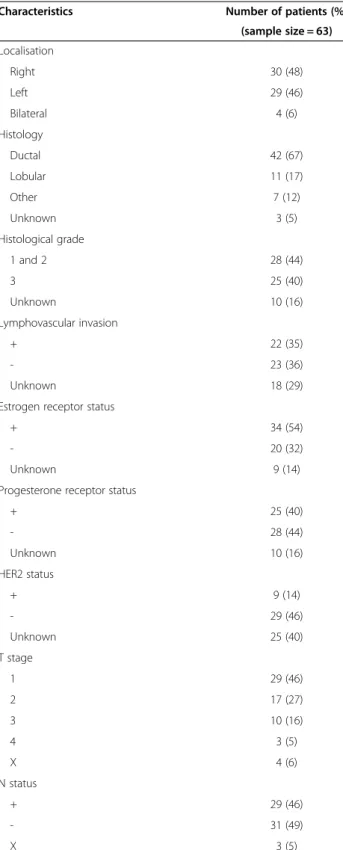
Patient characteristics at the time of initial diagnosis are summarized in Table 1.
Diagnostic performance
Relapse involving at least one site was confirmed in 42 of the 63 patients (67%). CI was positive for disease in
Table 1 Patient characteristics at the time of initial diagnosis
Characteristics Number of patients (%)
(sample size = 63)
Localisation
Right 30 (48)
Left 29 (46)
Bilateral 4 (6)
Histology
Ductal 42 (67)
Lobular 11 (17)
Other 7 (12)
Unknown 3 (5)
Histological grade
1 and 2 28 (44)
3 25 (40)
Unknown 10 (16)
Lymphovascular invasion
+ 22 (35)
- 23 (36)
Unknown 18 (29)
Estrogen receptor status
+ 34 (54)
- 20 (32)
Unknown 9 (14)
Progesterone receptor status
+ 25 (40)
- 28 (44)
Unknown 10 (16)
HER2 status
+ 9 (14)
- 29 (46)
Unknown 25 (40)
T stage
1 29 (46)
2 17 (27)
3 10 (16)
4 3 (5)
X 4 (6)
N status
+ 29 (46)
- 31 (49)
X 3 (5)
37 of these patients, yielding a patient sensitivity of 88%, whereas PET/CT was positive for disease in 40, corre-sponding to a patient sensitivity of 95%.
Table 2 shows comparison of extent of suspected re-lapse as assessed before and after PET/CT. Downstaging by PET/CT was confirmed to be correct in 12/14 pa-tients (one patient had a suspicious bony lesion on bone scan that was non 18F-FDG-avid, but confirmed to be malignant; one patient showed suspicious mediastinal
lymph nodes on CT that were non 18F-FDG-avid but
confirmed to be metastasis of breast cancer by path-ology), while upstaging with PET/CT was confirmed in 5/5 patients.
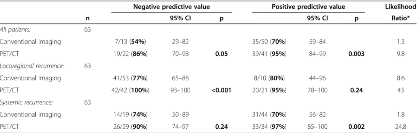
On final diagnosis, 20 patients (32%) had a loco-regional recurrence and 36 (57%) had a systemic recurrence (14 patients had both loco-regional and systemic recur-rence). CI was truly positive for loco-regional recurrence in 8/20 patients (40%) and systemic recurrence in 30/36 patients (83%). PET/CT was truly positive for loco-regional recurrence in 20/20 patients (100%) and systemic disease in 32/36 patients (89%). Among the 20 patients experiencing loco-regional recurrence, only 3 had local re-lapse only, for whom both PET/CT and CI were positive. PET/CT detected regional relapse non-detected by CI in 12 patients (3 in axillary nodes only, 9 in extra-axillary nodes). PET/CT had significantly higher positive predict-ive values when compared with CI for determination of
loco-regional, systemic and global recurrence, and higher negative predictive value for loco-regional and global re-currence (Table 3). 18F-FDG PET/CT found incidental malignancies in 2 patients (one patient had a primary esophageal cancer and one patient had a gastrointestinal stromal tumor).
Impact on management
The PET/CT results had a high impact on management in 30 patients (48%) of whom the treatment intent was modified in 24 patients (including from invasive diagno-sis to observation for 11 patients, according to negative PE/CT results despite suspicious CI findings). For the 6 remaining patients, the treatment intent was not modi-fied after PET/CT (palliative for 5 patients, curative for 1 patient), but the modalities of therapy were changed.
PET/CT had a medium impact on management in 6/63 patients (9%). Management changes in these pa-tients primarily included changes in radiation treatment volume as a result of more extensive disease detected by PET/CT. All these patients had a palliative treatment in-tent, which was not modified by PET/CT.
PET/CT had a low impact (i.e. did not change the planned management) in 27/63 patients (43%) for whom the relapse extent was concordant with that found on CI (19 patients) or the documentation of a different
Table 2 Comparison of extent of suspected relapse as assessed before and after PET/CT
Pre-PET/CT extent of relapse
Post-PET/CT extent of relapse % less disease by PET/CT
% same status
% more disease by PET/CT Negative LR only Systemic Total
Negative 8 1 3 12 - 66 33
LR only 1 6 1 8 12.5 75 12.5
Systemic 12 1 30 43 30 70
-Total 21 8 34 63 22 70 8
LR = Loco-Regional; PET/CT = Positron Emission Tomography/Computerized Tomography.
Table 3 Diagnostic accuracy of conventional imaging (CI) and PET/CT in detecting relapse of disease in all patients, according to the histological type, and for locoregional and systemic recurrence
Negative predictive value Positive predictive value Likelihood
n 95% CI p 95% CI p Ratio*
All patients: 63
Conventional Imaging 7/13 (54%) 29–82 35/50 (70%) 59–84 1.3
PET/CT 19/22 (86%) 70–98 0.05 39/41 (95%) 84–99 0.003 9.8
Locoregional recurrence: 63
Conventional Imaging 41/53 (77%) 65–88 8/10 (80%) 44–96 8.6
PET/CT 42/42 (100%) 93–100 <0.001 20/21 (95%) 78–100 0.24 43
Systemic recurrence: 63
Conventional imaging 14/19 (74%) 50–89 31/44 (70%) 56–82 1.8
PET/CT 26/29 (90%) 74–97 0.24 33/34 (97%) 85–100 0.002 24.8
distribution of disease did not alter the planned treat-ment (2 patients), or both PET/CT and CI findings were negative (6 patients). In no case was the PET/CT result apparently ignored.
Prediction of survival by CI and PET/CT
Survival data were analyzed with a close-out date of September, 4th 2010. The median follow-up time was 5.1 years (range 0.8-8.6 years). All 63 patients entered into the study had a known status at the close-out date. All patients with negative PET/CT findings were followed for a minimum of 2 years after the scan (except one patient who died 2 months after the scan, because
of complications of an esophageal primary tumor discov-ered on PET/CT).
Thirty-eight patients (60%) were deceased with a median survival of 3.4 years (95% CI 2.5 to 5.0 years) (Figure 1). The estimated 2-year OS was 66.4% (95% CI 53.2% to 76.6%) and the estimated 5-year OS was 37.3% (95% CI 24.3% to 50.3%).
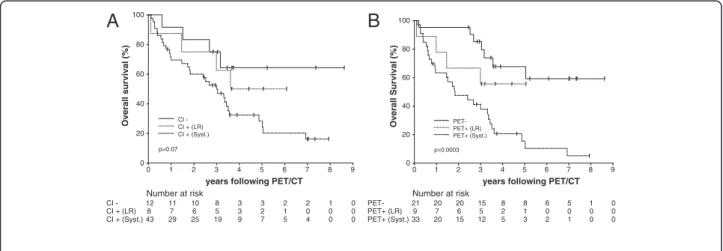
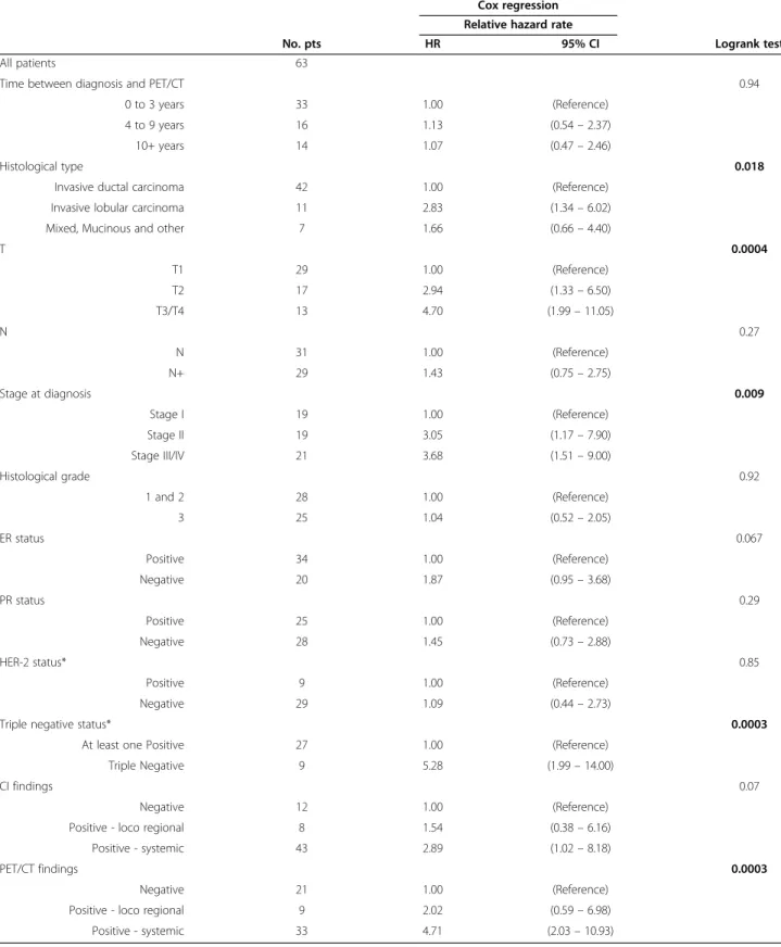
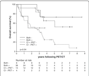
On univariate analysis, PET/CT status (negative, posi-tive LR or posiposi-tive systemic) was strongly associated with survival (log-rank test: p = 0.0003 for the entire model; p = 0.0001 for comparison of negative results and positive for systemic disease; p > 0.05 for other single comparisons) (Figure 2). Patients with systemic disease according to PET/CT had a 4.7-fold in the risk of death when compared with patients with negative PET/CT findings, while patients with only loco-regional recur-rence had a 2-fold increase in the risk of death (Table 4). In contrast, CI status (negative, positive LR or positive systemic) did not significantly predict OS (log-rank test: p = 0.07 for the entire model; p > 0.05 for all single com-parisons) (Figure 2). Moreover, patients with positive CI findings but negative PET/CT findings had similar es-timated survival to patients in whom both tests were negative, whereas patients with negative CI findings but positive PET/CT results had comparable estimated sur-vival to patients in whom both procedures were positive for recurrence (Figure 3).
Initial stage of the disease at the time of diagnosis, histological type of cancer and triple negative status (hormone receptor and HER-2 negativity) were also pre-dictors of survival on the univariate analysis (Table 4). Time between initial diagnosis and the restaging PET/ CT, initial histological grade, estrogen receptor status, progesterone receptor status and overexpression of Human Epidermal Growth Factor Receptor-2 (HER-2)
0 20 40 60 80 100
Overall Survival (%)
0 1 2 3 4 5 6 7 8 9
years following PET Number at risk
63 47 41 32 15 12 8 6 1 0
Figure 1Estimated overall survival (+/- 95% confidence interval) for all 63 patients.
0 20 40 60 80 100
Overall survival (%)
0 1 2 3 4 5 6 7 8 9
years following PET/CT
Number at risk
12 11 10 8 3 3 2 2 1 0 CI
-8 7 6 5 3 2 1 0 0 0 CI + (LR)
43 29 25 19 9 7 5 4 0 0 CI + (Syst.)
0 20 40 60 80 100
Overall Survival (%)
0 1 2 3 4 5 6 7 8 9
years following PET/CT
Number at risk
21 20 20 15 8 8 6 5 1 0
PET-9 7 6 5 2 1 0 0 0 0 PET+ (LR)
33 20 15 12 5 3 2 1 0 0 PET+ (Syst.)
A
B
Table 4 Univariate predictors of overall survival
Cox regression Relative hazard rate
No. pts HR 95% CI Logrank test
All patients 63
Time between diagnosis and PET/CT 0.94
0 to 3 years 33 1.00 (Reference)
4 to 9 years 16 1.13 (0.54–2.37)
10+ years 14 1.07 (0.47–2.46)
Histological type 0.018
Invasive ductal carcinoma 42 1.00 (Reference)
Invasive lobular carcinoma 11 2.83 (1.34–6.02)
Mixed, Mucinous and other 7 1.66 (0.66–4.40)
T 0.0004
T1 29 1.00 (Reference)
T2 17 2.94 (1.33–6.50)
T3/T4 13 4.70 (1.99–11.05)
N 0.27
N 31 1.00 (Reference)
N+ 29 1.43 (0.75–2.75)
Stage at diagnosis 0.009
Stage I 19 1.00 (Reference)
Stage II 19 3.05 (1.17–7.90)
Stage III/IV 21 3.68 (1.51–9.00)
Histological grade 0.92
1 and 2 28 1.00 (Reference)
3 25 1.04 (0.52–2.05)
ER status 0.067
Positive 34 1.00 (Reference)
Negative 20 1.87 (0.95–3.68)
PR status 0.29
Positive 25 1.00 (Reference)
Negative 28 1.45 (0.73–2.88)
HER-2 status* 0.85
Positive 9 1.00 (Reference)
Negative 29 1.09 (0.44–2.73)
Triple negative status* 0.0003
At least one Positive 27 1.00 (Reference)
Triple Negative 9 5.28 (1.99–14.00)
CI findings 0.07
Negative 12 1.00 (Reference)
Positive - loco regional 8 1.54 (0.38–6.16)
Positive - systemic 43 2.89 (1.02–8.18)
PET/CT findings 0.0003
Negative 21 1.00 (Reference)
Positive - loco regional 9 2.02 (0.59–6.98)
Positive - systemic 33 4.71 (2.03–10.93)
*The information is available for only 38 patients.
were not significant predictors of survival. On multivari-ate analysis, positive PET/CT findings (for loco-regional and/or systemic recurrence) remained an independent predictor of OS when adjusting for age, histological sub-type and initial stage (model 1, Table 5). In contrast, positive CI findings did not independently predict OS (model 2, Table 5).
Discussion
For patients with possible recurrent breast cancer (BC), early detection and adequate localization of recurrent disease are essential for guiding optimal therapy and prognostication. Patients with isolated loco-regional re-currence are able to benefit from curative salvage ther-apy, whereas palliative treatment is generally indicated for patients with distant relapse.
Several studies have shown the relevance of 18F-FDG PET/CT in detecting distant metastasis in patients with clinical suspicion of recurrence [6,8,12,13,18-20], and in patients with documented loco-regional recurrence [5]. Our study confirmed that18F-FDG PET/CT is an accur-ate technique for the appropriaccur-ate detection of relapse, when compared with CI alone. Of particular economic and clinical importance was the observation that 12 pa-tients (19%) in this series who were suspected to have relapsed by conventional evaluation subsequently re-ceived no active treatment after a negative 18F-FDG PET/CT evaluation and demonstrated an excellent prog-nosis. In contrast, of the 8 patients with negative CI find-ings, 1 patient was found to have locoregional relapse and 3 patients were found to have systemic recurrence on
18
F-FDG PET/CT. In the current study, 18F-FDG PET/
CT had an impact on therapeutic management in 57% of patients; in particular, the treatment intent was changed in 38% of patients. This result is consistent with other studies which showed the important impact of18F-FDG PET/CT on therapeutic management in patients with suspicion of recurrent BC [5,8,12,18,19]. Most of these previous studies reported that 18F-FDG PET/CT was highly accurate for detecting recurrent disease in patients with negative or in-conclusive CI findings [5,18,19]. In contrast, in our study, CI findings were consistent with relapse in the majority of patients (51/63) but18F-FDG PET/CT downstaged 12 of them, providing a better negative predictive value when compared to CI alone (Table 3). These findings suggested that18F-FDG PET/CT was not only effective for early de-tection of relapse in patients with negative CI findings, but also yielded a better characterization of CI findings in pa-tients with a high suspicion of relapse.
As BC was not a funded indication of18F-FDG PET/
CT in Australia during the study period, referring clinicians were likely to use PET/CT in patients for whom there was clinical uncertainty with respect to the appropriate management. Although this patient selection
0 20 40 60 80 100
Overall survival (%)
0 1 2 3 4 5 6 7 8 9
years following PET/CT Number at risk
8 8 8 6 3 3 2 2 1 0
Both -
38 24 19 15 7 4 2 1 0 0
Both +
13 12 12 9 5 5 4 3 0 0
CI + /PET
-4 3 2 2 0 0 0 0 0 0
CI - /PET +
Both - Both + CI + /PET -CI - /PET +
p=0.04
Figure 3Kaplan-Meier estimate of overall survival (OS) stratified by combination of Conventional imaging (CI) and PET/CT results.
Table 5 Multivariate predictors of overall survival
No of observed deaths HR 95% CI P
Model 1 36/58
Age (per 10 years) 1.4 1.1–1.7 0.02
Invasive ductal carcinoma vs. other type 2.6 1.3–5.3 0.007
Initial stage II, III, IV vs. I 4.1 1.7–10.3 0.002
PET/CT positive vs negative 3.5 1.5–8.2 0.005
Model 2 36/58
Age (per 10 years) 1.4 1.1–1.8 0.01
Invasive ductal carcinoma vs. other 2.9 1.4–5.8 0.003
Initial stage II, III, IV vs. I 5.4 2.1–13.4 <0.001
Conventional Imaging positive vs. negative 2.0 0.7–5.7 0.22
could have introduced biases in the evaluation of the im-pact of PET/CT findings on patient management, it also showed the value of18F-FDG PET/CT in patients whose disease status could not be adequately determined using CI alone.
The more accurate prognostic information derived from PET/CT results when compared with CI findings underpinned the value of PET/CT in the management
of patients with recurrent BC. Both 18F-FDG PET/CT
and CT are not optimal modalities for detection of local recurrence, when compared with mammography, ultra-sound or MRI. However, detection of additional distant metastases in patients with documented loco-regional recurrence is essential in order to optimize management and stratify prognosis. Of note was the favourable sur-vival of patients with isolated loco-regional recurrence according to PET/CT, when compared with patients with systemic relapse.
In our study, CT performed with PET was not of diag-nostic quality. However, all patients included in this study had diagnostic CT before undergoing FDG PET/CT.
While Dirisamer et al. showed in a retrospective study that the association of FDG PET and contrast-enhanced (ce) CT could improve restaging of breast cancer when compared with ceCT or FDG PET alone [6], there is no evidence in the literature of the superiority of FDG PET/ ceCT for restaging of breast cancer when compared with FDG PET associated to non-diagnostic, low dose CT. In our study, the outcome data validate our approach in that FDG PET/CT results were more often correct than conventional imaging (including diagnostic CT) when discordant, and stratified prognosis whereas conven-tional imaging did not (Figure 2). If we hadn’t ignored the conventional imaging findings (including diagnostic CT) when discordant with FDG PET/CT, the accuracy wouldn’t have been enhanced and the prognostic value of FDG PET/CT incorporating a low dose, non-contrast CT wouldn’t have been superior to that of conventional imaging. These results have implications for the report-ing of FDG PET combined with ceCT, which is per-formed by some facilities as a routine procedure and suggest that significant clinical weight should be placed on the PET findings even when discordant with the ceCT appearances.
One of the limitations of our study was that the CI procedures were not standardized and were selected on the basis of clinical findings. However, this represented routine clinical practice and did not detract from the results. Although comparison of a masked reading of PET/CT with a masked reading of CI techniques might be appropriate if PET/CT were to be suggested as a re-placement for CI, the main purpose of this study was to evaluate the incremental diagnostic and prognostic value of PET/CT in routine practice. The results of this
retrospective study would, however, justify a randomized trial in which patients with clinical risk or suspicion of relapse would be stratified to have either CI or FDG PET/CT as the initial restaging procedure [21]. Finally, we did not compare PET/CT and CI findings with histo-pathological findings in most patients. For 50 patients (79%), the final disease status was determined clinically and/or with follow-up imaging. Nevertheless, since most of the patients were followed up for a long period of time, the survival analysis would be the best validation of diagnostic accuracy.
Conclusion
Our findings support the value of 18F-FDG PET/CT in providing incremental information that influence patient management and refine prognostic stratification in the setting of suspected recurrent breast cancer. The prog-nostic stratification provided by this technique empha-sizes the crucial role of18F-FDG PET/CT in optimizing treatment choices in this setting. Further multicentric studies are needed to confirm this role in particular in patients with high suspicion of relapse.
Competing interests
The authors declare that they have no competing interests.
Authors’contributions
AC, SD, KM and RJH were involved in the concept and design of the study. AC SD, and ED were involved in data collection, history review and follow-up. KM was the imaging lead on the study and with RJH verified classification of scan results. GT was the study statistician and contributed to analysis of the primary data. SD, MM and BC were the clinical leads for assessing the impact criteria and verifying classifications of PET/CT impact. All authors read and approved the final manuscript.
Funding
A.C. was supported by a fellowship from the Fondation de France, and by grants from the French Ligue contre le cancer and from the Conseil Régional de Bourgogne.
Author details
1Centre for Cancer Imaging, Peter MacCallum Cancer Centre, East Melbourne, Australia.2Department of Radiation Oncology, Peter MacCallum Cancer Centre, East Melbourne, Australia.3Centre for Biostatistics and Clinical Trials, Peter MacCallum Cancer Centre, East Melbourne, Australia.4The Sir Peter MacCallum Department of Oncology, the University of Melbourne, Parkville, Australia.5Department of Nuclear Medicine, Centre Georges-François Leclerc, 1 rue Professeur Marion, Dijon Cedex 21079, France.
Received: 20 March 2014 Accepted: 20 March 2014 Published: 22 April 2014
References
1. van Dongen JA, Voogd AC, Fentiman IS, Legrand C, Sylvester RJ, Tong D, van der Schueren E, Helle PA, van Zijl K, Bartelink H:Long-term results of a randomized trial comparing breast-conserving therapy with mastectomy: European organization for research and treatment of cancer 10801 trial. J Natl Cancer Inst2000,92(14):1143–1150.
2. Avril N, Rose CA, Schelling M, Dose J, Kuhn W, Bense S, Weber W, Ziegler S, Graeff H, Schwaiger M:Breast imaging with positron emission tomography and fluorine-18 fluorodeoxyglucose: use and limitations. J Clin Oncol2000,18(20):3495–3502.
CF:Biologic correlates of (18) fluorodeoxyglucose uptake in human breast cancer measured by positron emission tomography.J Clin Oncol 2002,20(2):379–387.
4. Wahl RL, Cody RL, Hutchins GD, Mudgett EE:Primary and metastatic breast carcinoma: initial clinical evaluation with PET with the radiolabeled glucose analogue 2-[F-18]-fluoro-2-deoxy-D-glucose. Radiology1991,179(3):765–770.
5. Aukema TS, Rutgers EJ, Vogel WV, Teertstra HJ, Oldenburg HS, Vrancken Peeters MT, Wesseling J, Russell NS, Valdés Olmos RA:The role of FDG PET/ CT in patients with locoregional breast cancer recurrence: a comparison to conventional imaging techniques.Eur J Surg Oncol2010,36(4):387–392. 6. Dirisamer A, Halpern BS, Flory D, Wolf F, Beheshti M, Mayerhoefer ME,
Langsteger W:Integrated contrast-enhanced diagnostic whole-body PET/CT as a first-line restaging modality in patients with suspected metastatic recurrence of breast cancer.Eur J Radiol2010,73(2):294–299. 7. Eubank WB, Mankoff D, Bhattacharya M, Gralow J, Linden H, Ellis G, Lindsley
S, Austin-Seymour M, Livingston R:Impact of FDG PET on defining the extent of disease and on the treatment of patients with recurrent or metastatic breast cancer.AJR Am J Roentgenol2004,183(2):479–486. 8. Evangelista L, Baretta Z, Vinante L, Cervino AR, Gregianin M, Ghiotto C,
Saladini G, Sotti G:Tumour markers and FDG PET/CT for prediction of disease relapse in patients with breast cancer.Eur J Nucl Med Mol Imaging2011,38(2):293–301.
9. Grahek D, Montravers F, Kerrou K, Aide N, Lotz JP, Talbot JN:[18F]FDG in recurrent breast cancer: diagnostic performances, clinical impact and relevance of induced changes in management.Eur J Nucl Med Mol Imaging2004,31(2):179–188.
10. Pan L, Han Y, Sun X, Liu J, Gang H:FDG-PET and other imaging modalities for the evaluation of breast cancer recurrence and metastases: a meta-analysis.J Cancer Res Clin Oncol2010,136(7):1007–1022. 11. Pennant M, Takwoingi Y, Pennant L, Davenport C, Fry-Smith A, Eisinga A,
Andronis L, Arvanitis T, Deeks J, Hyde C:A systematic review of positron emission tomography (PET) and positron emission tomography/computed tomography (PET/CT) for the diagnosis of breast cancer recurrence. Health Technol Assess2010,14(50):1–103.
12. Radan L, Ben-Haim S, Bar-Shalom R, Guralnik L, Israel O:The role of FDG-PET/CT in suspected recurrence of breast cancer.Cancer2006, 107(11):2545–2551.
13. Veit-Haibach P, Antoch G, Beyer T, Stergar H, Schleucher R, Hauth EA, Bockisch A:FDG-PET/CT in restaging of patients with recurrent breast cancer: possible impact on staging and therapy.Br J Radiol2007, 80(955):508–515.
14. Vranjesevic D, Filmont JE, Meta J, Silverman DH, Phelps ME, Rao J, Valk PE, Czernin J:Whole-body (18)F-FDG PET and conventional imaging for predicting outcome in previously treated breast cancer patients.J Nucl Med2002,43(3):325–329.
15. Evangelista L, Cervino AR, Ghiotto C, Al-Nahhas A, Rubello D, Muzzio PC: Tumor marker-guided PET in breast cancer patients-a recipe for a perfect wedding: a systematic literature review and meta-analysis.Clin Nucl Med 2012,37(5):467–474.
16. Santiago JF, Gonen M, Yeung H, Macapinlac H, Larson S:A retrospective analysis of the impact of 18 F-FDG PET scans on clinical management of 133 breast cancer patients.Q J Nucl Med Mol Imaging2006,50(1):61–67. 17. Kalff V, Hicks RJ, MacManus MP, Binns DS, McKenzie AF, Ware RE, Hogg A, Ball DL:Clinical impact of (18)F fluorodeoxyglucose positron emission tomography in patients with non-small-cell lung cancer: a prospective study.J Clin Oncol2001,19(1):111–118.
18. Filippi V, Malamitsi J, Vlachou F, Laspas F, Georgiou E, Prassopoulos V, Andreou J:The impact of FDG-PET/CT on the management of breast cancer patients with elevated tumor markers and negative or equivocal conventional imaging modalities.Nucl Med Commun2011,32(2):85–90. 19. Grassetto G, Fornasiero A, Otello D, Bonciarelli G, Rossi E, Nashimben O,
Minicozzi AM, Crepaldi G, Pasini F, Facci E, Mandoliti G, Marzola MC, Al-Nahhas A, Rubello D:18 F-FDG-PET/CT in patients with breast cancer and rising Ca 15-3 with negative conventional imaging: a multicentre study.Eur J Radiol 2010,80(3):828–833.
20. Murakami R, Kumita S, Yoshida T, Ishihara K, Kiriyama T, Hakozaki K, Yanagihara K, Iida S, Tsuchiya S:FDG-PET/CT in the diagnosis of recurrent breast cancer.Acta Radiol2011,53(1):12–16.
21. Hicks RJ:Should positron emission tomography/computed tomography be the first rather than the last test performed in the assessment of cancer?Cancer Imaging2012,12(2):315–323.
doi:10.1186/1470-7330-14-13
Cite this article as:Cochetet al.:The utility of18F-FDG PET/CT for suspected recurrent breast cancer: impact and prognostic stratification.
Cancer Imaging201414:13.
Submit your next manuscript to BioMed Central and take full advantage of:
• Convenient online submission
• Thorough peer review
• No space constraints or color figure charges
• Immediate publication on acceptance
• Inclusion in PubMed, CAS, Scopus and Google Scholar
• Research which is freely available for redistribution