TECHNICAL UNIVERSITY OF CLUJ-NAPOCA
ACTA TECHNICA NAPOCENSIS
Series: Applied Mathematics, Mechanics, and Engineering Vol. 60, Issue IV, November, 2017
AN INNOVATIVE AUTOMATED INSTRUMENT FOR ROBOTICALLY
ASSISTED BRACHYTHERAPY USED IN CANCER TREATMENT
Iosif BIRLESCU, Florin CRACIUN, Călin VAIDA, Bogdan GHERMAN, Doina PISLA
Abstract: The paper presents the design of a novel brachytherapy automated instrument with 6 needles meant to be used within a medical robotic system as an end effector. Based on the design requirements derived from the medical procedure, the medical instrument uses a needle storage mechanism and a gripper which have a synchronized motion in order to perform needle loading, insertion and releasing. The functionality of the instrument is explained and the experimental model is presented. Experimental scenarios are also presented followed by conclusion and proposed future research which aims to optimize the medical instrument and validate it for the medical procedure.
Key words: medical instrument, multiple needle insertion, medical robot, brachytherapy.
1. INTRODUCTION
Brachytherapy is a local radiotherapy used in oncology, where a radioactive seed is placed within or near a cancer tumor [1] in order to deliver a high radioactive dose to the tumor to neutralize the cancer [2]. Being a local therapy, the majority of the radioactive dose is absorbed by the tumor, with reduced unwanted dose on the adjacent tissue [1] (reducing thus the unwanted side-effects of radiotherapy such as radiation poisoning and radiation induced cancers), which represents a very important advantage of brachytherapy.
To fully exploit the brachytherapy advantages, precise methods for radioactive seed delivery are required [2]. Robotic assisted brachytherapy is a method intended to offer increased accuracy in seed delivery [3] since a robotic system eliminates the involuntary clinician movement and is capable of a more refined and precise motion range. Moreover, in order to further improve the procedure accuracy and reliability, robotic assisted brachytherapy uses medical imaging techniques (also to validate the seed position within the tissue) such as: magnetic resonance imaging (MRI), computerized tomography (CT), and ultrasound
imaging (UI). Each imaging method offers advantages and disadvantages: MRI offers very good soft tissue contrast, however it is challenging to develop a robotic system to be MRI compatible (due to the high strength magnetic field near the MRI, magnetic metals use is forbidden, and the conductive materials should be used with increased care); CT offers very good image resolution, but the patient is exposed to an extra radiation dose from the CT; UI offers lower quality imaging but is highly available in medical facilities.
single needle manual insertion and seed delivery) robotic system for lung brachytherapy using ultrasound imaging [7]. Other robotic systems for brachytherapy have been developed in CESTER research center within the Technical University of Cluj-Napoca. Pisla et al proposed the PARA-BRACHYROB parallel robotic system for brachytherapy (prostate, lung, liver, breast) [8]. The robot has 5 DoF, it has been designed to work with CT and offers automatic needle insertion and manual seed delivering [9]. According to the developed protocol, PARA-BRACHYROB is capable of inserting one brachytherapy needle for the desired procedure (lung, liver breast, prostate brachytherapy), followed by the reloading of another needle in the robot’s end-effector since usually the procedure requires more needles.
The present paper proposes a multi-needle brachytherapy medical instrument for automatic needle insertion and manual seed delivery (the seed delivery is performed by medical experts). The instrument is designed to be mounted on the PARA-BRACHYROB parallel robotic system (to serve as an end effector) in order to be suitable for the brachytherapy procedure guided by CT imaging.
The paper content is structured as follows: Section 2 presents the design requirements and the current limitations; Section 3 presents the proposed medical instrument in detail, breaking the entire system into subsystems to point out the functionality; Section 4 contains information about the experimental model of the instrument; Section 5 deals with conclusions and future work for the development of the medical instrument.
2. DESIGN REQUIREMENTS AND
LIMITATIONS
Due to the high incidence of cancer patients, where the treatment options are limited or close to none, brachytherapy represents a viable option. Furthermore this procedure requires high precision for the positioning of the seeds, and so an automated device is required. Due to the fact that cancer tumors do not have a regular size and shape and can differ a lot from patient to patient, sometimes one single insertion of a radioactive seed is not enough to cover the entire area of the
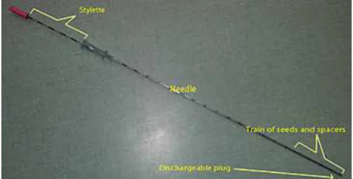
tumor and this is the case where multiple needle insertions from different angles and orientation and at different depths are required. This instrument has been designed to work with Elekta interstitial brachytherapy needles such as the ones in Fig.1, but with few reconfigurations, the instrument can work with other types of needles also. Another peculiarity of this automated medical instrument is that it is designed to be mounted on the PARA-BRACHYROB parallel robot.
Fig. 1. Brachytherapy needle example
The robotic assisted protocol (Fig. 2) for the insertion procedure of multiple brachytherapy needles inside the target area which is represented by an interest zone inside the patient’s tumor, requires a few steps:
1. The first step in a robotic brachytherapy procedure consists in performing the homing operation for the entire robotic system. 2. While in the homing position, the
brachytherapy instrument should have the magazine loaded by a clinician with up to 6 brachytherapy needles.
3. Once the loading by the clinician of the brachytherapy needles has been done, the robotic system with the brachytherapy instrument can be positioned at the initial skin penetration point also called as insertion point.
4. At this point, the brachytherapy instrument, will load in the claws a brachytherapy needle from the magazine needle holder.
6. When the pre-insertion position of the needle is validated, the actual insertion of the needle takes place.
7. Brachytherapy needle is inserted inside the patient’s body towards the target point. 8. At this point, if the needle has reached the
target position (validated by the CT), the instrument releases the needle that has inserted, and retracts entirely allowing access to the needle for seed delivering. If the desired position has not been reached or if during the insertion the needle deflects from the imposed trajectory, it is retracted and re-inserted on the right path.
9. Another needle is loaded from the magazine holder into the brachytherapy instrument and the steps 4 – 8 are repeated until there all the required needles have been inserted and placed into the tumor.is no need for more needle insertion inside the patient, or all the needles have been used, in which case, the protocol goes back to step 1.
10.After all the required needles have been inserted the inner rod of the needles is retracted by a clinician and direct access from the radiation source to the tumor will been achieved via the needle.
Fig. 2 Brachytherapy needle insertion protocol
One of the limitations of the needle insertion instrument consists in the required number of needles, since some medical brachytherapy procedures can require up to 12 needles inserted inside the patient’s tumor, which is quite a large number, especially considering the tight space inside the CT.
Another issue that will have to be addressed in designing the final solution of the brachytherapy instrument consists in the possibility to sterilize it, but this drawback can
be solved if the instrument actuators and materials used are sterilizable.
3. THE BRACHYTHEAPY INSTRUMENT
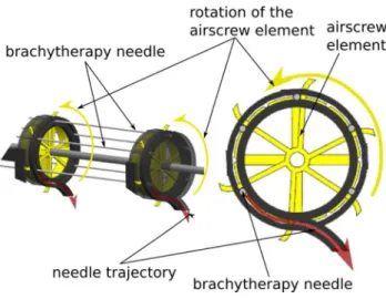
The medical instrument (Fig. 4) has two main mechanical subsystems designed to load one needle at a time, from a magazine and then insert it on la linear path until the established depth. The first subsystem is the needle storage mechanism (Fig. 3) with a total capacity of 6 brachytherapy needles and the second subsystem is the loading/insertion mechanism (with the main component being the needle gripper).
The two subsystems (actuated by two rotatory motors) work together with a synchronized motion as follows:
1. With the actuation of motor 1, both the needle storage airscrew element, and the needle loading gripper will rotate. The needle is guided (by the storage airscrew element) through a circular channel (Fig. 3) and at the end of the channel it is grabbed by the gripper. This synchronous motion is provided by the actuation of two shafts simultaneously (both with motor 1). The two shafts rotate in opposite directions with different speeds (1:6) due to the usage of a planetary gear. This makes possible the loading stage of a single needle, and the mounting of 6 needles. 2. After the loading stage, the needle is fixed in
the gripper, and the pre-insertion stage begins (actuated by motor 2 through a screw/nut mechanism). I.e. the needle is moved on a linear path until it enters from the needle tip guiding element. From here the insertion stage is performed until a predefined depth, followed by the release of the needle with the actuation of motor 1 (30o opposite of the loading direction). The shaft that guides the gripper allows both the translation on a linear path and the rotation of the gripping claws. 3. With the needle inserted the loading
One critical aspect that has to be pointed out is that after the needle is released, the robot has to retract the medical instrument on the same linear path that has been used for the needle insertion until the needle exits the needle tip guiding element hole, in order to leave the needle inside the tissue without causing trauma.
Fig. 3. The needle storage subsystem
4. THE EXPERIMENTAL MODEL
The cad model of the brachytherapy instrument is presented in Figure 5. The chosen actuators were from Maxon motors [10] with an output torque of 0.2 Nm. In order to increase the efficiency of the screw/nut mechanism with balls from SKF [11] and rail/sledge with balls from TBI motion [12] were used with reduced friction coefficients. All the other components of the medical instrument were 3D printed with
ABS plastic material within the CESTER research center on a Stratasys FORTUS 380mc production system.
Fig. 4. The brachytherapy instrument (experimental model)
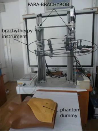
Figure 6 illustrates the brachytherapy instrument mounted on the PARA-BRACHYROB robotic system. The authors have performed preliminary experiments with the medical instrument as follows:
1. The determination of the tissue resistance to needle insertion was performed with a ZWICK/ROELL testing pressing equipment [13], by inserting the brachytherapy needles into a pig leg (in vitro). Out of 10 experimental trials the maximum resistance to needle insertion was 12 N. The experiments were necessary prior to building the prototype in order to find the output torque needed for the mechanism to function.
2. A slight incision on the skin before inserting the needle proved a more linear increase in the tissue resistance. This small incision is valuable for the insertion accuracy since it reduces the risk of bending the needle. 3. To determine if the loading mechanism is
reliable, 50 experimental trials were conducted by only testing the needle loading stage. Out of the 50 trials 45 were successful (90% success rate), while in the other trials the needle load failed due to various reason (friction between the needle and the circular path in which is guided, improper gripping due to elasticity of the ABS). All the discovered issues will be addressed to optimize the functionality of the medical instrument.
Fig. 6. Experimental model mounted on the parallel robot PARA-BRACHYROB
4. A set of experiments were performed based on a medical scenario using a human torso and ballistic gel. In this stage of development the accuracy of the needle placement was neglected. The scenario was built to analyze the behavior of the robotic system with the brachytherapy mounted. I.e. the needle was loaded and the robot guided the instrument to
the insertion point (located on the abdomen of the phantom dummy). Next the needle was inserted through the dummy’s abdomen into a gel. The needle was released and the robotic system retracted the instrument on la linear path to fully free the needle. A second needle was loaded and inserted using different points (insertion and target), and the robot retracted again the instrument. Out of 20 trials using of this experimental scenario no collisions were reported (between the inserted needle and the brachytherapy instrument). Further testing and experimental data is required to test the accuracy of the instrument as well.
5. CONCLUSION
Based on the design requirements of the brachytherapy medical procedure, a novel automated medical instrument was proposed. The instrument is capable of holding 6 brachytherapy needles which are loaded automatically, one by one and then inserted to a predefined depth.
Despite the mechanical complexity of the medical instrument, the experimental data shows good results in the functionality; however optimizations have to be made in order to further improve the reliability of the instrument. Experimental scenarios were performed on a human torso, with the medical instrument mounted on the PARA-BRACHYROB medical robot .
Further research is proposed to determine the accuracy and to optimize the medical instrument.
6. ACKNOWLEDGMENTS
8. REFERENCES
[1] Gerbaulet, Alain; Pötter, Richard., Mazeron, Jean-Jacques., Meertens, Harm., Limbergen, Erik Van, eds. (2002). The GEC ESTRO
handbook of brachytherapy, ESTRO, OCLC
52988578, Leuven, Belgium, 2002.
[2] Stewart AJ et al., Radiobiological concepts for brachytherapy. In Devlin P. Brachytherapy, Applications and Techniques. Philadelphia, 2007
[3] Yu Y1., Podder T., Zhang Y., Ng WS., Misic V., Sherman J., Fu L., Fuller D., Messing E., Rubens D., Strang J., Brasacchio R., Robot-assisted prostate brachytherapy,
Medical Image Computing and Computer Assisted Intervention.;9(Pt 1):41-9,2006 [4] Tarun K. Podder, et al., AAPM and
GEC-ESTRO guidelines for image-guided robotic brachytherapy: Report of Task Group 192, Medical Physics, Volume 41, Issue 10, pp i-vi, DOI: 10.1118/1.4895013 October 2014 [5] Dan Stoianovici et al., ‘‘MRI Stealth’’
robot for prostate interventions, Minimally Invasive Therapy. 2007; 16:4; 241–248, Urology Robotics, Johns Hopkins Medicine, Baltimore, MD, USA
[6] Podder et al., MIRAB: An Image-Guided
Multichannel Robot for Prostate
Brachytherapy, International Journal of Radiation OncologyBiologyPhysics, DOI: 10.1016/j.ijrobp.2010.07.1876, November 2010
[7] Trejos et al., MIRA V: An Integrated System for Minimally Invasive RobotAssisted
Lung Brachytherapy, IEEE International
Conference on Robotics and Automation, Pasadena, CA, USA, May 19-23, 2008
[8] Doina Pisla, Dragos Cocorean, Calin Vaida, Bogdan Gherman, Adrian Pisla, Nicolae Plitea, Application oriented design and simulation of an innovative parallel robot for brachytherapy, ASME 2014 International Design Engineering Technical Conferences and Computers and Information in Engineering Conference, DOI: 10.1115/DETC2014-35047, August 2014, [9] Cocorean D., Development of new parallel
robots for brachytherapy (Published in Romanian), PHD Thesis, Cluj-Napoca, 2015 [10] http://www.maxonmotor.com/, 2017 [11] http://www.skf.com, 2017
[12] http://www.tbimotion.com.tw/, 2017 [13] https://www.zwick.com/, 2017
Un instrument inovativ pentru brahiterapia asistată robotic utilizat in tratamentul cancerului
Lucrarea prezintă un nou instrument automatizat multi-ac de brahiterapie cu 6 ace destinat a fi utilizat de un sistem robotizat medical ca si efector. Pe baza cerințelor derivate din procedura medicală, instrumentul medical utilizează un mecanism de stocare al acelor și un dispozitiv de prindere care are o mișcare sincronă pentru a efectua încărcarea, introducerea și eliberarea acului. Funcționalitatea instrumentului este explicatăși este prezentat modelul prototip. Sunt prezentate și scenarii experimentale urmate de concluziile și cercetările viitoare propuse care vizează optimizarea instrumentului medical și validarea acestuia pentru procedura medicală.
Iosif BIRLESCU, PhD Student, Technical University of Cluj-Napoca, Research Center for Industrial Robots Simulation and Testing, [email protected].
Florin CRACIUN, PhD Student, Technical University of Cluj-Napoca, Research Center for Industrial Robots Simulation and Testing, [email protected].
Călin VAIDA, PhD, Assoc. Prof., Technical University of Cluj-Napoca, Research Center for Industrial Robots Simulation and Testing, [email protected].
Bogdan GHERMAN, PhD, Lect., Technical University of Cluj-Napoca, Research Center for Industrial Robots Simulation and Testing, [email protected].