Determination of human absorbed dose from
[
153Sm]-Samarium maltolate based on distribution
data in rats
INTRODUCTION
Projecting radiotracer distribution and dosimetry across species from small animals
such as rats to humans can be useful for accelerating the development of radioactive
compounds to be used in clinical settings (1-2). Preclinical studies of radiopharmaceutical
behavior are useful for predicting pharmacokinetics and metabolism in humans.
Animal studies provide valuable distribution
therapeutic radiopharmaceuticals (1). Owing to
the introduction of new therapeutic radiocompounds to deliver high doses of radiation to selected target organs with an aim
of minimizing unwanted radiation to surrounding healthy tissue (5), there is a need for
ef%icient evaluation of the time-dependent distribution of such tracers in animals (2-4).
Concerning to the recent project on the production and evaluation of lanthanide-based
labeled compound using samarium-153 for
targeted radionuclide therapy (TRT)
H. Rezaeejam
1, A. Hakimi
2*, A.R. Jalilian
3, P. Abbasian
2,
S. Shirvani-Aran
3, M. Ghannadi-Maragheh
31Department of Medical Physics and Biomedical Engineering, Faculty of Medicine, Tehran University of Medical
Sciences, Tehran, Iran
2Department of Energy Engineering and Physics, Amir Kabir University of Technology, Tehran, Iran 3Nuclear Science and Technology Research Institute, Tehran, Iran
ABSTRACT
Background: Therapeu c radiopharmaceu cals are designed to deliver high doses of radia on to selected target organs with an aim of minimizing unwanted radia on to surrounding healthy ssue. Due to the poten al of
targeted radiotherapy to treat a wide range of malignant condi ons, [153Sm]-samarium maltolate was developed for possible therapeu c
applica ons. Materials and Methods: The organ radia on-absorbed doses have been evaluated for human based on animal data. A%er intravenous administra on of 153Sm-Mal to four groups of rats, they were sacrificed at exact me intervals and the percentage of injected dose per gram of each organ was calculated by direct coun ng from rat data. Then S values for 153Sm by using specific absorbed frac ons were calculated. By taking advantage of
the formula on that Medical Internal Radia on Dose suggests, radia on-absorbed doses for all organs were calculated and extrapolated
from rat to human. Results: From rat data, it is es mated that a 185-MBq injec on of 153Sm-Mal into a human might result in the highest absorbed dose in the lymphoma ssues (liver 176.3, lungs 68, spleen 66.8 and sternum 19 mGy), especially in liver respect to the other ssues. Conclusion:These results suggest 153Sm-Mal as an efficiently new therapeu c agent in order to overcome possible lympha c malignancies.
Keywords: Absorbed dose, biodistribution, M I R D, internal dosim etry,
153Sm-Mal. * Corresponding author:
Mr. Amir Hakimi, Fax: +98 21 22803280
E-mail:[email protected] Revised: June 2014
Accepted: July 2014
Int. J. Radiat. Res., April 2015; 13(2): 173-180
►
Original article
DOI: 10.7508/ijrr.2015.02.008
[153Sm]-samarium maltolate was performed (7). Radioisotopes with medium-energy beta
emissions and half-life of a few days are attractive candidates for targeted irradiation (8).
Samarium-153 has favorable radiation characteristics, medium-energy beta particle
emissions (Emax = 808 keV) which is desirable for treatment, and range of about 3.0 mm in tissue has potential for targeted tumor radiotherapy (9). It emits medium-energy gamma photon (103 keV) in addition to particle
emissions which make it suitable for monitoring the therapy with imaging, and for continuous follow-up of the absorbed dose distribution (5).
The use of targeted radionuclide therapy (TRT) for the treatment of cancer necessitates
the development of dosing schedules that minimize the exposure of healthy tissues to radiation while maximizing the radiation dose
received by the tumor (10). The aim in targeted radiotherapy is the selective delivery of radiation to cancer cells in a way that causes
minimal toxicity to surrounding normal tissues. The basis for successful radionuclide therapy is selective concentration and prolonged retention of the radiopharmaceutical within the tumor (11).
Maltol (3-Hydroxy-2-methyl-4-pyron) is commonly formed when sugars are heated.
Maltol loses its hydroxyl proton at neutral to basic pH levels, forming the maltolate anion; this anionic molecule forms a strong bidentate/
tridentate chelate with gallium, iron, zinc, aluminum, vanadium and many other metals (12).
Most of maltolate metal complexes (e.g. Gallium-maltolate (13) and 177Lu-Maltolate (12))
are reported as biologically active compounds. Some of maltolate metal complexes are reported as biologically active compounds for lymphoma treatment (15) and apoptotic cell death (16).
Treatment with radiopharmaceuticals may result in abnormalities in other organs so it is
important to estimate organ doses (17). Dosimetry represents meanwhile a precious
guide for the selection of radionuclides as well
as for therapy optimization (18). There exist various methods to estimate internal doses and
new studies promises new perspective to come
(19). Internal Commission on Radiological Protection (ICRP) and Medical Internal
Radiation Dose (MIRD) methodologies are the
most commonly used internal dosimetry methods. The various ICRP and MIRD internal
dosimetry models are similar in terms of their
assumptions and de%ining equations. This similarity is obscured by differing terminology
and notation, and these differences contribute to confusion in understanding these models (20).
The Committee on Medical Internal Radiation Dose of the Society of Nuclear Medicine developed a methodology to perform
radiation absorbed dose calculations. These calculations are performed to assess the risks
associated with the administration of radiopharmaceuticals for medical studies including imaging, therapy, and metabolic applications. The MIRD technique is a computational methodology that facilitates absorbed dose calculations to speci%ied target
organs from radioactive decays that occur in source organs. The source organs contain the radioactive material, and the target is the organ in which the dose is calculated. The target and
source organs can be the same tissue. In subsequent discussion, the terms tissue and
organ are used interchangeably. To specify the MIRD methodology, it is necessary to de%ine several terms which detailed descriptions were presented in methods.
In this investigation, a precise description of organ distribution has been used owing to a
large amount of distribution data (source organs in dosimetry calculations); efforts were
taken to estimate the effective radiation dose absorbed into human organs following intra vascular administration of 153Sm-Mal by using the distribution data for normal rats.
MATERIALS
AND METHODS
Samarium-153 was produced by neutron irradiation of 1 mg of enriched [152Sm]Sm2O3 (152Sm, 98.7% from ISOTEC Inc.) according to reported procedures (19) in the Tehran Research Reactor at thermal neutron %lux of 5×10¹³ ncm-2s
-1 for 5d. 153Sm is produced according to the reaction 152Sm (n,γ) 153Sm by σ=206 barns for
thermal neutron and disintegrates via 3 main
routes by 100% β- emission to levels in 153Eu. 153Sm is not a carrier free radioisotope and its
speci%ic activity was 22-28 GBq/mg. The irradiation 153Sm was dissolved in 200 μl of 1.0
M HCl, to prepare [153Sm]153SmCl3 and diluted to the appropriate volume with ultra pure water, to produce a stock solution.
Chromatography paper (Whatman No. 2) was obtained from Whatman (Maidstone, UK). Radio
-chromatography was performed using a Bioscan AR-2000 radio TLC scanner (Bioscan,
Paris, France). A high purity germanium (HPGe) detector coupled with a Canberra™ (model GC1020-7500SL) multichannel analyzer and a
dose calibrator ISOMED 1010 (Dresden, Germany) were used for counting distributed
activities in rat organs. All other chemical reagents were purchased from Merck
(Darmstadt, Germany). Calculations were based on the 103 keV peak for 153Sm. All values were expressed as mean ± standard deviation (Mean±SD) and the data were compared using
Student’s t-test. Statistical signi%icance was de%ined as Pvalue less than 0.05.
Preparation of 153Sm-Mal
The labeling was developed in ethanolic media. Brie%ly, 153SmCl3 (111 MBq, 0.1 ml) was
added to a borosilicate vial and dried by warming (50°C) under a nitrogen %low for about
15 minutes. Then, maltol (30mg, 0.25 mmol) dissolved in absolute ethanol (1 ml) was added
to the dried residue and the mixture agitated
and incubated at 60°C for 2 hours. The radiochemical purity (21) of free samarium and
Sm-MAL were determined by counting Whatman No.2 sheets as stationary phase using
various mobile phases (A:
ammo-nia:water:methanol (2:40:20), B: 1mM DTPA aqueous solution, C: %10 ammonium acetate:methanol system, 1:1). After obtaining the desired radiochemical purity, the ethanolic solution was concentrated by warming 40–50°C to 0.05 ml and then diluted to a 5% solution by adding 1 ml of normal saline.
Biodistribution studies of radiopharmaceutical The distribution of 153Sm-Mal among tissues was determined for untreated rats. Five male
rats each were sacri%iced by CO2 asphyxiation (after anesthesia induction using propofol/ xylazine mixture) at 2, 4, 24, 28 and 96 h (3 rats in each time) after injection of 4.80±0.18 MBq of
%inal complex through their tail vein and exsanguinated, and the tissues (blood, heart,
lung, spleen, intestine, kidneys, liver, muscle and bone) were rapidly removed. During the entire study, autoclaved food and drinking water were available ad libitum. Animal studies were carried
out in accordance with the UK Biological Council’s Guidelines of the Use of Living Animals
in Scienti%ic Investigations, 2nd edition (22) (approved by Iranian Ministry of Health and Medical Education).
Activity measurement
The activity in the syringes was measured
before the injection and then after administration of the radiopharmaceutical
products with the well-type ionization chamber (CRC-15R, Capintec, Ramsey, New Jersey, USA), all measurements were background subtracted
and then averaged together. Uncertainties in the determinations were minimal, because each assay collected at least 1000 count.
For each of these measurements, three samples were weighed and then counted by
HPGe detector to determine the percentage of
injected activity per gram (%IA/g) (which was equivalent to the percentage of injected activity
per gram %IA/gr ≡ Percentage injected dose per
gram %ID/gr); all the organ activity measurements were normalized to injected activity. All samples were
background-subtracted and nondecay-corrected to the time of killing, and then similar samples were averaged together.
To convert the counts to activity, following formula was used:
(1) Where the “area” term is the number of
counts in the selected window (in the con%ined energy) and the t represents the counting time in this study; it was 100s and the term “Eff” is the detector ef%iciency (in the selected energy) were given from the detector manual and the term
“Br” is de%ined as the decay yield of the selected gamma energy.
The 153Sm activity concentration at time t,
Ctissue (t) was then calculated the percentage of
injected activity per gram of tissue (%IA/g) with equation (2):
(2) Where Atissue(t) is the 153Sm activity in the
sample, Mtissue is the mass of the sample and Atotal is the total activity of 153Sm injected into the rat. Estimate the human absorbed dose from rat data
To extrapolate biokinetic data from animals
to humans different methods have been previously used (23). The proposed method of
Sparks and Aydogan (24) on the basis of percent
injected dose (or injected activity) per gram (%ID/gr) unit was used as follows:
(3)
The mean weight of the organs harvested
from the animals and standard weights established for the adult male human organs
(25-27) was used. The rat’s organ masses and the
ration of organ mass to body mass in humans
and in rats are shown in table 1 according to peters and Boyd (28).
Dosimetric calculations
For each group of rats, the non-decay corrected time activity curves were generated
for the source organs including the followings: the lung, kidney, spleen, liver, a trabecular bone and blood.
The distribution data were plotted out, and the points were %itted to a single exponential equation by the least-squares linear regression
method from radioactivity from individual organs, %itted a mono-exponential curve.
The non-decay-corrected time-activity curves were extrapolated to in%inity by %itting the tail of
each curve to a mono-exponential and integrated, resulting in a concentration integral
for each organ. The data points representing the
percentage-injected dose (%ID/organ) were created and %itted to a mono-exponential, a bi-exponential or an uptake-and-clearance
curve. The rat data were assumed to approximate the biokinetic parameters in the
human.
The use of tables presented in the Medical Internal Radiation Dose (MIRD) No. 11 (29) which
are used to compute the absorbed dose, it is necessary to calculate the cumulated source activity (Ah) in each organ according to the
equation (4):
(4)
The liver, kidneys, blood, spleen, lung, stomach, Lower large Intestine, colon, muscle,
heart, sternum and trabecular bone were used
as the primary source organs. The MIRD formulation was applied to calculate the
absorbed radiation dose for various organs (30). (5)
Where the mean absorbed dose
stated in (mGy) to a target organ rk from a
radiopharmaceutical distributed identically in a source organ rh has been formulated by the
MIRD committee. The S (rk ← rh) expressed in
(mGy/MBq-s) represented the speci%ic absorbed
fraction of energy for the target organ rk for particles emitted in the source organ rh(31).
Table 1.The rat’s organ masses and the ra o of organ mass to body mass in human and in rat.
Tissues Rat (%) Weight (g) Humana (%)
Adrenal 0.03 0.04 0.02
Urine 0.06 0.09 0.52
Intes ne 0.60 0.90 0.22
Kidney 0.78 1.17 0.44
Liver 4.28 6.42 2.57
Lung 0.66 0.98 1.42
Muscle 53.4 80.16 68.6
Ovary 0.03 0.04 0.02
Pancreas 0.31 0.47 0.14
Bone 1.00 1.5 1.43
Skin 19.9 29.9 13.0
Spleen 0.36 0.54 0.53
a Human data according to ICRP 22 (23).
Radiation-absorbed dose calculation
Methods compatibles with those recommended by the MIRD Committee of the
Society of Nuclear Medicine were used to determine the absorbed doses to the human normal organs and the whole body (32-33).
RESULTS
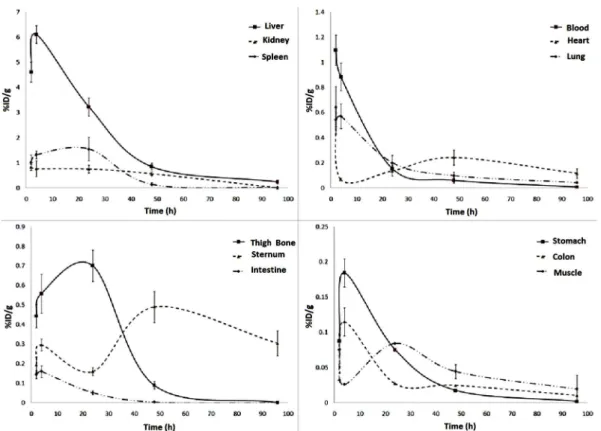
Table 2 demonstrated the accumulation of 153Sm-Mal in the major organs. In addition the clearance curves from each organ of the rats shown in %igure 1. The results showed that most of the activity was accumulated in the liver. The absorption of the colon, Lower large Intestine, muscle and stomach was absolutely negligible,
and it was very interesting because the samarium-153 chelates e.g. 153Sm-EDTMP, 153Sm
-NTMP, 153Sm-HEEDTMP, 153Sm-NBTP and 153Sm
-NTA major absorption were in the skeletal system (34), but in complex 153Sm-Mal
the major absorption was in the liver. It means
that the absorption of new complex is completely different from other samarium-153
chelates that are potential therapeutic bone agents.
The radiation absorbed organ doses estimated in humans according to the biological
data from the rat study are shown in table 3.
Table 2.Distribu on of 153Sm-Mal in different me points.
Organs Time (Mean
a ±SD)
2 h 4 h 24 h 48 h 96h
Blood 1.0969 ± 0.125 0.8861 ± 0.11 0.1543 ± 0.03 0.0607 ± 0.02 0.008 ± 0.001
Heart 0.6494 ± 0.16 0.0694 ± 0.01 0.1351 ± 0.04 0.2426 ± 0.06 0.1163 ± 0.04
Lung 0.5423 ± 0.11 0.5717 ± 0.10 0.2018 ± 0.06 0.0980 ± 0.03 0.0433 ± 0.007
Stomach 0.0874 ± 0.01 0.1846 ± 0.02 0.0754 ± 0.00 0.0175 ± 0.00 0.0019 ± 0.000
Colon 0.0335 ± 0.00 0.1152 ± 0.02 0.0273 ± 0.00 0.0249 ± 0.00 0.0103 ± 0.00
Intes)ne 0.1446 ± 0.02 0.1599 ± 0.03 0.0517 ± 0.01 0.0042 ± 0.00 0.0004 ± 0.000
Liver 4.6164 ± 0.4 6.1051 ± 0.35 3.2293 ± 0.37 0.8590 ± 0.13 0.2439 ± 0.08
Spleen 1.0166 ± 0.3 1.3231 ± 0.16 1.5545 ± 0.47 0.1482 ± 0.06 0.023 ± 0.003
Kidney 0.8232 ± 0.12 0.7729 ± 0.31 0.7591 ± 0.16 0.5676 ± 0.09 0.012 ± 0.004
Muscle 0.0753 ± 0.02 0.0261 ± 0.00 0.0842 ± 0.00 0.0446 ± 0.01 0.01962 ± 0.02
Sternum 0.1938 ± 0.04 0.2968 ± 0.03 0.1601 ± 0.02 0.6909 ± 0.08 0.304 ± 0.064
Thigh Bone 0.4438 ± 0.06 0.5590 ± 0.10 0.7005 ± 0.08 0.0896 ± 0.02 0.0012 ± 0.000
a Data were presented as the percentage of administrated ac vity per gram (non-decay corrected).
The rats’ biodistribution data were projected into the humans. A 185-MBq (5-mCi) injection of 153Sm-Mal into the human body might result in an estimated absorbed dose of 53.22 mGy for the total body and the highest absorbed dose was observed in the liver with 151.14 mGy followed by spleen, lungs, kidneys, bone and red marrow tissues received 64.01, 52.35, 34.04, 18.87 and 18.13 mGy respectively.
Table 3. The Human absorbed dose es ma on.
Organs Absorbed dose
(mGy/MBq)a
Absorbed dose (mGy)b
Adrenals 0.0164 3.0340
Bladder wall 0.00696 1.2876
Bone 0.119 22.015
Brain 0.00723 1.3375
Breasts 0.00816 1.5096
Stomach wall 0.01003 1.8555
Heart wall 0.0220 4.070
Kidneys 0.244 45.140
Liver 0.953 176.305
Lungs 0.368 68.080
Ovaries 0.00779 1.4411
Pancreas 0.0153 2.8305
Red marrow 0.0996 18.426
Skin 0.00711 1.3153
Spleen 0.361 66.785
Testes 0.00669 1.2376
Thyroid 0.00721 1.3338
Uterus 0.00784 1.4504
Total body 0.3483 64.4355
a Might had some over/under es ma on according to table 3. b 185 MBq Injected Ac vity
DISCUSSION
In rat studies, the tracer was mostly accumulated in the liver, which was the major
organ of accumulation throughout the study as
shown in table 2. The liver to blood activity concentration ratio was about 20 at 24 h and up
to 14 at 48 h post-injection. The livers to muscle ratios were more than 30 at 24 and about 20 at 48 h post-injection.
There are four methods using animal data to
predict human residence times: %irst, no extrapolation assumes that the percentage of
injected activity (%IA ≡ %ID) at any time in the human organ is the same as in the animal organ. Second, relative organ mass extrapolation study was carried out, third using physiological time extrapolation and fourth, using a combination of the mass and time methods by applying both the mass and physiological time extrapolations to the animal data (24).
The amount of the radiation-absorbed dose to a human was calculated by determining the
distributions in rats, obtaining a cumulated
activity value, multiplying by the ratio of organs to extrapolate to humans (using relative organ mass extrapolation) and then multiplying the
converted rats’ cumulative activity to the S factor table of 153Sm (35-36). For humans, the highest radiation-absorbed doses per unit-administered activity were calculated for
liver (0.953 mGy/MBq), lungs (0.361 mGy/MBq) and spleen (0.368 mGy/MBq). The absorbed doses to the kidneyes, bone and red marrow were less than 0.2 mGy per MBq. The total body dose was estimated to be 0.348 mGy/MBq as
shown in table 3. The discrepancy between different animal models is important if absorbed
dose estimations for humans are derived from animal models. Obviously the distribution of a radiopharmaceutical parameter varies not only from rodents to humans, but also between rats and mice (37-39).
Extrapolation between various animal species is more reliable than extrapolation between animals and humans (24), but previous
studies have demonstrated the usefulness of
using animal distribution as a model for absorbed dose estimations in humans (40-43).
Figure 1. The clearance curves from each organ of the rats.
Using physiological time extrapolation improves the mean result. However, there appears to be some increase in severe overestimates (more than 10-fold) with the time
extrapolation method (24). Using the physiological time extrapolation is not valid for
this study because one cannot estimate the exact value resident time. Therefore, for this study using relative organ mass extrapolation is more precise than physiological time extrapolation.
New therapeutic radiopharmaceutical signi%icantly accumulated in liver and increased
after administration for 4 h. Blood and heart uptake showed their maximum at %irst 2 h and
then decreased as shown in %igure 1.
CONCLUSION
Radiation dosimetry for 153Sm-Mal was estimated for humans based on distribution data
of 153Sm-Mal in normal rats. The distribution of 153Sm-Mal in rats demonstrated signi%icant liver
uptake and low muscle and blood uptake; however, the kidney uptake is somewhat high.
The hypothesis for high uptake of the kidney is excretion of the complex by the urinary system 2 h post-injection till 48 h post-injection.
Although further dosimetry work should be performed on humans as this compound become useful in the clinic, these estimates can be used to predict potential absorbed doses in humans and for planning human studies.
Con'licts of interest: none to declare.
REFERENCES
1. Shanehsazzadeh S, Jalilian AR, Sadeghi HR, Allahverdi M
(2009) Determina on of human absorbed dose of 67GA-DTPA-ACTH based on distribu on data in rats.
Radia on protec on dosimetry, 134(2): 79-86.
2. Moghaddam AK, Jalilian AR, Haya V, Shanehsazzadeh S (2010) Determina on of human absorbed dose of 201Tl
(III)-DTPA-HIgG based on biodistribu on data in rats.
Radia on protec on dosimetry, 141(3): 269-274.
3. Abbasian P, Foroghy M, Jalilian AR, Hakimi A Shirvani-Arani
S (2014) Modeling the me dependent biodistribu on of Samarium-153 ethylenediamine tetramethylene phosphonate using compartmental analysis. Reports of Prac cal Oncology & Radiotherapy, 19(3):214-220.
4. Sardari D and Hakimi A (2012) Modeling the me dependent distribu on of a new 153Sm complex for targeted radiotherapy purpose. Reports of Prac cal Oncology and Radiotherapy, 17(6):358-362.
5. Naseri Z, Hakimi A, Shirvani-Arani S, Jalilian AR, Bahrami-Samani A, Nema Kharat M, Ghannadi-Maragheh M (2012) Prepara on and quality
control of 153Sm-[tris (1, 10-phenanthroline) samarium (III)] complex. Int J Radiat Res, 10(1): 59-62.
6. Naseri Z, Hakimi A, Jalilian AR, Nema Kharat A, Shirvani-Arani S, Bahrami-Saman, Ghannadi-Maragheh M
(2012) Synthesis, quality control and biological evalua on
of tris [(1, 10-phenanthroline)[153Sm] samarium (III)] trithiocyanate complex as a therapeu c agent.
Radiochimica Acta, 100(4): 267-271.
7. Naseri Z, Hakimi A, Jalilian AR, Kharat AN, Bahrami-Samani A, Ghannadi-Maragheh M (2011) Prepara on and quality control of the [153Sm]-samarium Maltolate Complex as a
Lanthanide Mobiliza on Product in Rats. Scien a pharmaceu ca, 79(2): 265.
8. Anderson PM, Wiseman GA, Dispenzieri A, Arndt CA, Hart-mann LC, Smithson WA, Bruland OS (2002) High-dose sa-marium-153 ethylene diamine tetramethylene phospho-nate: low toxicity of skeletal irradia on in pa ents with osteosarcoma and bone metastases. Journal of Clinical Oncology, 20(1): 189-196.
9. Hakimi A, Jalilian AR, Arani SS, Samani AB, Maragheh MG,
Arbabi A (2010) Prepara on and quality control of 153Sm-[tris (1, 10-phenanthroline) samarium (III)] complex as a therapeu c compound. Iranian Journal of
Nuclear Medicine, 18(Suppl 1): 111.
10. Ferl GZ, Kenanova V, Wu AM, DiStefano JJ (2006) A two- ered physiologically based model for dually labeled
single-chain Fv-Fc an body fragments. Molecular cancer therapeu cs, 5(6): 1550-1558.
11. Ersahin D, Doddamane I, Cheng D (2011) Targeted radionuclide therapy. Cancers, 3(4): 3838-3855.
12. Bernstein LR, Tanner T, Godfrey C, Noll B (2000) Chemistry and pharmacokine cs of gallium maltolate, a compound with high oral gallium bioavailability. Metal Based Drugs, 7: 33-48.
13. Chitambar CR, Purpi DP, Woodliff J, Yang M, Wereley JP (2007) Development of gallium compounds for treatment of lymphoma: gallium maltolate, a novel hydroxypyrone gallium compound, induces apoptosis and circumvents lymphoma cell resistance to gallium nitrate. Journal of Pharmacology and Experimental Therapeu cs, 322(3): 1228-1236.
14. Hakimi A, Jalilian AR, Arani SS, Samani, AB, Maragheh MG,
Arbabi A (2010) Produc on and evalua on of Lute um-177 Maltolate as a possible therapeu c agent.
Iranian Journal of Nuclear Medicine, 18(Suppl 1): 143.
15. Thompson KH, Barta CA, Orvig C (2006) Metal complexes of maltol and close analogues in medicinal inorganic chem-istry. Chemical Society Reviews, 35(6): 545-556.
16. Eiko S, Iho Y, Yamada T, Suzuki Y, Ohyashiki T (2007
Involvement of NO genera on in aluminum-induced cell death. Biological and Pharmaceu cal Bulle n 30, no. 8: 1390-1394.
17. Shahbazi-Gahrouei D and Nikzad S (2011) Determina on
of organ doses in radioiodine therapy using medical internal radia on dosimetry (MIRD) method. Int J Radiat
Res,8(4): 249-252.
18. Schuchardt C, Kulkarni HR, Prasad V, Zachert C, Müller D,
Baum RP (2013 The Bad Berka dose protocol: Compara ve results of dosimetry in pep de receptor
radionuclide therapy using 177Lu-DOTATATE, 177Lu-DOTANOC, and 177Lu-DOTATOC. In Theranos cs,
Gallium-68, and Other Radionuclides (pp. 519-536). Springer Berlin Heidelberg.
19. Cremonesi M, Ferrari M, Di Dia A, BoOa F, De Cicco C, Bo-dei L, Paganelli G (2011) Recent issues on dosimetry and
radiobiology for pep de receptor radionuclide therapy. Journal of Nuclear Medicine and Molecular
Imag-ing, 55(2): 155-167.
20. Bevelacqua JJ (2005) Internal dosimetry primer. Radia on Protec on Management,22(5): 7.
21. Jalilian AR, HakimA, Garousi J, Bolourinovin F, Kamali-Dehghan M, Aslani G (2008) Development of
[201Tl](III) oxinate complex for in-vitro cell labeling. Int J Radiat Res,6(3): 145-150.
22. Manual for produced radioisotopes, IAEA, Vienna (2003) IAEA-TECDOC-1340, ISBN 92-0-101103-2, ISSN 1011-4289,
© IAEA (2003), P. 71, printed by the IAEA in Austria, January 2003.
23. ICRP, (1992) Radiological Protec on in Biomedical Research. ICRP Publica on 62. Ann. ICRP 22 (3).
24. Sparks RB and Aydogan B (1999) Comparison of the effec veness of some common animal data scaling techniques in es ma ng human radia on dose. In Sixth
Interna onal Radiopharmaceu cal Dosimetry Symposium. Stelson AS, editor. Oak Ridge: Associated Universi es (pp. 705-716).
25. Richmond CR (1986)ICRP publica on 23. Interna onal Journal of Radia on Oncology* Biology* Physics, 12(3): 433.
26. Loevinger R, Budinger TF, Watson EE (1988) MIRD primer
for absorbed dose calcula ons. New York: Society of Nuclear Medicine.
27. Cristy M and Eckerman KF (1987) Specific Absorbed Frac ons of Energy at Various Ages from Internal Photon
Sources: 1, Methods. Atomic Energy Research Establishment.
28. Peters JM and Boyd EM (1966) Organ weights and water levels of the rat following reduced food intake. The Jour-nal of Nutri on, 90(4): 354-360.
29. Snyder WS, Ford MR, Warner GG, Watson SB (1975) S" absorbed dose per unit cumulated ac vity for selected
radionuclides and organs. MIRD Pamphlet No. 11. Society of Nuclear Medicine, New York.
30. Snyder WS, Ford MR, Warner GG (1978) MIRD pamphlet no. 5, revised: Es mates of specific absorbed frac ons for photon sources uniformly distributed in various organs of
a heterogeneous phantom. New York, NY: Society of Nuclear Medicine.
31. ICRU, Methods of assessment of absorbed dose in clinical
use of radionuclides. Report No. 32 (Washington, DC: ICRU) (1979).
32. BenedeOo AR (1986) NCRP Report No. 83, The Experimental Basis for Absorbed-Dose Calcula ons in
Medical Uses of Radionuclides Na onal Council on Radia on Protec on and Measurements, Bethesda, NCRP
Publica ons, 1985. Journal of Nuclear Medicine, 27(8): 1378-1379.
33. Goeckeler WF, Edwards B, Volkert WA, Holmes RA, Simon J, Wilson D (1987) Skeletal localiza on of samarium-153 chelates: poten al therapeu c bone agents. Journal of Nuclear Medicine, 28(4): 495-504.
34. Goeckeler WF, Edwards B, Volkert WA, Holmes RA, Simon J, Wilson D (1987) Skeletal localiza on of samarium-153 chelates: poten al therapeu c bone agents. Journal Nucle-ar Medicine, 28(4): 495-504.
35. Radfar E, Jalilian AR, Yousefnia H, Bahrami-Samani A, Ghannadi-Maragheh M (2012) A compara ve study of preliminary dosimetry for human based on distribu on data in rats with 111In, 90Y, 153Sm, and 177Lu labeled rituximab. Nuclear Technology and Radia on Protec on, 27(2): 144-151.
36. Logan KW, Volkert WA, Holmes RA (1987) Radia on dose calcula ons in persons receiving injec on of samarium-153 EDTMP. J Nucl Med, 28: 505-509.
37. SchmiO A, Bernhardt P, Nilsson O, Ahlman H, Kölby L, Maecke HR, Forssell-Aronsson E (2004) Radia on therapy of small cell lung cancer with 177Lu-DOTA-Tyr3-octreotate in an animal model. Journal of Nuclear Medicine, 45(9): 1542-1548.
38. de Jong M, Breeman WA, Bernard BF, Bakker WH, Schaar
M, van Gameren A, et al. (2001) [177Lu-DOTA0, Tyr3] octreotate for somatosta n receptor-targeted radionuclide therapy. Interna onal Journal of Cancer, 92
(5): 628-633.
39. Lewis JS, Wang M, Laforest R, Wang F, Erion JL, Bugaj JE, Anderson CJ (2001) Toxicity and dosimetry of 177Lu-DOTA-Y3-octreotate in a rat model. Interna onal Journal of Can-cer, 94(6): 873-877.
40. Palm S, Enmon RM, Matei C, Kolbert KS, Xu S, Zanzonico PB, Sgouros G (2003) Pharmacokine cs and biodistribu on
of 86Y-trastuzumab for 90Y dosimetry in an ovarian carcinoma model: correla ve MicroPET and MRI. Journal
of Nuclear Medicine, 44(7): 1148-1155.
41. Deterding TA, Votaw JR, Wang CK, Eshima D, Eshima L, Keil R, et al. (2001) Biodistribu on and radia on dosimetry of the dopamine transporter ligand [18F] FECNT. Journal of Nuclear Medicine, 42(2): 376-381.
42. Bélanger MJ, Krause SM, Ryan C, Sanabria-Bohorquez S, Li W, Hamill TG, Burns HD (2008) Biodistribu on and
radia-on dosimetry of [18F] F-PEB in nradia-onhuman primates. Nu-clear Medicine Communica ons, 29(10): 915-919.
43. Kesner AL, Hsueh WA, Czernin J, Padget H, Phelps ME, Silverman DH (2008) Radia on dose es mates for [18F] 5-Fluorouracil derived from PET-based and ssue-based methods in rats. Molecular Imaging and Biology, 10(6): 341 -348.