SURGERY
CASE REPORTSMULTIPLE MYELOMA WITH BILATERAL HUMERUS LOCATION
CASE REPORT
O. Alexa, B. Veliceasa
University of Medicine and Pharmacy “Grigore T. Popa” – Iasi Faculty of Medicine
Discipline of Orthopedics and Traumatology
MULTIPLE MYELOMA WITH BILATERAL HUMERAL LOCATION: CASE REPORT (Abstract): Multiple myeloma, also known as Kahler’s disease or myelomatosis, is a type of cancer that begins in plasma cells. It is the second most common hematologic malignancy (13%) and accounts for 1% of all cancers. A mnemonic sometimes used to remember the common tetrad of multiple myeloma is CRAB: C = Calcium (elevated), R = Renal failure, A = Anemia, B = Bone lesions. Case report: We present the case of an 70- year-old male with bilateral humeral location. Diagnosis was difficult because of atypical X -ray findings in the right shoulder, the classic finding being punched-out lesions. ESR, blood, renal and liver tests were atypical. Conclusion: Although the diagnosis of multiple myeloma is easy, being based on blood and kidney tests and X-ray examination, in some cases an accurate diagnosis is obtained only by pathological examination. Keywords: MULTIPLE MYELOMA, PLASMA CELLS, HUMERUS.
Multiple myeloma (MM) is a cancer of plasma B cells, also known as Kahler's disease or myelomatosis. It is the second most common hematologic cancer (13%) and accounts for 1% of all cancers (1).
The exact cause of multiple myeloma is not known, and most myeloma patients are elderly.
It is located in the bone marrow, espe-cially in the vertebrae, ribs, skull, pelvis and femur and is accompanied by numerous clinical manifestations including anemia, bone lesions, hypercalcemia, renal dysfunc-tion and compromised immune funcdysfunc-tion. A mnemonic sometimes used to remember the common tetrad of multiple myeloma is
CRAB: C = Calcium (elevated), R = Renal failure, A = Anemia, B = Bone lesions” (2).
Diagnosis is based on the presence of
neoplastic plasma cells in the bone marrow or elsewhere extramedullary together with the presence of multiorgan dysfunctions.
CASE REPORT
Patient, aged 70 years, reported about two months earlier a traction injury of the left upper limb and about 1.5 months ago one of the right upper limb for which he presented at the Câmpulung Moldovenesc Hospital, Suceava County, where he was diagnosed with pathological fracture of the middle third of the right humeral shaft and of the proximal third of the left humeral shaft, both treated by plaster splint. Be-cause after about two months both fractures were not consolidated, the patient was referred to our department.
30 years ago and surgical removal of a rib segment about 15 years ago, but was unable to provide detailed information. The patient had a first-degree relative with leukemia.
Physical examination showed an in-crease in volume of the left shoulder and right arm in the middle third. Radiographs showed in the right humerus a relatively well-defined osteolytic area of approxi-mately 30 mm accompanied by fracture (fig. 1A), and in the left humerus a
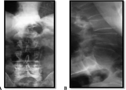
poorly-defined osteolytic area covering the entire proximal humeral epiphysis, with moderate periosteal reaction and extension to the neighboring soft tissues (fig. 1B). Thoracic radiography showed the absence of the lateral arch of the VIII-th right rib of about 8 cm, and lumbar column radiography re-vealed no osteolytic lesions (fig. 2). Ab-dominal and pelvic ultrasound revealed hepatic steatosis, homogeneous spleen and prostatic hypertrophy.
A B
Fig. 1. X-ray - anterior-posterior view: A – right arm, B – left shoulder
A B
Laboratory tests detected a slight increase in granulocyte count (70,1/%), moderately increased ESR (16 mm/h); total proteins and the other biological tests were normal.
Scintigraphy revealed hyperfixing sites projected in areas of the middle 1/3 of the right humeral shaft, proximal third of the left humerus, very heterogeneous, which appeared to exceed the bone contour. It also detected linear hyperfixed areas at the level of the xiphoid appendix; moderate hyperfixing sites in the posterior arches of the CIX and CX right ribs; heterogeneous hyperfixing in the lumbar spine with mod-erate hyperfixing al the level of the right costovertebral joints CVIII, CIX and CX. It was concluded that in the context of recent
trauma and in the absence of other patho-logical hyperfixing sites, a traumatic etiol-ogy seemed more likely but a tumoral eti-ology could not be ruled out. (fig. 3).
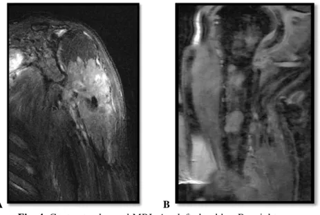
Contrast enhanced MRI showed the presence of an expansive, solid, heteroge-neous formation in the upper third of the left humerus, which disrupted the bone cortical with invasion of the brachial biceps and triceps muscles; with invasion of the left scapula, mainly the superficial muscle plane (supra and infraspinatus muscles), and subscapular muscle (fig. 4A). In the right humerus multiple well-defined sites of “gadolinophilic” lesions of 2 to 30 mm, disseminated intraosseously in the upper two-thirds were seen (fig. 4B).
A B
Fig. 4. Contrast enhanced MRI: A – left shoulder; B – right arm. Pathological examination of
hematoxy-lin-eosin stained slides revealed bone tissue of osteolytic appearance and tumor frag-ments consisting of a monomorphic plasma
cell population, which, in the left humerus, also infiltrated the muscle, these findings being conclusive for the diagnosis of mul-tiple myeloma.
A B
Fig. 5. Multiple myeloma, H & E staining (A - Ob. 20x, B – Ob. 90x) DISCUSSION
The most common locations of MM in-clude the ribs, vertebrae, pelvis and skull cap (3). In our case it was located in the humerus.
In our patient the diagnosis was difficult
due to the development of an atypical radi-ologic image picture in the left shoulder; radiologically, MM is characterized by the presence of "punched-out" osteolytic le-sions prevalently located in the shaft of long bones (4).
MM diagnosis can be made with pretty much certainty on the basis of laboratory tests. In our case these were not conclusive enough. MM is characterized by normo-chromic anemia, normocytic anemia, elevat-ed ESR, hypercalcemia, elevatelevat-ed serum proteins, Bence-Jones protein in urine + (5), while our patient presented only a slight increase in granulocyte counts (70,1/%) and a moderately increased ESR (16 mm/h), kidney and liver tests being normal.
In our patient, diagnostic certainty came from pathological examination performed on tissue biopsy taken from the left shoulder and proximal third of the right humerus.
MM is a malignant neoplasm of plasma cells that remains incurable by convention-al chemotherapy, median survivconvention-al being 2-3 years (6). It is usually chemosensitive, but frequently enters a plateau phase of variable duration (6).
The presented case had an atypical pro-gression with quite important extra-medullary extension. The cases with ex-traosseous extension of an intramedullary tumor nodule as a result of cortical erosion and subsequent spread beyond the perioste-um are not rare, bur soft tissues involve-ment is exceptional occurring in rare forms of IgD MM (7).
REFERENCES
1. Raab MS, Podar K, Breitkreutz I, Richardson PG, Anderson KC. Multiple myeloma. Lancet 2009; 374 (9686): 324–339.
2. International Myeloma Working Group. Criteria for the classification of monoclonal gammopathies, multiple myeloma and related disorders: a report of the International Myeloma Working Group. Br J Haematol, 2003; 121 (5):749–757.
3. Edmonson A.S. Myeloma. In: Campbell's Operative Orthopaedics, Vol 2: 6th Edition By the C.V Mosby Company, 1980, 1339-1345.
4. Bland KI, Daly JM, Karakousis CP. Common Malignant Tumours of Bone. In: Surgical Oncology: Contemporary Principles and Practice, McGraw-Hill, 2001, 375-377.
5. Russell, RCG, William NS, Bulstrode CJK. Bone Tumours in Bailey and Love's Short Practice of Surgery: 24th Edition Arnold; 2004, 431-440.
6. Pratt G. Molecular aspect of multiple myeloma, J Clin Pathol: Mol Pathol, 2002; 55: 273-283. 7. Jowitt SN, Jacobs A, Batman PA, Sapherson DA. Atypical progression of multiple myeloma with