Bulletin of the Chemists and Technologists of Macedonia, Vol. 25, No. 2, pp. 121–126 (2006)
GHTMDD – 490 ISSN 0350 – 0136
Received: September 25, 2006 UDC: 661.183.7
Accepted: December 11, 2006
Original scientific paper
PREPARATION AND CHARACTERIZATION OF SOL-GEL PROCESSED SPRAY
DRIED SILICA XEROGEL MICROPARTICLES AS CARRIERS OF HEPARIN SODIUM
Packa Antovska1, Maja Cvetkovska2, Katerina Goračinova1
1Institute of Pharmaceutical Technology and Biopharmacy, Faculty of Pharmacy,
Sts Cyril and Methodius University, Vodnjanska 17, 1000 Skopje, Republic of Macedonia
2Institute of Organic and Polymer Engineering, Faculty of Technology and Metallurgy,
Sts Cyril and Methodius University, Rudjer Bošković 16, 1000 Skopje, Republic of Macedonia [email protected]
A spray drying technique was used for xerogel microparticles preparation from acetic acid catalyzed tetraeth-oxysilane (TEOS) – based silica sol, TEOS co-hydrolyzed with methyltriethtetraeth-oxysilane (METES), or α,ω -silane-terminated poly(ethylene glycol). The effect of TEOS modification on the heparin dissolution rate, particle degrada-tion rate, as well as particle size and morphology was investigated. Smooth, spherical microparticles in the range of 1.02 – 13.51 μm, were produced by the spray drying method, which showed slow degradation rate and controlled re-lease of heparin over prolonged period of time, dependent on the polymer’s composition.
Key words: silica xerogel; microparticles; spray drying; controlled release; heparin
ПОДГОТОВКАИКАРАКТЕРИЗАЦИЈАНАМИКРОЧЕСТИЧКИОДСИЛИКА-КСЕРОГЕЛ
КАКОНОСАЧИНАХЕПАРИНДОБИЕНИСОСОЛ-ГЕЛ-ПРОЦЕСИСУШЕЊЕ CO СПРЕЈУВАЊЕ
Микрочестичкиодксерогелседобиениодрастворнатетраетоксисилан (TEOS), TEOS ко-хидролизи -рансометилтриетоксисилан (METES) илиполиетиленгликол (PEG) сокрајнитриетоксисиланскигрупи, со оцетнакиселинакакокатализатор, сосол-гел-процесисушењесоспрејување. Испитуванеефектотнаовие модификации во формулацијатана микрочестичките базиранина TEOS врз степенот на ослободувањена хепаринотинивнатадеградација, какоиврзголеминатаначестичкитеинивнатаморфологија.
Добиенитемикрочестичкисемазнии сферични, содимензиивоинтервалотод 1,02 μm до 13,51 μm. Тестовитезадисолуцијаидеградацијанатритеформулациинамикрочестичкипокажаадекасекарактеризи -раатсобавенпроцеснадеградацијаисоконтролираноослободувањенахепаринвоподолгвременскипери -од, зависноодупотребениотпрекурсорзаксерогелот.
Клучнизборови: cилика-ксерогел; микрочестички; su{ewe so sprejuvawe; контролираноослободување; хепарин
INTRODUCTION
The sol-gel process, one of the fastest growing fields in material chemistry, has opened new pos-sibilities for linking ceramics, glasses, and other inorganic materials on one hand, with bioactive molecules on the other hand, forming a novel, wide-scope family of biochemical reactive materials [1]. The sol-gel process involves the manufacture of inorganic matrices through the formation of a
colloidal suspension, which is called sol. After ge-lation, the wet gel forms a globally connected solid matrix, which after drying forms the dry gel state, called xerogel. Drug molecules incorporated into the sol state are located within the porous silica xerogel network [2].
thick and thin films) [3–4]. These silica matrices are chemically inert, hydrophilic, and inexpensive to synthesize. They also exhibit higher mechanical strength, enhanced thermal stability, and negligible swelling in organic solvents compared to most or-ganic polymers. Other advantages of silica sup-ports include biocompatibility and resistance to microbial attack [5].
The sol-gel process has been used as matrix for catalysts in hybrid materials such as ceramic-polymer, ceramic-metal composites or biosensors in diagnostic applications [6]. In the field of biosen-sors, Narang et al. [7], encapsulated glucose oxidase for a period of over 2 months. Growth factor TGF-β1 [8], as well as several enzymes has been shown
to retain their activity in dried silica xerogel matri-ces [9]. Conventional drug molecules have also been incorporated into silica gel matrices [10].
A major concern with the use of artificial or-gans and biomedical devices is the untoward interac-tions of blood upon contact with foreign surface. Local, controlled release of an appropriate antico-agulant at the site of trauma would be a good ap-proach that would prevent thrombus formation [11]. For inhibition of blood coagulation in extra-corporeal circulation procedures, the anticoagulant heparin is of interest due to its specificity, low col-lateral responses, and good tolerance by the organ-ism [12].
The purpose of the present study was to evaluate the suitability of sol-gel produced mi-croparticles as a carrier material for a controlled release of heparin. TEOS based silica xerogels were designed as carriers for heparin, but organi-cally modified systems were evaluated as well.
EXPERIMENTAL
Materials
Silica xerogel particles were prepared by us-ingthe following reagents: tetraethoxysilane (TEOS, Merck), deionised water, acetic acid (CH3COOH,
Sigma), ammonium hydroxide (NH4OH, Merck)
and heparin sodium salt (Dongcheng biochemi-cals). Particular heparin sodium injectable grade is derived from porcine intestinal mucosa with de-clared potency of 175 IU/mg. Organically modi-fied silica xerogels included methyltriethoxysilane (METES, Fluka) and α,ω-silane-terminated PEG (synthesized for the purpose of this work using isocyanate chemistry) [13].
Two moles of γ-isocyanatopropyltriethoxysi-lane (Fluka) were reacted with 1 mol α,ω-hydroxy-terminated poly(ethylene glycol) (PEG, Fluka, Mw= 600 g/mol; dried for 48 h at 40 ˚C) in vacuum oven, at 80 ˚C under dry nitrogen, until complete disappearance of the absorptions of the hydroxyl and isocyanate groups in the IR spectrum, at cca 3500 cm–1 and cca 2280 cm–1, respectively, in
fa-vor of the urethane linkage (3384 cm–1). The
reac-tion was completed in 24 h.
Preparation of silica xerogels
TEOS based silica xerogels
Silica sol containing heparin was prepared by a two-step sol-gel process using acetic acid as a catalyst. The molar ratio of the silica sol was TEOS: H2O:CH3COOH = 0.05:5:0.004.
After the hydrolysis of TEOS had proceeded to a point where a homogenous solution was formed, pH was adjusted up to 4.5-4.6 using 1 M solution of NH4OH and heparin sodium was
dis-solved directly into the sol. The concentration of heparin in the silica sol was 1.39 % corresponding to 36.36 % in spray-dried silica xerogel. Hydro-lyzed silica sol was spray dried with a mini spray dryer (B-290, Büchi Labortechik AG; Switzer-land). The spray drying process parameters were: inlet temperature 135 ˚C; pump 15; aspirator set-ting 90; spray flow 600 NL/h; pump setset-ting 3–4 ml/min; outlet temperature: 60 ˚C. A 0.77 mm noz-zle was used throughout the experiments.
Organically modified silica xerogels
Modification of the TEOS based xerogel was made by co-hydrolysis of TEOS with METES or α,ω-silane-terminated PEG. Partial substitution of TEOS was made with 10 % of the organically modified alkoxy silanes.
Organically modified silica xerogels were prepared using the procedure described in the pre-vious section.
Evaluation of the heparin loaded silica xerogel microparticles
Particle size and particle size distribution determination
tometry using Malvern mastersizer 2000S Hydro. The samples were dispersed under pressure in a special gas chamber intended for particle size de-termination in a dry state.
Morphology
The shape and surface morphology of the mi-croparticles was determined from micrographs taken with scanning electron microscope (Jeol SEM 6400 Japan).
Determination of heparin entrapment in silica xerogels
The percent of the incorporated heparin in sil-ica xerogels was determined by dispersing 50 mg sample in 25 ml 0.02 M NaOH, stirred with mag-netic stirrer at temperature not exceeding 60 ˚C until clear solution was obtained.
Toluidine blue test
The total amount of heparin was measured by a colorimetric toluidine blue method [14] modified for the present study; 0.005 % toluidine blue solu-tion was prepared in 0.01 M HCl containing 0.2 % NaCl.
A 2 ml of sample in 0.02 N NaOH were pipet-ted in test tubes, 2 ml of toluidine blue solution were added, and the mixture was vigorously vor-texed for 30 s. Four ml of hexane was then added to the tubes and they were shaken for another 30 s to separate the heparin-dye complex formed. The aqueous layers were sampled for measurement and diluted if necessary. The absorbance was measured at 631 nm within 30 minutes with UV-Vis-Spec-trophotometer – Perkin Elmer Lambda 16.
Release of heparin in vitro
The dissolution test from different silica xe-rogel matrices was carried out in a shaking water bath at 37 ˚C. The average weight of the evaluated samples was 50 mg.
Simulated body fluid (SBF) was used as a dissolution medium. SBF was prepared by dissolv-ing the followdissolv-ing reagents: NaCl (136.8 mM); Na-HCO3 (4.2 mM); KCl (3.0 mM); K2HPO4 × 3H2O
(1.0 mM); MgCl2 × 6H2O (1.5 mM); CaCl2 ×
2H2O (2.5 mM); Na2SO4 (0.5 mM) in deionised
water.
The solution was buffered with tris(hydroxy-methyl)aminomethane (TRIZMA) and hydrochlo-ric acid (HCl) at pH 7.40. The composition of
in-organic ions corresponded to that of human blood plasma [15].
Silica xerogel microparticles were immersed in 10 ml SBF in a polyethylene tube with a screw cap. Alternatively, a 5ml sample was removed from each tube and replaced immediately with fresh SBF in predefined time intervals.
Toluidine blue test
The total amount of heparin dissolved was also measured by the colorimetric toluidine blue method. Standard heparin solution was prepared by diluting 20 mg heparin to 20 ml deionised water.
The standard dilutions were between 5 and 30 μg of heparin in SBF.
A 1.3 ml of toluidine blue solution and 1.3 ml of sample in SBF were pipetted in test tubes, then vigorously vortexed for 30 s. A 2.6 ml of hexane was then added to the tubes and they were shaken for another 30 s to separate the heparin-dye com-plex formed. The aqueous layers were sampled for measurement and diluted if necessary. The absorb-ance was measured at 631 nm within 30 minutes with UV-Vis-Spectrophotometer.
Degradation of xerogel
The degradation process of the silica xerogel microparticles was evaluated using unloaded mi-croparticles immersed in SBF, by measuring the dissolved Si(OH)4 as a molybdenum blue complex
by UV-Vis-Spectrophotometer at 815 nm [16].
RESULTS AND DISSCUSION
Preparation of microparticles
At first, unloaded microparticles were ob-tained by spray-drying method. Examination by scanning electron microscopy showed that un-loaded silica microparticles were spherical, with no visible pores on the surfaces.
The microparticle yields of three different xe-rogel formulations (n = 3 batches for each formula-tion) were calculated from the theoretical amount of silica gel (SiO2). Unloaded microparticles were
produced with the following yields: TEOS based: 39.16 %; TEOS/METES based: 27.50 %; and TEOS/α,ω-silane-terminated PEG based micropar-ticles: 47.89 %.
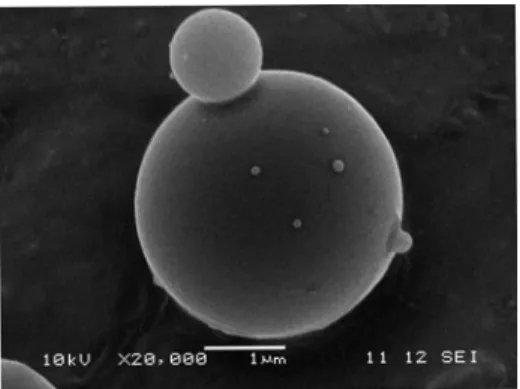
spray-drying. Examination by SEM showed that loaded silica microparticles were with spherical shape and smooth surfaces, regardless of the com-position of the silica network. (Figs. 1, 2).
Fig.1. Scanning electron micrograph of TEOS based loaded microparticles
Fig. 2. Scanning electron micrograph of TEOS/α,ω -silane-terminated PEG loaded microparticles.
Particle size measurement showed that TEOS based microparticles have Dv (0.1) bellow 1.78 μm and Dv (0.9) bellow 13.51 μm. As for the TEOS/METES based microparticles, Dv (0.1) was bellow 1.08 μm and Dv (0.9) was bellow 7.68 μm. For TEOS/α,ω-silane-terminated PEG, Dv (0.1) was bellow 1.02 μm and Dv (0.9) was bellow 4.89 μm.
It has been shown that hydrogen bonds be-tween organic polymers prone to hydrogen bond-ing with the silanol functions formed by hydrolysis of tetraethoxysilane have a decisive effect on the formation and final preparation of the obtained hybrid materials [17]. Thus, it can be expected that PEG increases the average particle size in the silica sols due to hydrogen bonding, but also hinders in some way the polycondensation process during spray-drying process, resulting in formation of smaller particles.
The yields of loaded microparticles were as follows: TEOS based: 48.26 %; TEOS/METES based: 40.48 %; and TEOS/α,ω-silane-terminated PEG based microparticles: 71.19 %.
Experimentally determined loading efficiency of heparin sodium in the microparticles was 84.11 % in TEOS based, 98.63 % in TEOS/METES based, and 90.88 % in TEOS/α,ω-silane-terminated PEG based microparticles.
The differences in loading efficiency of hepa-rin sodium into the silica microparticles are proba-bly due to the incorporation of different precursors in the silica sol. In fact, incorporation of ormosils into hydrolysis combined with TEOS results in decreased hydrophilicity of the network and changes in porosity [18]. Organic groups linked to the oxide network by stable chemical bonds change the inner structure by reducing the degree of cross-linking [19]. Hence, higher loading effi-ciency would be expected in the organically modi-fied systems. Also, according to Ahola et al. [2] when pure PEG was incorporated as an additive into an acetic acid catalyzed TEOS based silica xerogel, the pore volume and the specific area of pores in silica xerogel samples were decreased, which is a factor involved in the efficacy of load-ing of the active substance durload-ing preparation.
Incorporation of heparin into the microparti-cles was confirmed by means of FTIR spectro-scopy, comparing the O–H bending mode of hepa-rin SO3–(H3O)+ groups located at 1623 cm–1 [20],
as well as by TG analysis and comparison of the weight losses of unloaded and loaded microparti-cles at temperatures above 180 °C.
Release of heparin and degradation of silica microparticles
The in vitro release of heparin from the three different formulations is shown in Fig. 3.
After 14 days the cumulative percent of re-leased heparin was: 32.11 % from TEOS micro-particles, 27.34 % from TEOS/METES microparti-cles, and 24.27 % from TEOS/α,ω-silane-termina-ted PEG microparticles.
Dissolution curves show an initial burst re-lease of heparin sodium (in the first hour) followed by a slower release pattern in all three systems. The plot of the release was linear with respect to the square root of time for TEOS, TEOS/METES and TEOS/α,ω-silane-terminated PEG (R: 0.989; 0.988; 0.971), respectively.
The increased initial burst may be associated with sufficiently high amount of drug close to the surface of the gel network [21. When the drug was depleted from the surface, the silica gel degrada-tion became a factor involved in the heparin re-lease rate.
The results from the in vitro release of hepa-rin show that addition of ormosils decreased the release rate of heparin from the microparticles. This is probably a result of an increased hydropho-bicity when METES is introduced in the silica network, or it is due to the already discussed de-crease of pore volume in the system with TEOS/α,ω-silane-terminated PEG.
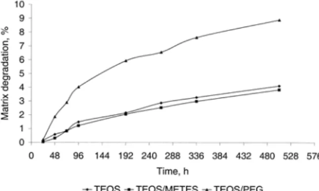
Silica gel degradation of the microparticles is shown in Fig 4. After 21 days the amount of the silica gel left was 91.10 % for TEOS/α,ω-silane-terminated PEG system, and even 95.89 % for TEOS based and 96.17 % for TEOS/METES based xerogels.
Fig 4. Degradation of different xerogel formulations
Degradation plots were fitted to first order ki-netics model and the following values for R were obtained: (0.977; slope = 3.65·10–5) for TEOS micro-particles, (0.978; slope = 3.49·10–5) for TEOS/METES
microparticles, and (0.945; slope = 7.73·10–5) for TEOS/α,ω-silane-terminated PEG microparticles.
The cumulative percent of degraded silica gel network was similar when TEOS and TEOS/METES microparticle were compared. Al-most two times higher amount of degraded silica gel was obtained in the case of TEOS/α,ω-silane-terminated PEG microparticles. This is probably due to the increased hydrophilicity of the silica network and enhanced intake of water into the sys-tem, which favors hydrolysis as a mechanism of degradation of the silica xerogel network.
Heparin was released faster than the silica gel microparticles were degraded (Fig 5). Literature data suggests that for highly water soluble drugs, the release rate is controlled mainly by diffusion through the matrix, whereas the erosion process is the main factor for release of low water-soluble drugs. Thus, the release rate is probably a combi-nation of diffusion of the dissolved heparin through the xerogel network and degradation-generated pores in the xerogel, and surface erosion over time.
Fig 5. In vitro release of heparin and degradation of silica gel microparticles
Compared with silica xerogel monoliths, spray dried silica gels degrade more slowly [2]. During spray-drying at an increased temperature (135 ˚C), the hydrolyzed silica sol is very rapidly dried into small particles which most likely results in a more condensed structure.
CONCLUSIONS
the advantage of being single-step process which can be readily scaled up.
It has been shown that heparin release from the microparticles may be controlled by addition of organically modified precursors that cause changes in the microstructure of the silica network.
Acknowledgement: This work was supported by
the Ministry of Education and Science of the Republic of Macedonia and the Turkish Scientific Technical Re-search Council (TUBITAK), Turkey.
REFERENCES
[1] P. J. Brinker, G. W. Scherer, Sol-Gel Science: The Physics and Chemistry of Sol-Gel Processing, Academic Press, New York, 1990.
[2] M. Ahola, P. Kortesuo, I. Kangasniemi, J. Kiesvaara, A. Yli-Urpo, Silica xerogel carrier material for controlled re-lease of toremifene citrate, Int. J. of Pharm., 195, 219– 227 (2000).
[3] D. Avnir, S. Braun, Biochemical Aspects of Sol-gel Sci-ence and Technology, Kluwer Academic Publishers, Bos-ton, 1996.
[4] J. Livage, Bioactivity in sol-gel glasses, C. R. Acad. Sci.,
322, 417–427 (1996).
[5] R. B. Bhatia, C. J. Brinker, Aqueous Sol-Gel Process for Protein Encapsulation, Chem. Mater., 12, 2434–2441 (2000),
[6] S. Braun, S. Rappoport, R. Zusman, D. Avnir and M. Ottolenhgi, Biochemically active sol-gel glasses: the trapping of enzymes. Mater. Lett., 10, 1–5 (1990). [7] V. Narang, P. N. Prasad, F. V. Bright, K. Ramanyhan, N.
D. Kumar, B. D. Malhotra, M. N. Kamalasan, S. Chandra, Glucose sensor based on a sol-gel derived platform, Anal. Chem., 66, 3139–3144 (1994).
[8] S. B. Nicoll, S. Radin, E. M. Santos, R. S. Tuan, P. Ducheyne, In vitro release kinetics of biologically active transforming growth factor-1 from a novel porous glass carrier, Biomaterials, 18, 853–859 (1997).
[9] S. A. Yamanaka, F. Nishida, L. M. Ellerby, C. L. Nishida, B. Dunn, J. S.Valentine, J. I. Zink, Enzymatic activity of
glucose oxidase encapsulated in transparent glass by the sol-gel method, Chem. Mater., 4, 495–497(1992). [10] K. Unger, H. Rupprecht, B. Valentin, W. Kircher, The use
of porous and surface modified silicas as drug delivery and stabilizing agent, Drug Dev. Ind. Pharm., 9, 69–91 (1983). [11] L. Thunberg, G. Backstrom, U. Lindahl, Further
charac-terization of the antithrombin binding sequence in hepa-rin, Carbohydr. Res., 100, 393–410 (1982).
[12] Z. Chen, R. F. Zhang, M. Kodama, T. Nakaya, Antico-agulant Surface Prepared by the Heparinization of Ionic Polyurethane Film, J. Appl. Polym Sci., 76, 382–390 (2000).
[13] S. Cuney, J. F. Gerard, J. P. Pascault, G. Vigeier, Or-ganic-rich hybrid O/I systems based on isocyanate chem-istry, Mat. Res. Soc. Symp. Proc., 435, 143–154 (1996). [14] P. K. Smith, S. Mallia, G. T. Hermanson, Colorimetric
method for the assay of heparin content in immobilized heparin preparations, Anal. Biochem., 109, 466–473 (1980).
[15] J. Gramble, Chemical anatomy: Physiology and pathol-ogy of extracellular fluid, 6th ed. Cambridge: Harvard
University Press, 1967
[16] O. G. Koch, G. A, Koch-Dedic, Handbuch der Spurenan-alyse, 2nd Edition, Vol 1, Springer-Verlag, Berlin, 1974.
[17] D. Tian, Ph. Qubois, R. Jerome, Biodegradable and bio-compatible inorganic-organic hybrid materials. I. Synthe-sis and characterization, J. Polym. Sci. A: Polym. Chem.,
35, 2295–2309 (1977).
[18] M. Ahola, E. S. Säilynoja, M. H. Raitavuo, M. M. Vaahtio, J. I. Salonen, A. Yli-Urpo, In vitro release of heparin from silica xerogels, Biomaterials, 22, 2163–2170 (2001). [19] H. Schmidt, Organic modification of glass structure. New
glasses or new polymers, Non-Cryst. Solids, 112, 419– 423 (1989)
[20] N. S. Harada, H. T. Oyama, J. R. Bartoli, D. Gouvea, I. A. Cestari and S. H. Wang, Quantifying adsorption of heparin on a PVC substrate using ATR-FTIR, Polym. Int., 54, 209–214 (2005).