The Spine and Scapula Stabilizing (S3) Brace Has an Effect on Posture and Muscle Activity in Overhead Athletes with Poor Posture
Ashley Kahlil Cole, LAT, ATC
A thesis submitted to the faculty of the University of North Carolina at Chapel Hill in partial fulfillment of the requirements for the degree of Master of Arts in the
Department Exercise and Sport Science (Athletic Training).
Chapel Hill 2008
Approved by:
Advisor: Dr. Bill Prentice
Reader: Dr. Darin Padua
Reader: Mrs. Melanie McGrath, MS, ATC
Reader: Ms. Shana Harrington, PT
ABSTRACT
Ashley K. Cole: The Spine and Scapula Stabilizing (S3) Brace Has an Effect on Posture and Muscle Activity in Overhead Athletes with Poor Posture
(Dr. Bill Prentice)
The purpose of this study was to determine whether or not the S3 scapular
stabilizing brace corrects the posture of participants with FHRSP. In addition, this
study determined whether or not wearing the S3 scapular stabilizing brace has an
effect on the muscle activity of participants with FHRSP while performing six
scapular stabilization exercises. Posture was measured using a digital camera and
Adobe Photoshop to determine both the forward head and rounded shoulder angles.
Muscle activity was measure for the upper trapezius, middle trapezius, lower
trapezius, and serratus anterior using the average EMG recorded during Y’s, T’s,
W’s, shoulder extension, forward flexion, and scapular punches. This study found
that there were significant changes in FSA and upper, middle, and lower trapezii
EMG activity caused when wearing the S3 brace compared to not wearing it.
However, this study did not find significant differences in the treatment group
TABLE OF CONTENTS
List of Tables ...vi
List of Figures ...vii
List of Figures ...vii
List of Abbreviations... viii
List of Abbreviations... viii
Chapter 1 ... 1
Introduction ... 1
Statement of Purpose... 5
Independent Variables ... 5
Dependent Variables... 5
Research Hypothesis ... 6
Null Hypothesis ... 7
Alternate Hypotheses ... 8
Operational Definitions ... 8
Assumptions... 11
Delimitations... 12
Limitations ... 12
Chapter 2 ... 13
Anatomy ... 14
Acromioclavicular Joint ... 15
Glenohumeral Joint... 16
Scapulothoracic Joint... 18
Glenohumeral Stabilizers... 19
Scapular Stabilizers ... 20
Kinematics... 23
Posture... 28
Muscular balance ... 31
EMG Analysis... 35
S3 scapular stabilization brace... 37
Chapter 3 ... 40
Subjects ... 40
Study Design ... 41
Instruments... 41
Posture ... 41
EMG ... 42
Motion Analysis... 43
S3 Brace ... 43
Procedures ... 44
Screening... 44
Counterbalancing... 45
Posture ... 45
Exercises ... 47
Data Processing and Reduction ... 49
Statistical Analyses ... 49
Chapter 4 ... 51
Descriptive Statistics ... 51
Posture... 52
EMG ... 53
Chapter 5 ... 56
Posture... 56
EMG ... 58
Upper Trapezius ... 58
Lower Trapezius ... 60
Middle Trapezius ... 60
Serratus Anterior... 61
The S3 Brace... 63
Limitations ... 65
Future Research... 67
Conclusion ... 68
Appendix A: Tables... 69
Appendix B: Figures... 74
Appendix C: Manuscript... 94
LIST OF TABLES
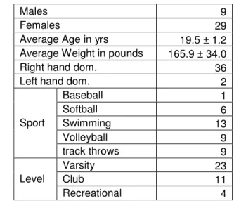
Table 1: Means and numbers for subject characteristics ... 70
Table 2: Means and standard deviations for posture measurements in degrees... 71
Table 3: Means and standard deviations for forward flexion normalized EMG in
percentages ... 71
Table 4: Means and standard deviations for shoulder extension normalized EMG in
percentages ... 72
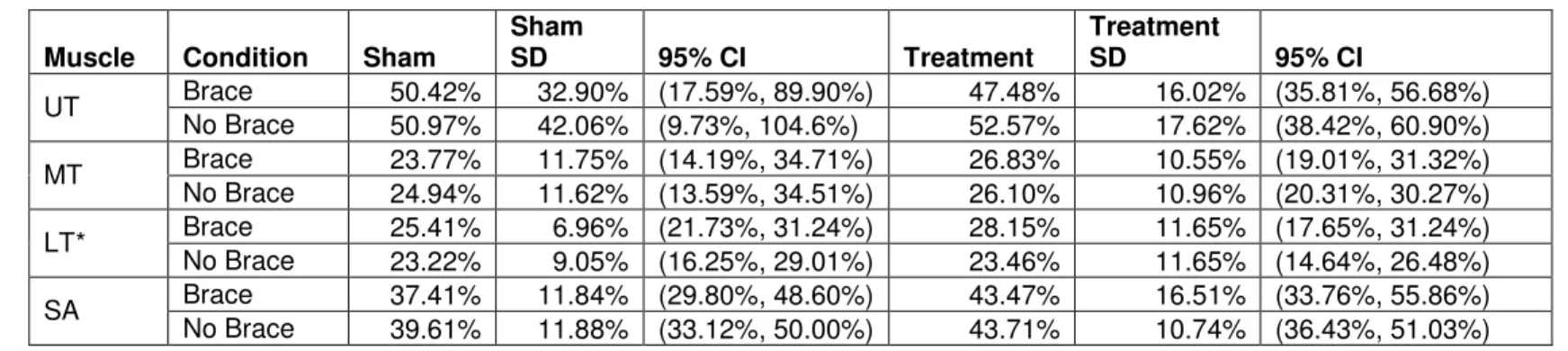
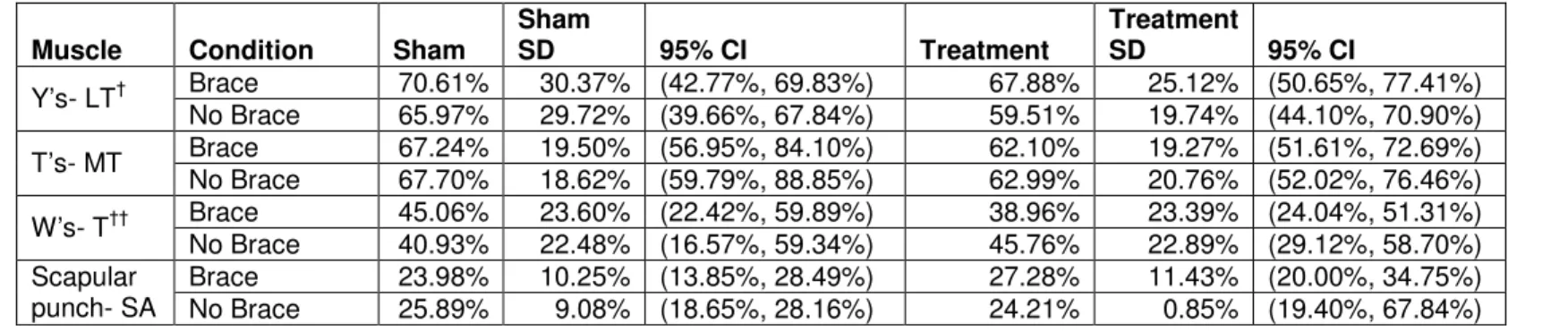
Table 5: Means and standard deviations for normalized EMG in percentages ... 72
LIST OF FIGURES
Figure 1: Head and Shoulder Angle Measures ... 75
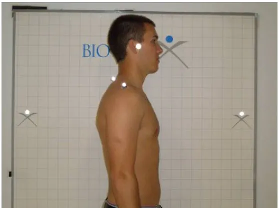
Figure 2: Postural Screening ... 76
Figure 3: Brace Application... 77
Figure 4: Y ... 78
Figure 5: T ... 79
Figure 6: W ... 80
Figure 7: Scapular Punch ... 82
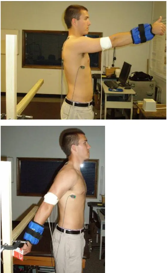
Figure 8: Forward Flexion ... 83
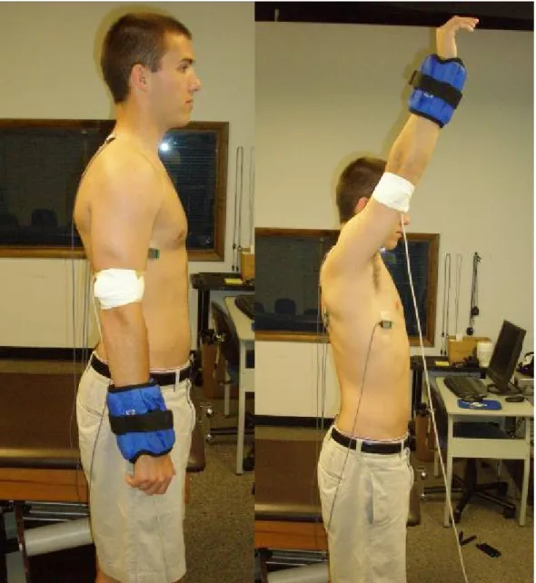
Figure 9: Shoulder Extension... 84
Figure 11: EMG electrode placement ... 85
Figure 14:Forward flexion average normalized EMG activity... 86
Figure 15: Shoulder extension average normalized EMG activity... 87
Figure 16: Y’s average normalized EMG activity ... 87
Figure 17: T’s average normalized EMG activity ... 88
Figure 18: W’s average normalized EMG activity ... 90
Figure 19: Scapular punches average normalized EMG activity... 90
Figure 20: Average postural measurements ... 92
LIST OF ABBREVIATIONS
C7 Seventh cervical vertebrae
FHA Forward head angle
FHP Forward head posture
FHRSP Forward head, rounded shoulder posture
FSA Forward shoulder angle
FSP Forward shoudler posture
LT Lower trapezius
MT Middle trapezius
ROM Range of motion
SA Serratus anterior
CHAPTER 1
Introduction
Shoulder injuries are a common and disabling condition among many athletes.
Shoulder injuries account for 8-20% of all athletic injuries and many of these are
classified as “overuse” (Terry & Chopp, 2000). Shoulder injuries are particularly
common in overhead athletes. Nearly 50% of baseball pitchers experience shoulder
or elbow pain significant enough to prevent participation at some point in their
careers (Myers, Laudner, Pasquale, Bradley, & Lephart, 2005). Recent NCAA injury
surveillance system research has shown that shoulder injuries account for 39.4% of
all injuries in baseball with shoulder injuries accounting for 28.3% of all injuries
resulting in a time loss of 10 or more day. Similar studies were done for softball and
women’s volleyball with shoulder injuries accounting for 15.8% and 21.7% of overall
injuries (Agel, Palmieri-Smith, Dick, Wojtys, & Marshall, 2007; Dick et al., 2007;
Marshall, Hamstra-Wright, Dick, Grove, & Agel, 2007). Thus finding ways to treat
and prevent shoulder injuries are critical to the sports medicine profession.
Research has shown that alterations in scapular motion occur in 68-100% of
patients with shoulder injury (Terry & Chopp, 2000; Warner, Micheli, Arslanian,
Kennedy, & Kennedy, 1992). Many studies have looked at the relationship between
syndrome. Studies have consistently shown that patients with shoulder impingement
syndrome present with decreased scapular upward rotation, decreased posterior
tipping/tilting (sometimes referred to as increased anterior tipping), and increased
medial/internal rotation (Borstad & Ludewig, 2002; Ebaugh, McClure, & Karduna,
2006; Hebert, Moffet, McFadyen, & Dionne, 2002; Ludewig & Cook, 2000;
Lukasiewicz, McClure, Michener, Pratt, & Sennett, 1999). It has been suggested that
a decrease in the amount of scapular posterior tilt may reduce the size of the
subacromial space which subjects the rotator cuff tendons to greater compressive
forces (Lukasiewicz, McClure, Michener, Pratt, & Sennett, 1999)
A specific postural anomaly, forward head rounded shoulder posture, may
also play a role in the development of shoulder pain. Forward head posture is
defined as the tragus (ear lobe) being in front of the plumb line while the rest of the
body remains in alignment (Lewis, Green, & Wright, 2005; Lewis, Wright, & Green,
2005). Rounded/ forward shoulder posture is described as the acromion of the
shoulder being located in front of the plumb line while the rest of the body remains in
alignment (Lewis, Wright, & Green, 2005). These two postural abnormalities often
occur in conjunction with one another and are thought to be related to many overuse
injuries in the shoulder. One study found that when healthy patients adopted a
slouched position this significantly increased scapular anterior tilt and upward
rotation in neutral position, when compared to the neutral position during upright
posture (Finley & Lee, 2003). These specific scapular alterations are believed to be
related to the development of shoulder pathology. It may also be argued that
encroachment of the supraspinatus tendon and the development of shoulder
pathology (Finley & Lee, 2003). Furthermore, Greigel-Morris et al. found that the
incidence of pain increased in subjects with more severe postural abnormalities
including kyphosis and interscapular pain, FHP and right cervical pain, FHP and left
cervical pain, FHP and headache, FHP and interscapular pain, left rounded shoulder
posture (LRSP) and interscapular pain, and right rounded shoulder posture (RRSP)
and interscapular pain (Griegel-Morris, Larson, Mueller-Klaus, & Oatis, 1992).
Muscular balance plays a significant role in proper posture and normal scapular
kinematics. Having muscular balance means that anterior and posterior, and medial
and lateral muscles are equal in strength and move in an appropriate sequence with
one another. One way to correct scapular positioning and FHRSP is to correct
muscular imbalances surrounding the shoulder complex. Weakness of the
scapulothoracic muscles has been shown to potentially lead to abnormal positioning
of the scapula, disturbances in the scapulothoracic rhythm, and generalized
shoulder dysfunction (Voight & Thomson, 2000). Several studies have examined
muscle strength in the scapular muscles, balance between scapular muscles, and
latent reaction times between muscles in overhead athletes with impingement.
Results of these studies have shown that athletes with shoulder pathology showed
decreased force output, decreased muscle activity during concentric isokinetic
retraction movements which was accompanied by a change in normal activation
patterns in the trapezius muscle activity (Cools, Declercq, Cambier, Mahieu, &
Witvrouw, 2007; Cools, Witvrouw, Danneels, & Cambier, 2002; Cools, Witvrouw,
& Cambier, 2004). Thus it may be possible to alleviate shoulder pain and
dysfunction by properly rehabilitating the musculature surrounding the scapula, to
achieve normal scapular motion and to restore proper alignment within the neck and
shoulder girdle.
The Scapular Stabilizing System (S3) brace is “a spine and scapula
stabilizing brace designed to improve posture, reduce pain, and increase range of
motion." The company designed the S3 brace to "trigger the body to correct
improper posture by re-educating and re-engineering the musculo-skeletal system
surrounding the shoulders and spine" as well as signaling the neuroreceptors in the
skin to engage in proper posture. The company also states that the S3 brace
“addresses and lends instant relief to fatigue and poor spine alignment associated
with unnatural body position at the computer.” There have been no published studies
yet on this brace although Uhl et al. performed a study on the prototype of the S3
brace that has not yet been published. Fifteen healthy subjects and 15 subjects with
scapular dyskinesis were used in this study. The results found that the brace
increased posterior tipping by 3 degrees in the first and last 30 degrees of motion,
decreased upward rotation in the dominant arm by 4 degrees at 90 degrees of
elevation, while increasing upward rotation in the non-dominant arm by 2 degrees in
the first and lat 40 deg of elevation. The S3 also decreased internal rotation by 3.5
degrees during the lowering phase of elevation. The authors concluded that the S3
brace affected the scapular kinematics at rest and in the lower ranges of motion and
that the increased posterior tipping and decreased internal rotation from wearing the
appears to be a new way to help correct scapular position and motion to help treat
individuals with shoulder pathology.
Statement of Purpose
The purpose of this study was to determine whether or not proper application
of the S3 scapular stabilization brace changes the posture of participants with
FHRSP. In addition, this study determined whether or not the wearing the S3
scapular stabilization brace has an effect on the EMG activity of patients with
FHRSP while performing six scapular stabilization exercises. Comparing the sham
group to the treatment group determined whether any changes in EMG seen were
due to the corrective straps or if merely wearing a compressive garment produced
changes in EMG activity.
Independent Variables
1. Wearing the S3 brace with straps properly applied and tensioned
2. Wearing the S3 brace not using the proper strap set up to correct posture
(sham treatment)
3. Not wearing the S3 brace
Dependent Variables
1. Average EMG present in the serratus anterior, upper trapezius, middle
trapezius, and lower trapezius during exercises
Research Hypothesis
1. It was hypothesized that participants who wore the S3 brace properly applied
would have an increase in the average EMG in the serratus anterior, lower
trapezius, and middle trapezius as compared to when they were not wearing
the brace.
2. It was hypothesized that participants who wore the S3 brace properly applied
would have a decrease in average EMG in the upper trapezius as compared
to when they were not wearing the brace
3. It was hypothesized that the treatment group would have a significant
increase in average EMG in the serratus anterior, lower trapezius, and middle
trapezius and a decrease in the average EMG of the upper trapezius when
compared to the sham group.
4. It was hypothesized that the sham group would have no change in average
EMG in the serratus anterior, lower trapezius, middle trapezius, and upper
trapezius after application of the S3 brace.
5. It was hypothesized that participants who wore the S3 brace properly applied
would have a decrease in the forward head and rounded shoulder angles
compared to when they were not wearing the brace, which signifies improved
posture.
6. It was hypothesized that the treatment group would have a significant change
7. It was hypothesized that the sham group would have no change in forward
head and rounded shoulder angle after application of the brace.
Null Hypothesis
1. Participants who wore the S3 brace properly applied would have no change in
the average EMG in the serratus anterior, lower trapezius, and middle
trapezius as compared to when they are not wearing the brace.
2. Participants who wore the S3 brace properly applied would have no change in
average EMG in the upper trapezius as compared to when they are not
wearing the brace
3. The treatment group would have no significant difference in average EMG in
the serratus anterior, lower trapezius, middle trapezius, and upper trapezius
compared to the sham group.
4. The sham group would have no significant difference in average EMG in the
serratus anterior, lower trapezius, middle trapezius, and upper trapezius after
application of the brace.
5. Participants who wore the S3 brace properly applied would have a no change
in the forward head and rounded shoulder angles compared to when they are
not wearing the brace, which signifies no improvement in posture.
6. The treatment group would have a no difference in forward head and rounded
shoulder angle compared to the sham group.
7. The sham group would have no difference in forward head and rounded
Alternate Hypotheses
1. Participants who wore the S3 brace properly applied would have a change in
the average EMG in the serratus anterior, lower trapezius, and middle
trapezius as compared to when they are not wearing the brace.
2. Participants who wore the S3 brace properly applied would have a change in
average EMG in the upper trapezius as compared to when they are not
wearing the brace
3. The treatment group would have a significant difference in average EMG in
the serratus anterior, lower trapezius, middle trapezius, and upper trapezius
compared to the sham group.
4. The sham group would have a significant difference in average EMG in the
serratus anterior, lower trapezius, middle trapezius, and upper trapezius after
application of the brace.
5. Participants who wore the S3 brace properly applied would have a change in
the forward head and rounded shoulder angles compared to when they are
not wearing the brace, which signifies no improvement in posture.
6. The treatment group would have a difference in forward head and rounded
shoulder angle compared to the sham group.
7. The sham group would have a difference in forward head and rounded
shoulder angle after application of the brace.
1. Sham treatment- A sham treatment was defined as the patient wearing the S3
scapular stabilization brace without the Velcro straps being properly applied.
This gave the feeling that he/she was wearing something that would correct
his/her posture without the actual corrective measures being applied. To
create this effect a longer Velcro strap was attached to the brace. This longer
strap did provide enough tension to make an effective change in the patient’s
posture.
2. Scapular stabilization exercises- Scapular stabilization exercises included
those exercises that strengthen the muscles responsible for maintaining
proper positioning of the scapula during shoulder movement. The exercises
that have been chosen for this study are scapular punches, forward flexion,
shoulder extension, Y’s, T’s, and W’s.
a. Scapular punches were performed lying supine on a table with the arm
in 90° of flexion. The patient then protracted the scapula by raising the
fist towards the ceiling.
b. Y’s were described as an arm raise above the head with the upper
extremity in line with the lower trapezius muscle fibers in the prone
position (Ekstrom, Donatelli, & Soderberg, 2003). This exercise was
performed lying prone on a table with arms hanging down in front and
palms facing each other. Arms were in the 10 and 2 o’clock position (at
about 125°) and thumbs were raised towards the ceiling. Arms were
c. T’s were described as shoulder horizontal extension with external
rotation in the prone position (Ekstrom, Donatelli, & Soderberg, 2003).
This exercise was performed lying prone on a table with arms hanging
down in front. Arms were raised out to the side in horizontal extension
until they were parallel to the floor.
d. W’s were described as prone external rotation with shoulder abducted
to 90° and elbow flexed to 90°. This exercise was performed while
lying prone on a table with arms hanging down in front. Arms were
raised so that brachium was parallel to the floor with the elbow bent to
90°. The arms were then externally rotated.
e. Forward flexion was performed in the sagittal plane. The exercise
began with the arm at 0° of flexion and it was elevated with the forearm
in a neutral position (thumb facing ceiling) in the sagittal plane to full
shoulder flexion(Myers et al., 2005).
f. Shoulder extension was performed in the sagittal plane. The exercise
began with the arm at 90° of flexion with the forearm in a neutral
position (thumb facing ceiling) and was moved into full shoulder
extension and then back to 90°(Myers et al., 2005).
3. Poor posture was defined as having forward head, rounded shoulder
positioning. Reflective markers were placed over the tragus (ear), acromion,
and C7 spinous process. Pictures were taken in the sagittal view of each
subject and measurements were taken using the pictures. Forward head
46° relative to the vertical line extending from C7 to the line connecting C7 to
the tragus. Rounded shoulder position was described as having a forward
head angle of greater than or equal to 46° relative to the vertical line
extending from C7 to the line connecting C7 to the acromion (Sawyer, 2006;
Thigpen, 2006). Postural alignment criteria were based on a study done by
Thigpen in which he screened 310 individuals from the university population.
Those with FHA ≥ 46º and FSA ≥ 46º were determined to have the worst posture Sawyer, 2006; Thigpen, 2006).
4. Overhead athlete- An overhead athlete was described as an athlete who
competes in a NCAA, club, or recreational overhead sport for at least 3-4
days per week for 1 hour a day or more. Overhead sports were those in which
repetitive overhead activity were required including baseball, softball,
swimming, volleyball, tennis, water polo, javelin, shot put, and discus.
5. EMG- Electromyography was used to assess muscles activity during the
scapular punches, forward flexion, shoulder extension, Y’s, T’s, and W’s.
Maximal voluntary isometric contraction (MVIC) readings were taken using
EMG to determine what percentage of each patient’s MVIC the EMG reads
during the exercise. The average value of the EMG was used to normalize
MVIC readings.
6. Average EMG- Average smoothed and rectified EMG amplitude during the
exercise. This value was normalized to each subject’s MVIC.
1. Participants did not know the difference between the S3 brace treatment and
sham groups
2. All exercises activated the muscles which they were intended to activate
3. Subjects were able to complete the exercise protocol
4. Individuals gave the same amount of effort whether wearing the S3 brace or
not wearing it.
5. EMG was a valid and reliable measuring device and is properly calibrated
6. Researchers could reliably apply the brace.
Delimitations
1. Subjects will be truthful about their history of upper extremity injury
2. Subjects are all overhead athletes
3. Analysis will be performed on the subjects dominant arm for his/ her sport
Limitations
1. Variability of EMG readings between subjects
2. The pressure of the brace may affect EMG readings
3. Exercises were performed in a lab setting with wires attached which may
CHAPTER 2
Shoulder injuries account for 8-20% of all athletic injuries. Many of these
injuries are classified as overuse (Terry & Chopp, 2000). Research has shown that
alterations in scapular motion occur in 68-100% of patients with shoulder injury
(Terry & Chopp, 2000; Warner, Micheli, Arslanian, Kennedy, & Kennedy, 1992). The
prevalence of these injuries tends to increase with age as studies have indicated
that 21-34% of elderly people are inflicted with shoulder injury, and in about 30% of
these cases that injury led to disability (Chakravarty & Webley, 1993; Chard,
Hazleman, Hazleman, King, & Reiss, 1991). Athletes involved in overhead sports
are at increased risk of developing shoulder injury, but researchers and clinicians
believe that many of these injuries (particularly overuse injuries) may be preventable
(Lewis, Wright, & Green, 2005; Myers, Laudner, Pasquale, Bradley, & Lephart, 2005;
Myers et al., 2005). Nearly 50% of baseball pitchers experience shoulder or elbow
pain significant enough to prevent participation at some point in their careers (Myers
et al., 2005). Research indicates that shoulder impingement is the most common
source of shoulder pain. Lukasiewicz and colleagues found that 16-40% of patients
complaining of shoulder pain had signs and symtoms consistent with impingement
(Ludewig & Cook, 2000; Lukasiewicz, McClure, Michener, Pratt, & Sennett, 1999).
Closely related to incidence of shoulder pain is the incidence of postural
sample of 88 healthy volunteers ages 20-50 66% presented with forward head
posture, 38% presented with thoracic kyphosis, 73% presented with a right rounded
shoulder, and 66% presented with a left rounded shoulder (Griegel-Morris, Larson,
Mueller-Klaus, & Oatis, 1992). This study also investigated the relationship of pain to
postural abnormalities. It was found that the incidence of pain increased in subjects
with more severe postural abnormalities and a significant relationship was found
between forward head and left cervical pain, forward head and headache, forward
head and interscapular pain, left rounded shoulder and interscapular pain and right
rounded shoulder and interscapular pain (Griegel-Morris, Larson, Mueller-Klaus, &
Oatis, 1992). Shoulder pain and postural abnormalities are common and debilitating
problems for the general population and especially for athletes. Improving posture
and decreasing the incidence of shoulder pain may facilitate quicker return to play/
daily activities, as well as decreasing the amount of time lost from sports
participation and/or daily activities.
Anatomy
Sternoclavicular Joint
The sternoclavicular joint is the only true articulation between the upper
extremity and the axial skeleton (Terry & Chopp, 2000). It is a saddle joint formed by
the articulation of the medial end of the clavicle and the upper portion of the sternum.
Stability is provided by the surrounding ligamentous structures (Terry & Chopp,
2000). There are three ligaments that provide stability along with the interarticular
connects to the inferior surface of the medial clavicle. Its anterior fibers resist upward
rotation while its downward fibers resist downward rotation (Terry & Chopp, 2000).
The intercalvicular ligament connects the clavicle with the capsular ligament and
upper sternum. This ligament acts as a checkrein against excessive downward
rotation of the clavicle (Terry & Chopp, 2000). Finally, the capsular ligament covers
the anterosuperior and posterior aspects of the sternoclavicular joint. The heavier,
stronger anterior portion is the primary stabilizer against upward displacement which
is caused by a downward force on the distal end of the clavicle. The intra-articular
disc acts as a checkrein against medial displacement of the proximal clavicle (Terry
& Chopp, 2000). While providing stability is essential the SC joint must be
sufficiently mobile to full range of motion in the upper limb. The SC joint allows 45° of
elevation and 10°of depression(Neumann, 2002). When the clavicle is elevated due
to glenohumeral flexion or abduction it rotates around its longitudinal axis
approximately 40-50°. Additionally 15-30 degrees of protraction and retraction are
available at the joint (Neumann, 2002).
Acromioclavicular Joint
The acromioclavicular joint is a diarthroidal joint connecting the lateral border
of the clavicle to the medial border of the acromion. High axial loads are transferred
through this small area and as a result contact stresses are high and may result in
eary failure(Terry & Chopp, 2000). Stability is provided mainly through the static
stabilizers composed of the capsule, intrarticular discs and ligaments (Terry &
the fibers of the upper trapezius (Moore, 2006). There are three ligaments which
provide stability at the AC joint. The acromioclavicluar ligament connects the distal
clavicle to the proximal acromion and strengthens the joint superiorly (Moore, 2006).
The coracoacromial ligament connects the coracoid process to the acromion
process. This ligament, along with the acromioclavicular ligament provide the
primary restraint to posterior translation (Terry & Chopp, 2000). The coracoclavicular
ligament is actually composed of a pair of ligaments, the trapezoid and the conoid.
These ligaments are the primary restraint to vertical displacement of the clavicle
(Terry & Chopp, 2000). While the SC joint permits relative extensive motion of the
clavicle, which guides the scapula, the AC joint permits subtle and slight movements
of the scapula. These slight movements are physiologically important as they
provide the maximum extent of mobility at the scapulothoracic joint (Neumann,
2002). The AC joint allows up to 30 degrees of scapular upward rotation, this motion
places a significant stretch on the inferior capsule and coracoclavicular joint.
Horizontal and sagittal plane rotational adjustments also occur at the AC joint
allowing between 10 and 30° of motion. These adjustments enhance both the
quantity and quality of movement at the scapulothoracic joint (Neumann, 2002).
Glenohumeral Joint
The glenohumeral joint is a ball-and-socket joint that provides extreme
mobility, but lacks stability. At any given time only 25-30% of the humeral head is in
contact with the glenoid fossa (Terry & Chopp, 2000). The stabilizing effect of the
forces, which produce a concavity-compression effect directed toward the glenoid
center(Terry & Chopp, 2000). Biomechanical dysfunction results in a loss of this
precise constraint of the center of rotation, more simply stated results in instability
(Terry & Chopp, 2000). Instability can occur in anterior, posterior, or inferior
directions (or a combination of these) and may range from mild subluxation to
dislocation (Terry & Chopp, 2000). The glenoid articular cartilage is thicker at the
periphery which creates significant articular surface conformity and resultant stability.
This articular conformity provides the foundation for the concavity-compression
effect provided by the rotator cuff and surrounding musculature(Terry & Chopp,
2000). The glenoid labrum, a dense fibrous structure located at the glenoid margin,
serves to extend the conforming articular surfaces which increases the contact
surface area and adds stability. The labrum enhances stability by deepening the
concavity of the glenoid socket and also acts as an anchor point for the
capsuloligamentous structures.
The glenohumeral joint capsule has a surface area approximately twice that
of the humeral head allowing for extensive range of motion. The joint capsule
tightens or “winds up” in various extremes of position and the capsuloligamentous
structures reciprocally tighten and loosen during rotation of the arm to limit
translation (Terry & Chopp, 2000). There are four ligaments that make up the
glenohumeral ligament complex, the coracohumeral, the superior glenohumeral, the
middle glenohumeral, and the inferior glenohumeral ligaments. The coracohumeral
ligament is a thick band of capsular tissue that originates from the base of the lateral
in adduction and constrains the humeral head on the glenoid (Terry & Chopp, 2000).
The superior glenohumeral ligament extends from the anterosuperior edge of the
glenoid to the top of the lesser tuberosity. It is consider similar in function as the
coracohumeral ligament and together these ligaments stabilize the humeral head
from inferior translation in adduction and from posterior translation in forward flexion,
adduction, and internal rotation (Terry & Chopp, 2000). The middle glenohumeral
ligament is rather variable in its orientation and is absent in 8-30% of patients. Its
function is to limit anterior translation of the humeral head in the lower ranges of
abduction (60-90°) and inferior translation in the adducted position at the side. The
inferior glenohumeral ligament is the thickest and most consistent ligament. It is
often described as having an anterior band, axillary pouch, and posterior band(Terry
& Chopp, 2000). The anterior band is the thickest portion and the primary stabilizer
against anterior translation of the humeral head in abduction and external rotation. In
this position, the complex moves anteriorly and becomes a barrier to anterior
translation. Injury to the inferior glenohumeral ligament through repetitive
microtrauma or single traumatic episode plays an integral role in recurrent stability
(Terry & Chopp, 2000).
Scapulothoracic Joint
Although the scapulothoracic joint is not a true articulation, it represents the
space between the convex surface of the posterior thoracic wall and the concave
surface of the anterior scapula (Terry & Chopp, 2000). This space is occupied by
of the scapula on the underlying thorax (Terry & Chopp, 2000). The scapula serves
as the bony foundation and the scapulothoracic articulation allows increased
shoulder movement beyond the 120 degrees offered solely by the glenohumeral
joint (Terry & Chopp, 2000). There are approximately 2 degrees of glenohumeral
elevation for every 1 degree of scapulothoracic elevation, although the actual ratio
varies for any portion of the arc of motion (Terry & Chopp, 2000). There are
seventeen muscles that attach to or originate from the scapula and function to
stabilize it and provide motion. Two of the most important are the serratus anterior
and the trapezius. The serratus anterior maintains the medial angle against the
chest wall, while the trapezius helps rotate and elevate the scapula synchronously
with glenohumeral motion (Terry & Chopp, 2000). The motions available at the
scapulothoracic joint are elevation/depression, protraction/retraction, and
upward/downward rotation. Scapular elevation and depression occurs as a result of
composite SC and AC joint rotations. Protraction and retraction occur through a
summation of horizontal plane rotations at both the SC and AC joints (Neumann,
2002). Upward rotation occurs as a summation of clavicular elevation at the SC joint
and scapular upward rotation at the AC joint. These dual rotations allow a total of 60
degrees of scapular rotation. Downward rotation occurs as the opposite of upward
rotation (Neumann, 2002).
Glenohumeral Stabilizers
The rotator cuff muscles are the primary dynamic stabilizers of the
teres minor, and subscapularis and together they act as a dynamic steering
mechanism for the humeral head (Terry & Chopp, 2000). The rotator cuff muscles
act as regulators of the dynamic joint stability and controllers of glenohumeral
arthrokinematics (Neumann, 2002). Contraction of the rotator cuff muscles results in
concavity-compression, and asymmetric contraction acts to cause humeral head
rotation during shoulder motion (Terry & Chopp, 2000). The supraspinatus originates
from the supraspinatus fossa and inserts on the superior aspect of the greater
tuberosity. It stabilizes the glenohumeral join and abducts the arm, along with the
deltoid (Terry & Chopp, 2000). The infraspinatus originates from the infraspinatus
fossa and inserts on the greater tuberosity as well. The teres minor originates from
the mid to upper regions of the axillary border of the scapula and inserts on the
greater tuberosity. Together the infraspinatus and teres minor provide the primary
external rotation force and stabilize the glenohumeral joint against posterior
subluxation. The subscapularis muscle is the only anterior rotator cuff muscle (Terry
& Chopp, 2000). Originating the in the subscapularis fossa and inserting on the
lesser tuberosity of the humerus, it functions as an internal rotator. Although the long
head of the biceps tendon is not a rotator cuff muscle it functions as a humeral head
depressor, and may reduce anterior translation and increase torsional rigidity of the
joint which resists external rotation (Terry & Chopp, 2000).
Scapular Stabilizers
The trapezius has an extensive origin from the base of the skull to the upper
scapular spine (Terry & Chopp, 2000). The trapezius has three different parts: the
upper trapezius, lower trapezius, and middle trapezius which each provide a slightly
different action. It functions mainly as a scapular retractor and elevator of the lateral
angle of the scapula and is innervated by the spinal accessory nerve (Terry & Chopp,
2000). The serratus anterior originates from the bodies of the first 9 ribs and the
anterolateral aspect of the thorax and inserts in three portions from the superior to
the inferior angle of the scapula (Terry & Chopp, 2000). Activation of the serratus
anterior causes protraction and upward rotation and it is innervated by the long
thoracic nerve. Injuries to the long thoracic nerve often result in a winged scapula
(Terry & Chopp, 2000). Two other important scapular muscles include the rhomboids
and the levator scapulae. The rhomboids include the major, which originates from
the spinous processes of C7-T1, and the minor, which originates on the spinous
processes of T2-T5. They insert on the medial aspect of the scapula and retract and
elevate the scapula. The levator scapulae originate on the transverse processes of
the cervical spine and inserts on the superior angle of the scapula (Terry & Chopp,
2000). This muscle elevates the superior angle resulting in upward and medial
rotation of the scapula. The trapezii muscles and the serratus anterior work
separately and together to create movement and the scapulothoracic articulation.
The upper trapezius along with the levator scapulae and the rhomboids is
responsible for elevation of the clavicle. The lower trapezius along with the lattisimus
dorsi, the pectoralis minor, and the subclavius depress the scapula (Neumann,
2002). The serratus anterior is the prime protractor of the scapula, while the middle
these muscles perform separate and opposite functions alone, the serratus anterior
and all parts of the trapezius cooperate to produce upward rotation of the scapula
(Neumann, 2002).
Force couples allow muscles that perform different individual motions to act
together as a unit to perform a single motion. One such force couple is that of the
upper trapezius, lower trapezius, and lower serratus anterior in producing upward
rotation (Neumann, 2002). During glenohumeral abduction the upper trapezius
upwardly rotates the scapula by its attachment to the clavicle, the serratus anterior is
the most effective upward rotator due to its large moment arm, and the lower
trapezius has been shown to be particularly active during the later phase of shoulder
abduction (Neumann, 2002). The middle trapezius is robbed of its leverage and
therefore, does not contribute to the upward rotation torque. It does, however,
contribute a needed retraction force on the scapula, which along with the rhomboids
helps to balance the protraction effect of the serratus anterior (Neumann, 2002). A
force couple is also present between the deltoid and the rotator cuff muscles during
glenohumeral abduction. The deltoid rolls the humeral head upward, while the
supraspinatus compresses the humeral head into the glenoid fossa. Simultaneously,
the subscapularis, infraspinatus, and teres minor exert a downward force on the
humeral head to counteract the excessive superior translation (Neumann, 2002).
These force couples allow for normal shoulder kinematics, but are disturbed when
one muscle becomes overactive while the other muscles become weak and under
Kinematics
To maintain joint congruency the scapula has a high degree of 3 dimensional
mobility that includes its ability to upwardly/downwardly rotate, internally/externally
rotate, tip anteriorly/posteriorly, elevate/depress, and protract/retract (Myers,
Laudner, Pasquale, Bradley, & Lephart, 2005). It is important for the scapula to have
coordinated elevation and upward rotation with the humerus in order to maintain
sufficient subacromial space as the humerus is elevated to 90°, thus avoiding
impingement of the rotator cuff in this position (Myers, Laudner, Pasquale, Bradley,
& Lephart, 2005). Additionally, proper 3D position of the scapula relative to the
humerus and trunk is vital for muscle function due to the fact that the scapula acts as
the common point of attachment of the rotator cuff and primary humeral movers as
well as several scapular stabilizers (Myers, Laudner, Pasquale, Bradley, & Lephart,
2005). The scapula index can be used to define normal resting position of the
scapula. The formula used is [(scapular notch to coracoid process/ posterolateral
angle of the acromion to thoracic spine) x 100]. A normal scapular index ranges
60.45 to 66.73 (Borstad, 2006). Ludewig and Cook defined normal scapular resting
positions as 40° of medial rotation, 11° of upward rotation, and 10° of posterior
tipping (Ludewig & Cook, 2000).
With shoulder abduction the scapula should go through a specific set of
postural changes. Lukasiewicz et. al. found the normal posterior tilt angle at rest to
be about 12°, in 90 degrees of abduction it was found to be about 22°, and at
maximum abduction it was found to be about 34°. The normal upward rotation angle
maximum abduction it was approximately 40°. The normal values for internal rotation
were found to be approximately 47° at rest, 41° in 90° of abduction, and 39° at
maximum abduction. For scapular elevation normal resting position was found to be
about 10 centimeters between C7 and the centroid of the scapula at rest, about 8
centimeters between the C7 and the centroid of the scapula in 90° of abduction, and
about 7 centimeters between C7 and the centroid of the scapula at maximum
abduction. Medial-lateral positioning was the final variable measured. It was defined
as the centimeters of horizontal difference between C7 and the centroid of the
scapula. At rest the position was 12 centimeters, at 90° of abduction it was about 11
centimeters, and at maximum abduction it was about 10 centimeters (Lukasiewicz,
McClure, Michener, Pratt, & Sennett, 1999).
Scapular dyskinesis is defined as observable alterations in the position of the
scapula and the patterns of scapular motion in relation to the thoracic cage (Kibler &
McMullen, 2003). Scapular dyskinesis occurs with a large number of injuries in the
shoulder and it often caused by injuries that result in the inhibition or disorganization
of activation patterns in the scapular stabilizing muscles. It may be caused by a
resting posture of excessive thoracic kyphosis and increased cervical lordosis,
commonly referred to as forward head rounded shoulder posture (FHRSP). This
condition causes excessive scapular protraction and acromial depression, increasing
the potential for impingement (Kibler & McMullen, 2003).
Scapular dyskinesis frequently occurs as a result of alterations in muscle
activation or coordination. The motion of the scapula results from patterned muscle
normal muscular force couples of the scapulohumeral region are disturbed then the
scapular kinematics will change as well. Most nonphysiologic motion and thus
abnormal mechanics that occur with the scapula can be traced to alterations in
function of the muscles that control it (Kibler & McMullen, 2003). Inflexibility or
contracture of the muscles and ligaments around the shoulder can also affect the
position and motion of the scapula. Tightness in the pectoralis minor or in the short
head of the biceps can create an anterior tilt and forward pull on the scapula. Lack of
full internal rotation of the glenohumeral joint, caused by tightness in the capsule or
musculature, affects the normal motion of the scapulothoracic articulation through a
“wind up” effect (Kibler & McMullen, 2003). This “wind up” effect causes the glenoid
and scapula to be pulled forward and inferior by the moving rotating arm. This
creates an excessive amount of protraction of the scapula as the arm continues into
an adducted position. The ellipsoidal shape of the upper portion of the thorax then
causes the scapula to move disproportionately anteriorly and inferiorly around the
thorax with more scapular protraction (Kibler & McMullen, 2003). Scapular
dyskinesis can cause a loss of control over scapular retraction/protraction, which
may result in impingement as the scapula rotates downward and forward. It may
also cause loss of elevation control, and loss of kinetic chain function (Kibler &
McMullen, 2003).
It has been proposed that fatigue of the external rotators may be related to
scapular dyskinesis. Impairments in the external rotators have been reported in
subjects with shoulder impingement syndrome (Ebaugh, McClure, & Karduna, 2006).
completed an external rotation fatigue protocol demonstrated less external rotation
of the humerus (Ebaugh, McClure, & Karduna, 2006). More importantly the subjects
had less posterior tilt of the scapula in the beginning phase of arm elevation, and
more scapular upward rotation and clavicular rotation in the mid-ranges of arm
elevation (Ebaugh, McClure, & Karduna, 2006). All of these alterations have been
associated with impingement syndrome. It was concluded from this study that
performing an external rotation fatigue protocol results in altered scapulothoracic
and glenohumeral kinematics (Ebaugh, McClure, & Karduna, 2006).
Many studies have looked at the relationship between altered scapulothoracic
kinematics and impingement syndrome. Studies have consistently shown that
patients with shoulder impingement syndrome present with decreased posterior
tipping/tilting (sometimes referred to as increased anterior tipping), and increased
medial/ internal rotation (Borstad & Ludewig, 2002; Ebaugh, McClure, & Karduna,
2006; Hebert, Moffet, McFadyen, & Dionne, 2002; Ludewig & Cook, 2000;
Lukasiewicz, McClure, Michener, Pratt, & Sennett, 1999). Su et al. studied
swimmers before and after practice and found that before practice scapular
kinematics were the same in healthy subjects and those with impingement. After
practice there were significant decreases in scapular upward rotation in those
subjects with impingement syndrome, while practice resulted in no significant
changes for healthy swimmers (Su, Johnson, Gracely, & Karduna, 2004). The
majority of studies also found decreased upward rotation in subjects with
impingement when compared to healthy subjects; however, Lukasiewicz et al.
2002; Hebert, Moffet, McFadyen, & Dionne, 2002; Ludewig & Cook, 2000;
Lukasiewicz, McClure, Michener, Pratt, & Sennett, 1999). It has been suggested that
a decrease in the amount of scapular posterior tilt may reduce the size of the
subacromial space which subjects the rotator cuff tendons to greater compressive
forces (Lukasiewicz, McClure, Michener, Pratt, & Sennett, 1999). Karduna et al
studied the effects of scapular orientation on contact forces in the subacromial space
using cadavers. It was found that posterior tilt and external rotation did not affect
subacromial space, but upward rotation did (Karduna, Kerner, & Lazarus, 2005). An
increase in scapular upward rotation was found to decrease subacromial clearance.
Endo et al. found through radiographic assessment, that in patients with
impingement syndrome, upward rotation was impaired at the painful arc angle of
abduction. This resulted is reduced available subacromial clearance as the shoulder
was abducted (Endo, Ikata, Katoh, & Takeda, 2001). In conclusion, results in the
research disagree as to whether upward rotation contributes to impingement
syndrome; however, more recent research has found that there are differences in
upward rotation between healthy shoulders and those with shoulder impingement
syndrome.
Altered scapulothoracic function is often a result of muscular imbalance and
may contribute to shoulder instability and impingement. Warner et al used Moire
topographic analysis to determine static and dynamic differences in scapulothoracic
function, the authors specifically studied scapulothoracic asymmetry which is
indicative of scapulothoracic dysfunction. Moire topography uses an optical effect
a point light source. The line shadows cast by the grid conform to the surface
topography of the subject. Fringe patterns are formed that appear as contour lines o
the subject and as long as the subject is kept parallel to the apparatus the lines will
accurately reflect asymmetry of the scapulothoracic area (Warner, Micheli, Arslanian,
Kennedy, & Kennedy, 1992). This study found that when performing a dynamic
movement 64% of patients with anterior shoulder instability and 100% of patients
with shoulder impingement syndrome demonstrated either asymmetry, increased
topography, or frank scapular winging during shoulder flexion whereas only 18% of
the control group demonstrated asymmetry or increased topography (Warner,
Micheli, Arslanian, Kennedy, & Kennedy, 1992). The static Moire test demonstrated
57% of patients with impingement syndrome had asymmetry with the affected side
being higher than the unaffected side. Thirty-two percent of the patients in the
instability group demonstrated asymmetry with the affected side being lower than the
unaffected side (Warner, Micheli, Arslanian, Kennedy, & Kennedy, 1992). The
findings of this study reiterate that there is a relationship between abnormal
scapulothoracic motion, and glenohumeral instability and impingement syndrome. It
remains undetermined whether or abnormal scapulothoracic motion causes
glenohumeral instability and impingement syndrome or whether it is a result of these
disorders.
Posture
Ideal posture maintains the structural integrity and optimum alignment of each
force couple relationships, and joint kinematics (Clark, 2006). Ideal posture is
frequently measured using a plumb line to determine the alignment of specific points
on the body to one another. The points of reference used to measure ideal posture
include the lobe of the ear, the seventh cervical vertebrae, the acromion process, the
greater trochanter, just anterior to the midline of the knee, and slightly anterior to the
lateral malleolus. These points form a theoretical line (and should all be aligned
along the plumb line) around which the body is balanced in perfect skeletal
alignment, yielding equal weight distribution and maximum joint stability
(Griegel-Morris, Larson, Mueller-Klaus, & Oatis, 1992). Forward head posture is defined as
the tragus (ear) being in front of the plumb line while the rest of the body remains in
alignment. The exact angle can be calculated by taking a lateral-medial picture of
the subject and measuring the angle made between the vertical plane and the line
starting at C7 and running through the tragus of the ear (Figure 1) (Lewis, Green, &
Wright, 2005; Lewis, Wright, & Green, 2005). Rounded/ forward shoulder posture is
described as the acromion of the shoulder being located in front of the plumb line
while the rest of the body remains in alignment. The exact angle can be calculated
by taking a lateral-medial picture of the subject and measuring the angle made
between the vertical plane and the line starting at C7 and running through the
midpoint of the shoulder (Figure 1) (Lewis, Green, & Wright, 2005; Lewis, Wright, &
Green, 2005). These two postural abnormalities often occur in conjunction with one
another and are thought to be related to many overuse injuries in the shoulder.
While researchers suggest that FHRSP is related to changes in scapular
of motion in 16 healthy patients with full pain-free range of motion and no history of
shoulder pathology. When compared to an upright posture, adopting a slouched
position significantly increased scapular anterior tip and upward rotation in the
neutral position (Finley & Lee, 2003). Although the stated changes in scapular
movement were small the study clearly showed that a slouched posture with
increased thoracic kyphosis would lead to a decreased posterior tip and decreased
lateral rotation of the scapula. The author hypothesized that more pronounced
alterations in scapular motion would be present with greater humeral elevation
(Finley & Lee, 2003) especially given the previously stated research on dynamic
scapular kinematics. However, Lewis et al found that there is no distinct pattern of
postural deviation when comparing asymptomatic subjects without impingement
syndrome to symptomatic subjects with impingement syndrome (Lewis, Green, &
Wright, 2005). It was found that forward head posture (FHP) was not related to
forward shoulder posture (FSP), thoracic kyphosis, protraction, GH flexion, or GH
abduction. It was also found that thoracic kyphosis was not related to protraction,
GH flexion, or GH abduction (Lewis, Green, & Wright, 2005). From these findings
the authors concluded that static posture in asymptomatic subjects and subjects with
shoulder impingement syndrome does not follow a set pattern. The authors state
that posture may appear to be faulty, yet the individual may be flexible and capable
of large ranges of movement (Lewis, Green, & Wright, 2005). Greenfield et al. found
that protraction, retraction, midthoracic curvature, and scapular symmetry were not
significantly different between healthy subjects and those with overuse injuries. The
one another. Based on several confounding variables that may have affected the
outcome, the authors deduced that their findings regarding the influence of posture
to shoulder injury were inconclusive (Greenfield et al., 1995).
It has been found that FHRSP may have some effect on shoulder overuse
injuries, although this relationship is still unclear. It has been found that a decrease
in posterior tipping and lateral rotation of the scapula has been associated with
glenohumeral impingement and instability. It may also be argued that repetitive
humeral elevation in a slouched posture may increase the likelihood of
encroachment of the supraspinatus tendon and the development of shoulder
pathology (Finley & Lee, 2003). Furthermore, although Griegel-Morris et al. did not
find a relationship between the severity of postural deviations and the severity and
frequency of pain in the thoraco-cervial-shoulder region they found that the
incidence of pain increased in subjects with more severe postural abnormalities
including kyphosis and interscapular pain, FHP and right cervical pain, FHP and left
cervical pain, FHP and headache, FHP and interscapular pain, LRSP and
interscapular pain and RRSP and interscapular pain (Griegel-Morris, Larson,
Mueller-Klaus, & Oatis, 1992). These postural abnormalities are seemingly related to
pain in the thoraco-cervical-shoulder region, but no clear relationship has been
determined.
Muscular balance
Muscular balance plays a significant role in proper posture and normal
and medial and lateral muscles are equal in strength and move in an appropriate
sequence with one another. The National Academy of Sports Medicine (NASM)
divides muscles into two groups: the movement system that encompasses those
muscles that are prime movers, and the stabilization system whose primary job is to
stabilize other parts of the body (Clark, 2006). The movement system is
characterized as being prone to develop tightness, readily activated during most
functional movements, and overactive in fatigue situations or during new movement
patterns. The stabilization group is characterized as being prone to weakness and
inhibition, less activated in most functional movement patterns, and fatigues easily
during dynamic activities (Clark, 2006). The upper extremity movement group
consists of the pectoralis major and minor, latissimus dorsi, teres major, upper
trapezius, levator scapulae, sternocleidomastoid, and scalenes. The upper extremity
stabilization group includes the serratus anterior, middle and lower trapezii,
rhomboids, teres minor, infraspinatus, posterior deltoid, longus coli/capitus, and
deep cervical stabilizers (Clark, 2006).
Common muscular imbalances include tight anterior shoulder musculature
and weak scapular stabilizer musculature, including an overactive upper trapezius
and an under active middle trapezius and serratus anterior. It is common that
inhibition and/ or weakness of the scapular stabilizers is caused by a direct-blow
trauma; microtrauma-induced strain in the muscles; fatigue from repetitive tensile
forces; or inhibition by painful conditions around the shoulder (Kibler & McMullen,
Weakness of the middle trapezius or serratus anterior disrupts the resting position of
the scapula (Neumann, 2002).
As previously stated, the upper trapezius often becomes overactive while the
serratus anterior becomes under active. This can lead to a shoulder-shrugging
motion with upward rotation of the scapula, which causes excess superior translation
of the scapula with less efficient upward rotation and reduced posterior tipping
(Ludewig, Hoff, Osowski, Meschke, & Rundquist, 2004). Clinical consequences of
these alterations may include subacromial impingement, associated subacromial
bursitis, and rotator cuff or biceps tendonitis (Ludewig, Hoff, Osowski, Meschke, &
Rundquist, 2004). Upper-extremity distortion pattern is described by the NASM and
is characterized by rounded shoulders and a forward head position. This pattern is
common in individuals who sit a lot or who develop pattern overload from
uni-dimensional training products (Clark, 2006). Common short muscles of interest
include the pectoralis minor, pectoralis major, and the upper trapezius. Common
lengthened muscles of interest include the lower trapezius, serratus anterior, and the
rhomboids (Clark, 2006). This positioning results in protraction of the scapula, and
narrowing of the subacromial space which may lead to shoulder impingement
syndrome.
It has been suggested in the literature and is commonly used in clinical
practice that exercise may be helpful in correcting poor shoulder posture. Wang et al.
investigated this theory in twenty asymptomatic patients with forward shoulder
posture. The patients performed 5 exercises with therabands (scapular retraction,
one stretch (corner pec stretching) (Wang, McClure, Pratt, & Nobilini, 1999). The
subjects were called once a week to encourage compliance and a log was given to
them to record how often they did the exercises, however it was not stated how often
the subjects completed the exercises. The authors found that there were significant
gains in isometric force for both external and internal rotation as well as horizontal
abduction (Wang, McClure, Pratt, & Nobilini, 1999). They also found that resting
scapular posture did not change and that the scapula showed less superior
translation after the exercise program.
Ludewig et al. compared the activation of the upper trapezius and serratus
anterior when healthy subjects and those with mild shoulder dysfunction performed a
standard push up plus compared to modified versions on the knees, elbows, and
against a wall (Ludewig, Hoff, Osowski, Meschke, & Rundquist, 2004). The
hypothesis that the standard push up plus would maximally activate the serratus
anterior for both groups was correct. The standard push up with a plus also had a
low upper trapezius/ serratus anterior ratio, signifying that the serratus anterior was
highly activated proportionally to the upper trapezius being minimally activated
(Ludewig, Hoff, Osowski, Meschke, & Rundquist, 2004). It was found that selective
activation of the serratus anterior with minimal activation of the upper trapezius may
improve the relative strength of the serratus anterior and improve the balance of
these two muscles in patients with shoulder dysfunction (Ludewig, Hoff, Osowski,
Meschke, & Rundquist, 2004). Overall, subjects with shoulder dysfunction
responded similarly to healthy subjects across all exercise conditions. In clinical
the serratus anterior with minimal activation of the upper trapezius is desired the
standard push up plus is an optimal exercise. Those patients who are not ready to
begin the standard push up plus may benefit from a progression beginning with the
wall push up plus, to the elbow push up plus, to the knee push up plus (Ludewig,
Hoff, Osowski, Meschke, & Rundquist, 2004). The push up plus is a very beneficial
exercise in strengthening the serratus anterior, which is crucial to restoring normal
movement patterns and decreasing rounded shoulder posture. Decreasing rounded
shoulder posture will help to restore the scapula to a normal resting position.
EMG Analysis
Electromyography (EMG) is commonly used to analyze muscle activation
levels and patterns during exercise. The most common muscles analyzed in regards
to scapular stabilization are the upper trapezius, lower trapezius, middle trapezius,
and serratus anterior (Decker, Hintermeister, Faber, & Hawkins, 1999; Ekstrom,
Donatelli, & Soderberg, 2003; Ludewig, Hoff, Osowski, Meschke, & Rundquist, 2004;
Moseley, Jobe, Pink, Perry, & Tibone, 1992; Moynes, Perry, Antonelli, & Jobe, 1986;
Townsend, Jobe, Pink, & Perry, 1991). EMG has been used to analyze several
rehabilitation exercises that are intended to improve scapular function and
stabilization (Decker, Hintermeister, Faber, & Hawkins, 1999; Ekstrom, Donatelli, &
Soderberg, 2003; Ludewig, Hoff, Osowski, Meschke, & Rundquist, 2004; Moseley,
Jobe, Pink, Perry, & Tibone, 1992; Moynes, Perry, Antonelli, & Jobe, 1986;
Townsend, Jobe, Pink, & Perry, 1991). EMG data is most commonly reported as a
the scapular stabilizers report the mean and/or peak values during a movement.
(Cools, Declercq, Cambier, Mahieu, & Witvrouw, 2007; Decker, Hintermeister, Faber,
& Hawkins, 1999; Ekstrom, Donatelli, & Soderberg, 2003; Ludewig, Hoff, Osowski,
Meschke, & Rundquist, 2004; Moseley, Jobe, Pink, Perry, & Tibone, 1992; Myers et
al., 2005; Townsend, Jobe, Pink, & Perry, 1991). Many articles also studied a ratio of
the MVIC of the upper trapezius compared to the lower trapezius, middle trapezius,
and serratus anterior (Cools, Declercq, Cambier, Mahieu, & Witvrouw, 2007;
Ludewig, Hoff, Osowski, Meschke, & Rundquist, 2004).
The exercises that produced the most muscle activity in the serratus anterior
included the scapular punches, push up with a plus, and the dynamic hug, shoulder
flexion, and shoulder abduction with the best exercise being the flexion and
abduction (Ekstrom, Donatelli, & Soderberg, 2003; Moseley, Jobe, Pink, Perry, &
Tibone, 1992). However, when looking at literature that focused primarily on serratus
anterior exercises it was found that dynamic hugs, push-ups with a plus, and
serratus anterior punches were the best exercises (Decker, Hintermeister, Faber, &
Hawkins, 1999; Ludewig, Hoff, Osowski, Meschke, & Rundquist, 2004). Exercises
that produced the most activity in the upper trapezius were the shoulder shrug,
rowing, and abduction in the plane of the scapula above 120 deg (Ekstrom, Donatelli,
& Soderberg, 2003; Moseley, Jobe, Pink, Perry, & Tibone, 1992). Exercises that
produced the most activity in the middle trapezius were horizontal abduction, prone
arm raise overhead, and abduction in the plane of the scapula (Ekstrom, Donatelli, &
Soderberg, 2003), (Decker, Hintermeister, Faber, & Hawkins, 1999; Moseley, Jobe,
trapezius were prone arm raise overhead in line with the plane of the scapula,
shoulder external rotation at 90 deg of abduction, and abduction (Ekstrom, Donatelli,
& Soderberg, 2003; Moseley, Jobe, Pink, Perry, & Tibone, 1992; Myers et al., 2005).
Cools et al studied the balance of the trapezius muscles in overhead athletes
with impingement syndrome compared to those without impingement syndrome. It
was found that participants with impingement syndrome showed significantly higher
EMG activity in the upper trapezius of their injured side compared to the dominant
side of the control group (Cools, Declercq, Cambier, Mahieu, & Witvrouw, 2007). It
was also found that within each group a significant increase in EMG activity on the
injured side of the patient group was found (Cools, Declercq, Cambier, Mahieu, &
Witvrouw, 2007). There were no interaction effects for middle trapezius muscle
activity. In regards to lower trapezius muscle activity, there were significant group
effects, but no significant interaction effects. Regardless of the side, there was
decreased muscle activity in the lower trapezius in the patient group compared to
the control group (Cools, Declercq, Cambier, Mahieu, & Witvrouw, 2007). This study
supports the idea that in patients with impingement syndrome the upper trapezius is
over active while the lower trapezius is under active which may cause excess
superior translation of the scapula with less efficient upward rotation and reduced
posterior tipping (Ludewig, Hoff, Osowski, Meschke, & Rundquist, 2004).
S3 scapular stabilization brace
The bracing company AlignMed has come out with a new brace designed to
The Scapular Stabilizing System (S3) brace is “a spine and scapula stabilizing brace
designed to improve posture, reduce pain, and increase range of motion." The
company designed the S3 brace to "trigger the body to correct improper posture by
re-educating and re-engineering the musculo-skeletal system surrounding the
shoulders and spine" as well as signaling the neuroreceptors in the skin to engage in
proper posture. The company also states that the S3 brace “addresses and lends
instant relief to fatigue and poor spine alignment associated with unnatural body
position at the computer.” There have been no published studies yet on this brace
although Uhl et al. performed a study on the prototype of the S3 brace that has not
yet been published. The abstract from this study states that the objective was to
evaluate the S3 brace on scapular kinematics at rest and during active arm elevation.
Fifteen healthy subjects and 15 subjects with scapular dyskinesis were used in this
study. The results found that the brace increased posterior tipping by 3 degrees in
the first and last 30 degrees of motion, decreased upward rotation in the dominant
arm by 4 degrees at 90 degrees of elevation, while increasing upward rotation in the
non-dominant arm by 2 degrees in the first and lat 40 deg of elevation. The S3 also
decreased internal rotation by 3.5 degrees during the lowering phase of elevation.
The authors concluded that the S3 brace affected the scapular kinematics at rest
and in the lower ranges of motion and that the increased posterior tipping and
decreased internal rotation from wearing the brace may assist the scapular muscles
in controlling scapular motion.
Forward head rounded shoulder posture causes excessive scapular
other chronic shoulder injuries. If the S3 brace decreases the shoulder angle this
may improve scapular motion by restoring normal posterior tipping, upward rotation,
and medial rotation. If the scapula is returned to optimal positioning while wearing
the S3 brace then the scapular stabilizers may be more effectively strengthened
while performing strengthening exercises. Theoretically, due to improved scapular
positioning the lower trapezius, middle trapezius, and serratus anterior activity will
increase and upper trapezius activity will decrease. Improved scapular positioning
caused by wearing the S3 brace may improve muscular activation of the scapular
stabilizers. As a result the scapular stabilizers may be more effectively strengthened
while performing strengthening exercises. In conclusion, improving posture will help
to decrease the incidence of shoulder pain as well as decreasing the amount of time
lost from sports participation and/ or daily activities. This will improve the playing