The Journal of Infectious Diseases
M A J O R A R T I C L E
Identi
fi
cation of
Chlamydia trachomatis
Antigens
Recognized by T Cells From Highly Exposed Women Who
Limit or Resist Genital Tract Infection
Ali N. Russell,1,aXiaojing Zheng,1,aCatherine M. O’Connell,1Harold C. Wiesenfeld,3Sharon L. Hillier,3Brandie D. Taylor,4Michelle D. Picard,5 Jessica B. Flechtner,5Wujuan Zhong,2Lauren C. Frazer,1and Toni Darville1
1
Department of Pediatrics, and2
Department of Biostatistics, University of North Carolina–Chapel Hill;3
Department of Obstetrics, Gynecology, and Reproductive Sciences, University of Pittsburgh School of Medicine and Magee-Womens Research Institute, Pennsylvania;4
Department of Epidemiology and Biostatistics, Texas A&M Health Science Center, College Station; and5
Genocea Biosciences, Cambridge, Massachusetts
Background. Natural infection induces partial immunity toChlamydia trachomatis. Identification of chlamydial antigens that induce interferonγ(IFN-) secretion by T cells from immune women could advance vaccine development.
Methods. IFN-γproduction by CD4+and CD8+peripheral blood T cells from 58 high-risk women was measured after coculture with antigen-presenting cells preincubated with recombinantEscherichia coliexpressing a panel of 275 chlamydial antigens. Quantile median regression analysis was used to compare frequencies of IFN-γresponses in women with only cervical infection to those in women with endometrial infection and frequencies in women who remained uninfected for over 1 year to those in women who developed incident infection. Statistical methods were then used to identify proteins associated with protection.
Results. A higher median frequency of CD8+T-cell responses was detected in women with lower genital tract chlamydial in-fection, compared with those with upper genital tract chlamydial infection (13.8% vs 9.5%;P =.04), but the CD4+T-cell response frequencies were not different. Women who remained uninfected displayed a greater frequency of positive CD4+T-cell responses (29% vs 18%;P< .0001), compared with women who had incident infection, while the frequencies of CD8+T-cell responses did not differ. A subset of proteins involved in central metabolism, type III secretion, and protein synthesis were associated with protection. Conclusions. Investigations in naturally exposed women reveal protective responses and identify chlamydial vaccine candidate antigens.
Keywords. Chlamydia trachomatis; genital; women; vaccine; antigen.
Chlamydia trachomatiscauses the world’s most prevalent bacte-rial sexually transmitted infection, with approximately 3.0 mil-lion new infections in the United States [1] and 90 million new infections globally each year [2]. Adolescents and young adults are at greatest risk for infection [3]. Despite accurate diagnostic tests and effective antibiotic therapies, it remains an important global health problem. Most infections are asymptomatic, lead-ing to undetected and untreated infections and sustained high prevalence. Although only a small proportion of infected women develop severe morbidities, including chronic pelvic pain, ectopic pregnancy, and tubal factor infertility, their abso-lute number is great because infection prevalence is high, driv-ing annual healthcare costs in the United States to an estimated $517 million [4]. Although antibiotics clear infection,
destructive pathology is not ameliorated. Thus, a vaccine is urgently needed.
Evidence for naturally acquired immunity to chlamydia in humans is accumulating, including a documented decreased prevalence with increasing age despite continued exposure [5], decreased infection concordance between sex partners with increasing age of the partners, and lower bacterial loads among individuals with a history of infection [6]. Furthermore, in a prospective study of 200 women, those whose chlamydial infections cleared spontaneously between testing and treatment were less likely to become reinfected during the follow-up peri-od [7]. Human vaccine data are derived solely from trials of live and inactivated whole-cell vaccines administered to children living in trachoma-endemic areas in the 1960s, when short-term and serovar-specific protection against reinfection was ob-served [8–10]. Animal models have illuminated the importance of T cells in control of infection [11]. Resolution of infection with attenuated chlamydial strains leads to partial immunity from reinfection and to significant protection from disease in mice [12] and nonhuman primates [13]. Data from the mouse model indicate that IFN-γ–producing CD4+T-helper type 1 cells play a central role in infection clearance [14–16], while antichlamydia antibody contributes to resistance to reinfection Received 24 August 2016; accepted 4 October 2016; published online 12 October 2016.
Presented in part: Chlamydia Basic Research Society Meeting, New Orleans, Louisiana, March 2015.
a
A. N. R. and X. Z. are co–first authors.
Correspondence: T. Darville, University of North Carolina at Chapel Hill, 8340B MBRB, CB 7509, 111 Mason Farm Rd, Chapel Hill, NC 27599-7509 ([email protected]).
The Journal of Infectious Diseases®
2016;214:1884–92
[17]. Although CD8+T cells are dispensable for immunity in the mouse genital tract model [18], a role for CD8+T cells in vaccine-induced protection from ocular infection was demon-strated in macaques [19] andC. trachomatis–specific human CD8+T-cell clones inhibit chlamydial replication [20], indicat-ing a need to further explore their contribution to protection.
In this study, we build on previous investigations [21,22] by determining the relative contribution of IFN-γ–producing CD4+and CD8+T cells to the control of infection and to pro-tection against incident infection in highly exposed women [23]. We selected a panel of 275 chlamydial proteins to probe antigen-specific CD4+and CD8+T-cell IFN-γresponses in women whose extent of infection was defined at enrollment and who were followed longitudinally for reinfection. We hy-pothesized that women who had infection limited to the cervix would exhibit a greater frequency of positive responses to a spe-cific subset of proteins as compared to women in whom infec-tion ascended to their upper genital tract. Addiinfec-tionally, we hypothesized that women who remained uninfected over 1 year would recognize a specific subset of antigens with in-creased frequency, compared with women who developed inci-dent infection. Our overall goal was to use a minimally biased screening assay to probe peripheral blood samples available from women with evidence of natural immune protection to identify candidate vaccine T-cell antigens and thereby advance vaccine development.
METHODS
Patient Populations
Blood from 97 women recruited into a previously described T-Cell Response Against Chlamydia (TRAC) cohort was pro-cessed for analysis by a T-cell IFN-γassay [23]. The institutional review boards for human research of the University of Pitts-burgh and the University of North Carolina approved the study protocol. All participants provided written informed con-sent at the time of enrollment and agreed to be contacted to re-turn for follow-up visits 1, 4, 8, and 12 months after enrollment. TRAC was a longitudinal cohort study investigating cellular responses important for protection against incidentC. tracho-matis genital tract infection. A total of 225 women aged 15– 35 years with lower genital tract sexually transmitted infection (STI) or who were at risk for STI were enrolled in the study from 3 urban sites in Pittsburgh, Pennsylvania, during 2011–2015. Eligibility criteria included clinical evidence of mucopurulent cervicitis, diagnosis of chlamydial infection prior to treatment, or reported sexual contact with an individual with a recent di-agnosis of chlamydial urethritis or nongonococcal urethritis. Women with a diagnosis of pelvic inflammatory disease (PID), according to the Centers for Disease Control and Preven-tion guidelines [24], were excluded from this study and offered enrollment in a parallel study comparing PID antibiotic regimens.
At enrollment, study personnel obtained demographic data and a standardized medical history. Each participant completed a questionnaire, and cervical and endometrial swab samples were obtained for microbiologic testing [23]. Peripheral blood mononuclear cells (PBMCs) were obtained for T-cell assays. Participants returned for cervical infection screening and PBMC collection 1, 4, 8, and 12 months after enrollment. A standardized questionnaire was administered at each follow-up visit, addressing the participant′s interim medical and sexual history. PBMCs available from thefirst 97 women enrolled in the cohort were processed for assay; assay failure occurred for 39 women, owing to poor cell viability or inadequate cell num-bers after expansion, leavingfindings from 58 women for com-parative analysis.
Definition of Clinical and Microbiological Subgroups
Women were assigned to groups according to the extent of their infection at enrollment. Women with a cervical swab positive for chlamydia by nucleic acid amplification testing (NAAT) but with a negative chlamydia NAAT result for the endometrial sample were defined as Cervix+. Women who tested positive for chlamydia at the cervix and in the endometrium were defined as Endo+. Among 58 women analyzed, 16 (28%) were uninfected, 27 (47%) had lower genital tract infection (the Cervix+ group), and 15 (26%) had upper genital tract infection (the Endo+ group). No participants tested positive solely in the endometri-um. The chlamydial cervical burden was estimated via quanti-tative NAAT, using genomic DNA extracted from reserved cervical swab eluates as a template [23].
Women were also grouped on the basis of their infection sta-tus throughout follow-up. The susceptible group contained women who tested positive for chlamydial infection at any fol-low-up visit or who reported having an infection between visits that was diagnosed at an outside clinic. Women who tested neg-ative at 4 study follow-up visits and never reported having an infection between visits composed the follow-up–negative group. Within these groups, 31 women (53%) were follow-up negative, and 27 (47%) were susceptible.
Selection and Preparation of Antigen Panel
exposure to an infected person. An immune response was deemed “ineffective”in women with acute chlamydial PID or individuals with a diagnosis of chlamydia who did not clear infection by the time they returned for treatment >2 weeks later [21].
ATLAS T-Cell Screen
PBMCs were isolated from heparinized whole blood by density gradient centrifugation and stored in liquid nitrogen. CD14+ monocytes, CD4+T cells, and CD8+T cells were isolated by magnetic bead sorting [21]. Monocyte-derived dendritic cells (MDDCs) were generated to serve as antigen-presenting cells (APCs), while CD8+and CD4+T cells were nonspecifically ex-panded with anti-CD3/CD28 Dynabeads (ThermoFisher) and 20 U/mL recombinant human interleukin 2 for 6 to 7 days. The cells were rested for 48 hours with no cytokine, and the ex-pansion beads were removed≥4 hours before the cell-based assay. MDDCs were added to theEscherichia coliexpression panel in duplicate at a target cell ratio of 1:100 MDDCs to bac-teria. After 2 hours of incubation at 37°C, the MDDC-bacte-rium layer was fixed with 0.1% paraformaldehyde and washed; 4 × 10⁴ T cells were added to each well of pulsed MDDCs and cocultured for 24 hours. This method results in antigen presentation within <2 hours [25]. Cell-free superna-tants were harvested in duplicate and assayed via the OptEIA human IFN-γenzyme-linked immunosorbent assay (Becton Dickinson, Franklin Lakes, NJ). Positive-control wells con-tained T cells stimulated with phytohemagglutinin (PHA) at 5μg/mL. Negative-control wells containedE. coliexpressing greenfluorescent protein (GFP) [21]. Each candidate protein was expressed in 2 separateE. colistrains for optimal antigen presentation to CD4+and CD8+T cells.
Statistical Analysis
Four-parameter logistic curvefitting was used to calculate the concentration of IFN-γ. A positive response to antigen was de-fined as a mean IFN-γresponse to antigen greater than the sum of the mean response to GFP (+2 SDs). To compare T-cell re-sponses in the Endo+ group to those in the Cervix+ group, as well as T-cell responses in the susceptible group to those in the follow-up–negative group, we used quantile (median) regres-sion analyses. Quantile regresregres-sion provides more-robust estima-tions against response measurement outliers and unbiased estimates even when equal variance assumptions are violated. To determine specific proteins that were significantly more fre-quently recognized in Cervix+ women, we used the Fisher exact test of cross-sectional enrollment data. To identify specific pro-teins associated with resistance to incident infection over time, we used mixed-effects logistic regression for repeated measures. The covariate“sex with a new partner since last visit”was ad-justed in the model. All analyses were conducted using R (ver-sion 3.1.1). Immunodominant proteins were defined as the 20 proteins that elicited the highest CD4+or CD8+T-cell response
frequencies among the 58 evaluable participants in the cohort. For clinical and bacterial burden data, if outcomes were contin-uous with a normal distribution, the Studentttest was conduct-ed; for ordinal or interval outcomes, the Wilcoxon signed ranks test was used; and for categorical outcomes, theχ2test was used unless cell sizes were <5, in which case the Fisher exact test was used. APvalue of .05 was used to determine significance for all comparisons except for identification of potentially important protein antigens, for which we used aPvalue of .10.
RESULTS
Baseline Patient Characteristics
We processed PBMCs from 97 women from the TRAC cohort for T-cell analyses; data for 58 who had evaluable T-cell re-sponses are reported. Their sociodemographic characteristics are similar to those previously reported for the entire cohort (Supplementary Table 2) [23]. Follow-up rates were high, with 74% attending all 4 visits, and 93% attending at least 3.
Sexual Exposure and Sexual Behavior in the Clinical Groups
STI exposure data were similar in women in the Cervix+ and Endo+ groups (Supplementary Table 3). A comparison of sus-ceptible and follow-up–negative women revealed no difference in condom use frequency (approximately 50% of opportunities for each group). Gonococcal infection during follow-up was as-sociated with reinfection (P= .04, by the Fisher exact test). In addition, 78% of susceptible women reported having a new
Figure 1. Comparison of cervicalChlamydia trachomatisburden between women with lower genital tract (the Cervix+ group) and those with upper genital tract (the Endo+ group) infection at enrollment. The lower, middle, and upper lines of the box correspond to the 25th, 50th, and 75th percentiles, respectively, for each group. The x-axis indicates the clinical groups, and they-axis shows the log10bacterial copies
sex partner during follow-up, compared with 29% of follow-up– negative women (P= .0006, by theχ2test).
CD4+and CD8+T-Cell Responses Among Women in the Endo+ and Cervix+ Groups
Women in the Cervix+ group also had a lower mean cervical bacterial burden when compared to women in the Endo+ group (P= .02, by thettest; Figure1). This could indicate either a shorter duration or superior control of infection. Evaluation of questionnaires revealed that the median time between the most recent sexual encounter and the diagnosis of infection did not differ for women in the Cervix+ (9 days) and Endo+ (12 days) groups (P= .16, by the Wilcoxon test). Thus, more-recent infec-tion was insufficient to explain the lower cervical burden in the Cervix+ group, which implied a possible role for the immune response in limiting infection.
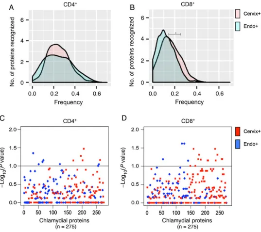
A comparison of median response frequencies for CD4+T cells revealed no difference between the Cervix+ and Endo+ groups (25% and 23.8%, respectively;P= .53; Figure 2A). However, a higher median frequency of CD8+T-cell responses was detected in the Cervix+ group, compared with the Endo+ group (13.8% vs 9.5%;P= .04; Figure2B). Examination of the response frequencies to individual proteins revealed that CD4+T cells from women in the Cervix+ group recognized 5 proteins with a significantly higher frequency (P< .10), compared with the Endo+ group (Figure2Cand Table1), and CD8+T cells from the Cervix+ group recognized a subset of 14 proteins with a higher frequency (P< .10) than those from the Endo+ group (Figure2Dand Table 1). Three of the antigens recognized by CD8+T cells with increased fre-quency (CT461, CT511, and CT529) were among the top 20 immunodominant proteins associated with CD8+T cells in Figure 2. Distribution of relative frequencies of positive CD4+and CD8+T-cell responses to a panel of chlamydial antigens, determined by ATLAS technology, among women with lower genital tract (Cervix+) and upper genital tract (Endo+) infection at enrollment.AandB, Per protein distributions of positive CD4+(A) and CD8+(B) T-cell responses to chlamydial proteins in the panel among women in the Cervix+ and Endo+ groups. Thex-axis is the frequency of positive T-cell responses in the 2 groups of patients; they-axis is the smooth fit curve of the number of proteins among the 275 proteins examined that were recognized at a particular frequency.A, The median positive response frequency of CD4+T cells from the Cervix+ group was similar to that for the Endo+ group (25% and 23.8%, respectively;P= .53).B, CD8+T cells from the Cervix+ group recognize a subset of chlamydial proteins more frequently than those from the Endo+ group (median response frequency, 13.8% and 9.5%, respectively;P= .04).CandD, Plots of the relative significance of the differential CD4+(C) and CD8+(D) T-cell responses to each protein in the panel among women in the Endo+ and Cervix+ groups. Thex-axis represents chlamydial proteins in the panel, and they-axis is the inverse log10(Pvalue), with the dashed horizontal lines representingPvalues of .10.C, CD4
+
the cohort, but none of the significant CD4+ T-cell– recog-nized antigens were among the immunodominant proteins associated with CD4+T cells. These data suggest that specific antigens drive host defense mechanisms in T cells during ac-tive chlamydial infection to prevent ascension to the upper genital tract.
CD4+and CD8+T-Cell Responses Among Follow-up
–Negative and Susceptible Women
We compared the response frequencies between follow-up– neg-ative and susceptible women and found that follow-up–negative women had a significantly greater median frequency of CD4+ T-cell responses (28.6%) as compared to susceptible women (18%;P< .0001; Figure3A). Examination of response frequen-cies to individual proteins revealed that a subset of 37 proteins were recognized by CD4+T cells of follow-up–negative women with a higher frequency (P< .10) (Figure3Cand Table2) than observed for susceptible women. In contrast to CD4+T-cell re-sponses, the median positive response rate of CD8+T cells in follow-up–negative and susceptible women did not differ (8.7% and 12.5%, respectively;P= .07; Figure3B), and no chla-mydial proteins were recognized with increased frequency by CD8+ T cells of follow-up–negative women (Figure 3D). These data suggest that a specific subset of proteins recognized by CD4+T cells may reduce susceptibility to reinfection, and these proteins should be further explored as vaccine candidates.
These proteins are summarized in Table2. Four were among the top 20 antigens recognized by CD4+T cells from the entire cohort: CT034, CT128, CT130, and CT368. No chlamydial pro-teins were recognized with increased frequency by CD8+T cells from follow-up–negative women as compared to susceptible women.
DISCUSSION
In this study, we identified specific antigens recognized by CD4+ and CD8+T cells associated with protective immunity among a high-risk cohort of women. Women in this cohort were highly sexually active, with the majority reporting irregular use of con-doms (93%) and priorC. trachomatisinfection (66%). The high rate of active infection (72%) and sampling of upper genital tract tissue provided an opportunity to compare responses of women in the Cervix+ and Endo+ groups.
Despite a lower mean antigen burden, T cells from women in the Cervix+ group recognized chlamydial proteins with greater (for CD8+T cells;P= .04) or equivalent (for CD4+T cells) fre-quency as compared to women in the Endo+ group. Recogni-tion of specific proteins by CD4+ and CD8+ T cells was associated with improved control of infection. Thesefindings indicate an important role for antigen-specific T-cell responses in preventing ascension of chlamydia to the upper genital tract. This is significant, given that chronic disease sequelae in women are primarily due to Fallopian tube scarring.
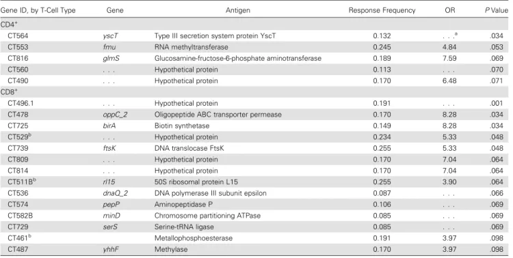
Table 1. Antigens Associated With Control of Lower Genital Tract Infection at Enrollment
Gene ID, by T-Cell Type Gene Antigen Response Frequency OR PValue
CD4+
CT564 yscT Type III secretion system protein YscT 0.132 . . .a
.034
CT553 fmu RNA methyltransferase 0.245 4.84 .053
CT816 glmS Glucosamine-fructose-6-phosphate aminotransferase 0.189 7.59 .069
CT560 . . . Hypothetical protein 0.113 . . . .070
CT490 . . . Hypothetical protein 0.170 6.48 .071
CD8+
CT496.1 . . . Hypothetical protein 0.191 . . . .001
CT478 oppC_2 Oligopeptide ABC transporter permease 0.170 8.28 .034
CT725 birA Biotin synthetase 0.149 8.28 .034
CT529b
. . . Hypothetical protein 0.234 5.33 .048
CT739 ftsK DNA translocase FtsK 0.255 5.33 .048
CT809 . . . Hypothetical protein 0.170 7.04 .064
CT814 . . . Hypothetical protein 0.170 7.04 .064
CT511Bb
rl15 50S ribosomal protein L15 0.255 3.90 .064
CT536 dnaQ_2 DNA polymerase III subunit epsilon 0.087 . . . .066
CT574 pepP Aminopeptidase P 0.106 . . . .069
CT582B minD Chromosome partitioning ATPase 0.085 . . . .069
CT729 serS Serine-tRNA ligase 0.085 . . . .069
CT461b
Metallophosphoesterase 0.191 3.97 .098
CT487 yhhF Methylase 0.170 3.97 .098
Abbreviations: ID, identifier; OR, odds ratio. a
The denominator is 0.
The high follow-up rate in our study provided the opportu-nity to compare participants who subsequently acquiredC. tra-chomatisand those who remained uninfected over 12 months of follow-up. We found that CD4+T cells are the key players in resistance to reinfection. Significantly greater CD4+T-cell re-sponse frequencies were detected in women who remained un-infected, compared with susceptible women (P< .0001), and a subset of 37 proteins were recognized more frequently by CD4+T cells from follow-up–negative women. Because having sex with a new partner is a known risk factor and was associated with acquisition (Supplementary Table 3), we adjusted for this in our statistical analysis. Nevertheless, one third of follow-up– negative women reported a new or multiple different partners during follow-up. It is possible that some of the women that we defined as follow-up negative were simply not exposed dur-ing follow-up. However, the fact that higher rates of positive CD4+T-cell responses were coupled with enhanced responses to a specific subset of proteins in follow-up–negative women
implies that these women were less susceptible because of either increased past exposure or a more robust response to a single past infection.
Our data suggest differing roles for CD8+and CD4+T cells in response to chlamydial genital tract infection. CD8+T cells may contribute to limiting the bacterial burden and ascension of in-fection, while CD4+T cells appear to drive protection from re-infection. In each case, limiting acute infection or preventing repeat infection was dependent on having a T-cell response to a specific subset of proteins, rather than on achieving a thresh-old frequency of responses to random proteins. This points to-ward certain proteins being better drivers of protective adaptive responses. Notably, some of these antigens are among the most frequently recognized proteins within the entire cohort.
Because the panel of proteins selected for evaluation was based on criteria derived via our prior study [21], several pro-teins reported in murine vaccine studies to induce protection were not assessed. Immunization with native major outer Figure 3. Distribution of relative frequencies of positive CD4+and CD8+T-cell responses to a panel of chlamydial antigens, determined by ATLAS technology, among
susceptible and follow-up–negative women, as defined by infection status during 1 year of follow-up.AandB, Per protein distributions of positive CD4+(
A) and CD8+(
B) T-cell responses to chlamydial proteins in the panel among susceptible and follow-up–negative women. Thex-axis is the frequency of positive T-cell responses in the 2 patient groups; they-axis is the smooth fit curve of the number of proteins among the 275 proteins examined that were recognized at a particular frequency.A, CD4+T cells from
follow-up–negative women recognized a subset of chlamydial proteins more frequently than susceptible women (median response frequency, 28.6% and 18%, respectively;P< .0001). B, The median positive response frequencies of CD8+T cells were similar among follow-up
–negative and susceptible women (8.7% and 12.5%, respectively;P= .07).CandD, Plots of the relative significance of the differential CD4+(
C) and CD8+(
D) T-cell response to each protein in the panel among susceptible and follow-up–negative women. Thex -axis represents chlamydial proteins in the panel, and they-axis is the inverse log10(Pvalue), with the dashed horizontal line representingP= .10.C, CD4+T cells from follow-up–
negative women recognized 14 chlamydial proteins significantly more frequently (P< .05) and 37 proteins more frequently (P< .10) than those in susceptible women.D, No proteins were recognized with increased frequency by CD8+T cells from follow-up
membrane protein (MOMP) protects against oviduct disease in the murine model when delivered with adjuvant [26]. This pro-tein was not included in our current screen because in the prior screen it elicited a low frequency of T-cell responses [21], de-spite MOMP immunoglobulin G (IgG) seropositivity in 40% of the cohort (unpublished data). A prior study that examined T-cell IFN-γresponses to 116 recombinant chlamydial antigens in infected patients revealed responses to inner membrane and cytoplasmic proteins only; MOMP was recognized only by IgG [27]. These studies suggest that MOMP elicits poor T-cell re-sponses in humans despite induction of antibody. Murine
studies reveal T-cell responses to MOMP [28], but differences in cellular processing machinery in mice and humans at the level of the proteasome and in major histocompatibility com-plex lead to differences in antigen presentation [29]. Other pro-teins that have induced protection in mouse vaccine models that were not included in our screen included CPAF [30]; polymor-phic membrane proteins E, F, and G [31,32]; OmcB and rl16 [33]; and CT823 [34]. CrpA, IncA (an immunodominant, inclusion membrane-associated protein), and PmpD were examined in our panel, but response frequencies were not sig-nificantly different among the subgroups. A hypothetical Table 2. Antigens Associated With Protection FromChlamydia trachomatisInfection During 1 Year of Follow-up
Gene ID, by T Cell-Type Gene Antigen Response Frequency OR PValue
CD4+
CT308 atpA V-type ATP synthase subunit A 0.259 4.55 .018
CT020 lepB Signal peptidase I 0.291 4.41 .019
CT313 tal Transaldolase 0.327 3.54 .025
CT034a
ytfF Cationic amino acid transporter 0.370 2.92 .032
CT156 . . . Hypothetical protein 0.255 4.08 .034
CT360 . . . Hypothetical protein 0.182 4.93 .035
CT134 . . . Hypothetical protein 0.259 3.31 .036
CT264 msbA ABC transporter ATP binding protein/permease 0.236 4.77 .039
CT421 . . . Hypothetical protein 0.283 2.97 .039
CT307 atpB V-type ATP synthase subunit B 0.273 3.20 .042
CT368a
aroC Chorismate synthase 0.382 2.69 .042
CT004 gatB Aspartyl/glutamyl-tRNA amidotransferase subunit 0.327 2.87 .044
CT005 . . . Hypothetical protein 0.315 2.95 .046
CT218 surE 5’-nucleotidase SurE 0.291 3.47 .050
CT757 mraY Phospho-N-acetylmuramoyl-pentapeptide transferase 0.236 4.39 .051
CT319 rI11 50S ribosomal protein L11 0.273 3.03 .052
CT386 . . . Metal dependent hydrolase 0.273 3.42 .054
CT130a
glnQ ABC amino acid transporter ATPase 0.455 2.38 .054
CT647 . . . Hypothetical protein 0.204 4.30 .056
CT017B . . . Hypothetical protein 0.236 4.44 .057
CT338 . . . Hypothetical protein 0.236 3.37 .059
CT002 gatC Glutamyl-tRNA amidotransferase subunit C 0.236 3.30 .060
CT351 . . . Hypothetical protein 0.273 3.32 .061
CT022 rI31 50S ribosomal protein L31 0.291 2.68 .065
CT133 . . . rRNA methylase 0.345 2.44 .068
CT687 . . . Cysteine desulfurase-like protein 0.236 4.00 .069
CT821 sucC Succinyl-CoA ligase subunit beta 0.236 3.18 .071
CT315 rpoB DNA-directed RNA polymerase subunitβ 0.309 2.42 .072
CT144 . . . Hypothetical protein 0.255 3.12 .074
CT318 rI1 50S ribosomal protein L1 0.309 3.13 .075
CT104 Fabl Enoyl-acyl-carrier protein reductase 0.164 6.19 .080
CT128a
Adk Adenylate kinase 0.382 2.23 .082
CT252C Lgt Prolipoprotein diacylglycerol transferase 0.273 2.68 .082
CT382 aroG Phospho-2-dehydro-3-deoxyheptonate aldolase 0.259 3.03 .084
CT717 . . . Type III secretion system ATP synthase 0.241 2.81 .086
CT808 cafE Rne/Rng family ribonuclease 0.145 5.61 .097
CT359 . . . Hypothetical protein 0.309 2.38 .098
CD8+
. . . .
None . . . .
Abbreviations: ID, identifier; OR, odds ratio. a
protein, CT144, that we found to be protective when adminis-tered as a vaccine in mice [34] was recognized with a greater fre-quency by CD4+ T cells from follow-up–negative women (P= .07). Tarp, which has been found to induce high titers of antibodies in humans and protective immunity against upper genital tract pathologies in mice [35], was investigated and was more frequently recognized by CD8+T cells from women in the Cervix+ group, compared with those in the Endo+ group (P= .11), although our small sample size likely limited our abil-ity to detect statistically significant differences in these respons-es. Because testing was limited to one third of the proteome, we may have missed antigens important in protective immunity. However, the current panel was based on prior human screens, which allows for inclusion of proteins that might have been omitted if the literature, animal studies, and predictive algo-rithms were used as the determinants [21].
The proteins of recognized function associated with control or resistance to infection in our cohort are involved in protein synthesis, central metabolism, and type III secretion (Tables1
and 2). This is consistent with our previous investigation, using ATLAS [21]. Many of the antigens identified then as in-ducing“effective”responses (ie, spontaneous clearance or lack of infection despite confirmed exposure) were also genes in-volved in central metabolism. Three proteins associated with ef-fective immunity in that cohort (CT002, CT252, and CT757) were also associated with resistance to infection in this cohort. Additionally, CT564 and CT725, specifically associated with in-fection control in our TRAC cohort, were associated with effec-tive immunity previously [21]. In the prior study, CD8+T-cell responses were associated with effective immune responses in men and women, which is consistent with a role for these cells in prevention of ascension in this female only cohort. These proteins are essential for intracellular survival of
C. trachomatis; since this pathogen lives within epithelial cells and interacts intimately with the endoplasmic reticulum, pep-tides of these essential proteins are likely shuttled through the vesicular system and loaded into major histocompatibility com-plex molecules. A limitation of the ATLAS screen is our inabil-ity to confirm that each protein was successfully expressed and presented by human APCs. Confirmation that a full-length pro-tein is expressed inE. coliis derived from a cell-based assay in which a B3Z T-cell hybridoma is tested for recognition of a C-terminal SIINFEKL-tag inserted into each clone. The antigens and tag are processed and presented to the T cells by murine APCs [36]. However, we cannot be certain that the human APCs presented each protein in comparable fashion.
Strengths of this study included comprehensive data on de-mographic, clinical, and sexual characteristics, high rate of fol-low-up, and a stringent requirement to complete 4 follow-up visits over 1 year to be considered in the follow-up–negative subgroup. A further strength is the nonbiased evaluation of over one third of the chlamydial genome. Classifying upper
and lower genital tract infection at enrollment allowed us to identify antigens important for control of infection. Additional-ly, following these women longitudinally over 1 year allowed us to identify antigens specifically associated with protection against subsequent infection. Several of the proteins identified were immunodominant, suggesting that this technology may be capable of identifying novel vaccine candidates with the po-tential for broad recognition by immunized individuals.
Supplementary Data
Supplementary materialsare available athttp://jid.oxfordjournals.org. Consisting of data provided by the author to benefit the reader, the posted materials are not copyedited and are the sole responsibility of the author, so questions or comments should be addressed to the author.
Notes
Acknowledgments. We thank the women who agreed to participate in this study; Alison Collins, Ingrid Macio, Melinda Petrina, Carol Priest, Abi Jett, and Lorna Rabe, for their efforts in the clinic and/or laboratory; and the staff at the Allegheny County Health Department STD Clinic, for their support.
Financial support. This work was supported by the National Institute of Allergy and Infectious Diseases (grants U19 AI084024 and U19 AI113170).
Potential conflicts of interest. All authors: No reported conflicts. All authors have submitted the ICMJE Form for Disclosure of Potential Con-flicts of Interest. Conflicts that the editors consider relevant to the content of the manuscript have been disclosed.
References
1. Satterwhite CL, Torrone E, Meites E, et al. Sexually transmitted infections among US women and men: prevalence and incidence estimates, 2008. Sex Transm Dis
2013; 40:187–93.
2. World Health Organization (WHO). Global prevalence and incidence of selected curable sexually transmitted infections: overview and estimates, 2001. Geneva, Switzerland: WHO,2001.
3. Miller WC, Ford CA, Morris M, et al. Prevalence of chlamydial and gonococcal infections among young adults in the United States. JAMA2004; 291:2229–36. 4. Owusu-Edusei K Jr., Chesson HW, Gift TL, et al. The estimated direct medical cost
of selected sexually transmitted infections in the United States, 2008. Sex Transm Dis2013; 40:197–201.
5. Arno JN, Katz BP, McBride R, et al. Age and clinical immunity to infections with
Chlamydia trachomatis. Sex Transm Dis1994; 21:47–52.
6. Batteiger BE, Xu F, Johnson RE, Rekart ML. Protective immunity toChlamydia trachomatisgenital infection: evidence from human studies. J Infect Dis2010; 201(suppl 2):S178–89.
7. Geisler WM, Lensing SY, Press CG, Hook EW III. Spontaneous resolution of gen-ital Chlamydia trachomatis infection in women and protection from reinfection. J Infect Dis2013; 207:1850–6.
8. Woolridge RL, Grayston JT, Chang IH, Cheng KH, Yang CY, Neave C. Field trial of a monovalent and of a bivalent mineral oil adjuvant trachoma vaccine in Tai-wan school children. Am J Ophthalmol1967; 63:1645–50.
9. Woolridge RL, Grayston JT, Chang IH, Yang CY, Cheng KH. Long-term follow-up of the initial (1959–1960) trachoma vaccinefield trial on Taiwan. Am J Ophthal-mol1967; 63:1650–5.
10. Sowa S, Sowa J, Collier LH, Blyth WA. Trachoma vaccinefield trials in The Gam-bia. J Hyg (Lond)1969; 67:699–717.
11. Rank RG, Whittum-Hudson JA. Protective immunity to chlamydial genital infec-tion: evidence from animal studies. J Infect Dis2010; 201(suppl 2):S168–77. 12. O’Connell CM, Ingalls RR, Andrews CW Jr, Skurlock AM, Darville T.
Plasmid-deficientChlamydia muridarumfail to induce immune pathology and protect against oviduct disease. J Immunol2007; 179:4027–34.
13. Kari L, Whitmire WM, Olivares-Zavaleta N, et al. A live-attenuated chlamydial vaccine protects against trachoma in nonhuman primates. J Exp Med2011; 208:2217–23.
15. Igietseme JU, Ramsey KH, Magee DM, Williams DM, Kincy TJ, Rank RG. Reso-lution of murine chlamydial genital infection by the adoptive transfer of a biovar-specific TH1 lymphocyte clone. Reg Immunology1993; 5:317–24.
16. Gondek DC, Olive AJ, Stary G, Starnbach MN. CD4+ T cells are necessary and sufficient to confer protection against chlamydia trachomatis infection in the mu-rine upper genital tract. J Immunol2012; 189:2441–9.
17. Morrison SG, Morrison RP. A predominant role for antibody in acquired immunity to chlamydial genital tract reinfection. J Immunol2005; 175: 7536–42.
18. Morrison RP, Feilzer K, Tumas DB. Gene knockout mice establish a primary protective role for major histocompatibility complex class II-restricted responses inChlamydia trachomatisgenital tract infection. Infect Immun1995; 63:4661–8.
19. Olivares-Zavaleta N, Whitmire WM, Kari L, Sturdevant GL, Caldwell HD. CD8+ T cells define an unexpected role in live-Attenuated vaccine protective immunity against Chlamydia trachomatis infection in macaques. J Immunol 2014; 192:4648–54.
20. Gervassi AL, Probst P, Stamm WE, Marrazzo J, Grabstein KH, Alderson MR. Functional characterization of class Ia- and non-class Ia-restricted Chlamydia-re-active CD8+ T cell responses in humans. J Immunol2003; 171:4278–86. 21. Picard MD, Bodmer JL, Gierahn TM, et al. Resolution of chlamydia trachomatis
infection is associated with a distinct T cell response profile. Clin Vaccine Immu-nol2015; 22:1206–18.
22. Grubaugh D, Flechtner JB, Higgins DE. Proteins as T cell antigens: methods for high-throughput identification. Vaccine2013; 31:3805–10.
23. Russell AN, Zheng X, O’Connell CM, et al. Analysis of factors driving incident and ascending infection and the role of serum antibody in chlamydia trachomatis gen-ital tract infection. J Infect Dis2016; 213:523–31.
24. Workowski KA, Berman SM. Centers for Disease Control and Prevention sexually transmitted disease treatment guidelines. Clin Infect Dis2011; 53(suppl 3): S59–63.
25. Hu PQ, Tuma-Warrino RJ, Bryan MA, et al. Escherichia coli expressing recombi-nant antigen and listeriolysin O stimulate class I-restricted CD8+ T cells following uptake by human APC. J Immunol2004; 172:1595–601.
26. Farris CM, Morrison SG, Morrison RP. CD4+ T cells and antibody are required for optimal major outer membrane protein vaccine-induced immunity to Chlamydia muridarum genital infection. Infect Immun2010; 78:4374–83.
27. Follmann F, Olsen AW, Jensen KT, Hansen PR, Andersen P, Theisen M. Antigenic profiling of a Chlamydia trachomatis gene-expression library. J Infect Dis2008; 197:897–905.
28. Finco O, Frigimelica E, Buricchi F, et al. Approach to discover T- and B-cell an-tigens of intracellular pathogens applied to the design of Chlamydia trachomatis vaccines. Proc Natl Acad Sci USA2011; 108:9969–74.
29. Sesma L, Alvarez I, Marcilla M, Paradela A, Lopez de Castro JA. Species-specific differences in proteasomal processing and tapasin-mediated loading influence peptide presentation by HLA-B27 in murine cells. J Biol Chem 2003; 278:46461–72.
30. Murthy AK, Chambers JP, Meier PA, Zhong G, Arulanandam BP. Intranasal vac-cination with a secreted chlamydial protein enhances resolution of genital Chla-mydia muridaruminfection, protects against oviduct pathology, and is highly dependent upon endogenous gamma interferon production. Infect Immun
2007; 75:666–76.
31. Yu H, Jiang X, Shen C, et al.Chlamydia muridarumT-cell antigens formulated with the adjuvant DDA/TDB induce immunity against infection that correlates with a high frequency of gamma interferon (IFN-gamma)/tumor necrosis factor alpha and IFN-gamma/interleukin-17 double-positive CD4+ T cells. Infect Immun2010; 78:2272–82.
32. Yu H, Jiang X, Shen C, Karunakaran KP, Brunham RC. NovelChlamydia muri-darumT cell antigens induce protective immunity against lung and genital tract infection in murine models. J Immunol2009; 182:1602–8.
33. Olsen AW, Theisen M, Christensen D, Follmann F, Andersen P. Protection against Chlamydia promoted by a subunit vaccine (CTH1) compared with a primary in-tranasal infection in a mouse genital challenge model. PloS One2010; 5:e10768. 34. Picard MD, Cohane KP, Gierahn TM, Higgins DE, Flechtner JB. High-throughput proteomic screening identifiesChlamydia trachomatisantigens that are capable of eliciting T cell and antibody responses that provide protection against vaginal challenge. Vaccine2012; 30:4387–93.
35. Wang J, Chen L, Chen F, et al. A chlamydial type III-secreted effector protein (Tarp) is predominantly recognized by antibodies from humans infected with
Chlamydia trachomatisand induces protective immunity against upper genital tract pathologies in mice. Vaccine2009; 27:2967–80.