ⓒ 2017 The Korean Society of Neurogastroenterology and Motility
Introduction
High-resolution manometry (HRM) provides an intuitive and panoramic view of the proximal digestive physiology from the phar-ynx to the stomach (Fig. 1). As compared to conventional manom-etry, HRM determines more comfort and speediness to the test, reduces inter-observer variability, and compensates movements’ ar-tefacts. These improvements allow a better evaluation of sphincters relaxation, and the identification of segmental defects of peristalsis not covered by the spacing of sensors in conventional systems.1,2
The more comprehensive and more appealing to the eyes HRM plots provoked the imagination of esophageal physiologists to create new investigative parameters and reclassify esophageal motility disorders. This led to the development of an algorithmic
scheme for the diagnosis of esophageal motility disorders in esopha-geal pressure topography plots, under the name of the Chicago classification.3 This allowed for improved recognition of motility
disorders, and easier interpretation than conventional manometry line tracings.
The Chicago classification was recently revised (version 3.0) to exclude some previous parameters without clear clinical application, define parameters to be used, and classify esophageal motility disor-ders.4
The aim of this study was to review the Chicago classification version 3.0, and assess the clinical implications of the parameters and disorders defined by this classification.
Understanding the Chicago Classification:
From Tracings to Patients
Francisco Schlottmann,1* Fernando A Herbella,2 and Marco G Patti1
1Department of Surgery and Center for Esophageal Diseases, University of North Carolina, Chapel Hill, NC, USA; and 2Department of Surgery,
Escola Paulista de Medicina, Federal University of Sao Paulo, Sao Paulo, Brazil
Current parameters of the Chicago classification include assessment of the esophageal body (contraction vigour and peristalsis), lower esophageal sphincter relaxation pressure, and intra-bolus pressure pattern. Esophageal disorders include achalasia, esophagogastric junction outflow obstruction, major disorders of peristalsis, and minor disorders of peristalsis. Sub-classification of achalasia in types I, II, and III seems to be useful to predict outcomes and choose the optimal treatment approach. The real clinical significance of other new parameters and disorders is still under investigation. (J Neurogastroenterol Motil 2017;23:487-494)
Key Words
Chicago classification; Esophageal achalasia; Esophageal motility disorders; High-resolution manometry
Received: February 22, 2017 Revised: April 26, 2017 Accepted: June 9, 2017
This is an Open Access article distributed under the terms of the Creative Commons Attribution Non-Commercial License (http://creativecommons. org/licenses/by-nc/4.0) which permits unrestricted non-commercial use, distribution, and reproduction in any medium, provided the original work is properly cited.
*Correspondence: Francisco Schlottmann, MD
University of North Carolina at Chapel Hill, 4030 Burnett Womack Building, 101 Manning Drive, CB 7081, Chapel Hill, NC 27599-7081, USA
Tel: +1-919-966-8436, Fax: +1-919-966-8440, E-mail: [email protected]
Parameters Evaluated by High-resolution
Manometry
Esophageal Body
Contraction vigour
HRM allows the evaluation of contractility not only by am-plitude measurement at fixed points, but through a combination of amplitude, time and length of the whole peristaltic wave. This
parameter is the distal contractile integral (DCI). DCI value is cal-culated as the product of the mean amplitude of contraction in the distal esophagus (mmHg) times the duration of contraction (sec-onds), times the length of the distal esophageal segment (cm) ex-ceeding 20 mmHg for the region spanning from the transition zone to the proximal aspect of the lower esophageal sphincter (LES) (Fig.
2). DCI classifies waves as failed (DCI < 100 mmHg ∙ sec ∙ cm), weak (DCI 100-450 mmHg ∙ sec ∙ cm), ineffective (failed or weak), normal (DCI 450-8000 mmHg ∙ sec ∙ cm), or hypercontractile (DCI > 8000 mmHg ∙ sec ∙ cm).5
Peristalsis
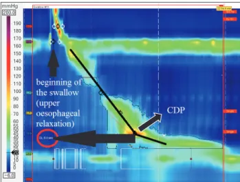
HRM evaluates peristalsis by the distal latency (DL) which measures objectively the timeframe of the wave from the beginning of the swallow (upper esophageal sphincter relaxation) to an inflection 180
160
140
120
100
80
60 50 40 30 20 10 0
10 20 mmHg
200.0
25.0
Figure 1. Normal high-resolution manometry plot (left) compared to the same swallow at the conventional ma-nometry (right).
190 180 170 160 150 140 130 120 110 100 90 80 70 60 55 50 45 40 35 30 25 15 10 5 0 20 mmHg 200.0
6.0
Figure 3. Distal latency (DL) measures objectively the time frame of the wave from the beginning of the swallow (upper esophageal relax-ation) to the contractile deceleration point (CDP).
190 180
170 160
150
140 130
120 110
100 90
80
70 60 55 50 45 40 35 30 25 15 10 5 0 20 mmHg
200.0
8.0
of the peristaltic axis known as the contractile deceleration point (Fig. 3). Premature contractions are defined with a DL < 4.5 seconds.
Fragmented contractions are considered segmental defects (break in the 20 mmHg isobaric contour > 5 cm) with normal contraction vigour.
Table summarizes esophageal body parameters evaluated with HRM.
Lower Esophageal Sphincter
Lower esophageal sphincter pressure
The Chicago classification did not define parameters for LES
length or basal pressure, but simply recommended assessment of pressure as an average of inspiratory and expiratory values for 3 normal respiratory cycles. Relaxation, however, is measured not by the nadir pressure as previously done with conventional manometry, but with the integrated relaxation pressure (IRP) that corresponds to the mean pressure of 4 seconds of greatest post deglutitive
relax-140
130
120
110
100
90
80
70
60
50
40 35 30 25
15 10 5 0 5 20 mmHg
150.0
10.0
Figure 4. Integrated relaxation pressure (IRP) corresponds to the mean pressure of 4 seconds of greatest post deglutitive relaxation in a 10 seconds gap, triggered at the beginning of a swallow. Note dia-phragmatic contraction pressure (*) during relaxation excluded from analysis.
Type I Type II Type III
Figure 5. Achalasia subtypes. Type I: absence of esophageal pressurization; Type II: panesophageal pressurization; Type III: premature contrac-tions (distal latency < 4.5 seconds).
Table. Characterization of Esophageal Contractility
Contraction vigour
Failed DCI < 100 mmHg ∙ sec ∙ cm
Weak DCI 100-450 mmHg ∙ sec ∙ cm
Ineffective Failed or weak
Normal DCI 450-8000 mmHg ∙ sec ∙ cm
Hypercontractile DCI > 8000 mmHg ∙ sec ∙ cm
Contraction pattern Premature DL < 4.5 sec
Fragmented Break > 5 cm in the 20 mmHg isobaric contour with normal DCI
ation in a 10 seconds gap, triggered at the beginning of a swallow (Fig. 4). This metric compensates for diaphragmatic contraction during LES relaxation, and eliminates pseudo-relaxation due to movement artefacts.
Intrabolus Pressure Pattern
Abnormal intrabolus pressure corresponds to regions of esophageal pressurization > 30 mmHg. It may be panesophageal (whole esophageal body), compartmentalized (from the contractile deceleration point to the esophagogastric junction [EGJ]) or EGJ pressurization (between the LES and the diaphragm).
Esophageal Motility Disorders
Achalasia
Achalasia is defined by aperistalsis and abnormal LES
relax-140
130
120
110
100
90
80
70
60
50
40 35 30 25
15 10 5 0 5 20 mmHg 150.0
10.0
Figure 7. Hypercontractile esopha-gus (jackhammer esophaesopha-gus). Distal contractile integral (DCI) > 8000
mmHg ∙ sec ∙ cm in at least 20% of swal -lows and normal distal latency (DL).
140 130
120 110 100
90 80 70
60 50 40 35 30 25 20 15 10 5 0 5 mmHg 150.0
10.0
Figure 8. Absent contractility. Aperistal-sis in the setting of normal lower esoph-ageal sphincter relaxation (integrated relaxation pressure < 10 mmHg).
140
130
120
110
100
90
80
70
60
50
40 35 30 25
15 10 5 0 5 20 mmHg 150.0
10.0
ation (IRP > 15 mmHg). The disease is further classified into 3 subtypes (Fig. 5).
Type I: incomplete LES relaxation, aperistalsis and absence of esophageal pressurization.
Type II: incomplete LES relaxation, aperistalsis and panesoph-ageal pressurization in at least 20% of swallows. Type III: incomplete LES relaxation and premature
contrac-tions (DL < 4.5 seconds) in at least 20% of swal-lows.
Esophagogastric Junction Outflow Obstruction
EGJ obstruction is characterized by an impaired LES relax-ation (IRP > 15 mmHg) with normal or weak peristalsis.
Major Disorders of Peristalsis
Distal esophageal spasm
Distal esophageal spasm (DES) is defined by premature con-tractions (DL < 4.5 seconds) in at least 20% of swallows with a normal IRP (Fig. 6).
Hypercontractile esophagus
Hypercontractile esophagus (jackhammer esophagus) is
char-acterized by DCI > 8000 mmHg ∙ sec ∙ cm in at least 20% of swal -lows and normal DL (Fig. 7).
Absent contractility
Absent contractility is characterized by aperistalsis in the setting of normal LES relaxation (IRP < 10 mmHg) (Fig. 8).
Minor Disorders of Peristalsis
Ineffective esophageal motility
Ineffective esophageal motility is defined by ≥ 50% ineffective
140
130
120
110
100
90
80
70
60
50
40 35 30 25
15 10 5 0 5 20 mmHg
150.0
10.0
Figure 9. Ineffective esophageal motility. Failed or weak peristalsis in at least ≥ 50% of swallows. DCI, distal contractile integral.
190 180
170
160
150 140
130
120
110 100
90
80
70 60 55 50 45 40 35 30 25 20 15 10 5 0 mmHg 200.0
7.0
swallows (failed or weak––DCI < 450 mmHg ∙ sec ∙ cm) (Fig. 9).
Fragmented peristalsis
Fragmented peristalsis is defined by ≥ 50% fragmented con -tractions with normal contraction vigour (Fig. 10).
Impact of High-resolution Manometry on
the Management of Esophageal Motility
Disorders
The Chicago classification reclassified motility disorders previ-ously defined by conventional manometry.6 The clinical value of this
new classification is under scrutiny. A recent publication claims that the Chicago classification has a higher threshold for abnormality, resulting in fewer patients classified as abnormal motility.7 On the
other hand, a recent study that randomized patients with dyspha-gia to undergo either HRM or conventional manometry, found a higher proportion of manometric diagnosis in the HRM arm.8
Achalasia
The current main therapeutic options for achalasia include pneumatic dilatation (PD), peroral endoscopic myotomy (POEM), and laparoscopic Heller myotomy (LHM).9
The Chicago classification may help to predict the result of treatment. Pandolfino and colleagues10 reported that type II
achala-sia patients were significantly more likely to respond to PD (91%) or LHM (100%), as compared to type I (56% overall) and type III (29% overall). Concordantly, Salvador et al11 evaluated 246
con-secutive patients who underwent LHM and found that treatment failure rates were significantly different among the subtypes of acha-lasia: type I (14.6%), type II (4.7%), and type III (30.4%) (P = 0.0007). A recent meta-analysis encompassing 9 studies and 727 patients also showed that type II achalasia was associated with the best prognosis after PD and LHM, while type III achalasia had the worst prognosis.12
The Chicago classification may also help selecting the best ini-tial approach for patients with achalasia. Kumbhari and colleagues13
reported that in patients with type III achalasia, clinical response was achieved more frequently after POEM (98.0%), as compared to LHM (80.8%) (P = 0.01). Recently, Khashab et al14 reported
their experience with POEM for the treatment of 54 patients with type III achalasia refractory to medical therapy, and showed a 96.3% clinical success rate. Hence, while in type I and II achalasia both PD and LHM remain as good treatment alternatives, type III achalasia seems to be better managed with POEM, probably due
to the ability to a longer myotomy of the thoracic esophagus. Overall, sub-classification of achalasia in types I, II, and III with the Chicago classification seems to be useful to predict out-comes and choose the optimal treatment approach for this motility disorder.
Esophagogastric Junction Outflow Obstruction
The definition of EGJ outflow obstruction based solely on the IRP with the exclusion of achalasia allows this diagnosis to be superimposed to other diagnoses dependent of the esophageal body motility. It may be caused by an anatomical abnormality at the cardia (hiatal hernia, diseases of the esophageal wall, etc) or be idio-pathic with normal anatomy.
Similar to achalasia, treatment is directed towards relief of the obstruction and can be accomplished by botulinum toxin injection, PD, LHM, or POEM. Both botulinum injection and PD showed good relief of dysphagia but with ephemeral duration.15
Scherer and colleagues,16
among 1000 HRM diagnosed 16 patients (1.6%) with EGJ outflow obstruction and treated them with botulinum toxin injection, PD or LHM. Only the 3 patients treated with LHM responded well.16
Interestingly, Pérez-Fernández et al17
reported that over one-third of the patients with EGJ outflow obstruction presented a spontaneous resolution of the symptoms, concluding that surgical treatment should be considered with special caution in these patients. In the setting of an anatomic abnormality such as a hiatal hernia, surgical correction is associated with long-lasting and excellent results.18
EGJ outflow obstruction is now recognized as a distinct entity in the Chicago classification. However, the real clinical significance of this diagnosis is still uncertain. In fact, it could be an early or incomplete expression of a variant of achalasia. Thus, the exact sig-nificance and clinical management of these patients remains unclear.
Major Disorders of Peristalsis
As the symptoms and the manometric picture of esophageal motility disorders can be due to gastroesophageal reflux disease (GERD), it is of paramount importance to rule out abnormal reflux by pH monitoring. If GERD is present, either medical or surgical treatment should be directed towards the control of the reflux.19
The management of DES remains elusive. The Chicago classi-fication defines this disorder with a new parameter: the DL. Previ-ous reports showed good results in patients with “diffuse esophageal spasm” (Richter classification)6
who underwent LHM with an extended myotomy.20,21
this definition became publicized showed that botulinum toxin injection in the esophageal body was superior to placebo to relieve dysphagia in patients with DES, and that POEM is a promising treatment for these patients.22
The definition of hypercontractile esophagus (jackhammer) was updated in the last version of the Chicago classification to
include only cases with ≥ 20% of swallows with a DCI > 8000 mmHg ∙ sec ∙ cm, excluding a single altered swallow from the
definition. Pharmacological relaxation of the smooth muscle with phosphodiesterase-5 inhibitor or anticholinergic agents has shown symptomatic improvement.23
Studies focusing on surgical therapy for hypercontractile esophagus based on the new classification are not available. Previous reports not using HRM or the Chicago classification, however, showed acceptable outcomes after surgical myotomy.24,25
Absent contractility is mostly diagnosed in patients with con-nective tissue diseases. There is no specific treatment to restore or improve peristalsis in these patients. Associated GERD is usually the target of therapy.26
Minor Disorders of Peristalsis
Therapeutic options for ineffective esophageal motility are still limited, as no effective treatment is available to restore impaired esophageal smooth muscle contractility.27
Treatment directed to-wards GERD is helpful when dysmotility is secondary to this dis-ease.
The concept of fragmented peristalsis changed radically from previous versions to the version 3.0. Only large breaks (> 5 cm) with normal peristalsis are included. This is more clinically relevant, since incomplete bolus transit is observed in 100% of the cases of large breaks but only in 16% of small breaks.28
It is unclear how to treat this finding since there are no studies focusing on the treat-ment for this disease under these criteria. There are no studies evaluating changes in the motility pattern after therapy for GERD as well, since both conditions are frequently associated.
Conclusions
HRM and the Chicago classification certainly contributed to a better definition of esophageal motility disorders. Particularly for achalasia, sub-classification in types I, II, and III seems to be useful to predict outcomes and choose the optimal treatment approach. The real clinical significance of other new parameters and disorders is still under investigation.
Financial support: None.
Conflicts of interest: None.
Author contributions: Francisco Schlottmann, Fernando A Herbella, and Marco G Patti planned conception and design, drafted the article, and approved the final article.
References
1. Herbella FA, Patti MG. Can high resolution manometry parameters for achalasia be obtained by conventional manometry? World J Gastrointest Pathophysiol 2015;15:58-61.
2. Herbella FA, Armijo PR, Patti MG. [A pictorial presentation of 3.0 Chicago classification for esophageal motility disorders.] Einstein (Sao Paulo) 2016;14:439-442.[English, Portuguese]
3. Kahrilas PJ, Ghosh SK, Pandolfino JE. Esophageal motility disorders in terms of pressure topography: the Chicago classification. J Clin Gastroen-terol 2008;42:627-635.
4. Kahrilas PJ, Bredenoord AJ, Fox M, et al. The Chicago classifica-tion of esophageal motility disorders, v3.0. Neurogastroenterol Motil 2015;27:160-174.
5. Roman S, Pandolfino JE, Chen J, Boris L, Luger D, Kahrilas PJ. Phe-notypes and clinical context of hypercontractility in high-resolution esoph-ageal pressure topography (EPT). Am J Gastroenterol 2012;107:37-45. 6. Richter JE. Oesophageal motility disorders. Lancet 2001;358:823-828. 7. Monrroy H, Cisternas D, Bilder C, et al. The Chicago classification 3.0
results in more normal findings and fewer hypotensive findings with no difference in other diagnoses. Am J Gastroenterol 2017;112:606-612. 8. Roman S, Huot L, Zerbib F, et al. High-resolution manometry improves
the diagnosis of esophageal motility disorders in patients with dysphagia: a randomized multicenter study. Am J Gastroenterol 2016;111:372-380. 9. Herbella FA, Moura EG, Patti MG. Achalasia 2016: treatment
alterna-tives. J Laparoendosc Adv Surg Tech A 2017;27:6-11.
10. Pandolfino JE, Kwiatek MA, Nealis T, Bulsiewicz W, Post J, Kahrilas PJ. Achalasia: a new clinically relevant classification by high-resolution manometry. Gastroenterology 2008;135:1526-1533.
11. Salvador R, Costantini M, Zaninotto G, et al. The preoperative mano-metric pattern predicts the outcome of surgical treatment for esophageal achalasia. J Gastrointest Surg 2010;14:1635-1645.
12. Ou YH, Nie XM, Li LF, Wei ZJ, Jiang B. High-resolution manomet-ric subtypes as a predictive factor for the treatment of achalasia: a meta-analysis and systematic review. J Dig Dis 2016;17:222-235.
13. Kumbhari V, Tieu AH, Onimaru M, et al. Peroral endoscopic myotomy (POEM) vs laparoscopic heller myotomy (LHM) for the treatment of type III achalasia in 75 patients: a multicenter comparative study. Endosc Int Open 2015;3:E195-E201.
15. van Hoeij FB, Smout AJ, Bredenoord AJ. Characterization of idiopathic esophagogastric junction outflow obstruction. Neurogastroenterol Motil 2015;27:1310-1316.
16. Scherer JR, Kwiatek MA, Soper NJ, Pandolfino JE, Kahrilas PJ. Functional esophagogastric junction obstruction with intact peristalsis: a heterogeneous syndrome sometimes akin to achalasia. J Gastrointest Surg 2009;13:2219-2225.
17. Pérez-Fernández MT, Santander C, Marinero A, Burgos-Santamaría D, Chavarría-Herbozo C. Characterization and follow-up of esophagogas-tric junction outflow obstruction detected by high resolution manometry. Neurogastroenterol Motil 2016;28:116-126.
18. DeLay K, Austin GL, Menard-Katcher P. Anatomic abnormalities are common potential explanations of manometric esophagogastric junction outflow obstruction. Neurogastroenterol Motil 2016;28:1166-1171. 19. Herbella FA, Raz DJ, Nipomnick I, Patti MG. Primary versus
second-ary esophageal motility disorders: diagnosis and implications for treat-ment. J Laparaoendosc Adv Surg Tech A 2009;19:195-198.
20. Patti MG, Gorodner MV, Galvani C, et al. Spectrum of esophageal motility disorders: implications for diagnosis and treatment. Arch Surg 2005;140:442-448; discussion 448-449.
21. Leconte M, Douard R, Gaudric M, Dumontier I, Chaussade S, Dous-set B. Functional results after extended myotomy for diffuse oesophageal
spasm. Br J Surg 2007;94:1113-1118.
22. Roman S, Kahrilas PJ. Distal esophageal spasm. Curr Opin Gastroen-terol 2015;31:328-333.
23. Hong YS, Min YW, Rhee PL. Two distinct types of hypercontractile esophagus: classic and spastic jackhammer. Gut Liver 2016;10:859-863. 24. Patti MG, Pellegrini CA, Arcerito M, Tong J, Mulvihill SJ, Way LW.
Comparison of medical and minimally invasive surgical therapy for primary esophageal motility disorders. Arch Surg 1995;130:609-615; discussion 615-616.
25. Herbella FA, Tineli AC, Wilson JL Jr, Del Grande JC. Surgical treat-ment of primary esophageal motility disorders. J Gastrointest Surg 2008;12:604-608.
26. Menezes MA, Herbella FA, Patti MG. Laparoscopic antireflux surgery in patients with connective tissue diseases. J Laparoendosc Adv Surg Tech A 2016;26:296-298.
27. Scheerens C, Tack J, Rommel N. Buspirone, a new drug for the manage-ment of patients with ineffective esophageal motility? United European Gastroenterol J 2015;3:261-265.