University of Warwick institutional repository: http://go.warwick.ac.uk/wrap
A Thesis Submitted for the Degree of PhD at the University of Warwick
http://go.warwick.ac.uk/wrap/60381
This thesis is made available online and is protected by original copyright. Please scroll down to view the document itself.
Application of Analytical Techniques for
the Study of Metal-based Anticancer
Complexes
A Thesis Submitted for the Degree of
Doctor of Philosophy
Ruth J. McQuitty, BSc. MSc.
Supervisor: Prof. Peter J Sadler, FRS
University of Warwick, Department of Chemistry
Contents I Acknowledgements IX
Declaration and publication list XII Abstract XIV Abbreviations XVII
2.2.2 Methods 55 2.2.2.1 Cell culture 55 2.2.2.2 Platinum accumulation in A2780 human ovarian carcinoma cells 55 2.2.2.3 Inductively coupled plasma mass spectrometry (ICP-‐MS) instrumentation and calibrations 56 2.2.2.4 UV-‐Vis spectroscopy 57 2.2.2.5 Light sources 57 2.2.2.6 Chromatography 57 2.2.2.7 Mass spectrometry 59
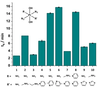
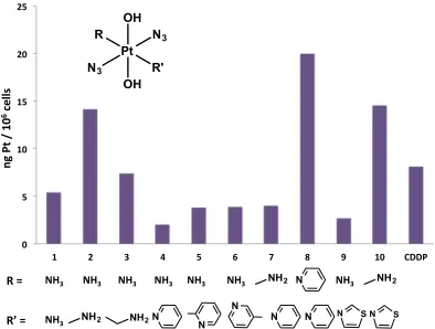
2.3 Results 60 2.3.1 Correlation between lipophilicity and cellular accumulation of platinum complexes 61 2.3.1.1 Lipophilicity 61 2.3.1.2 Platinum accumulation in A2780 cells 63 2.3.2 Study of photoactivatable Pt(IV) diazido complex
interaction with oligonucleotides by chromatography 67
2.3.2.1 Interaction of t,t,t-‐[Pt(N3)2(OH)2(py)2) with
complexes 77 2.3.3 Photoactivatable Pt(IV) diazido complex interaction with an oligonucleotide by mass spectrometry 79
2.3.3.1 Interaction of trans,trans,trans-‐[Pt(N3)2(OH)2(py)2] and oligo 1 79 2.3.3.2 Platinum oligonucleotide interaction by mass spectrometry – 15N labelled complexes 84 2.4 Discussion 87 2.4.1 Lipophilicity 87 2.4.2 Platinum accumulation in A2780 cells 92 2.4.3 Study of photoactivatable Pt(IV) diazido complex
interaction with oligonucleotides by chromatography 97 2.4.4 Sequence selectivity of Pt(IV) diazido complex
oligonucleotide binding 98 2.4.5 The effects of altering the wavelength of activation on the binding of Pt(IV) diazido complexes to a series of
oligonucleotides 101 2.4.6 Photoactivatable Pt(IV) diazido complex interaction with an oligonucleotide by mass spectrometry 102
2.5 Conclusions 104
Chapter 3 Chiral chromatography of organometallic anticancer
Complexes 114 3.1 Introduction 115 3.2 Experimental 117
3.2.1 Materials 117 3.2.2 Methods 118 3.2.2.1 Sample preparation 118 3.2.2.2 HPLC 118 3.3 Results 119
3.3.1.1 Stationary phase CHIRALPAK IA, complex 11 and the
stability of the enantiomers of 11 121 3.3.1.2 Stationary phase CHIRALPAK IA, complexes 12-‐17 124 3.3.1.3 Solution stability of the enantiomers of complex 17 127 3.3.2 Stationary phase CHIRALPAK IC, complexes 18-‐24 130 3.3.2.1. Complexes 18-‐21: separation of enantiomers and stability of 18 131 3.3.2.2 Complexes 22 and 23: separation of enantiomers and stability of 23 134 3.3.2.3. Complex 24: using a system that can be separated by other means 136
3.6 References 147
Chapter 4 Photonic crystal fibre mass spectrometry 150 4.1 Introduction 151 4.1.1 Photoactivatable metal-‐based drugs 151 4.1.2 Hyphenated mass spectrometry techniques 152 4.1.3 Photonic crystal fibres 153 4.2 Experimental 155
4.2.1 Materials 155 4.2.2 Methods 156 5.2.2.1 Sample preparation 156 4.2.2.2 Mass spectrometry 156 4.2.2.3 Light sources 157 4.3. Results 157
4.3.1 Photonic crystal fibre mass spectrometry (PCF-‐MS) system design and development 157
4.3.1.1 PCF-‐MS system design and development: metallic coupling devices 158 4.3.1.2 PCF-‐MS system design and development: plastic microfluidic coupling devices 165 4.3.1.3 Design of PCF-‐MS system:
using ‘off-‐the-‐shelf’ chips 173 4.3.2 Vitamin B12 -‐ a model for PCF-‐MS system validation in the positive-‐ion mode 177
4.3.2.1 Photoaquation of vitamin B12 pH 1.7: conventional methods 178 4.3.2.2 Photoaquation of vitamin B12 pH 1.7: PCF-‐MS system 179 4.3.2.3 Photoaquation of vitamin B12 pH 7.9: conventional methods 179 4.3.2.4 Photoaquation of vitamin B12 pH 7.9: PCF-‐MS system 182 4.3.3 PCF-‐MS system validation: sodium nitroprusside -‐ a model reaction for system validation in the negative-‐ion mode 183 4.3.4 PCF-‐MS system application: photoactivation of ruthenium anticancer complexes 186 4.3.5 PCF-‐MS system application as a rapid microreactor
screening technique to gain insights into the mechanism of action of photoactivatable drugs 189 4.4 Discussion 200
4.4.1. Photonic crystal fibre mass spectrometry (PCF-‐MS) system design and development 200
coupling devices 202 4.4.1.4 PCF-‐MS system design and development: microfluidic devices using ‘off-‐the-‐shelf’ chips 203 4.4.2 PCF-‐MS system validation: vitamin B12 -‐ a model reaction for system validation in the positive mode 204 4.4.3 PCF-‐MS system validation: sodium nitroprusside -‐ a model reaction for system validation in the negative mode 205 4.4.4 PCF-‐MS system application: photoactivation of ruthenium potential anticancer complexes 206 4.4.5 PCF-‐MS system application: PCF-‐MS as a rapid microreactor screening technique to gain insights into the mechanism of action of photoactivatable drugs 207 4.5 Conclusions 209 4.6 References 211
Chapter 5 Conclusions and future work 213 5.1 Conclusions 214 5.2 Future work 217 5.3 References 221
Appendix III part A: desalting methods 235 Appendix III part B: method optimisation of the separation of free oligonucleotide from oligonucleotide with platinum adducts 240 Appendix III part C: oligonucleotide quality control (QC) procedure 248
I would like to thank Professor Peter J. Sadler FRS for giving me this fantastic
opportunity. Your guidance and support over the past few years has been invaluable.
Thank you for pushing me to do my best and making me realise that I am capable of
more that I imagined.
I would also like to thank Dr Abraha Habtemariam for all of his advice and
helpful discussions. More importantly I would like to thank him for his wisdom in all
things, not just chemical, and his unique ability to radiate calmness to all those
around him. I very much doubt that I will have the privilege to work with anyone so
kind again.
To Isolda and Louisa thank you for the coffee, the laughs, the friendship and
the gin. Thank you for Spain, lazy Sundays and always being there. A girl could not
ask for a better duo to make a trio with. Isolda I will be eternally grateful to you and
your sofa. I don’t know what I would have done without you both and of course the
arepas that changed my life! I would also like to thank Dr Luca Salassa for the coffee,
the comedy, the pasta, the hospitality and the words of wisdom.
To PJS group members past and present I would like to say thank you for
letting me be part of this big, wonderfully dysfunctional family. We have had our
moments over the years but I can honestly say that it has been an absolute pleasure
and I do not know how I would have made it through the last few years without you
all.
Dr Ana Pizarro, what do I say? Thank you for getting me in to this mess in the
them. Ana, you are brave in all aspects of life and give hope to those of us who are
less so, that one day we might be too. Most importantly thank you for your honesty
and for always listening.
I would like to thank Dr Sarah Unterkofler for her seemingly endless patience
and great skill with optical equipment. My thanks also go to Dr Tijmen Euser and Prof
Philip Russell for their collaboration and input throughout my PhD. For their help
with all things NMR I would like to thank Dr Ivan Prokes and Mr Edward Tunnah. To
Dr Lijiang Song I will be forever grateful for his constant help with all thing mass
spectrometry and chromatography related. On the same note I would like to thank
Mr Philp Aston for all of his help over the years and reminding me that ‘it’s normal
for an analytical chemist to like things clean’.
To my dear friend and flatmate Becky, thank you for the late night chats and
the telly evenings. Our lovely little flat was an island of peace in the ocean of chaos
that was the past few years. My Wells people, I love you all! Thank you for all of the
welcome distractions and some of the unwelcome ones too. To Vale, my dear,
geographically we may be apart but I have drawn great strength from the knowledge
that you are with me in spirit. We have had some wonderful trips these past few
years, the memories of which have kept me going during the hard times. I will be
forever grateful to you and Antonio for our week on the magical island of Achill. I
look forward to all of the ones that we have yet to make. Thank you for listening and
truly being a friend.
To gramps, thank you for inspiring me to study what I love and nanna, thank
as a sister. Mum and dad, I was born eternally in your debt. Each day that debt
grows more and more. Thank you so much for everything. This would have been
impossible without your constant love and support.
I hereby declare that the work contained in this thesis is the original
work of the author, except where specific reference is made to other sources, with the nature and extent of the author’s contribution indicated (as appropriate) where work was based on collaborative research. The work has not been submitted, in whole or in part, for any other degree, diploma or other qualification. A list of research papers published during the term of study is given below.
Ruth J. McQuitty September 2013
1. R. J. McQuitty, S. Unterkofler, T. G. Euser, A. Habtermariam, P. St. J. Russell and P. J. Sadler Rapid screening of photoactivateable drugs: photonic crystal fibre microflow reactor coupled with ESI mass spectrometry (manuscript in preparation 2013).
3. S. Unterkofler, R. J. McQuitty, T. G. Euser, N. J. Farrer, P. J. Sadler, and P. St. J. Russell Optofluidic hollow-‐core photonic crystal fiber
nanoflow reactor for online photochemical reaction analysis Optics Letters 2012, 37, 1952.
4. G. Ragazzon, I. Bratsos, E. Alessio, L. Salassa, A. Habtemariam, R. J. McQuitty, G. J. Clarkson and P. J. Sadler Design of Photoactivatable Metallodrugs: Selective and Rapid Light-‐induced Ligand Dissociation from Half-‐Sandwich [Ru([9]aneS3)(N–N')(py)]2+ Complexes Inorg. Chim.
Acta 2012, 393, 230.
5. L. Ronconi, A. M. Pizarro, R. J. McQuitty; P. J. Sadler Insights into the Acid–Base Properties of Pt(IV)–Diazidodiam(m)inedihyroxido Complexes from Multinuclear NMR Spectroscopy Chem. Eur. J. 2011, 17, 12051.
Transition metal coordination complexes show great promise as novel
therapeutic agents with new mechanisms of action, but their characterisation,
and identification of their target sites present significant challenges. In this
thesis a variety of new analytical methods is explored for the study of platinum,
ruthenium, osmium and iridium anticancer complexes.
High performance liquid chromatography (HPLC) was used to determine
the relative hydrophobicity of a series of photoactivatable Pt(IV) diazido
complexes of the general type trans,trans,trans-‐[Pt(N3)2(OH)2(R)(R’)].
Interestingly the hydrophobicities did not follow trends based on literature Log
P values of individual ligands and did not correlate with the cellular uptake or
antiproliferative activity of the drugs. Other factors such as the quantum yield
of the complex, and the type of DNA adducts appear to be more important for
their efficacy.
Chromatography and high-‐resolution mass spectrometry were used to
study the formation of platinum adducts on DNA when the most active complex
trans,trans,trans-‐[Pt(N3)2(OH)2(pyridine)2], 8 was irradiated in the presence of
short single strand oligonucleotides 14 bases in length. Complex 8 was found to
bind to the oligonucleotides as a {Pt(pyridine)2}2+ adduct. Modifying the
wavelength of activation from UVA to 420 nm had no effect on the type of
adduct formed, but the higher energy irradiation achieved maximum levels of
DNA platination more quickly. Changing the sequence of the oligonucleotide
suggested that the photoactivated form of 8 does not favour the formation of
the case with other trans-‐platinum complexes.
Chiral chromatography using cellulose-‐ and amylose-‐based stationary
phases successfully separated the enantiomers of a series of organometallic
‘piano stool’ anticancer complexes. This appears to be the first successful
separation of facially chiral Ru(II) arene complexes, the enantiomers of which
were stable in solution for over 3 h. In contrast, separated cyclopentadienyl
Ir(III) complexes with chiral metal centres epimerized within 2 h in solution at
ambient temperature. Under similar conditions the enantiomers of the Os(II)
arene complex [Os(η6-‐p-‐cym)(4-‐(2-‐pyridylazo)-‐N,N-‐dimethylaniline)I]+ remained stable, as did those of the ruthenium-‐based complex [Ru(9,10-‐
diydrophenanthrene)(en)Cl]+. It was shown that it is possible to separate the diasteriomers of [Ru(η6-‐para-‐cymene)(iminopyridine)I], that can also be resolved by crystallisationtechniques, and hence, decrease the time required to
separate the enantiomers. This work will therefore allow exploration of the
biological properties of some of these enantiomers
A novel technique for the rapid irradiation and detection of light-‐
senstive species was developed. Photonic crystal fibers (PCFs) were coupled to a
mass spectrometer using HPLC tubing and fittings. This continuous flow method
of analysis was validated using the photaquation of cyanocobalamin. The PCF
system was compared to the conventional cuvette-‐based approach. No
significant difference in the species detected by MS could be found, but the PCF
system had the advantage of requiring 20 times less sample (25 μL), and only 15
photoactivateable ruthenium-‐based drug [{(η6-‐indan)RuCl}
2(μ-‐2,3-‐dpp)]2+ with a
range of small molecules that acted as models for intracellular components, e.g.
5’GMP for DNA. The nucleobase binding properties were consistent with those
previously reported with plasmid DNA by Magennis et al: a small amount of binding took place in the dark in view of the aquation of the mondentate
leaving groups but this dramatically increased upon photoactivation and loss of
the arene ligands. The complex was also found to bind to glutathione (GSH),
which is known to detoxify metal-‐based drugs, an observation possibly
explaining its poor anticancer activity.
ESI electrospray ionization
MALDI matrix assisted laser desorption ionisation
HR-‐MS high-‐resolution mass spectrometry
RP-‐HPLC reverse-‐phase high performance liquid chromatography
NP-‐HPLC normal-‐phase high performance liquid chromatography
SP stationary phase
MP mobile phase
py pyridine
LMCT ligand-‐to-‐metal charge transfer
PVC polyvinyl chloride
PTFE polytetrafluoroethylene
PEEK polyether ether ketone
DDW doubly deionised water
DACH diaminocyclohexane
GMP guanosine monophosphate
PDT photodynamic therapy
PCF photonic crystal fiber
ICP-‐MS inductively coupled plasma mass spectrometry
NMR nuclear magnetic resonance
UV-‐Vis ultraviolet-‐visable (spectrum)
TFA trifluoroacetic acid
DDW doubly deionised water
CTR1 copper transporter protein 1
Cpxbph tetramethyl(biphenyl)-‐cyclopentadiene
Cp* pentamethylcyclopentadiene
p-‐cym para-‐cymene
bip biphenyl
flu fluorene
phent phenanthrene
Impy iminopyridine
en ethylenediamine
azpy-‐F 4-‐(2-‐ pyridylazo)-‐N,N-‐fluoro
azpy-‐NMe2 4-‐(2-‐ pyridylazo)-‐N,N-‐dimethylaniline)
tz thiazole
pycarboxphen 3-‐pyridinecarboxaldehyde, 5-‐phenyl-‐
py,4-‐(4tfmph) pyridine,4-‐[4-‐(trifluoromethyl)phenyl]-‐
py,4-‐(2tfmph) pyridine, 4-‐(2-‐fluorophenyl)-‐
dfphpy 2-‐(2,4-‐difluorophenyl)pyridine)
benzq 7,8-‐benzoquinoline
phenpy 2-‐phenylpyridine
bmp 2,2ʹ′-‐bipyrimidine
μ-‐2,3-‐dpp 2,3-‐bis(2-‐pyridyl)pyrazine
VWD variable wavelength detector
DEA diethylamine
Chapter 1
Introduction
1.1Metals in Medicine
Traditionally the pharmaceutical industry has focused on the design and
development of organic drugs. Although metals and metal-‐based compounds have
been used for medicinal purposes for centuries, such as mercury and arsenic in the
treatment of syphilis,1 their use has not been widespread. The discovery of the
antiproliferative activity of the platinum-‐based compound cisplatin, cis-‐
[Pt(NH3)2Cl2], by Rosenberg et al. in 1968 created a new wave of interest in metal-‐
based therapies,2,3 bringing inorganic medicinal chemistry to the forefront. Since then the search for metal ions for medicinal purposes has expanded to a wide
range fields in therapy and diagnosis.
1.1.1 Antimicrobial agents
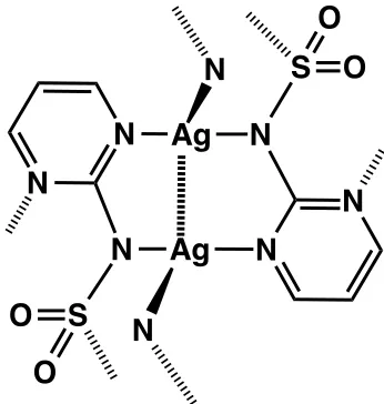
Compounds of silver have been used as antimicrobial agents for a number
of years, often in the form of AgNO3.4 Silver has the advantage of being toxic at low
levels to bacteria, but much higher levels are required to achieve toxicity in human
cells. One compound in clinical use for the treatment of burns is the polymeric
compound silver sulfadiazine, Figure 1.1, which like many silver antimicrobial
agents has been incorporated into various materials such as bandages,5,6 medical
instruments,7 shoe insoles, fabrics8,9 and even washing machines and food.10 Often
the silver compounds are incorporated into materials such as functionalised
N
N
N
N
Ag
N
N
S
Ag
N
N
S
O
O
O
O
Figure 1.1 Structure of silver sulfadiazine.
Several other metal compounds have also shown efficacy as antimicrobial
agents. Copper nanoparticles were shown to be inferior in activity to silver
nanoparticles by Ruparelia et al. (2008), but a mixed nanoparticle of both metals
was found to be the most effective against the four strains of bacteria tested.14
Amino acid-‐based complexes of Co(II), Cu(II), Ni(II) and Zn(II) were tested for their
antibacterial and antifungal activity in Gram-‐positive, Gram-‐negative bacteria and
fungal strains by Chohan et al. (2006). They were shown to be active towards E.
coli, B. subtilis, S. flexenari, S. aureus, P. aeruginosa, and S. typhi bacteria, and for
antifungal activity against T. longifusus, C. albicans, A. flavus, M. canis, F. solani, and
C. glaberata.15 The antibacterial properties of a series of Cu(II), V(IV) and Ni(II) with
Schiff bases obtained through the condensation of 4-‐amino-‐1,5-‐dimethyl-‐2-‐phenyl-‐
1H-‐3-‐pyrazol-‐3(2H)-‐one (anti-‐pyrine) with 2-‐hydroxybenzaldehyde, 4-‐hydroxy-‐5-‐
methoxyisophthalaldehyde and 4,5-‐dihydroxy isophalaldehyde, respectively, were
investigated by Rosu et al. (2010) along with their antiproliferative activity. The
[image:26.595.267.440.73.255.2]lines.16 The ruthenium-‐based complexes [Ru(phenanthroline)
2(dipyrido[3,2-‐d:2’,3’-‐
f]quinoxaline)]2+, [Ru(bipyridine)
2(dipyrido[3,2-‐a:2’,3’-‐c](6,7,8,9-‐tetrahydro)
phenazine)]2+, and [Ru(2,9-‐Me
2phenathroline)2(dipyrido[3,2-‐ a:2’,3’-‐c]phenazine)]2+
are active towards both Gram-‐positive and Gram-‐negative bacteria.17 Ruthenium
complexes have also shown promise in the treatment of the parasistic illness
Chagas’ disease.18 Another metal that has shown antiparasitic activity is gold; Au(I) chloroquinoline complexes are active against quinolone-‐resistant strains of
malaria.19 A series of gold phosphine compounds has shown antimicrobial activity in Gram-‐positive bacteria, with the potency dependent on the structure of the
phosphine ligands.20
1.1.2 Gold Antiarthritic drugs
Compounds of gold are also used to treat rheumatoid arthritis. There are
several forms of injectable gold antiarthritic gold drugs, including di-‐sodium
aurothiomalate, aurothioglucose and sodium aurothiopropanol sulfonate.21 The
orally active drug auranofin (Figure 1.2) gained approval for use in the clinic several
decades ago. Its mechanism of action is thought to involve the induction of heme
oxygenase-‐1 and the generation of reactive oxygen species (ROS), as explored in
detail by Kim et al.22 It is known to react readily with thiols such as glutathione (GSH) and human serum albumin (HSA).23 Using both X-‐ray absorption near-‐edge
spectroscopy (XANES) and extended X-‐ray absorption fine structure (EXAFS),
Messori et al. studied the interaction of auranofin with bovine serium albumin
(BSA) and human serum apotransferrin.24 They confirmed the findings of earlier
subsequently binds to the Cys-‐34 residue of BSA,25,26 and the loss of the same
ligand is thought to facilitate binding to apotransferrin.27 In both cases the Au(I)
oxidadtion state is conserved upon protein binding.
O
O
O
O
S
O
O
O
O
O
Au
P
Figure 1.2 The structure of auranofin.
Auranofin has also displayed antiparasitic characteristics, thought to be
derived from its ability to inhibit parasitic enzymes. The crystal structure of
auranofin bound to trypanothione reductase, a key enzyme of the parasite
Leishmania infantum, was reported by Ilari et al.28 The structure showed that auranofin was bound to two cysteine residues of the protein, Cys-‐52 and Cys-‐57,
with the thiosugar moiety of auranofin binding to the trypanothione binding site
within the protein inhibiting the action of the enzyme. Auranofin has also shown
promising anticancer activity against chronic lymphocytic leukemia.29
1.1.3 Bismuth antiulcer drugs
Bismuth has a long history in the treatment of ulcers.21 These drugs, including bismuth subsalicylate (BSS), colloidal bismuthcitrate (CBS), and ranitidine
bismuth-‐based drugs are thought to be metalloproteins such as transferrin which
can transport bismuth, and the enzyme urease in bacteria.30 A study by Cun et al.
has shown that Bi3+ binds to the histidine-‐ and cysteine-‐rich domain at the C-‐
terminus of the heat shock protein HspA, normally responsibly for binding Ni2+ ions
found in Helicobacter pylori.31 A proteomic study of Helicobacter pylori exposed to
bismuth subcitrate found that the drug induced oxidative stress in the cells,
suggesting that this may also be a key part of the mechanism of action of this class
of drug.32 Tsang et al. monitored the bismuth levels of individual bacterial cells exposed to antiulcer drugs, and found that the uptake of bismuth was affected by
the presence of ferric ions,33 indicating that bismuth is taken up into the cells via iron transport pathways.
1.1.4 Metal-‐based insulin mimics
The signaling hormone insulin is essential for the metabolism of
carbohydrates and fat.34 Insulin release is prompted by high blood glucose levels and the subsequent high levels of insulin cause the liver and gut to increase the
uptake of glucose. It is this function of insulin, inducing the storage of glucose, that
vanadium and other metal compounds have been shown to mimic. Their efficacy is
believed to ascribable due to their interaction with phosphotyrosine phosphatases
(PTPases) and tyrosine kinases that regulate insulin receptor binding.35
The vanadium complex bis(maltolato)oxovanadium(IV) (BMOV), Figure 1.3,
is an orally-‐active insulin mimic shown to be effective at reducing glucose levels in
blood plasma.36 To improve the uptake and efficacy of this class of drug, new
incorporate vanadium in different oxidation states. Rehder et al. synthesised and
studied in detail 22 V(V) and V(IV) complexes containing –OO, -‐ON, -‐OS, -‐NS and –
ONS donor ligands.37 They found that the oxidation state of the metal had little
effect on the activity of the complexes since the oxidation states of vanadium
readily interconvert in cells. The most active compounds contained –ON ligands.
This agrees with the findings of Gätjens et al. who synthesised a range of vanadium
complexes derived from 5-‐carboalkoxypicolinates that were shown to be more
active than BMOV.38
O
O O
O O
O V O
Figure 1.3 Structure of bis(maltolato)oxovanadium(IV) (BMOV).
The complexes of several other metals have shown efficacy as insulin
mimics, such as molybdenum and tungsten. The Mo(VI) and W(VI) oxidation states
are very similar in configuration to V(V); their similarities are discussed by
Thompson et al. in their review of vanadium compounds as insulin mimics.34 Zinc complexes may also show some efficacy as an insulin mimic.34 The naturally occurring chromium-‐oligopeptide complex has been found to activate tyrosine
kinase and some C-‐based complexes have been developed to explore this
phenomenon further.21
[image:30.595.239.430.304.407.2]
1.1.5 Vasodilators
The release of NO from metal complexes, usually by means of
photoactivation, has been shown to induce vasodilation.39 The iron compound
sodium nitroprusside (SNP) has been approved for clinical use to control
hypertension.40 The structure of this compound is shown in Figure 1.4. Upon
irradiation with short wavelengths of light, the NO ligand of SNP is released first
and subsequently small amounts of the CN-‐ ligand can also be relased.41,39 There are also examples of ruthenium-‐,42,43 chromium-‐,44 nickel-‐,45 and copper-‐based46 NO-‐ releasing compounds, as well as those of other metals.47
Fe C
C
C N
C C
O
N
N
N N
N
2
-2Na+
Figure 1.4 The structure of sodium nitroprusside.
1.1.6 MRI contrast agents
Advancements in other fields of medicine have led to new opportunities for
inorganic drugs. The use of magnetic resonance imaging (MRI) in medical diagnosis
has created a demand for contrast agents to improve the images acquired. The
majority of the contrast agents in use are Gd(III)-‐based, though there are some iron
gadolinium-‐based agents is shown in Figure 1.5. There are various agents for the
targeting of different types of tissue, e.g. liver and brain. The different types of MRI
contrast agent and their uses are reviewed extensively by Yan et al.48
N
N
N
N
Gd3+
O
O-O
O-O
O-O
O-H O H
Figure 1.5 The structure of the Gd(III)-‐based contrast agent Dotarem®.
1.2Platinum-‐based drugs
1.2.1 Cisplatin
The serendipitous discovery of the anitproliferative properties of cisplatin,
Figure 1.6, sparked new interest in metal-‐based therapies in the late 1960s.2,3 In a little over a decade, it had gained FDA approval and had already begun to save
lives. Cisplatin is highly effective in the treatment of testicular, ovarian, bladder,
head and neck cancers.49 Platinum-‐based drugs (cisplatin, carboplatin and oxaliplatin) are so successful that sales are now in the order of $3 billion each year.
Thier mechanism of action, metabolism and biotransformation have been
Pt
Cl
Cl
H
3N
H
3N
Figure 1.6 The structure of the platinum-‐based anticancer drug cisplatin.
1.2.1.1Mechanism of action
The efficacy of cisplatin is thought to be attributable to its hydrolysis once it
enters the cell and the subsequent binding of a small amount of the drug to nuclear
DNA. Many different lesions are formed by the hydrolysed cisplatin: monoadducts,
interstrand crosslinks, 1,2-‐intrastrand crosslinks and 1,3-‐intrastrand crosslinks. The
adducts that are believed to induce apoptosis are the 1,2-‐d(GpG) and 1,2-‐d(ApG)
intrastrand crosslinks, forming 65% and 25% of lesions (determined by high
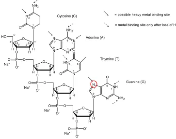
performance liquid chromatography), respectively.50 Cisplatin binds to the N7 atom of guanine (and to some extent adenine) more favourably than other nucleotide
binding sites since it is the most electron-‐rich, and it forms the most stable metal
ion-‐nucleobase complex at physiological pH, see Figure 1.7.51 In single-‐stranded
DNA, the N3 of cytidine and N1 of adenine are also possible binding sites for
platinum.52
The 1,2-‐(GpG) intrastrand crosslink causes a bending and unwinding of the
duplex DNA53 that is recognised by proteins. Two types of protein are known to
interact with cisplatin lesions on DNA: repair proteins that recognise DNA damage
and architectural proteins that are nuclear, or chromatin related proteins involved
proteins contain the high mobility group 1 box (HMG1) motif, a sequence of around
80 amino acids in length.
= metal binding site only after loss of H+
Adenine (A) Guanine (G) Cytosine (C) Thymine (T) Na+ Na+ Na+ Na+ 1’ 2’ 3’ 4’ 5’ 1 3 9
7 6
3 1 6 1 9 1
3 5
= possible heavy metal binding site
7
Figure 1.7 Possible binding sites for transition metal ions in a single strand
deoxytetranulceotide are indicated with arrows. Adapted from ref. 55
Ohndorf et al. reported the crystal structure of a HMG1 protein bound to a
strand of duplex DNA, of the sequence 5’(CCT CTC TG*G* ACC TTC C)/5’(GGA GAG
ACC TGG AAG G) (* indicates the position of platinum binding), containing a 1,2-‐
c(GpG) cisplatin adduct.56 The oligo-‐cisplatin adduct created a large enough bend in
the duplex for a phenylalanine (Phe) residue at position 37 to partially intercalate.
The binding of HMG1 was greatly reduced for a mutant protein in which Phe 37 was
substituted by alanine. This suggested that such intercalation stabilised this
modified DNA-‐protein complex. The bound HMG1 then interacts with mismatch
[image:34.595.113.476.143.431.2]enhances p53 DNA-‐binding activity.57 These protein interactions with the cisplatin
bound DNA-‐HMG1 complex and other proteins initiate signaling cascades that lead
to apoptosis in cells.
Figure 1.8 X-‐ray crystal structure showing the interaction of the HMG1 protein with the duplex 5’(CCT CTC TG*G* ACC TTC C)/5’(GGA GAG ACC TGG AAG G). From ref 56
1.2.1.2Cellular accumulation
Cisplatin uptake is not entirely understood, but it is known that it enters
cells by passive diffusion. The uptake and efflux of cisplatin is also thought to be
related to the high-‐affinity copper transporter CTR1.57 Mutation or deletion of the
CTR1 gene caused a reduction in the amount of Pt taken up by cells and an increase
in cisplatin resistance. Over-‐expression of CTR1 increases the levels of Pt taken up.58
Cisplatin was found to bind to the methionine residues of CTR1, perhaps allowing it
may also play a role in cisplatin uptake and efflux.60
1.2.1.3Mechanisms of resistance
Some types of cancer cells are intrinsically resistant to cisplatin, whilst
others can develop resistance after prolonged exposure to the drug. One of the
main sources of resistance is the reduced cellular accumulation of cisplatin by
decreased influx and increased efflux.61 Additionally, an increase in levels of
intracellular thiol e.g. GSH can detoxify cisplatin and increase efflux of platinum(II)
from the cell. Another means of resistance is increased adduct repair and tolerance
of Pt-‐DNA lesions resulting in failure to induce apoptosis.57 A diagram summarising the journey of cisplatin within the cell is shown in Figure 1.9.
[Cl$]&=&104&mM&
Pt Cl Cl H3N
H3N
Pt Cl Cl H3N
H3N
[Cl$]&=&24&mM&
[Cl$]&=&4&mM&
Pt
OH2
Cl
H3N
H3N
+
CTR1& Pt
S
Cl H3N
H3N
R
Hydrolysis&
Reac:on&with& intracellular&thiol& Pt&excre:on&
d(ApG) d(GpNpG)
d(GpG)
Passive&diffusion&
Figure 1.9 A diagram of cisplatin uptake, efflux and DNA binding (adapted from Reedijk et al.). 55