Functional analysis of the EsaB component of the
Staphylococcus aureus
Type VII secretion system
M. Guillermina Casabona,1Grant Buchanan,1Martin Zoltner,1Catriona P. Harkins,2Matthew T. G. Holden2and Tracy Palmer1,
*
Abstract
Type VII secretion systems (T7SS) are found in many bacteria and secrete proteins involved in virulence and bacterial competition. In Staphylococcus aureus the small ubiquitin-like EsaB protein has been previously implicated as having a regulatory role in the production of the EsxC substrate. Here we show that in the S. aureusRN6390 strain, EsaB does not genetically regulate production of any T7 substrates or components, but is indispensable for secretion activity. Consistent with EsaB being an essential component of the T7SS, loss of either EsaB or EssC are associated with upregulation of a common set of iron acquisition genes. However, a further subset of genes were dysregulated only in the absence of EsaB. Quantitative western blotting indicates that EsaB is present at very low levels in cells. Substitution of a highly conserved threonine for alanine or arginine resulted in a loss of EsaB activity and destabilisation of the protein. Taken together our findings show that EsaB is essential for T7SS activity in RN6390.
INTRODUCTION
Protein secretion systems are nanomachines employed by bac-teria to transport protein substrates across their cell envelopes. Gram-negative bacteria produce a number of different secre-tion machineries that export proteins involved in a wide vari-ety of processes including signalling, nutrient scavenging, host interaction and virulence [1]. The Type VII secretion system (T7SS) is found in some Gram-negative and many Gram-pos-itive bacteria, and is particularly common among organisms of the actinobacteria and firmicutes phyla [2]. The T7SS was initially described in the pathogenic mycobacteria Mycobacte-rium tuberculosisandMycobacterium bovis,where the ESX-1 T7SS was shown to be essential for virulence, due to the secre-tion of two major T-cell antigens EsxA (formerly known as ESAT-6) and EsxB (formerly known as CFP-10) [3–5]. EsxA and EsxB are founding members of the WXG100 protein fam-ily that appear to be exclusively linked to T7SSs, and all char-acterised T7 systems are associated with at least one family member. The presence of a membrane-bound ATPase of the SpoIIIE/FtsK family (termed EccC in actinobacteria and EssC in firmicutes) is another hallmark of all T7SSs [6]. In Myco-bacteria, three further membrane proteins EccB, EccD and EccE assemble with EccC to form a large 1.5 MDa core
complex [7, 8]. This complex further associates with a mem-brane-bound mycosin serine protease, MycP, that is essential for T7 protein secretion and for stability of the membrane complex [9].
Staphylococcus aureus, an opportunistic pathogen of humans and animals, also elaborates a T7SS that is distantly related to the T7SSs found in mycobacteria [10]. Mutational analysis has indicated that it plays an important role in persistence in mouse models of infection, intra-species com-petition and potentially iron homeostasis [10–15]. In com-monly-studied strains of S. aureus such as Newman, USA300 and RN6390, the secretion system is encoded by the 12 geneesslocus [10, 12, 16]. The first six genes at this locus encode essential components of the secretion machin-ery, including the WXG100 protein EsxA and the SpoIIIE/ FtsK ATPase EssC (Fig. 1a, b). However,S. aureusand other firmicutes lack homologues of EccB, EccD, EccE and MycP and instead have an apparently unrelated set of membrane-bound secretion components (EsaA, EssA and EssB in
S. aureus) [12, 17–19]. The sixth component of theS. aureus
T7SS is EsaB, which is predicted to be a small cytoplasmic protein of 80 amino acids that is structurally related to ubiq-uitin [20]. In S. aureus strains Newman and USA300, a
Received 17 June 2017; Accepted 14 November 2017
Author affiliations:1Division of Molecular Microbiology School of Life Sciences, University of Dundee, Dundee, UK;2School of Medicine, University of
St Andrews, St Andrews, KY16 9TF, UK.
*Correspondence:Tracy Palmer, [email protected]
Keywords:Staphylococcus aureus; protein secretion; T7SS; regulation.
Abbreviations:LB, Luria Bertani medium; TSB, Tryptic Soy Broth; T7SS, type VII secretion system; YFP, yellow fluorescent protein.
The RNA-Seq data from this study is submitted to the European Nucleotide Archive with accession number ERP009279 and in Array express under accession number E-ERAD-362.
One supplementary table is available with the online version of this article.
(a)
(b)
1 2
(c)
RT + - + - + - + - + - +
-Region 1 (esxA) Region 2 (esxC/B)
2000- 500-
1000- 200- 5000-bp
2000- 500-
1000- 200- 5000-bp
(d)
RN6390 RN6390∆esaB
T
o
to
a
lR
P
K
M
[image:2.595.125.467.67.630.2]RN6390 RN6390∆esaB
transposon insertion in esaBdoes not abolish secretion of T7 substrates but is linked with an increase in RNA tran-scripts covering the gene encoding the substrate EsxC [11]. By contrast, in-frame deletion ofesaBabolished EsxA and EsxC secretion in strain RN6390 but did not detectably affect production of these substrate proteins [12]. Similarly, inactivation of yukD, which encodes the Bacillus subtilis esaBhomologue, also abolished T7 secretion [17, 18].
In this study, we have addressed the role of EsaB in
S. aureusT7 secretion using strain RN6390. We show that EsaB does not regulate esxC transcripts or those of other
ess-encoded genes. Instead our findings show that EsaB behaves as an essential component of the T7SS. Interest-ingly, however, RNA-Seq analysis identified a subset of genes from the AirSR regulon that showed altered regula-tion in the absence of EsaB. This suggests that loss ofesaB
has additional unexpected effects onS. aureusphysiology.
METHODS
Bacterial strains and growth conditions
S. aureusstrain RN6390 (NCTC8325 derivative,rbsU,tcaR, cured of’11,’12,’13; [21]) and the isogenicDesaB,DessC andDesx(DesxA–esaG) strains [12] were employed in this study, along with strain Newman [22]. The esaB deletion strain is an in-frame deletion of the gene that maintains the first ten and final three codons ofesaB(as there is a 9 codon overlap between the end of essA and the start of esaB).
S. aureusstrains were cultured in Tryptic Soy Broth (TSB) at 37
C with shaking unless otherwise stated. For calcula-tion of cell numbers we estimated by dilucalcula-tion analysis that one unit at OD 600 nm corresponds to 6108c.f.u. for
strain. When required, chloramphenicol (Cml, final concen-tration 10 µg ml 1) was added for plasmid selection. E. coli
strain JM110 (Stratagene) was used for cloning purposes and BL21(DE3) [23] for EsaB overproduction and purifica-tion. E. coli was grown in Luria-Bertani (LB) medium at 37
C with agitation. When appropriate, ampicillin was used for plasmid selection (final concentration 125 µg ml 1).
Genetic constructs
All plasmids used in this study are listed in Table 1. TheesaB
gene with its own RBS was PCR amplified from S. aureus
RN6390 genomic DNA using primers EsaB-fw and EsaB-rev (Table S1, available with the online version of this article). The 0.3 kb HpaI/EcoRI restriction fragment was cloned into pRAB11 under control of the tetracycline inducible promoter, giving pRAB11-esaB. Clones were selected inE. coliand veri-fied by DNA sequencing. Plasmid pRAB11-esaB-YFP was generated by cloning the 0.3 kbHpaI/EcoRI restriction frag-ment into pRAB11-YFP [15]. Clones were selected inE. coli
and verified by DNA sequencing. Nucleotide variants ofesaB
were generated by the Quickchange site-directed mutagenesis protocol (Stratagene) using pRAB11-esaB or pRAB11-esaB-YFP as a template and primers listed in Table S1. Modified plasmids were digested usingDpnI for at least 1 h at 37
C and
transformed intoE. coli. Single point mutations were verified by DNA sequencing.
RNA isolation and RT-PCR
For RNA-Seq analysis, three biological repeats of the
S. aureus esaBstrain was grown aerobically in TSB up to an OD600 of 1 at which point mRNA was prepared (in three technical replicates). This experiment was carried out along-side the RN6390 andessCstrains [15] and followed identical methodology.
For RT-PCR, the indicatedS. aureusstrains were grown aer-obically in TSB and harvested at an OD600 of 1. At this point, the mRNA was extracted using the SV total RNA Iso-lation Kit (Promega) with some minor modifications. Cell samples were stabilised in 5 % phenol/95 % ethanol on ice for at least 30 min and then centrifuged at 2770g for 10 min. Cells were then resuspended in 100 µl of TE buffer containing 500 µg ml 1 lysostaphin and 50 µg ml 1 lyso-zyme and incubated at 37
C for 30 min. Subsequently, the manufacturer’s instructions were followed. Isolated RNA was subjected to a second DNase treatment using the DNA-free kit (Ambion). RNA was stored at 80
C until use. RT-PCR to probe transcription of genes in the indicated strains was carried out using 500 ng of mRNA as template with the indicated primers (Table S1). PCR products were visualized on 1 % agarose gels.
Purification of 6His-EsaB and generation of polyclonal antisera
The EsaB coding sequence (UniProt code ESAB_STAAM) was PCR amplified from a synthetic gene (codon optimised for E. coli K12 (Genscript)) using the primers EsaB-pET1 and EsaB-pET2 (Table S1) and cloned into theNdeI/XhoI site of a modified pET15b vector (Novagen). The plasmid produces an N-terminal His6-tagged protein with a TEV (tobacco etch virus) protease cleavage site. The protein was expressed and purified as described previously [24], except the tag-free EsaB was not collected in the flow-through of the negative purification but required a 30 mM imidazole elution. The final size exclusion chromatography step used a 24 ml HR 30/100 GL Superdex75 column (GE healthcare), equilibrated with 20 mM Tris pH 7.8, 100 mM NaCl and was calibrated with molecular mass standards (thyroglobu-lin, 670 kDa; g-globulin, 158 kDa; serum albumin, 67 kDa; ovalbumin; 44 kDa, myoglobin, 17 kDa; and vitamin B12, 1 kDa). Two mg purified EsaB (retaining a Gly–Ala–Ser– Thr sequence at the N-terminus after the cleavage step) was utilised as antigen to immunise rabbits for polyclonal anti-body production in a standard three injections protocol (Seqlab).
Secretion assays, subcellular fractionation and western blotting
2770g. Cells were washed twice with PBS, adjusted to and OD600of 1 and digested using 50 µg ml 1of lysostaphin by incubation at 37
C for 30 min. Supernatants were filtered using a 0.22 µm filter and TCA-precipitated in the presence of 50 µg ml 1deoxycholate, as described. ForS. aureus sub-cellular fractionation, cells were grown to mid-log phase with shaking and treated as previously described [12]. Briefly, cells were harvested by centrifugation and resus-pended in TSM buffer (50 mM Tris-HCl pH 7.6, 0.5 M sucrose, 10 mM MgCl2). Lysostaphin was added to a final concentration of 50 µg ml 1 and cells were incubated at 37
C for 30 min to digest the cell wall. At this point, proto-plasts were sedimented to recover the cell wall (supernatant fraction). Protoplasts were disrupted by sonication and the membrane was obtained after an ultracentrifugation step at 227 000g for 30 min and at 4
C. The supernatant was retained as the cytoplasmic fraction. Samples were boiled for 10 min prior to separation in bis-Tris gels and subse-quent western blotting.
Polyclonal antisera were used at the following dilutions: a-EsxA 1 : 2500 [12],a-EsxB 1 : 1000 [15], a-EsxC 1 : 2000 [12], a-EsaB 1 : 500, a-TrxA 1 : 20 000 [25] and a-SrtA (Abcam) 1 : 3000. Anti-GFP antibody was obtained from Roche and used according to manufacturer’s instructions.
RESULTS
EsaB does not regulate the level ofesxC
transcripts in strain RN6390
A previous study has shown that a transposon insertion in theesaB gene results in an increase in esxC transcripts in
the Newman and USA300 strain backgrounds, and a con-comitant increase in the EsxC polypeptide, implicating it as a regulator [11]. To investigate whether loss ofesaBby in-frame deletion affects the level of esxC mRNA in strain RN6390, we isolated mRNA from the parental strain and the isogenic esaB mutant, prepared cDNA and undertook reverse transcriptase PCR with primers covering eitheresxA
(the first gene at theesslocus, included as a negative con-trol) oresxC(Fig. 1a). It can be seen (Fig. 1c) that the level of transcripts for each of these genes was qualitatively simi-lar in the wild-type andesaBbackgrounds.
To examine this quantitatively, we undertook RNA-Seq analysis on RNA prepared from three biological repeats of the RN6390 andesaBstrains grown aerobically in TSB to an OD600of 1. Note that these experiments were performed at the same time as the RN6390 versusessCRNA-Seq analysis described in [15] and used the same RN6390 dataset. Fig. 1 (d) shows that the level of esxC transcripts were indistin-guishable between the wild-type and esaBstrains. Analysis of the transcript levels of the other genes at the ess locus indicates that in general they were also not significantly altered by the loss of esaB although there was a small increase in the level of essB. We conclude that there is no evidence thatesaBregulates the level ofesxC transcripts in RN6390.
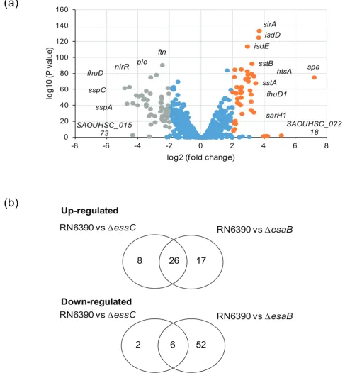
We next examined the entire transcript profile of theesaB
[image:4.595.47.549.79.357.2]mutant to investigate the transcriptional/post-transcrip-tional response to the loss of this small protein. We found 101 genes de-regulated in theesaBmutant compared to the parental strain (using a cut off of logFC >2 or < 2 and Table 1.Plasmids used in this study
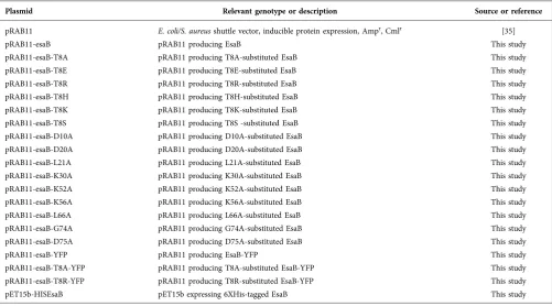
Plasmid Relevant genotype or description Source or reference
pRAB11 E. coli/S. aureusshuttle vector, inducible protein expression, Ampr, Cmlr [35]
pRAB11-esaB pRAB11 producing EsaB This study
pRAB11-esaB-T8A pRAB11 producing T8A-substituted EsaB This study
pRAB11-esaB-T8E pRAB11 producing T8E-substituted EsaB This study
pRAB11-esaB-T8R pRAB11 producing T8R-substituted EsaB This study
pRAB11-esaB-T8H pRAB11 producing T8H-substituted EsaB This study
pRAB11-esaB-T8K pRAB11 producing T8K-substituted EsaB This study
pRAB11-esaB-T8S pRAB11 producing T8S -substituted EsaB This study
pRAB11-esaB-D10A pRAB11 producing D10A-substituted EsaB This study
pRAB11-esaB-D20A pRAB11 producing D20A-substituted EsaB This study
pRAB11-esaB-L21A pRAB11 producing L21A-substituted EsaB This study
pRAB11-esaB-K30A pRAB11 producing K30A-substituted EsaB This study
pRAB11-esaB-K52A pRAB11 producing K52A-substituted EsaB This study
pRAB11-esaB-K56A pRAB11 producing K56A-substituted EsaB This study
pRAB11-esaB-L66A pRAB11 producing L66A-substituted EsaB This study
pRAB11-esaB-G74A pRAB11 producing G74A-substituted EsaB This study
pRAB11-esaB-D75A pRAB11 producing D75A-substituted EsaB This study
pRAB11-esaB-YFP pRAB11 producing EsaB-YFP This study
pRAB11-esaB-T8A-YFP pRAB11 producing T8A-substituted EsaB-YFP This study
pRAB11-esaB-T8R-YFP pRAB11 producing T8R-substituted EsaB-YFP This study
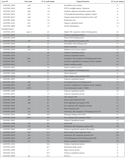
qvalue <0.05, as applied previously [15]), Fig. 2(a). Of these, 43 were upregulated by the loss of esaB whereas 58 were downregulated when esaB was absent – these genes are listed in Table 2. Interestingly, almost all of the genes that were differentially regulated in the essC mutant [15] were also similarly regulated in theesaBstrain (Fig. 3b), although there was a substantive subset of genes that were differen-tially expressed in theesaBmutant but not theessC strain (Table 2). It can be seen that almost all of the iron acquisi-tion genes, including those for heme acquisiacquisi-tion, staphylo-ferrin synthesis and uptake and ferrichrome import were commonly upregulated by loss of either esaB or essC
(Table 2). Furthermore six of the eight downregulated genes from theessCstrain were also down regulated in the esaB
strain (note that one of the two genes unaffected in theesaB
dataset is essC itself, which appears downregulated in the
essCdataset because it has been deleted). The finding that almost the entire subset of genes differentially regulated in the absence ofessC is also similarly altered by loss ofesaB
strongly suggests that EsaB is, like EssC, a component that is essential for activity of the secretion machinery in strain RN6390.
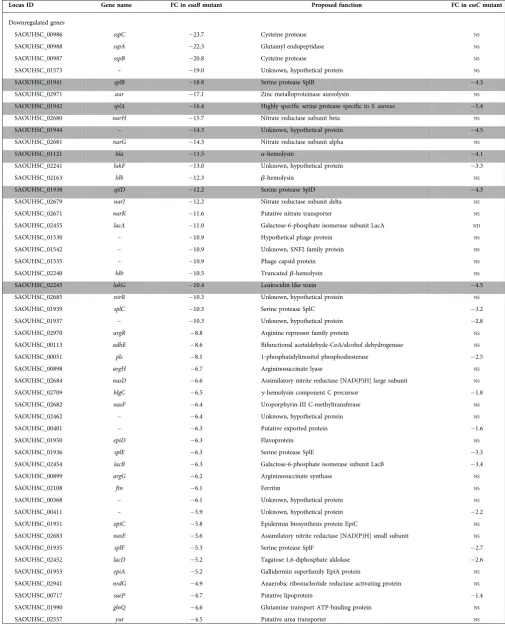
As mentioned above, a subset of transcripts were differen-tially expressed in the esaB but not the essC strain. These include downregulated genes required for anaerobic nitrate respiration (narGHJ/narK), some secreted proteases (sspA/ B/C,aur), capsular polysaccharide synthesis (capG/F/hysA), lactose metabolism (lacB/C/D) and antimicrobial peptide synthesis (epiA/C/D/P). Many of these genes are under con-trol of the essential two component regulatory system AirSR (formerly YhcSR) [26–29]. This observation indicates that EsaB has additional effects on S. aureus physiology. This could be indirect and arising from its role in T7 secretion, for example through altered membrane permeability when EsaB is absent. Alternatively, EsaB may have additional roles in the cell in addition to its requirement for T7 protein secretion.
EsaB is present at low amounts in cells of S. aureusRN6390
To explore the biological role of EsaB in T7 secretion, we overproduced recombinant EsaB with a cleavable His-tag in
E. coli. The purified protein eluted from gel filtration as a monomer, in agreement with structural analysis of the
B. subtilisEsaB homologue, YukD, which also appears to be monomeric [20]. Polyclonal antisera were raised against purified EsaB and the antibody was affinity purified against the EsaB antigen, before being used to detect the protein in whole cells ofS. aureus. Fig. 4(a) shows that although the purified antiserum could clearly recognise purified EsaB, it did not detect a band of the expected size of EsaB in whole cells. We have shown previously that expression of the T7SS genes in RN6390 is upregulated approximately 2–3-fold in the presence of exogenous hemin, and fourfold by hemin in a DessC background [15]. However, supplementation of either of these strains with hemin did not result in detect-able EsaB in the cellular fraction (Fig. 4b) and it could also
not be detected in cells of strain Newman (Fig. 4b). Probing a dilution series of purified EsaB indicated that the antibody was able to cross-react with as little as 25 ng of protein (Fig. 4a), which is equivalent to 1.61011EsaB molecules.
Since the antibody was unable to detect EsaB in whole cells of RN6390 from 9.6108colony forming units that were loaded onto the SDS gel, we conclude that are less than 170 molecules of EsaB per cell.
Since we were unable to detect native EsaB inS. aureuscell extracts, we constructed a series of tagged variants for which commercial antisera were available. To this end we intro-duced His6, Myc, hemagglutinin (HA) and Strep epitopes onto the N-terminus of EsaB, and His6, Myc, HA, mCherry or FLAG epitopes onto the C-terminus, but in each case were unable to detect the tagged protein (not shown). We also introduced His6 and His9 epitopes into two predicted loop regions internal to the EsaB sequence but again were unable to detect tagged EsaB (not shown). The only tag we introduced that allowed detection of EsaB was a C-terminal yellow fluorescent protein (YFP) tag. Fig. 4(c) shows that basal production of either native (untagged) EsaB or EsaB-YFP from plasmid vector pRAB11 was sufficient to restore secretion of the T7SS extracellular protein EsxA and of sub-strates EsxB and EsxC to the culture supernatant. Blotting the same cell samples for the presence of the YFP fusion protein (Fig. 4d) showed that it migrated at close to the pre-dicted mass (37 kDa) of the EsaB fusion. There was no evi-dence for degradation of the fusion protein even after prolonged exposure of the immunoblot (Fig. 4d). We con-clude that the YFP-tagged variant of EsaB probably retains functionality.
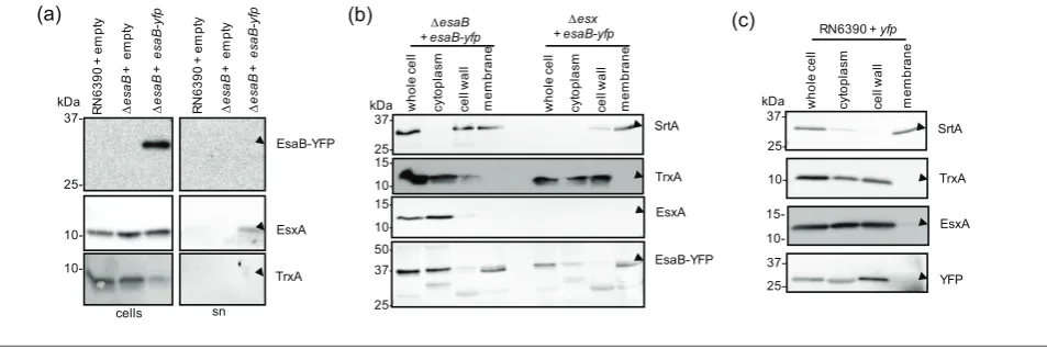
EsaB-YFP partially localises to the cell membrane EsaB is predicted to be a soluble cytoplasmic protein [10], and is known to share structural homology with ubiquitin [20]. Interestingly, a domain sharing the same fold is also associated with the actinobacterial T7SS, being found at the cytoplasmic N-terminus of EccD [30], indicating that such proteins may be essential features of all T7SSs. To determine the subcellular location of EsaB-YFP, we blotted secreted and whole cell sam-ples of the esaB mutant strain producing plasmid-encoded YFP with the YFP antiserum. Fig. 3(a) shows that EsaB-YFP was associated exclusively with the cellular fraction.
in the wild-type strain it did not localise to the membrane (Fig. 3c), indicating that membrane binding was unlikely to be mediated through the YFP portion of the fusion.
Next we tested whether EsaB-YFP localised to the mem-brane through interactions with memmem-brane components of the T7SS. To this end we repeated the fractionation in a
isdD
isdE
SAOUHSC_022 18 fhuD1
htsA spa
sarH1 sirA
sstA sstB
SAOUHSC_015 73
ftn
nirR plc
sspC fhuD
sspA
0 20 40 60 80 100 120 140 160
-8 -6 -4 -2 0 2 4 6 8
lo
g
1
0
(
P
v
a
lu
e
)
log2 (f old change)
Up-regulated
Down-regulated
26
8
17
2
6
52
RN6390 vs ∆
essC
RN6390 vs ∆
esaB
RN6390 vs ∆
essC
RN6390 vs ∆
esaB
(a)
[image:6.595.51.539.72.606.2](b)
Table 2.Genes differentially regulated (>log 2 fold) in the RN6390esaBdeletion mutant, sorted by ascending fold change (FC)
Genes highlighted in grey are also differentially regulated in theessCdeletion strain. The column on the right shows the fold change (FC) of the same gene in theessCdataset whereNSindicates no statistically significant change in expression level relative to the same gene in the wild-type dataset.
Locus ID Gene name FC inesaBmutant Proposed function FC inessCmutant
Downregulated genes
SAOUHSC_00986 sspC 23.7 Cysteine protease NS
SAOUHSC_00988 sspA 22.3 Glutamyl endopeptidase NS
SAOUHSC_00987 sspB 20.8 Cysteine protease NS
SAOUHSC_01573 – 19.0 Unknown, hypothetical protein NS
SAOUHSC_01941 splB 18.8 Serine protease SplB 4.3
SAOUHSC_02971 aur 17.1 Zinc metalloproteinase aureolysin NS
SAOUHSC_01942 splA 16.4 Highly specific serine protease specific toS. aureus 5.4
SAOUHSC_02680 narH 15.7 Nitrate reductase subunit beta NS
SAOUHSC_01944 – 14.3 Unknown, hypothetical protein 4.5
SAOUHSC_02681 narG 14.3 Nitrate reductase subunit alpha NS
SAOUHSC_01121 hla 13.5 a-hemolysin 4.1
SAOUHSC_02241 lukF 13.0 Unknown, hypothetical protein 3.3
SAOUHSC_02163 hlb 12.3 b-hemolysin NS
SAOUHSC_01938 splD 12.2 Serine protease SplD 4.3
SAOUHSC_02679 narJ 12.2 Nitrate reductase subunit delta NS
SAOUHSC_02671 narK 11.6 Putative nitrate transporter NS
SAOUHSC_02455 lacA 11.0 Galactose-6-phosphate isomerase subunit LacA ND
SAOUHSC_01530 – 10.9 Hypothetical phage protein NS
SAOUHSC_01542 – 10.9 Unknown, SNF2 family protein NS
SAOUHSC_01535 – 10.9 Phage capsid protein NS
SAOUHSC_02240 hlb 10.5 Truncatedb-hemolysin NS
SAOUHSC_02243 lukG 10.4 Leukocidin like toxin 4.5
SAOUHSC_02685 nirR 10.3 Unknown, hypothetical protein NS
SAOUHSC_01939 splC 10.3 Serine protease SplC 3.2
SAOUHSC_01937 – 10.3 Unknown, hypothetical protein 2.8
SAOUHSC_02970 argR 8.8 Arginine repressor family protein NS
SAOUHSC_00113 adhE 8.6 Bifunctional acetaldehyde-CoA/alcohol dehydrogenase NS
SAOUHSC_00051 plc 8.1 1-phosphatidylinositol phosphodiesterase 2.5
SAOUHSC_00898 argH 6.7 Argininosuccinate lyase NS
SAOUHSC_02684 nasD 6.6 Assimilatory nitrite reductase [NAD(P)H] large subunit NS
SAOUHSC_02709 hlgC 6.5 g-hemolysin component C precursor 1.8
SAOUHSC_02682 nasF 6.4 Uroporphyrin-III C-methyltransferase NS
SAOUHSC_02462 – 6.4 Unknown, hypothetical protein NS
SAOUHSC_00401 – 6.3 Putative exported protein 1.6
SAOUHSC_01950 epiD 6.3 Flavoprotein NS
SAOUHSC_01936 splE 6.3 Serine protease SplE 3.3
SAOUHSC_02454 lacB 6.3 Galactose-6-phosphate isomerase subunit LacB 3.4
SAOUHSC_00899 argG 6.2 Argininosuccinate synthase NS
SAOUHSC_02108 ftn 6.1 Ferritin NS
SAOUHSC_00368 – 6.1 Unknown, hypothetical protein NS
SAOUHSC_00411 – 5.9 Unknown, hypothetical protein 2.2
SAOUHSC_01951 epiC 5.8 Epidermin biosynthesis protein EpiC NS
SAOUHSC_02683 nasE 5.6 Assimilatory nitrite reductase [NAD(P)H] small subunit NS
SAOUHSC_01935 splF 5.3 Serine protease SplF 2.7
SAOUHSC_02452 lacD 5.2 Tagatose 1,6-diphosphate aldolase 2.6
SAOUHSC_01953 epiA 5.2 Gallidermin superfamily EpiA protein NS
SAOUHSC_02941 nrdG 4.9 Anaerobic ribonucleotide reductase activating protein NS
SAOUHSC_00717 saeP 4.7 Putative lipoprotein 1.4
SAOUHSC_01990 glnQ 4.6 Glutamine transport ATP-binding protein NS
Table 2.cont.
Locus ID Gene name FC inesaBmutant Proposed function FC inessCmutant
SAOUHSC_01949 epiP 4.4 Intracellular serine protease NS
SAOUHSC_00120 capG 4.4 UDP-N-acetylglucosamine 2-epimerase NS
SAOUHSC_01952 bsaB 4.4 Lantibiotic epidermin biosynthesis protein EpiB NS
SAOUHSC_03015 hisZ 4.4 ATP phosphoribosyltransferase regulatory subunit NS
SAOUHSC_00119 capF 4.4 Capsular polysaccharide biosynthesis protein CapF NS
SAOUHSC_02463 hysA 4.3 Hyaluronate lyase NS
SAOUHSC_02453 lacC 4.1 Tagatose-6-phosphate kinase 2.2
SAOUHSC_00608 adh1 4.1 Alcohol dehydrogenase NS
Upregulated genes
SAOUHSC_02767 opp-1A 4.0 Peptide ABC transporter substrate-binding protein 2.6
SAOUHSC_02655 – 4.2 Unknown, hypothetical protein 6.3
SAOUHSC_01292 – 4.4 Putative DNA-binding protein NS
SAOUHSC_00130 isdI 4.4 Heme-degrading monooxygenase IsdI 5.7
SAOUHSC_00176 – 4.5 Extracellular solute-binding protein NS
SAOUHSC_02435 sfaA 4.5 Putative transporter 6.7
SAOUHSC_02799 sarT 4.6 Staphylococcal accessory regulator NS
SAOUHSC_02432 – 4.8 Unknown, hypothetical protein 6.2
SAOUHSC_02245 – 4.9 Unknown, hypothetical protein 6.5
SAOUHSC_00652 fhuA 5.1 Ferrichrome ABC transporter ATP-binding protein FhuA 7.0
SAOUHSC_00071 sirC 5.3 Involved in staphyloferrin B transport into the cytoplasm 4.6
SAOUHSC_00131 – 5.3 Putative membrane protein 6.1
SAOUHSC_02821 – 5.8 Putative membrane protein NS
SAOUHSC_02719 – 6.2 ABC transporter ATP-binding protein 5.5
SAOUHSC_01920 – 6.3 Putative lipoprotein NS
SAOUHSC_02428 htsB 6.3 Heme transport system permease HtsB 5.4
SAOUHSC_00974 – 6.4 Unknown, hypothetical protein NS
SAOUHSC_01081 isdA 6.5 Iron-regulated heme-iron binding protein 5.4
SAOUHSC_00072 sirB 6.5 Involved in staphyloferrin B transport into the cytoplasm 7.4
SAOUHSC_02554 fhuD2 6.5 Ferric hydroxamate receptor 1 FhuD2 6.8
SAOUHSC_01090 – 6.7 Unknown, hypothetical protein 3.9
SAOUHSC_00973 – 7.9 Unknown, hypothetical protein NS
SAOUHSC_01086 isdF 8.5 ABC permease IsdF 6.1
SAOUHSC_01085 isdE 8.6 Heme-receptor lipoprotein IsdE 5.6
SAOUHSC_01089 isdG 8.7 Heme-degrading monooxygenase IsdG 4.7
SAOUHSC_01087 – 8.9 Iron compound ABC transporter permease 6.3
SAOUHSC_01082 isdC 8.9 Heme transporter IsdC 5.5
SAOUHSC_00748 sstC 9.6 Ferrichrome ABC transporter ATP-binding protein SstC 9.1
SAOUHSC_00545 sdrD 10.0 Fibrinogen-binding protein SdrD NS
SAOUHSC_02246 fhuD1 10.0 Iron compound ABC transporter FhuD1 8.0
SAOUHSC_00972 – 10.1 Unknown, hypothetical protein NS
SAOUHSC_01088 srtB 10.2 Sortase StrB 6.2
SAOUHSC_00747 sstB 10.4 Ferrichrome ABC transporter permease SstB 9.0
SAOUHSC_00070 sarH1 11.2 Unknown, hypothetical regulatory-like protein NS
SAOUHSC_02430 htsA 11.2 Heme transport system lipoprotein HtsA 10.5
SAOUHSC_00746 sstA 11.9 Ferrichrome ABC transporter permease SstA 10.9
SAOUHSC_01084 isdD 13.3 ATP-hydrolysing and heme-binding protein IsdD 6.2
SAOUHSC_00074 sirA 13.6 Receptor component of staphyloferrin B 16.3
SAOUHSC_01514 – 15.6 Unknown, hypothetical protein NS
SAOUHSC_02232 – 16.7 Hypothetical phage protein NS
SAOUHSC_02084 – 17.7 Phage repressor protein NS
SAOUHSC_02218 – 25.9 Unknown, hypothetical protein NS
(a)
(b)
[image:9.595.58.541.73.600.2](c)
(d)
Fig. 3.EsaB-YFP localises to the cytoplasm and membrane. (a) EsaB-YFP is not secreted inS. aureusstrain RN6390. RN6390 har-bouring empty pRAB11 and the isogenicDesaBstrain harbouring empty pRAB11 or pRAB11 encoding EsaB-YFP were cultured in TSB
strain carrying a chromosomal deletion in all twelve genes at the ess locus (Fig. 1a). However this did not alter the localisation of EsaB-YFP, which was still detected in both cytoplasm and membrane fractions (Fig. 3b). It is possible that EsaB-YFP localises to the membrane through interac-tion with addiinterac-tional membrane proteins. Alternatively, it may be that the membrane localisation arises as an artefact of the C-terminal YFP tag, since this tag is known to influ-ence protein behaviour (e.g. [31]).
Mutagenesis of conserved residues in EsaB
An alignment of EsaB homologues encoded across firmi-cutes (Fig. 5a) identifies a number of highly conserved amino acids. Many of these are hydrophilic and fall on one face of the predicted structure of EsaB including T8 (S. aureus numbering) which is highly conserved as either threonine or serine, and the invariant K56. The presence of an invariant lysine is intriguing since there are a number of highly conserved lysine residues on the structurally-related protein ubiquitin, which are used to assemble polyubiquitin chains [32]. To probe potential roles of these conserved resi-dues we mutated each of T8, D10, L21, K30, K52, K56, L66, G74 and D75 to alanine on plasmid-encoded EsaB and assessed whether the variant EsaB proteins were able to restore T7 secretion activity to theesaBdeletion strain.
Fig. 5(c) shows that alanine substitutions of each of these conserved residues was tolerated by EsaB with the exception
of T8A, which completely abolished EsaB activity. To test whether other side chain substitutions were permissive at T8, we subsequently constructed EsaB T8S, T8E, T8H, T8K and T8R substitutions. As seen in Fig. 5(d), in addition to T8A the T8R substitution also abolished EsaB activity, but the other substitutions resulted in active protein. Finally we attempted to assess whether the T8A and T8R inactivating substitutions altered the subcellular location of EsaB-YFP. However, when we introduced these into EsaB-YFP we found that they destabilised the protein as it was almost undetectable in whole cells (Fig. 5e), precluding further analysis. We are therefore unable to determine whether sub-stitution of T8 directly alters EsaB function or has an indi-rect effect by disrupting folding.
DISCUSSION
In this work we have investigated the role of EsaB in Type VII secretion. EsaB proteins are conserved in firmicutes that produce the T7SS and are encoded at the same loci. Previ-ous work had implicated EsaB in the regulation of esxC
transcripts [11], although this cannot be a conserved role for EsaB proteins as they are found in allS. aureusstrains, including the subset that do not encodeesxC[16]. Here we show that EsaB does not regulate esxC in strain RN6390, nor any of the other genes encoded at theesslocus. Instead, deletion of esaB is associated with upregulation of genes involved in iron acquisition, mirroring the upregulation of 37- 25- 10-sn cells R N 6 3 9 0 + e m p ty ∆ e sa B + e m p ty 10-∆ e sa B + e sa B -yf p kDa R N 6 3 9 0 + e m p ty ∆ e sa B + e m p ty ∆ e sa B + e sa B -yf p EsxA EsaB-YFP TrxA 37- 25- 15-
10-RN6390 + yfp
25- 37- 10-kDa 37- 25- 37- 25- 50- 15- 10 -∆esaB + esaB-yfp ∆esx + esaB-yfp kDa 15- 10-w h o le ce ll cyt o p la sm ce ll w a ll m e m b ra n e w h o le ce ll cyt o p la sm ce ll w a ll m e m b ra n e EsaB-YFP EsxA TrxA SrtA w h o le ce ll cyt o p la sm ce ll w a ll m e m b ra n e YFP EsxA TrxA SrtA
[image:10.595.68.545.66.225.2](a) (b) (c)
Fig. 4.EsaB is present in cells at low amounts. (a) Titration ofa-EsaB antibodies. The indicated amounts of purified EsaB, alongside 30 µl of OD6005 adjusted cells were loaded on a SDS-PAGE as indicated and blotted usinga-EsaB antibodies. Two exposures of the blot are shown. (b) RN6390 and the isogenicDessCstrain (top) or strain Newman (bottom panel) were grown aerobically in TSB
medium with or without hemin, as indicated, until an OD600of 2 was reached, at which point cells were harvested as described in Methods. In each case, for detection of EsaB, 25 µl of OD6002 adjusted cells were loaded and 25 ng of purified EsaB protein was loaded as a positive control. 5 µl of OD6002 adjusted cells were probed against anti-TrxA antisera as a cytoplasmic control. (c) RN6390 har-bouring empty pRAB11 (labelled RN6390), and the isogenicesaB deletion strain harbouring pRAB11 (labelledDesaB), or pRAB11
(a)
10-cells sn cells sn R N 6 3 9 0 + e m p ty ∆ e sa B + e m p ty ∆esaB Esa B E sa B -T 8 A E sa B -D 1 0 A E sa B -D 2 0 A E sa B -L 2 1 A E s a B -K 3 0 A E s a B -K 5 2 A E s a B -K 5 6 A E sa B -L 6 6 A E sa B -D 7 5 A E sa B -G 7 4 A α-EsxA α-TrxA kDa K30 T8 L66 L21 D20 G74 D75 K56 K52 D10(b)
(c)
(d)
[image:11.595.73.519.70.608.2]1 10 20 30 40 50 60 70 80 | | | | | | | | | Sau --MNQHVKVTFDFTNYNYGTYDLAVPAYLPIKNLIALVLDSLDISIF--DVN-TQIKVMTKGQLLVENDRLIDYQIADGDILKLL---- Slu --MSQHKKVTFDFSNYQHGSFDLAVPIFIPIKQLIPLIIESLDLEIY--DYK-NQIKVTTKDRLLLENDRLVDGKIADGDILKIL---- Lmo MAKNTHMNVTVDFTNWGASKYDLRIPVHQPIKALIINLAETLKIDYK--DLSKCTIKTTNKAILLSDDDKLTNFQIADGDILEIL---- Lgr MAAQTHINVTIDFSRWGAGNHDLRIPVHQPVKALIVNLADTLKLDYQ--DISKCSIKVTNKAILLSDNDKLTDYPIANGDILEIL---- Bce MAIQTHINVTVDFNKWNGNTYDLRIPNHQSIKYLLKNLLDTLKIDNH--EGSHFVIKVKNKSIVLTDNDRLIDHQITDGDILQVL---- Bam ----MYIDITIDLTQYNGSVFDLRLSDYHSVKKVIDIARQAQHVSVP--PREGYWIRVVNKETVFSGEQKLSDCGITNGDRLEIL---- Bsu ----MYIDITIDLKHYNGSVFDLRLSDYHPVKKVIDIAWQAQSVSMP--PREGHWIRVVNKDKVFSGECKLSDCGITNGDRLEIL---- Bli ----MYVDITIDLKHYDGSAFDLRLSDYHSVKKVIDIAWQAKSIPVP--PREGYWVRVTNKDAVFSGEYTLSQCGITTGDRLEIL---- Bhc ----MYIEVTVDLSKYEGGVFDLRLSDYHTFKKLADIAREAKGVTRH--QREGHWVRVVNKNRTYPGHLTMAHCGITSGDRLEIL---- Ssi ----MYIEVSIDLKQYNGECFDLRLSNSYSVKELIEIVWQVKSIPYP--PKEGFWVRNTNKQRVLSGNDHLVNSGITTGDRLEIL---- Sor --MEQHINITLRLRERD---VDIRIPRRIEVRRLVR-EVDTIFNPGI--KRKKYQLRIVNKGLLIDEGKHLSDYPMTTGDLVEIEEI-- Sga -MAFENIDITIEYQNKQIDLRISRGLTLIRLEKLLKELNILEQLGLNEDSDKKWELHLKNKNITVNRFEMLVNFPLGNGDILEIETYDI †* * * * † * * * † ** † Nter Cter α-TrxA 10-kDa 37- 20-W T -Y F P T 8 A -Y F P T 8 R -Y F P α-EsxA 10-α-GFP W T -Y F P T 8 A -Y F P T 8 R -Y F P cells sn 10-α-EsxA 10-R N 6 3 9 0 + e m p ty ∆ e sa B + e m p ty ∆ e sa B + E sa B ∆ e sa B + E s a BT 8 E ∆ e sa B + E sa B T 8 A kDa cells ∆ e sa B + E s a BT 8 H ∆ e sa B + E sa B T 8 S ∆ e sa B + E s a BT 8 K ∆ e sa B + E s a BT 8 R sn α-TrxA cells sn
10-(e)
Fig. 5.Site-directed mutagenesis of conserved residues of EsaB. (a) Sequence alignment of EsaB homologues from:
Sau-Staphylococ-cus aureus; Slu-StaphylococSau-Staphylococ-cus lugdunensis; Lmo -Listeria monocytogenes; Lgr-Listeria grayi; Bce -Bacillus cereus; Bam-Bacillus
amylo-liquefaciens; Bsu-Bacillus subtilis; Bli-Bacillus licheniformis; Bhc-Bhargavaea cecembensis; Ssi-Solibacillus silvestris; Sor-Streptococcus
oralis; Sga-Streptococcus gallolyticus. * indicate conserved residues and†indicates residues forming a potential hydrophobic patch that
acid-iron-acquisition genes seen when the core T7 component, EssC, is absent [15]. This supports the notion that EsaB is an essential component of the secretion machinery in RN6390 that is necessary for activity, and in agreement with this, deletion ofesaBprevented export of the T7-dependent extracellular proteins EsxA, EsxB and EsxC. This conclusion is also in agreement with related studies inB. subtilis, where the EsaB homologue YukD was shown to be essential for secretion of the WXG100 protein YukE [17, 18].
The precise role of EsaB in T7 secretion is unclear. Struc-tural analysis ofB. subtilisYukD shows that it shares a very similar fold to ubiquitin but that it lacks the ability to be conjugated with other proteins [20]. Interestingly, a struc-turally-related domain is associated with the actinobacterial T7SS, being found at the cytoplasmic N-terminus of the pol-ytopic EccD membrane component [30], suggesting that EsaB-like domains may be essential features of all T7SSs. Given its small size and the observation that highly con-served residues fall primarily on one face of the protein, we reasoned that EsaB may interact with one or more compo-nents of theS. aureusT7SS, potentially regulating activity. Post-translational regulation of theS. aureusT7SS has been suggested because in some growth conditions the secretion machinery is present but there is no or very little substrate secretion [12, 19]. Other protein secretion systems are also post-translationally regulated, for example the flagellar Type III secretion system is regulated through interaction of the FliI component with the second messenger cyclic di-GMP [33], and Type VI secretion systems are regulated by phosphorylation [34]. In this context, EsaB proteins contain a highly conserved threonine (or serine) residue close to their N-termini which we considered as a potential site for phosphorylation. Intriguingly, substitution of EsaB T8 for alanine abolished the function of EsaB, although introduc-tion of either the phospho-mimetic glutamate at this posi-tion or a positively charged lysine did not affect EsaB activity.
RNA-Seq analysis of theesaBmutant strain showed that in addition to a common set of genes showing similar regula-tion in theesaBand essCstrains, a further subset of genes were uniquely deregulated in theesaBmutant. Many of the genes in this EsaB-specific subset are part of the AirSR regu-lon [26–29]. The AirSR two component system responds to oxidation signals via a redox-active [2Fe-2S] cluster in the sensor kinase AirS to regulate diverse sets of genes involved in anaerobic respiration, lactose metabolism and capsule biosynthesis. It is possible that these genes are dysregulated indirectly, for example in the absence of EsaB the T7 machinery may be in a state that causes ion leakage or membrane stress. Alternatively it is possible that EsaB
interacts directly with a component of the Air system. In future it will be interesting to further decipher the roles of EsaB in T7 protein secretion andS. aureusphysiology.
Funding information
This study was supported by the Wellcome Trust (through Investigator Award 10183/Z/15/Z to TP and through Clinical PhD studentship sup-port to CPH through grant 104241/z/14/z), the Biotechnology and Bio-logical Sciences Research Council and the Medical Research Council (through grants BB/H007571/1 and MR/M011224/1, respectively).
Acknowledgements
We thank Dr Sarah Murdoch for calculating the relationship between OD 600 nm and c.f.u. for RN6390. Dr Francesca Short is thanked for her assistance with RNA-Seq data analysis and Professor Nicola R. Stanley-Wall for her advice with RNA extraction.
Conflicts of interest
The authors declare that there are no conflicts of interest.
References
1. Costa TR, Felisberto-Rodrigues C, Meir A, Prevost MS, Redzej A
et al.Secretion systems in Gram-negative bacteria: structural and mechanistic insights.Nat Rev Microbiol2015;13:343–359.
2. Unnikrishnan M, Constantinidou C, Palmer T, Pallen MJ. The
Enigmatic Esx proteins: looking beyond mycobacteria. Trends
Microbiol2017;25:192–204.
3. Hsu T, Hingley-Wilson SM, Chen B, Chen M, Dai AZet al.The
pri-mary mechanism of attenuation of bacillus Calmette-Guerin is a loss of secreted lytic function required for invasion of lung inter-stitial tissue.Proc Natl Acad Sci USA2003;100:12420–12425.
4. Pym AS, Brodin P, Majlessi L, Brosch R, Demangel C et al.
Recombinant BCG exporting ESAT-6 confers enhanced protection against tuberculosis.Nat Med2003;9:533–539.
5. Stanley SA, Raghavan S, Hwang WW, Cox JS.Acute infection and
macrophage subversion byMycobacterium tuberculosisrequire a specialized secretion system. Proc Natl Acad Sci USA2003;100: 13001–13006.
6. Pallen MJ.The ESAT-6/WXG100 superfamily–and a new
Gram-positive secretion system?Trends Microbiol2002;10:209–212.
7. Houben EN, Bestebroer J, Ummels R, Wilson L, Piersma SRet al.
Composition of the type VII secretion system membrane complex.
Mol Microbiol2012;86:472–484.
8. Beckham KS, Ciccarelli L, Bunduc CM, Mertens HD, Ummels R
et al.Structure of the mycobacterial ESX-5 type VII secretion sys-tem membrane complex by single-particle analysis.Nat Microbiol 2017;2:17047.
9. van Winden VJ, Ummels R, Piersma SR, Jimenez CR, Korotkov
KVet al.Mycosins are required for the stabilization of the ESX-1 and ESX-5 type VII secretion membrane complexes.MBio2016;7: e01471-16.
10. Burts ML, Williams WA, Debord K, Missiakas DM.EsxA and EsxB
are secreted by an ESAT-6-like system that is required for the pathogenesis ofStaphylococcus aureusinfections.Proc Natl Acad
Sci USA2005;102:1169–1174.
11. Burts ML, Dedent AC, Missiakas DM. EsaC substrate for the
ESAT-6 secretion pathway and its role in persistent infections of
Staphylococcus aureus.Mol Microbiol2008;69:736–746.
12. Kneuper H, Cao ZP, Twomey KB, Zoltner M, J€ager Fet al.
contribution of the Ess/Type VII protein secretion system to viru-lence across closely related Staphylocccus aureus strains. Mol
Microbiol2014;93:928–943.
13. Wang Y, Hu M, Liu Q, Qin J, Dai Yet al.Role of the ESAT-6
secre-tion system in virulence of the emerging community-associated
Staphylococcus aureuslineage ST398.Sci Rep2016;6:25163.
14. Cao Z, Casabona MG, Kneuper H, Chalmers JD, Palmer T.The
type VII secretion system of Staphylococcus aureus secretes a nuclease toxin that targets competitor bacteria. Nat Microbiol 2016;2:16183.
15. Casabona MG, Kneuper H, Alferes de Lima D, Harkins CP, Zoltner
Met al.Heme-iron plays a key role in the regulation of the Ess/ Type VII secretion system of Staphylococcus aureus RN6390.
BioRxiv2017.
16. Warne B, Harkins CP, Harris SR, Vatsiou A, Stanley-Wall Net al.
The Ess/Type VII secretion system of Staphylococcus aureus shows unexpected genetic diversity.BMC Genomics2016;17:222.
17. Baptista C, Barreto HC, S~ao-Jose C.High levels of DegU-P
acti-vate an Esat-6-like secretion system inBacillus subtilis.PLoS One 2013;8:e67840.
18. Huppert LA, Ramsdell TL, Chase MR, Sarracino DA, Fortune SM
et al.The ESX system inBacillus subtilismediates protein secre-tion.PLoS One2014;9:e96267.
19. Jager F, Zoltner M, Kneuper H, Hunter WN, Palmer T.€ Membrane
interactions and self-association of components of the Ess/Type VII secretion system ofStaphylococcus aureus. FEBS Lett2016; 590:349–357.
20. van den Ent F, Löwe J.Crystal structure of the ubiquitin-like
pro-tein YukD fromBacillus subtilis.FEBS Lett2005;579:3837–3841.
21. Novick RP, Ross HF, Projan SJ, Kornblum J, Kreiswirth Bet al.
Synthesis of staphylococcal virulence factors is controlled by a regulatory RNA molecule.Embo J1993;12:3967–3975.
22. Baba T, Bae T, Schneewind O, Takeuchi F, Hiramatsu K.Genome
sequence ofStaphylococcus aureusstrain Newman and compara-tive analysis of staphylococcal genomes: polymorphism and evo-lution of two major pathogenicity islands.J Bacteriol 2008;190: 300–310.
23. Studier FW, Moffatt BA.Use of bacteriophage T7 RNA polymerase
to direct selective high-level expression of cloned genes.J Mol Biol1986;189:113–130.
24. Zoltner M, Fyfe PK, Palmer T, Hunter WN. Characterization of
Staphylococcus aureusEssB, an integral membrane component of
the Type VII secretion system: atomic resolution crystal structure of the cytoplasmic segment.Biochem J2013;449:469–477.
25. Miller M, Donat S, Rakette S, Stehle T, Kouwen TRet al.
Staphylo-coccalPknB as the first prokaryotic representative of the
proline-directed kinases.PLoS One2010;5:e9057.
26. Yan M, Yu C, Yang J, Ji Y.The essential two-component system
YhcSR is involved in regulation of the nitrate respiratory pathway
ofStaphylococcus aureus.J Bacteriol2011;193:1799–1805.
27. Yan M, Hall JW, Yang J, Ji Y.The essential yhcSR two-component
signal transduction system directly regulates the lac and
opu-CABCD operons of Staphylococcus aureus. PLoS One 2012;7:
e50608.
28. Sun F, Ji Q, Jones MB, Deng X, Liang Het al.AirSR, a [2Fe-2S]
cluster-containing two-component system, mediates global oxy-gen sensing and redox signaling inStaphylococcus aureus.J Am
Chem Soc2012;134:305–314.
29. Hall JW, Yang J, Guo H, Ji Y.The AirSR two-component system
contributes toStaphylococcus aureussurvival in human blood and transcriptionally regulatessspABCoperon.Front Microbiol2015;6: 682.
30. Wagner JM, Chan S, Evans TJ, Kahng S, Kim Jet al.Structures of
EccB1 and EccD1 from the core complex of the mycobacterial ESX-1 type VII secretion system.BMC Struct Biol2016;16:5.
31. Swulius MT, Jensen GJ. The helical MreB cytoskeleton in
Escherichia coliMC1000/pLE7 is an artifact of theN-Terminal
yel-low fluorescent protein tag.J Bacteriol2012;194:6382–6386.
32. Li W, Ye Y.Polyubiquitin chains: functions, structures, and
mecha-nisms.Cell Mol Life Sci2008;65:2397–2406.
33. Trampari E, Stevenson CE, Little RH, Wilhelm T, Lawson DMet al.
Bacterial rotary export ATPases are allosterically regulated by the nucleotide second messenger cyclic-di-GMP. J Biol Chem 2015; 290:24470–24483.
34. Mougous JD, Gifford CA, Ramsdell TL, Mekalanos JJ.Threonine
phosphorylation post-translationally regulates protein secretion in
Pseudomonas aeruginosa.Nat Cell Biol2007;9:797–803.
35. Corrigan RM, Foster TJ. An improved tetracycline-inducible
expression vector for Staphylococcus aureus. Plasmid 2009;61: 126–129.
Edited by: D. Grainger
Five reasons to publish your next article with a Microbiology Society journal
1. The Microbiology Society is a not-for-profit organization.
2. We offer fast and rigorous peer review–average time to first decision is 4–6 weeks. 3. Our journals have a global readership with subscriptions held in research institutions around
the world.
4. 80% of our authors rate our submission process as‘excellent’or‘very good’.
5. Your article will be published on an interactive journal platform with advanced metrics.