organic papers
o1772
Wanget al. C12H10N2O2 doi:10.1107/S1600536806012244 Acta Cryst.(2006). E62, o1772–o1773 Acta Crystallographica Section E
Structure Reports Online
ISSN 1600-5368
2-Hydroxy-
N
-(2-pyridyl)benzamide
Wen-Hua Wang,aZhong-Lu You,aWei-Sheng Liua* and Da-Qi Wangb
aCollege of Chemistry and Chemical Engineering
and State Key Laboratory of Applied Organic Chemistry, Lanzhou University, Lanzhou 730000, People’s Republic of China, and
bDepartment of Chemistry, Liaocheng
University, Liaocheng, 252000, People’s Republic of China
Correspondence e-mail: [email protected]
Key indicators
Single-crystal X-ray study
T= 298 K
Mean(C–C) = 0.007 A˚
Rfactor = 0.046
wRfactor = 0.135 Data-to-parameter ratio = 7.4
For details of how these key indicators were automatically derived from the article, see http://journals.iucr.org/e.
Received 22 March 2006 Accepted 4 April 2006
#2006 International Union of Crystallography
All rights reserved
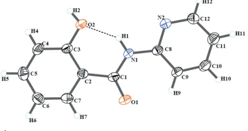
The molecule of the title compound, C12H10N2O2, is nearly planar, with a dihedral angle of 3.7 (2)between the planes of
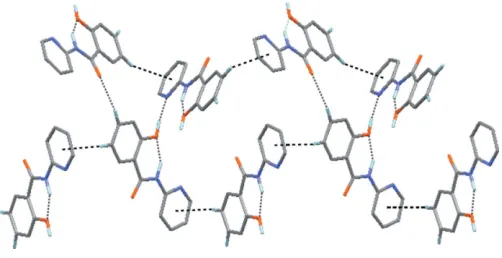
the benzene and pyridine rings. Molecules are linked by intermolecular O—H N and C—H O hydrogen bonds, as well as possible C—H interactions, forming a two-dimensional zigzag hydrogen-bonded network.
Comment
Molecular self-assembly by hydrogen bonds, e.g. C—H O, N—H O and C—H interactions, has emerged as an attractive approach in crystal engineering (Lu¨et al., 2005). The study of antimycobacterial properties of salicylanilides is of great interest, as salicylanilides can inhibit bacterial two-component systems, which can also be important in myco-bacteria. Salicylanilide and its derivatives exhibit biological and pharmacological activities (Waisseret al., 2004). The title compound, (I), was synthesized as a route to synthetic inter-mediates. We present here its crystal structure.
[image:1.610.207.460.580.714.2]In the molecule of the title compound, (I), bond lengths and angles (Table 1) are in normal ranges (Allenet al., 1987). The strong intramolecular N—H O hydrogen bond (Table 2) causes the formation of a pseudo-six-membered ring (C1–C3/ O2/H1/N1) (Fig. 1). The dihedral angle between the two planar rings [A(atoms C2–C7) andB(N2/C8–C12)] is 3.7 (2).
Figure 1
The crystal structure of (I) is stabilized by intermolecular O—H N and C—H O hydrogen bonds (Table 2), as well as possible T-shaped C—H interactions with a distance of 2.88 A˚ between the centroid of ring B and atom H6(C6) at (3
2 x, y, z 1
2), which result in the formation of a two-dimensional zigzag hydrogen-bonded network (Fig. 2).
Experimental
A mixture of pyridin-2-amine (0.94 g, 10.0 mmol) and 2-(methoxy-carbonyl)phenol (1.52 g, 10.0 mmol) was melted at 463–483 K for 2 h with an air condenser under an argon atmosphere. The reaction mixture was then heated under reflux in ethanol (10 ml) for 10 min, filtered and the product crystallized from the same solvent (yield 1.16 g, 54%; m.p. 484–485 K).
Crystal data
C12H10N2O2 Mr= 214.22
Orthorhombic,P212121 a= 6.1216 (16) A˚
b= 12.535 (3) A˚
c= 13.321 (4) A˚
V= 1022.2 (5) A˚3
Z= 4
Dx= 1.392 Mg m 3
MoKradiation = 0.10 mm1 T= 298 (2) K Needle, colourless 0.480.190.17 mm
Data collection
Bruker SMART CCD area-detector diffractometer
’and!scans
Absorption correction: multi-scan (SADABS; Sheldrick, 1996)
Tmin= 0.955,Tmax= 0.984
5265 measured reflections 1074 independent reflections 671 reflections withI> 2(I)
Rint= 0.106
max= 25.0
Refinement
Refinement onF2 R[F2> 2(F2)] = 0.046 wR(F2) = 0.135 S= 1.03 1074 reflections 145 parameters
H-atom parameters constrained
w= 1/[2(F
o2) + (0.062P)2
+ 0.1271P]
whereP= (Fo2+ 2Fc2)/3
(/)max< 0.001 max= 0.18 e A˚
3
[image:2.610.46.297.71.200.2]min=0.17 e A˚ 3
Table 1
Selected geometric parameters (A˚ ,).
N1—C1 1.353 (5) N1—C8 1.382 (5) N2—C12 1.340 (6)
N2—C8 1.342 (5) O1—C1 1.217 (5) O2—C3 1.358 (5)
C1—N1—C8 128.4 (4) C12—N2—C8 118.1 (4) O1—C1—N1 122.5 (5) O1—C1—C2 121.0 (4) N1—C1—C2 116.4 (4) O2—C3—C4 121.5 (4)
O2—C3—C2 118.5 (4) N2—C8—C9 121.5 (4) N2—C8—N1 113.4 (4) C9—C8—N1 125.2 (4) N2—C12—C11 123.5 (5)
Table 2
Hydrogen-bond geometry (A˚ ,).
D—H A D—H H A D A D—H A
N1—H1 O2 0.86 1.90 2.627 (5) 142 O2—H2 N2i
0.82 1.88 2.676 (5) 164 C5—H5 O1ii 0.93 2.50 3.384 (6) 159
Symmetry codes: (i)x1 2;yþ
1
2;zþ1; (ii)xþ1;yþ 1 2;zþ
1 2.
H atoms were positioned geometrically, with O—H = 0.82 A˚ , N— H = 0.86 A˚ and C—H = 0.93 A˚, and constrained to ride on their parent atoms, with Uiso(H) = xUeq(C,N,O), with x = 1.5 for the
hydroxyl H atom andx= 1.2 for all other H atoms. In the absence of significant anomalous scattering effects, Friedel pairs were averaged. Data collection:SMART(Bruker, 2000); cell refinement:SMART; data reduction: SAINT-Plus; program(s) used to solve structure:
SHELXS97(Sheldrick, 1997); program(s) used to refine structure:
SHELXL97 (Sheldrick, 1997); molecular graphics: DIAMOND
(Brandenburg, 2005); software used to prepare material for publi-cation:SHELXTL(Bruker, 2000).
The authors acknowledge financial support from the Natural Science Foundation of China (grant Nos. 20371022, 20431010 and 20021001), the Specialized Research Fund for the Doctoral Programme of Higher Education, and the Key Project of the Ministry of Education of China (grant No. 01170).
References
Allen, F. H., Kennard, O., Watson, D. G., Brammer, L., Orpen, A. G. & Taylor, R. (1987).J. Chem. Soc. Perkin Trans. 2, pp. S1–19.
Brandenburg, K. (2005).DIAMOND. Release 3.1a. Crystal Impact GbR, Bonn, Germany.
Bruker (2000). SMART (Version 5.0), SAINT-Plus (Version 6) and
SHELXTL(Version 6.1). Bruker AXS Inc., Madison, Wisconsin, USA. Lu¨, X.-Q., Jiang, J.-J., zur Loye, H.-C., Kang, B.-S. & Su, C.-Y. (2005).Inorg.
Chem.44, 1810–1817.
Sheldrick, G. M. (1996).SADABS. University of Go¨ttingen, Germany. Sheldrick, G. M. (1997). SHELXS97 and SHELXL97. University of
Go¨ttingen, Germany.
Waisser, K., Drazˇkova´, K., Kunesˇ, J., Klimesˇova´, V. & Kaustova´, J. (2004).Il Farmaco,59, 615–625.
Figure 2
supporting information
sup-1 Acta Cryst. (2006). E62, o1772–o1773
supporting information
Acta Cryst. (2006). E62, o1772–o1773 [https://doi.org/10.1107/S1600536806012244]
2-Hydroxy-
N
-(2-pyridyl)benzamide
Wen-Hua Wang, Zhong-Lu You, Wei-Sheng Liu and Da-Qi Wang
2-Hydroxy-N-(2-pyridyl)benzamide
Crystal data C12H10N2O2
Mr = 214.22
Orthorhombic, P212121
Hall symbol: P 2ac 2ab a = 6.1216 (16) Å b = 12.535 (3) Å c = 13.321 (4) Å V = 1022.2 (5) Å3
Z = 4
F(000) = 448 Dx = 1.392 Mg m−3
Mo Kα radiation, λ = 0.71073 Å Cell parameters from 903 reflections θ = 2.2–20.5°
µ = 0.10 mm−1
T = 298 K Needle, colorless 0.48 × 0.19 × 0.17 mm
Data collection
Bruker SMART CCD area-detector diffractometer
Radiation source: fine-focus sealed tube Graphite monochromator
φ and ω scans
Absorption correction: multi-scan (SADABS; Sheldrick, 1996) Tmin = 0.955, Tmax = 0.984
5265 measured reflections 1074 independent reflections 671 reflections with I > 2σ(I) Rint = 0.106
θmax = 25.0°, θmin = 2.2°
h = −7→7 k = −14→14 l = −15→10
Refinement Refinement on F2
Least-squares matrix: full R[F2 > 2σ(F2)] = 0.046
wR(F2) = 0.135
S = 1.04 1074 reflections 145 parameters 0 restraints
Primary atom site location: structure-invariant direct methods
Secondary atom site location: difference Fourier map
Hydrogen site location: inferred from neighbouring sites
H-atom parameters constrained w = 1/[σ2(F
o2) + (0.062P)2 + 0.1271P]
where P = (Fo2 + 2Fc2)/3
(Δ/σ)max < 0.001
Δρmax = 0.18 e Å−3
Δρmin = −0.17 e Å−3
Absolute structure: Flack (1983), no Friedel pairs
Absolute structure parameter: 10 (10)
Special details
Refinement. Refinement of F2 against ALL reflections. The weighted R-factor wR and goodness of fit S are based on F2,
conventional R-factors R are based on F, with F set to zero for negative F2. The threshold expression of F2 > σ(F2) is used
only for calculating R-factors(gt) etc. and is not relevant to the choice of reflections for refinement. R-factors based on F2
are statistically about twice as large as those based on F, and R- factors based on ALL data will be even larger.
Fractional atomic coordinates and isotropic or equivalent isotropic displacement parameters (Å2)
x y z Uiso*/Ueq
N1 1.0593 (6) 0.0508 (3) 0.4239 (3) 0.0423 (10) H1 1.0406 0.1143 0.4469 0.051* N2 1.3572 (6) 0.0537 (3) 0.5262 (3) 0.0427 (9) O1 0.9183 (6) −0.0704 (2) 0.3171 (3) 0.0644 (10) O2 0.8700 (6) 0.2389 (2) 0.4265 (3) 0.0637 (11) H2 0.8406 0.3009 0.4409 0.096* C1 0.9106 (8) 0.0175 (3) 0.3557 (3) 0.0420 (11) C2 0.7368 (7) 0.0945 (3) 0.3287 (3) 0.0399 (10) C3 0.7180 (7) 0.2007 (3) 0.3616 (3) 0.0421 (12) C4 0.5473 (8) 0.2626 (4) 0.3291 (4) 0.0561 (13) H4 0.5381 0.3331 0.3507 0.067* C5 0.3906 (9) 0.2239 (4) 0.2660 (4) 0.0587 (14) H5 0.2760 0.2673 0.2452 0.070* C6 0.4039 (8) 0.1197 (4) 0.2334 (4) 0.0564 (14) H6 0.2988 0.0920 0.1903 0.068* C7 0.5746 (7) 0.0575 (4) 0.2656 (3) 0.0474 (12) H7 0.5814 −0.0129 0.2438 0.057* C8 1.2375 (7) −0.0039 (3) 0.4615 (3) 0.0383 (11) C9 1.2951 (8) −0.1068 (3) 0.4358 (4) 0.0497 (13) H9 1.2126 −0.1457 0.3901 0.060* C10 1.4762 (9) −0.1499 (4) 0.4795 (4) 0.0581 (14) H10 1.5171 −0.2193 0.4636 0.070* C11 1.5982 (9) −0.0928 (4) 0.5460 (4) 0.0593 (14) H11 1.7222 −0.1219 0.5756 0.071* C12 1.5330 (8) 0.0076 (4) 0.5676 (4) 0.0508 (13) H12 1.6143 0.0467 0.6137 0.061*
Atomic displacement parameters (Å2)
U11 U22 U33 U12 U13 U23
supporting information
sup-3 Acta Cryst. (2006). E62, o1772–o1773
C7 0.053 (3) 0.047 (3) 0.042 (3) −0.005 (3) 0.000 (3) −0.006 (2) C8 0.043 (2) 0.032 (2) 0.040 (2) −0.004 (2) 0.007 (2) 0.001 (2) C9 0.060 (3) 0.033 (3) 0.057 (3) 0.004 (2) −0.006 (3) −0.009 (2) C10 0.070 (3) 0.038 (3) 0.066 (3) 0.007 (3) −0.001 (3) −0.007 (3) C11 0.066 (3) 0.052 (3) 0.060 (3) 0.010 (3) 0.000 (3) −0.008 (3) C12 0.052 (3) 0.051 (3) 0.049 (3) 0.000 (3) −0.003 (3) −0.005 (2)
Geometric parameters (Å, º)
N1—C1 1.353 (5) C5—C6 1.378 (6) N1—C8 1.382 (5) C5—H5 0.9300 N1—H1 0.8600 C6—C7 1.372 (6) N2—C12 1.340 (6) C6—H6 0.9300 N2—C8 1.342 (5) C7—H7 0.9300 O1—C1 1.217 (5) C8—C9 1.381 (6) O2—C3 1.358 (5) C9—C10 1.363 (6)
O2—H2 0.8200 C9—H9 0.9300
C1—C2 1.481 (6) C10—C11 1.362 (6) C2—C7 1.381 (6) C10—H10 0.9300 C2—C3 1.406 (5) C11—C12 1.351 (6) C3—C4 1.372 (6) C11—H11 0.9300 C4—C5 1.366 (6) C12—H12 0.9300 C4—H4 0.9300
Hydrogen-bond geometry (Å, º)
D—H···A D—H H···A D···A D—H···A N1—H1···O2 0.86 1.90 2.627 (5) 142 O2—H2···N2i 0.82 1.88 2.676 (5) 164
C5—H5···O1ii 0.93 2.50 3.384 (6) 159