2-Cyanoanilinium perchlorate
Li-Jing Cui* and Xin-Yuan Chen
College of Chemistry and Chemical Engineering, Southeast University, Nanjing 210096, People’s Republic of China
Correspondence e-mail: [email protected]
Received 22 January 2010; accepted 25 January 2010
Key indicators: single-crystal X-ray study;T= 298 K; mean(C–C) = 0.004 A˚;
Rfactor = 0.044;wRfactor = 0.117; data-to-parameter ratio = 16.2.
In the title compound, C7H7N2 +
ClO4
, the cation is almost planar (r.m.s. deviation = 0.042 A˚ ). In the crystal structure, the cations and anions are linked into a two-dimensional network parallel to (100) by N—H O hydrogen bonds.
Related literature
For the crystal structure of 2-cyanoanilinium chloride, see: Oueslati et al.(2005). For Cl—O distances, see: Messaiet al. (2009).
Experimental
Crystal data
C7H7N2+ClO4
Mr= 218.60
Monoclinic,P21=c a= 11.089 (2) A˚
b= 7.4561 (15) A˚
c= 13.872 (5) A˚ = 128.454 (18)
V= 898.2 (4) A˚3
Z= 4
MoKradiation = 0.42 mm1
T= 298 K
0.400.050.05 mm
Data collection
Rigaku Mercury2 diffractometer Absorption correction: multi-scan
(CrystalClear; Rigaku, 2005)
Tmin= 0.90,Tmax= 1.00
9026 measured reflections 2070 independent reflections 1761 reflections withI> 2(I)
Rint= 0.041
Refinement
R[F2> 2(F2)] = 0.044
wR(F2) = 0.117
S= 1.11 2070 reflections
128 parameters
H-atom parameters constrained max= 0.32 e A˚3
min=0.30 e A˚3
Table 1
Hydrogen-bond geometry (A˚ ,).
D—H A D—H H A D A D—H A
N2—H2A O4i
0.89 2.14 2.936 (2) 148
N2—H2B O4ii 0.89 2.24 3.007 (3) 144
N2—H2C O1iii
0.89 1.98 2.842 (2) 161
Symmetry codes: (i)xþ1;y;zþ1; (ii)x1;yþ1 2;z
1
2; (iii)x1;y;z. Data collection: CrystalClear (Rigaku, 2005); cell refinement: CrystalClear; data reduction:CrystalClear; program(s) used to solve structure:SHELXS97(Sheldrick, 2008); program(s) used to refine structure: SHELXL97 (Sheldrick, 2008); molecular graphics: SHELXTL(Sheldrick, 2008); software used to prepare material for publication:SHELXTL.
This work was supported by the Innovative Dissertation Fund of Southeast University.
Supplementary data and figures for this paper are available from the IUCr electronic archives (Reference: CI5023).
References
Messai, A., Direm, A., Benali-Cherif, N., Luneau, D. & Jeanneau, E. (2009).
Acta Cryst.E65, o460.
Oueslati, A., Kefi, R., Akriche, S. & Nasr, C. B. (2005).Z. Kristallogr. New Cryst. Struct.220, 365–366.
Rigaku (2005).CrystalClear. Rigaku Corporation, Tokyo, Japan. Sheldrick, G. M. (2008).Acta Cryst.A64, 112–122.
Acta Crystallographica Section E
Structure Reports Online
supporting information
Acta Cryst. (2010). E66, o467 [https://doi.org/10.1107/S1600536810003077]
2-Cyanoanilinium perchlorate
Li-Jing Cui and Xin-Yuan Chen
S1. Comment
Aniline derivatives attracted more attention as phase transition dielectric materials for their applications in
micro-electronics and memory storage. With the purpose of obtaining phase transition crystals of 2-aminobenzonitrile salts, its
interaction with various acids has been studied and we have obtained a series of new materials with this organic
molecule. In this paper, we describe the crystal structure of the title compound, 2-cyanoanilinium perchlorate.
The asymmetric unit is composed of a 2-cyanoanilinium cation and a perchlorate anion (Fig.1). The anion displays a
typical tetrahedral geometry around Cl atom and the Cl—O distances compare well with previously reported values
(Messai et al., 2009). The cation is almost planar (r.m.s. deviation 0.042 Å; maximum atomic deviation from coplanarity
is 0.073 (2) Å by atom N1). The C—NH3 [1.466 (2) Å] and C≡N [1.143 (3) Å] distances in the 2-cyanoanilinium cation
are longer compared to the corresponding distances in the crystal structure of 2-cyanoanilinium chloride (1.457 (4) Å,
1.137 (4) Å; Oueslati et al., 2005).
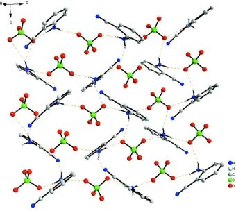
In the crystal structure, all the amine group H atoms are involved in N—H···O hydrogen bonds (Table 1). The N—H···O
hydrogen bonds link the ionic units into a two-dimensional network parallel to the ac plane (Fig. 2).
S2. Experimental
The commercial 2-aminobenzonitrile (3 mmol, 324 mg) was dissolved in a water-HClO4 (50:1 v/v) solution. The solvent
was slowly evaporated in air affording colourless crystals of the title compound suitable for X-ray analysis.
While the permittivity measurement shows that there is no phase transition within the temperature range (from 100 K to
400 K), and the permittivity is 7.8 at 1 MHz at room temperature.
S3. Refinement
All H atoms were initially located in a difference Fourier map. They were then constrained to an ideal geometry, with C–
Figure 1
The asymmetric unit of the title compound with the atomic numbering scheme. Displacement ellipsoids were drawn at
the 30% probability level.
Figure 2
The crystal packing of the title compound, showing a two-dimensional network parallel to the (100). H atoms not
[image:3.610.136.476.305.609.2]2-Cyanoanilinium perchlorate
Crystal data
C7H7N2+·ClO4− Mr = 218.60
Monoclinic, P21/c
Hall symbol: -P 2ybc
a = 11.089 (2) Å
b = 7.4561 (15) Å
c = 13.872 (5) Å
β = 128.454 (18)°
V = 898.2 (4) Å3 Z = 4
F(000) = 448
Dx = 1.617 Mg m−3
Mo Kα radiation, λ = 0.71073 Å Cell parameters from 1761 reflections
θ = 3.3–27.5°
µ = 0.42 mm−1 T = 298 K Needle, colourless 0.40 × 0.05 × 0.05 mm
Data collection
Rigaku Mercury2 diffractometer
Radiation source: fine-focus sealed tube Graphite monochromator
Detector resolution: 13.6612 pixels mm-1
CCD profile fitting scans
Absorption correction: multi-scan (CrystalClear; Rigaku, 2005)
Tmin = 0.90, Tmax = 1.00
9026 measured reflections 2070 independent reflections 1761 reflections with I > 2σ(I)
Rint = 0.041
θmax = 27.5°, θmin = 3.3°
h = −14→14
k = −9→9
l = −18→18
Refinement
Refinement on F2
Least-squares matrix: full
R[F2 > 2σ(F2)] = 0.044 wR(F2) = 0.117 S = 1.11 2070 reflections 128 parameters 0 restraints
Primary atom site location: structure-invariant direct methods
Secondary atom site location: difference Fourier map
Hydrogen site location: inferred from neighbouring sites
H-atom parameters constrained
w = 1/[σ2(Fo2) + (0.0456P)2 + 0.522P]
where P = (Fo2 + 2Fc2)/3
(Δ/σ)max = 0.001
Δρmax = 0.32 e Å−3
Δρmin = −0.30 e Å−3
Special details
Geometry. All esds (except the esd in the dihedral angle between two l.s. planes) are estimated using the full covariance matrix. The cell esds are taken into account individually in the estimation of esds in distances, angles and torsion angles; correlations between esds in cell parameters are only used when they are defined by crystal symmetry. An approximate (isotropic) treatment of cell esds is used for estimating esds involving l.s. planes.
Refinement. Refinement of F2 against ALL reflections. The weighted R-factor wR and goodness of fit S are based on F2,
conventional R-factors R are based on F, with F set to zero for negative F2. The threshold expression of F2 > σ(F2) is used
only for calculating R-factors(gt) etc. and is not relevant to the choice of reflections for refinement. R-factors based on F2
are statistically about twice as large as those based on F, and R- factors based on ALL data will be even larger.
Fractional atomic coordinates and isotropic or equivalent isotropic displacement parameters (Å2)
x y z Uiso*/Ueq
N2 0.10356 (18) 0.1622 (2) 0.40630 (16) 0.0351 (4)
H2A 0.0666 0.0523 0.3784 0.053*
H2B 0.0743 0.2332 0.3435 0.053*
N1 0.2247 (3) 0.4407 (3) 0.2725 (2) 0.0647 (7) C7 0.2722 (2) 0.1553 (3) 0.49390 (19) 0.0327 (4) C1 0.2853 (3) 0.3504 (3) 0.3573 (2) 0.0472 (6) C5 0.5007 (3) 0.0559 (4) 0.6835 (2) 0.0629 (8)
H5 0.5493 −0.0064 0.7572 0.075*
C2 0.3588 (2) 0.2429 (3) 0.4665 (2) 0.0386 (5) C6 0.3415 (3) 0.0628 (3) 0.6013 (2) 0.0469 (6)
H6 0.2827 0.0054 0.6189 0.056*
C4 0.5883 (3) 0.1403 (4) 0.6574 (3) 0.0662 (8)
H4 0.6952 0.1343 0.7133 0.079*
C3 0.5183 (3) 0.2326 (4) 0.5496 (3) 0.0547 (7)
H3 0.5775 0.2886 0.5320 0.066*
Cl1 0.91401 (6) 0.33466 (7) 0.57188 (4) 0.03477 (17) O4 0.8898 (2) 0.2197 (2) 0.64175 (15) 0.0471 (4) O3 0.7769 (2) 0.4305 (3) 0.48297 (16) 0.0681 (6) O2 1.0336 (2) 0.4577 (2) 0.65476 (18) 0.0610 (5) O1 0.9559 (2) 0.2236 (3) 0.51274 (17) 0.0574 (5)
Atomic displacement parameters (Å2)
U11 U22 U33 U12 U13 U23
N2 0.0340 (9) 0.0349 (9) 0.0403 (10) −0.0018 (7) 0.0250 (8) −0.0023 (7) N1 0.0733 (16) 0.0653 (15) 0.0617 (14) −0.0195 (13) 0.0451 (13) −0.0017 (12) C7 0.0326 (10) 0.0306 (10) 0.0365 (10) −0.0049 (8) 0.0223 (9) −0.0081 (8) C1 0.0499 (13) 0.0460 (13) 0.0576 (15) −0.0166 (11) 0.0393 (13) −0.0111 (12) C5 0.0518 (15) 0.0571 (16) 0.0415 (13) 0.0008 (13) 0.0101 (12) −0.0004 (12) C2 0.0391 (11) 0.0351 (11) 0.0491 (12) −0.0064 (9) 0.0311 (10) −0.0094 (9) C6 0.0476 (13) 0.0474 (13) 0.0384 (12) −0.0070 (11) 0.0232 (11) −0.0029 (10) C4 0.0314 (12) 0.0587 (16) 0.0718 (19) −0.0030 (12) 0.0140 (13) −0.0145 (15) C3 0.0393 (13) 0.0495 (14) 0.0762 (18) −0.0111 (11) 0.0364 (14) −0.0166 (13) Cl1 0.0407 (3) 0.0312 (3) 0.0334 (3) 0.0017 (2) 0.0235 (2) −0.00068 (19) O4 0.0561 (10) 0.0436 (9) 0.0532 (10) −0.0028 (8) 0.0397 (9) 0.0023 (8) O3 0.0669 (12) 0.0745 (14) 0.0424 (10) 0.0332 (11) 0.0238 (9) 0.0151 (9) O2 0.0660 (12) 0.0423 (10) 0.0638 (11) −0.0212 (9) 0.0349 (10) −0.0126 (8) O1 0.0724 (12) 0.0587 (11) 0.0635 (11) 0.0041 (9) 0.0534 (11) −0.0085 (9)
Geometric parameters (Å, º)
N2—C7 1.466 (2) C5—H5 0.93
N2—H2A 0.89 C2—C3 1.388 (3)
N2—H2B 0.89 C6—H6 0.93
N2—H2C 0.89 C4—C3 1.367 (4)
N1—C1 1.143 (3) C4—H4 0.93
C7—C6 1.364 (3) C3—H3 0.93
C7—C2 1.396 (3) Cl1—O3 1.4181 (18)
C1—C2 1.437 (4) Cl1—O2 1.4233 (18)
C5—C4 1.381 (4) Cl1—O1 1.4315 (17)
C7—N2—H2A 109.5 C7—C6—C5 118.8 (2)
C7—N2—H2B 109.5 C7—C6—H6 120.6
H2A—N2—H2B 109.5 C5—C6—H6 120.6
C7—N2—H2C 109.5 C3—C4—C5 120.2 (2)
H2A—N2—H2C 109.5 C3—C4—H4 119.9
H2B—N2—H2C 109.5 C5—C4—H4 119.9
C6—C7—C2 121.2 (2) C4—C3—C2 120.0 (2)
C6—C7—N2 118.99 (19) C4—C3—H3 120.0
C2—C7—N2 119.78 (19) C2—C3—H3 120.0
N1—C1—C2 177.1 (3) O3—Cl1—O2 109.53 (13)
C4—C5—C6 120.8 (3) O3—Cl1—O1 110.35 (12)
C4—C5—H5 119.6 O2—Cl1—O1 111.38 (12)
C6—C5—H5 119.6 O3—Cl1—O4 109.75 (12)
C3—C2—C7 119.0 (2) O2—Cl1—O4 108.07 (11)
C3—C2—C1 120.1 (2) O1—Cl1—O4 107.72 (11)
C7—C2—C1 120.8 (2)
C6—C7—C2—C3 1.0 (3) C4—C5—C6—C7 −0.3 (4) N2—C7—C2—C3 −179.0 (2) C6—C5—C4—C3 0.2 (4) C6—C7—C2—C1 −175.6 (2) C5—C4—C3—C2 0.5 (4) N2—C7—C2—C1 4.4 (3) C7—C2—C3—C4 −1.1 (4) C2—C7—C6—C5 −0.3 (4) C1—C2—C3—C4 175.5 (2) N2—C7—C6—C5 179.7 (2)
Hydrogen-bond geometry (Å, º)
D—H···A D—H H···A D···A D—H···A
N2—H2A···O4i 0.89 2.14 2.936 (2) 148
N2—H2B···O4ii 0.89 2.24 3.007 (3) 144
N2—H2C···O1iii 0.89 1.98 2.842 (2) 161