epithelial-mesenchymal interactions in COPD
patients
Jun Araya, … , David J. Erle, Stephen L. Nishimura
J Clin Invest.
2007;
117(11)
:3551-3562.
https://doi.org/10.1172/JCI32526
.
Squamous metaplasia (SM) is common in smokers and is associated with airway
obstruction in chronic obstructive pulmonary disease (COPD). A major mechanism of
airway obstruction in COPD is thickening of the small airway walls. We asked whether SM
actively contributes to airway wall thickening through alteration of epithelial-mesenchymal
interactions in COPD. Using immunohistochemical staining, airway morphometry, and
fibroblast culture of lung samples from COPD patients; genome-wide analysis of an in vitro
model of SM; and in vitro modeling of human airway epithelial-mesenchymal interactions,
we provide evidence that SM, through the increased secretion of IL-1
b
, induces a fibrotic
response in adjacent airway fibroblasts. We identify a pivotal role for integrin-mediated
TGF-
b
activation in amplifying SM and driving IL-1
b
–dependent profibrotic mesenchymal
responses. Finally, we show that SM correlates with increased severity of COPD and that
fibroblast expression of the integrin
a
vb
8, which is the major mediator of airway fibroblast
TGF-
b
activation, correlated with disease severity and small airway wall thickening in
COPD. Our findings have identified TGF-
b
as a potential therapeutic target for COPD.
Research Article
Pulmonology
Find the latest version:
Squamous metaplasia amplifies pathologic
epithelial-mesenchymal interactions
in COPD patients
Jun Araya,1 Stephanie Cambier,1 Jennifer A. Markovics,1 Paul Wolters,2 David Jablons,3
Arthur Hill,3 Walter Finkbeiner,1 Kirk Jones,1 V. Courtney Broaddus,2 Dean Sheppard,2
Andrea Barzcak,2 Yuanyuan Xiao,4 David J. Erle,2 and Stephen L. Nishimura1
1Department of Pathology, 2Department of Medicine and Lung Biology Center, Pulmonary Division, 3Department of Surgery, and 4Department of Epidemiology and Biostatistics, Center for Bioinformatics and Molecular Biostatistics, UCSF, San Francisco, California, USA.
Squamous metaplasia (SM) is common in smokers and is associated with airway obstruction in chronic
obstructive pulmonary disease (COPD). A major mechanism of airway obstruction in COPD is thickening of
the small airway walls. We asked whether SM actively contributes to airway wall thickening through alteration
of epithelial-mesenchymal interactions in COPD. Using immunohistochemical staining, airway
morphom-etry, and fibroblast culture of lung samples from COPD patients; genome-wide analysis of an in vitro model
of SM; and in vitro modeling of human airway epithelial-mesenchymal interactions, we provide evidence that
SM, through the increased secretion of IL-1
β
, induces a fibrotic response in adjacent airway fibroblasts. We
identify a pivotal role for integrin-mediated TGF-
β
activation in amplifying SM and driving IL-1
β
–dependent
profibrotic mesenchymal responses. Finally, we show that SM correlates with increased severity of COPD and
that fibroblast expression of the integrin
α
vβ
8, which is the major mediator of airway fibroblast TGF-
β
activa-tion, correlated with disease severity and small airway wall thickening in COPD. Our findings have identified
TGF-
β
as a potential therapeutic target for COPD.
Introduction
Chronic obstructive pulmonary disease (COPD) is the fourth-lead-ing cause of death worldwide and affects up to 50% of long-term smokers (1, 2). The major histopathologic correlates of airway obstruction in COPD are loss of alveolar walls and narrowing of the small airways (3). The relative contribution of each of these pathologic features to airway obstruction is not completely under- stood (4). However, recently, airway wall thickening has been iden- tified as a major independent predictor of the severity of physio-logic airway obstruction in COPD patients stratified by the criteria established by the Global Initiative for Obstructive Lung Disease (GOLD; http://www.goldcopd.com/), suggesting that airway-wall fibrosis contributes to airway obstruction (5). COPD is caused by the noxious effects of tobacco smoke, which leads to airway epithe- lial injury and the induction of changes such as squamous meta-plasia (SM), the reversible replacement of the normal columnar epithelium by squamous epithelium (6). In 1978, Cosio et al. noted that SM correlated with the severity of airway obstruction, sug-gesting that SM and airway obstruction may be linked (7).
Changes in airway epithelium have the potential to alter the finely balanced reciprocal interactions between the epithelium and mesenchyme that influences cellular differentiation, the so-called epithelial-mesenchymal trophic unit (8, 9). Thus, SM developing in response to pathologic stimuli may alter normal homeostatic autocrine and paracrine interactions between the epithelium and
the surrounding fibroblast sheath. Recently, we provided evidence for a homeostatic role for integrin-mediated TGF-β activation in a human model of the epithelial-mesenchymal trophic unit (10). In this model, fibroblastic integrin αvβ8-mediated TGF-β
activa-tion was found to regulate the fibrogenic phenotype as well as synthesis and secretion of HGF, which influences the prolifera-tion of adjacent airway epithelial cells (10).
TGF-β is a major fibrogenic cytokine regulating both extracellular matrix deposition and immune homeostasis that may link SM and airway obstruction. TGF-β is a major factor in driving SM of airway epithelial cells in vitro (11). Increased TGF-β can be found in the small airway epithelium of COPD patients, and its levels correlate with the severity of obstruction (12, 13). Polymorphisms in the TGF-β1 promoter have been shown to increase susceptibility to COPD (14). Adenoviral transfer of TGF-β1 to murine lung has been shown to increase fibroblast accumulation around airways, and airway epithe-lial transgenic expression of TGF-β causes peribronchiolar fibrosis, an effect that is reversible (15, 16). These results implicate TGF-β– mediated airway remodeling as a potential therapeutic target.
TGF-β is ubiquitously expressed in 3 isoforms in all mammalian cell types and tissues but is found almost entirely in a latent form, consisting of the noncovalent interactions between the latency-associated peptide (LAP) of TGF-β and the mature peptide of TGF-β and covalent interactions with latent TGF-β –binding pro-teins (17, 18). Thus, a major point of regulation of TGF-β func- tion is through its activation, which occurs by 2 general mecha-nisms, those that alter the conformation of LAP and those that result in the proteolytic cleavage of LAP (17, 19–21). The integrins
αvβ6 and αvβ8, heterodimers that have the αn integrin subunit in
common, can each bind to and activate latent TGF-β (20, 21) and together may account for the majority of TGF-β 1 activation dur-ing murine development (22). In the case of αvβ6, interaction with
Nonstandard abbreviations used: BEGM, bronchial epithelial growth medium; Col I, type I collagen; COPD, chronic obstructive pulmonary disease; EDC, epidermal differ-entiation complex; GOLD, Global Initiative for Obstructive Lung Disease; IL-1RA, IL-1 receptor antagonist; LAP, latency-associated peptide; MT1-MMP, membrane type I– MMP; P0, initial passage; P2, second passage; SM, squamous metaplasia.
Conflict of interest: The authors have declared that no conflict of interest exists.
latent TGF-β results in conformational alteration of LAP, allow-ing cell-associated active TGF-β to interact with TGF-β receptors on immediately adjacent cells (21). In the case of αvβ8, binding of
latent TGF-β results in the metalloproteolytic cleavage of LAP and release of active TGF-β from the cell surface, where it can then act as an autocrine or paracrine factor (20). Both αvβ6 and αvβ8 are
expressed by human airway epithelial cells; αvβ8 (but not αvβ6) is
expressed by human airway fibroblasts.
Of the many cytokines that have been implicated in the patho-genesis of COPD, IL-1β, a major proinflammatory cytokine, is of particular interest (23). IL-1β has been found in higher levels in alveolar macrophages from smokers and in cigarette smoke–treat-ed airway epithelial cells from COPD patients (24). Transgenic overexpression of IL-1β in airway epithelial cells leads to airway inflammation, emphysema, and airway-wall thickening (25).
Here we report a link between SM of airway epithelium and airway-wall thickening through a mechanism involving increased secretion of IL-1β and increased integrin-mediated activation of TGF-β.
Results
SM in the small airways of COPD patients. Involucrin is a component of the cornified envelope of stratified squamous epithelium and is
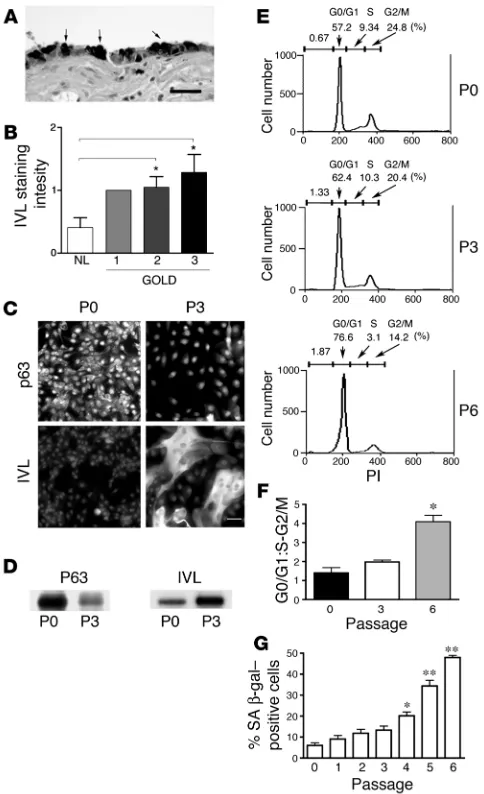
a marker of SM in the airway epithelium (26). In the human small airways, involucrin staining was found in superficial metaplastic airway epithelial cells in areas of cuboidal airway epithelium devoid of cilia as well as in areas of morphologically obvious SM, confirm-ing involucrin as a robust marker for SM (Figure 1A). Involucrin staining intensity in the small airway epithelium was significantly increased in patients with GOLD stages 2 and 3 COPD compared with normal controls (Figure 1B).
In vitro model of human airway epithelial squamous metaplasia. Human bronchial epithelial cells undergo SM during cell culture in the absence of retinoic acid (11). We found that in 2D cultures, in the low retinoic acid concentrations of our commercial airway epithelial medium preparation (0.3 nM; ref. 27), human bronchial epithelial cells phenotypically changed during serial passage from small poorly spread round cells to large and flattened squamous-like cells (Figure 1C). At the initial passage (P0), we determined that the cultures consisted mostly of basal cells, the progenitor cells of the human airway, because more than 95% of the cells expressed p63 (Figure 1C), a marker of basal cell differentiation (28). By the third passage (P3), the human bronchial epithelial cells displayed reduced p63 and strong involucrin staining (Figure 1C) typical of metaplastic squamous epithelial cells (29). Western blotting of human bronchial epithelial cell lysates confirmed the acquisition of a squamous cell phenotype during serial passage, since high p63 and low involucrin expression were found in P0 human bronchial epithelial cell lysates and low p63 and high involucrin expression were found in P3 human bronchial epithelial cell lysates (Figure 1D). These data are in agreement with previous studies using simi- lar media, demonstrating that the primary cell in 2D tracheobron-chial culture is initially the basal cell, which changes to an SM-like cell during serial passage (29).
[image:3.585.45.287.82.479.2]SM in COPD patients is not only characterized by the onset of epidermal differentiation but is also a hyperproliferative state (30). Thus, we characterized cell proliferation and senescence of seri-ally passaged human bronchial epithelial cells. The percentage of actively proliferating cells in the S and G2/M phases of the cell
Figure 1
SM correlates with increased GOLD stage of COPD in human lung samples, and SM can be induced during in vitro culture. (A) Involu-crin (IVL), a marker of epidermal differentiation (26), stains small air-way SM. Shown is a photomicrograph of metaplastic squamous cells (arrows) of a small airway from a COPD patient stained with anti-IVL. Scale bar: 50 μm. (B) IVL-staining intensity of lung samples from nor-mal versus COPD patients stratified according to GOLD (www.gold-copd.com), and assessed by grading digital images (0–3 scale) with 0 being absent; grade 1, 0–10%; grade 2, 11–20%; and grade 3, >20% cytoplasmic staining. *P < 0.05. (C) Photomicrographs of P0 compared with P3 human bronchial epithelial cells. Upper panels are stained with the basal cell marker anti-p63 and lower panels with anti-IVL. Scale bar: 50 μm. (D) Western blotting of 40 μg of P0 or P3 human bronchial epithelial cells total cell lysates for p63 and IVL. (E) Propidium iodide (PI) staining and flow cytometry of serially passaged human bronchial epithelial cells of a representative experiment (n = 3) showing the rela-tive proportion of cells in the G0/G1, S, and G2/M phases of the cell cycle. (F) Relative proportion (± SEM) of human bronchial epithelial cells (n = 3) in G0/G1 compared with S and G2/M phases of the cell cycle during serial passage. *P < 0.01. (G) Senescence-associated
cycle remained high (~ 30%) and did not significantly change dur- ing the P0 to P3 culture period, but by P6, proliferation had mark-edly decreased due to an accumulation of cells in the G0/G1 phase of the cell cycle (Figure 1, E and F). The fraction of senescent cells did not significantly change between P0 and P3 but increased sig-nificantly in subsequent passages (Figure 1G). We conclude that at P3, human bronchial epithelial cells in 2D culture approximate the hyperproliferative state of SM in vivo and avoid the in vitro phenomenon of replicative senescence.
Global analysis of gene expression during SM reveals induction of the pro-inflammatory mediators IL-1α and IL-1β. We analyzed the changes in human bronchial epithelial cell gene expression occurring during the culture period from P0 to P3 using high-density oligonucle-otide microarrays. In the transition from P0 to P3, there were 278 genes induced and 225 genes repressed (503 total) that reached statistical significance (http://www.ncbi.nlm.nih.gov/geo/, Geo accession number: GSE7557). Gene ontology analysis of the sta- tistically significant induced genes revealed enrichment of a num-ber of functional categories, including cell motility, cytoskeletal assembly, ectodermal development, IL-1 receptor binding, cell proliferation, and cell adhesion (Supplemental Table 1; supple-mental material available online with this article; doi:10.1172/ JCI32526DS1). Statistically significant repressed genes included only the immune response group (Supplemental Table 2).
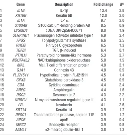
Of the 503 statistically significant differentially expressed genes, 93 and 62 were at least 2-fold induced or repressed, respectively. Most notably, IL-1β (13.4-fold), IL-1α (9.7-fold), and the TGF-β– responsive gene, plasminogen-activator inhibitor (SERPINE1,
6.9-fold), were the first, third, and sixth most highly induced genes, respectively (Table 1). Other genes known to be induced by TGF-β in epithelial cells that were also differentially expressed at least 2-fold in P3 airway epithelial cultures (see Table 1 and http:// www.ncbi.nlm.nih.gov/geo/, Geo accession number: GSE7557) included the integrin β6 (ITGB6) (31), the T cell differentiation protein (MAL) (32), the TGF-β inducible gene (BIGH3) (33), and laminin A3 (LAMA3) (32), suggesting increased autocrine TGF-β signaling during serial passage. In addition, a number of genes involved in epidermal differentiation (KRT6B, S100A8,
GJ1A, IVL, DSC2, KRT14, DESC1, A2ML) were highly induced during serial passage; 3 of these (KRT6, IVL, and KRT14 ) have been reported to label actively prolif-erating suprabasal cells in the squamous mucosa or in SM (34–37) (Table 1).
Immunocytochemistry confirmed the suprabasal dif-ferentiation state of P3 human bronchial epithelial cells since 80%, 61%, and 88% of cells expressed keratin 6, ker- atin 14, and involucrin, respectively (Supplemental Fig-ure 1). A high percentage (42%) of the involucrin-stained P3 cells were actively proliferating, since they were in the S or G2/M phase of the cell cycle (Supplemental Fig-ure 2). Finally, 76%, 67%, and 90% of IL-1β–positive P3 human bronchial epithelial cells coexpressed keratin 6, keratin 14, and involucrin, respectively (Supplemen-tal Figure 1). These data demonstrate that P3 human bronchial epithelial cells represent actively proliferating IL-1β–producing suprabasal squamous cells.
IL-1α and IL-1β are functionally redundant and bind to the same IL-1 receptor and endogenous soluble recep-tor antagonist (IL-1RA) but differ in that IL-1β is secreted whereas IL-1α exists in both a membrane-bound and cleaved (secreted) form. Increased levels of IL-1α and IL-1β in P3 human bronchial epithelial cells were confirmed by RT-PCR (Figure 2A), cytokine antibody array (Figure 2B), and ELISA (Figure 2C). The increase in IL-1β in the supernatant of P3 airway epithelial supernatants was approximately 30 pg/ml. This level is in 4-fold greater excess than found in synovial fluid from patients with rheumatoid arthritis (38). Finally, to confirm the physiological relevance of our 2D sys-tem, we used air-liquid interface culture conditions. Thus, human bronchial epithelial cells were grown on top of filters and only the basal surface of the cells was allowed to remain in contact with the medium, which simulates the anatomic arrangement in the airway in vivo (39). Human bronchial epithelial cells (P0) grown in air-liquid interface conditions for 21 days using SM-promoting medium conditions adopted a squamous metaplastic phenotype and expressed involucrin in the basal and suprabasal cells (Figure 2D) and expressed IL-1β (Figure 2E). Air-liquid interface cultures grown in airway-differentiating medium adopted a pseudostrati-fied ciliated columnar phenotype and expressed neither involucrin nor IL-1β (Figure 2D and Figure 2E).
[image:4.585.54.309.125.397.2]In vitro SM closely mimics metaplastic squamous epithelium in COPD in vivo. We characterized and confirmed the increased expression of IL-1β and the epidermal differentiation genes found using P0–P3 oligoarrays by immunohistochemical staining of tissue samples of human airway SM from COPD patients and compared the staining pattern with normal and wounded epidermis, squamous mucosa, and SM from a variety of mucosal sites. Antibodies to
Table 1
Twenty-five most highly induced genes during human bronchial epithelial cells’ serial passage
Gene Description Fold change BA
1 IL1B IL-1β 13.4 2.8
2 KRT6B Keratin 6B 12.0 2.0
3 IL1A IL-1α 9.7 2.0
4 S100A8 S100 calcium-binding protein A8 8.5 0.9 5 LYSMD1 cDNA DKFZp564C0671 8.0 1.9 6 SERPINE1 Plasminogen activator inhibitor type 1 6.9 2.4 7 FPGS Folylpolyglutamate synthase 6.6 2.5 8 RHCG Rh type C glycoprotein 6.5 1.3 9 TGFBI TGF, β-induced 6.4 0.1 10 PTHLH Parathyroid hormone-like hormone 5.3 2.7 11 NDUFA4L2 NADH:ubiquinone oxidoreductase 5.0 1.5 12 MAL Mal, T cell differentiation protein 4.9 2.1
13 GJA1 Connexin 43 4.9 0.5
14 FLJ21511 Hypothetical protein FLJ21511 4.5 1.4 15 GPX3 Glutathione peroxidase 3 4.5 0.5
16 CDA Cytidine deaminase 4.4 2.4
17 AREG Amphiregulin 4.4 1.6
18 DSC2 Desmocollin 2 4.3 2.2
19 NDRG1 N-myc downstream regulated gene 1 4.3 1.1
20 IVL Involucrin 4.1 2.6
21 KRT14 Keratin 14 3.9 1.7 22 DESC1 Transmembrane protease, serpine 11E 3.9 1.7
23 APOE apoE 3.9 0.4
24 MRC2 Endocytic receptor 3.9 0.8
25 A2ML1 α2-macroglobulin–like 1 3.8 1.3
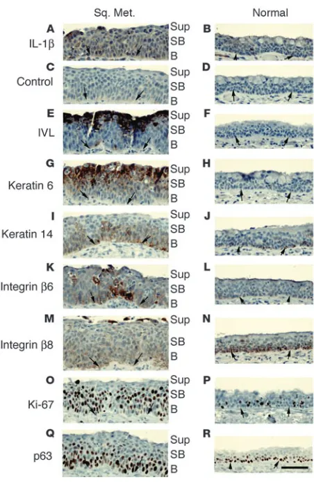
IL-1β stained basal, suprabasal, and superficial cells of metaplastic squamous airway epithelium (Figure 3A) but did not stain normal airway epithelium from COPD patients (Figure 3B). The staining was specific, as no staining was observed after preabsorption of the antibody with the immunogen (Figure 3C). Antibodies to involu-crin and keratin 6 stained the superficial and suprabasal cells of the squamous metaplastic epithelium (Figure 3, E and G) but not normal epithelium (Figure 3, F and H). Keratin 14 immunolocal-ized to the basal and suprabasal cells of SM (Figure 3I) and stained only the basal cells of the normal airway (Figure 3J). The basal and suprabasal cells of SM stained intensely with antibodies to integrin
β6 (Figure 3K) while the basal cells of the normal epithelium only lightly stained (Figure 3L); the opposite was true of integrin β8 (Figure 3, M and N). The basal and suprabasal cells of SM stained intensely with antibodies to the proliferation marker Ki-67 (Figure 3O) while fewer scattered basal cells of the normal airway stained (Figure 3P). The basal cell marker p63 (28) intensely stained basal cells and moderately labeled suprabasal cells of SM (Figure 3Q) while staining only basal cells of the normal airway (Figure 3R). A pattern of immunoreactivity similar to that seen in SM of the airways in COPD was seen in the squamous mucosa of the cervix, and in the epidermis at the edge of healing wounds (Supplemen-tal Data and Supplemental Table 3). We conclude that P3 human bronchial epithelial cells in 2D culture closely approximate SM in the airway in COPD as well as in other mucosal sites.
IL-1β released by squamous metaplastic epithelium increases αvβ8 inte-grin expression and αvβ8-mediated TGF-β activation in human airway fibroblasts. We hypothesized that IL-1β released by metaplastic
squamous epithelial cells contributed to a profi- brotic fibroblast phenotype by increasing fibro-blast TGF-β activation, a largely integrin αvβ8–
mediated process (10). Indeed, we found that
αvβ8 integrin expression (Figure 4, A and B) and αvβ8 integrin–mediated TGF-β activation (Figure
4C) in human airway fibroblasts was increased by treatment either with recombinant IL-1β (data not shown) or by coculture with squamous meta-plastic (P3) epithelial cells. Increased fibroblast
β8 expression caused by coculture with squamous metaplastic epithelial cells was IL-1 dependent, since IL-1RA, a physiological inhibitor of both IL-1α and IL-1β, almost completely inhibited the increase in β8 expression (Figure 4D). The airway epithelial cells were the sole source of IL-1 in the coculture system because neither IL-1α nor IL-1β mRNA could be detected in normal airway fibro-blasts by RT-PCR (data not shown).
IL-1β released by squamous metaplastic epithelium causes an αvβ8- and TGF-β–dependent switch to a fibrogenic fibroblast phenotype . Increased ECM pro-duction and increased fibroblast contractility are hallmarks of fibrotic responses seen in airway wall thickening, and increased type I collagen (Col I) and increased SMA (α SMA) are key bio-chemical markers of that response. To assess the contribution of autocrine αvβ8–mediated TGF-β
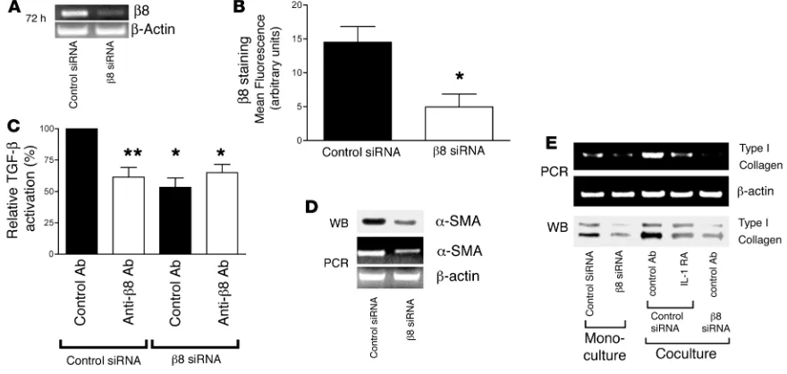
activation to the profibrotic fibroblast pheno-type, we used siRNA to knock down β 8 expres-sion in airway fibroblasts. Treatment of normal airway fibroblasts with β8 siRNA significantly reduced β8 expression as assessed by RT-PCR (Figure 5A) and flow cytometry (Figure 5B) and completely inhibited αvβ8
-medi-ated TGF-β activation (Figure 5C). These effects were specific to
β8 siRNA since they were not seen with control siRNA (Figure 5, A–C). Neutralizing anti-β8 and β 8 siRNA blocked only approxi-mately 50% of the total TGF-β activation as determined using a TGF-β neutralizing antibody that inhibits the function of all 3 TGF-β isoforms (Figure 5C). Thus, the incomplete inhibition of TGF-β activation could be due to activation of TGF-β2 by an
αvβ8-independent mechanism, since αvβ8 is only involved in the
activation of TGF-β1 and -β 3 (10, 20). Alternatively, the incom-plete inhibition may be due to the relatively poor efficacy of the
β8-neutralizing antibody and the incomplete silencing achieved with β8 siRNA (Figure 5A). Autocrine αvβ8-mediated activation of
TGF-β influenced the myofibroblast phenotype, since treatment of airway fibroblasts with β8 siRNA reduced αSMA expression (Figure 5D) and Col I secretion (Figure 5E). Coculture of airway fibroblasts with P3 human bronchial epithelial cells led to an increase in Col I transcription and protein production by airway fibroblasts (Figure 5E). The increased production of collagen was IL-1β– and fibroblast β8–dependent because the increase in Col I expression induced by coculture with P3 human bronchial epi-thelial cells could almost be completely inhibited by treatment of cocultures with IL-1RA (Figure 5E) or by transfection of airway fibroblasts with β8 siRNA (Figure 5E).
IL-1β–dependent increase in αvβ8-mediated activation of TGF-β is MT1-MMP dependent. IL-1β, while increasing β8 expression, also has
diverse functional effects, including increased transcription of mem-Figure 2
IL-1α and IL-1β are induced during SM. (A) RT-PCR of total RNA from P0 to P3 human bronchial epithelial cells grown in 2D culture, using primers to IL-1α and IL-1β and
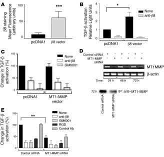
[image:5.585.42.344.83.232.2]brane type I–MMP (MT1-MMP) (40) and TGF-β (41), either of which could independently increase the efficiency of β8 integrin–mediated TGF-β activation (20). Indeed, treatment of airway fibroblasts with IL-1β also increased MT1-MMP activity (not shown). To determine whether the IL-1β–dependent increase in fibroblast αvβ8–mediated
TGF-β activation was due to increased β8 expression or to increased MT1-MMP activity, we overexpressed either β 8 or MT1-MMP in air-way fibroblasts. Transient β8 transfection significantly increased β8 integrin expression and αvβ8-mediated TGF-β activation (Figure 6,
A and B) and did not affect MT1-MMP activity or expression (not shown). Conversely, overexpression of MT1-MMP, which increased MT1-MMP activity more than 100-fold (data not shown) had no effect on β8 expression (data not shown) or αvβ8-mediated TGF-β
activation (Figure 6C). However, transfection of airway fibroblasts with MT1-MMP siRNA, which inhibited approximately 70% of MT1-MMP surface expression (Figure 6D) and activity (not shown), markedly inhibited αvβ8-mediated activation of TGF-β (Figure 6E).
In contrast, treatment of airway fibroblasts with IL-1β did not increase TGF-β transcription (ratio of TGF-β to β-actin transcript: nontreated, 0.97 ± 0.11; IL-1β treated, 0.87 ± 0.16, n = 3) or secretion (TGF-β activity as measured by TGF-β bioassay: control, 1269 ± 131; IL-1β treated, 1152 ± 129, n = 4). These results demonstrate that the endogenous level of functionally active MT1-MMP present in air-way fibroblasts is sufficient to support αvβ8-mediated activation of
TGF-β and that the IL-1β–induced increases in TGF-β activation are due to increased β 8 expression and not increased activity of MT1-MMP or expression of TGF-β.
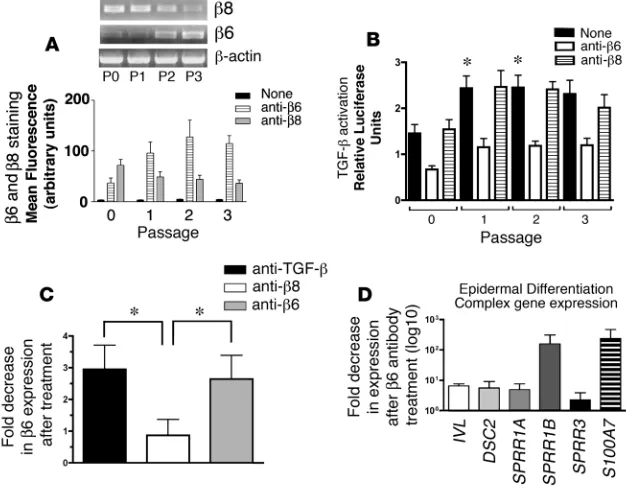
Increased TGF-β activation during serial passage of airway epithelial cells. We next addressed the role of integrin-mediated activation of TGF-β in driving SM. The TGF-β–activating integrin αvβ6
showed a dramatic increase in mRNA and surface expression during serial passage of airway epithelial cells. At P0, airway epi-thelial cells expressed little αvβ6. By P1, high levels of αvβ6 were
detected, which persisted throughout later passages (Figure 7A). The other TGF-β–activating integrin expressed by airway epithelial cells, αvβ8, showed high expression in P0 cells and demonstrated
a slight decrease during serial passage (Figure 7A). Approximately 50% of TGF-β activation by airway epithelial cells from P0 to P3 was accounted for by the αvβ6 integrin, with the αvβ8
integrin con-tributing little to the total TGF-β activation (Figure 7B). This is consistent with our previous observations that airway epithelial cells under these culture conditions require stimulation to sup-port full αvβ8-mediated activation of TGF-β (10). Anti-αvβ3 and
anti-αvβ5 integrin antibodies showed no effects on TGF-β
activa-tion (data not shown).
Airway epithelial β6 expression has previously been reported to be increased by exogenous active TGF-β (31). Here, increased β6 expression was partially mediated by autocrine αvβ6
–mediated acti-vation of TGF-β, since treatment of airway epithelial cells (P2) with neutralizing anti-β6 or TGF-β antibodies caused a decrease in β6
mRNA (Figure 7C). This effect was specific to αvβ6
-mediated activa-tion of TGF-β, since anti-β8 had no effect (Figure 7C). These results suggest that the integrin αvβ6 provides a self-amplifying autocrine
loop of TGF-β activation during the metaplastic process.
Role of autocrine and paracrine TGF-β signaling in regulation of squamous differentiation of airway epithelial cells. We investigated the role of airway epithelial integrin αvβ6–mediated autocrine TGF-β in promoting
the expression of a panel of 6 genes that were differentially expressed during serial passage and that include members of gene clusters on chromosome 1q21 known as the epidermal differentiation complex (EDC) (Table 1 and http://www.ncbi.nlm.nih.gov/geo/, series acces-sion GSE7557; IVL, DSC2, SPRR1A, SPRR1B, SPRR3, S100A7) (42, 43). Neutralizing anti-β6 decreased the transcript expression of all 6
tested members of the EDC (Figure 7D). These results suggest that autocrine αvβ6–mediated activation of TGF-β plays an active role in
promoting squamous differentiation.
We next investigated the role of αvβ8
-dependent paracrine secre-tion of active TGF-β by airway fibroblasts in regulation of human bronchial epithelial cell β6 expression. In P0 human bronchial epithelial cells, β6 transcript expression was found to be TGF-β dependent (Figure 8A). To determine the paracrine role of αvβ8
[image:6.585.47.273.79.426.2]-mediated activation of TGF-β by airway fibroblasts in regulation
Figure 3
IL-1β is coexpressed with integrins αvβ6 and αvβ8 in
hyperprolifera-tive suprabasal squamous metaplastic airway epithelium of COPD patients. Immunostaining of adjacent paraffin sections from an airway with a focus of SM (Sq. Met.: A, C, E, G, I, K, M, O, and Q) compared with relatively normal airway mucosa (B, D, F, H, J, L, N, P, and R) from a COPD patient using antibodies to (A and B) IL-1β; (C and D) IL-1β preabsorbed with the immunogenic peptide (control); (E and
F) the suprabasal markers anti-involucrin; (G and H) anti–keratin 6; (I and J) anti–keratin 14; (K and L) the TGF-β activating integrins β6; (M and N) and β8; (O and P) the cell proliferation marker Ki-67; (Q and
of human bronchial epithelial cell β 6 expression, P0 human bron-chial epithelial cells were cocultured with airway fibroblasts, which had been transfected with control or β8 siRNA. The P0 human bronchial epithelial cell expression of the β 6 transcript was sig-nificantly increased by coculture with control transfected airway fibroblasts compared with P0 human bronchial epithelial cells cul-tured alone (Figure 8A). The majority of this increase was due to
TGF-β, since the increase could be largely blocked by neutralizing anti–TGF-β (Figure 8A). Furthermore, the increase in expression of the human bronchial epithelial cells’ β6 transcript was partially dependent on αvβ8-mediated activation of TGF-β
[image:7.585.51.544.81.194.2]by airway fibro- blasts, since the majority of the increase in human bronchial epi-thelial cells’ β 6 expression induced by coculture with airway fibro-blasts could be blocked by transfection of fibroblasts with siRNA
Figure 4
Paracrine secretion of IL-1β by P3 human bronchial epithelial cells increases β8 expression and αvβ8-mediated TGF-β activation of adjacent
airway fibroblasts in a coculture model of the epithelial-mesenchymal trophic unit. Normal adult airway fibroblasts were cultured alone (Mono) or cocultured (Co) with P3 human bronchial epithelial cells grown on filter inserts. (A) Total fibroblast RNA was harvested and RT-PCR per-formed using primers specific for β8 or β-actin, as a control. Shown is a representative experiment of 3 with similar results. No cDNA indicates a no template control. (B) Fibroblasts (n = 3) in mono- or coculture were stained with anti-β8 and analyzed by flow cytometry. Shown is mean fluorescence in arbitrary units. (C) Monocultured fibroblasts or fibroblasts after 48-hour coculture with P0 or P3 human bronchial epithelial cells (n = 6) were cocultured with TGF-β reporter cells in the presence of neutralizing anti-β8, or no antibody (none). Shown is fold increase in TGF-β
activation relative to untreated fibroblasts in monoculture. (D) Airway fibroblasts (n = 3) were cocultured with P0 or P3 conditioned medium (CM) in the presence of no antagonist or different concentrations (5, 50, or 500 ng/ml) of IL-1RA, a naturally occurring soluble antagonist of IL-1α and IL-1β. The fibroblasts were stained with anti-β8 and analyzed using flow cytometry. Shown is the fold increase in β8 expression compared with matched airway fibroblasts grown without conditioned medium. **P < 0.001.
Figure 5
Autocrine αvβ8-mediated TGF-β activation regulates the airway fibroblast contractile phenotype and collagen production. siRNA to β8 or a control
siRNA were transfected into normal adult airway fibroblasts and 72 hours following transfection were analyzed. (A) RT-PCR using primers to
[image:7.585.73.513.434.643.2]to β8 (Figure 8A). β6 surface expression was also increased when P0 human bronchial epithelial cells were cocultured with control siRNA–treated airway fibroblasts (Figure 8B). This increase was significantly less when P0 human bronchial epithelial cells were cocultured with β8 siRNA–treated airway fibroblasts (Figure 8B).
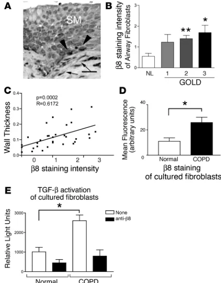
Increased β8 expression in small airway fibroblasts correlates with increasing severity of COPD and airway wall thickening. We used immunohistochemistry to evaluate the expression of β8 in the fibroblasts surrounding the small airways in a cohort of normal and COPD patients. We found that the β8 antibody strongly stained airway basal cells, as previously reported (44), as well as the subepithelial airway fibroblasts (Figure 9A). The intensity of
β8 staining correlated significantly with increasing severity of disease, as classified by GOLD (Figure 9B), and with increasing airway wall thickness (Figure 9C). We next evaluated lung fibroblasts harvested from normal and COPD patients for β 8 expres-sion. Lung fibroblasts harvested from COPD patients expressed significantly more β8 and activated more TGF-β than fibroblasts from normal patients (Figure 9, D and E).
Discussion
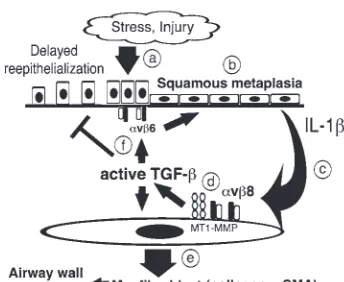
Association of SM with COPD was described nearly 30 years ago (7). How-ever, a mechanistic connection between SM and COPD may have been overlooked due to the long-held assumption that SM functions solely as a benign protective cellular reaction to injury. We have used recent advances in genome-wide analy-sis and gene silencing to parse out the complex role that SM plays in amplifying pathologic interactions between airway epithelium and fibroblasts in the evolu-tion of airway remodeling in COPD. We have provided evidence of bidirectional paracrine signaling, mediated by the integrins αvβ6 and αvβ8, effecting both
epithelial and mesenchymal differentia-tion in the adult airway (Figure 10). As such, amplification of integrin-mediated activation of TGF-β, by way of SM, may play a key role in derangement of mor-phoregulatory molecules and subsequent spatiotemporal interactions required to maintain homeostasis of the epithelial-mesenchymal trophic unit.
The SM that we observe in P3 human bronchial epithelial cells shares many similarities with SM in COPD. Thus, expression of IL-1β, basal, and suprabasal squamous epithelial markers, integrin subunits β6 and β8, and proliferation markers from our in vitro model mirror the immunohistochemical staining data of SM in human airways. These findings are consistent with the work of others sug-gesting that SM occurs through the increased recruitment of basal cells into an amplifying population of committed squamous lin-eage cells (29). This amplifying population occupies the suprabasal layer and expresses involucrin, keratin 6B, and keratin 14, which are known markers of actively proliferating suprabasal cells in normal squamous mucosa, SM either in the lung or other mucosal sites, and the epidermis at the edge of wounds (29, 30, 45–47).
[image:8.585.43.371.81.395.2]During in vitro SM, the most highly induced gene, from an unbi-ased genome-wide analysis approach, was the proinflammatory gene IL-1β. IL-1β has been shown to be expressed in the skin in vivo and in keratinocytes in 2D culture (48, 49), where it may rep-resent epidermal “activation” in response to epithelial injury and
Figure 6
Increased αvβ8-mediated activation of TGF-β in airway fibroblasts is mediated through increased
link innate immune responses to subsequent adaptive immune responses, ultimately promoting wound repair (50). Thus, the induction of IL-1β that occurs during SM of the airway epithelium may act as both an autocrine factor to influence gene expression to facilitate cell migration and wound closure and as a paracrine fac-tor to recruit inflammatory cells and to induce matrix production and contractility of adjacent fibroblasts (51, 52). The basal, supra-basal, and superficial cells of SM in COPD all express increased IL-1β, suggesting that both increased luminal and basal secretion of IL-1β are provided by the squamous metaplastic epithelium. This increased IL-1β secreted by SM may be an important addi-tional source of IL-1β, complementing the pool of IL-1β produced by immune effector cells.
We find that IL-1β secreted by metaplastic squamous airway epithelial cells promotes collagen expression by airway fibroblasts through increased fibroblast expression of αvβ8 and a subsequent
increase in TGF-β activation. In contrast, in a study utilizing a
coculture model of keratinocytes and dermal fibroblasts, myofi-broblast differentiation was TGF-β depen-dent but was antagonized by IL-1β (52). The discrepancy between these 2 studies likely rep-resents inherent cell type–specific differences between airway and dermal fibroblasts. For instance, we have found that unlike human airway fibroblasts, human dermal fibroblasts do not express αvβ8 (53). However, it is likely
that there are other differences between air-way epithelial cells and keratinocytes in the cofactors that they secrete that can modify fibroblast responses to IL-1β.
We, like others, have found that SM is an inevitable consequence of 2D but not air- liquid interface culture, indicating that cau-tion must be exercised when interpreting the results of many of the studies that have been published using human bronchial epithe-lial cells in 2D culture, as they may represent squamous metaplastic cells bearing little sim-ilarity to normal airway epithelium. On the other hand, 2D human bronchial epithelial cell culture provides an excellent model of the important in vivo phenomenon of SM.
We have created a simplified coculture model of the epithelial-mesenchymal tro-phic unit to study the reciprocal interactions between airway epithelial cells and the adja-cent fibroblasts. In vivo, these cell types are separated by a basement membrane, where movement of proteins may be physically impaired by pore size or interactions with matrix components. Thus, our model, which does not provide an intact basement mem-brane, may overestimate the importance of paracrine secretion of IL-1β and TGF-β in vivo. However, IL-1β and TGF-β are compact proteins that appear to move freely through basement membranes (54, 55), which increas- es the likelihood that the paracrine interac-tions that we observe in our coculture model are physiologically relevant.
In the epithelial-mesenchymal trophic unit, we propose an impor-tant role for integrin-mediated TGF-β activation in driving SM. However, blocking integrin-mediated TGF-β activation only delays and does not completely prevent squamous differentiation in our systems. This is expected since integrin-mediated TGF-β activation only accounts for approximately 50% of the total TGF-β activation by airway epithelial cells. This partial effect could be due to anti- body efficacy or other integrin-independent mechanisms of activat-ing TGF-β , such as matrix metalloproteases-2 and -9 (56), throm-bospondin-1 (19), or plasmin (57). Finally, we have not defined the mechanism that initiates integrin-mediated TGF-β activation by airway epithelial cells. We speculate that since normal airway epi-thelial cells express relatively high levels of αvβ8 and low levels of
the αvβ6 integrin, some environmental stimulus may activate the
pathways involved in airway epithelial αvβ8–mediated activation
of TGF-β, thus increasing TGF-β activation, which would increase
β6 expression and initiate SM. Our previous data support this
hypothesis. For instance, we have found that P0 fetal tracheal epi-Figure 7
αvβ6-mediated TGF-β activation forms a self-amplifying loop of increasing TGF-β activation
in human bronchial epithelial cells, which drives SM. Normal human bronchial epithelial cells in 2D culture (n = 3) were assessed during serial passage (P0–P3) for integrin αvβ6 and αvβ8
[image:9.585.44.357.85.330.2]thelial cells express αvβ8 but do not efficiently support αvβ8
-medi-ated activation of TGF-β, which correlates with low expression of MT1-MMP (10). When fetal tracheal epithelial cells are stimulated with phorbol esters, MT1-MMP activity increases coincident with increasing αvβ8-mediated activation of TGF-β (10). Therefore, we
speculate that environmental stimuli, such as tobacco smoke, may also increase αvβ8-mediated activation of TGF-β through a similar
mechanism and thus initiate TGF-β–dependent SM.
Methods
Cell culture, antibodies, and reagents. Adult airways were collected from first through fourth order bronchi from lobectomy specimens from resections performed for primary lung cancer or from normal lungs not used for transplantation. Informed consent was obtained from all surgical par- ticipants as part of an approved ongoing research protocol by the Univer-sity of California San Francisco Committee on Human Research, in full accordance with the declaration of Helsinki principles. Normal human bronchial epithelial cells were isolated as previously described (58). Freshly isolated adult human bronchial epithelial cells were plated onto rat-tail Col I–coated (10 μg/ml) dishes and incubated overnight; then the medium was changed to bronchial epithelial growth medium (BEGM) (Clonetics). Human bronchial epithelial cells were serially passaged to P6. For air-liquid interface culture, P0 airway epithelial cells were seeded onto human placen-tal collagen–coated filters (Sigma-Aldrich; Costar, Corning), as previously described using 2% Ultraser G–containing (Invitrogen) medium (58). Cells were cultured until confluent (as indicated by the ability to resist fluid movement through the epithelial layer), and then the medium on top of the filter was removed. To induce SM of air-liquid interface cultures, the medium was changed to BEGM (Clonetics), and the cultures were harvested at 3 and 21 days after exchange of the medium. Airway fibroblasts were cultured from the airway tissues remaining after epithelial cell isolation by the explant technique (58) and used P1 to P4. The TGF-β reporter cell line, TMLC (gift of John Munger and Dan Rifkin, New York University, New York, New York, USA) were cultured as previously described (59).
[image:10.585.48.285.82.211.2]Antibodies used were mouse anti-β 8 (clones 14E5 and 37e1; ref. 20), rab-bit anti-β8 (G17, Santa Cruz Biotechnology Inc.), anti-β3 (clone LM609, Chemicon), anti-β5 (clone P1F6, Chemicon), anti-β6 (10D5, ref. 60; B64B5, ref. 61), and mouse anti-involucrin, anti–keratin 6, anti–keratin 14, p63 (LabVision), rabbit anti–Ki-67 (Cell Marque Corp.), mouse anti–Col I (Southern Biotech), mouse anti-αSMA (Sigma-Aldrich), goat anti–IL-1β Figure 8
Fibroblast αvβ8-mediated activation of TGF-β contributes to β6
[image:10.585.304.531.447.738.2]expres-sion by adjacent human bronchial epithelial cells through a paracrine mechanism. (A) P0 human bronchial epithelial cells were cultured on filter inserts and were cultured alone (monoculture) or cocultured with control siRNA- or β8 siRNA–transfected airway fibroblasts in the bot-tom chamber treated with control antibody (control Ab) or neutralizing anti–TGF-β for 48 hours. Total human bronchial epithelial cell RNA was harvested, and β6 expression was determined by RT-PCR using primers to β6 or β-actin, as a control. Shown is a representative experi-ment of 3 showing similar results. (B) P0 human bronchial epithelial cells grown on filter inserts were similarly cocultured alone or with control siRNA (white bar) or β8 siRNA (black bar) transfected airway fibroblasts for 48 hours (n = 5). β6 surface expression was determined by flow cytometry using anti-β6. Shown is increase in surface expres-sion relative to human bronchial epithelial cells in monoculture. Shown is SEM. *P < 0.05.
Figure 9
and blocking peptide (Santa Cruz Biotechnology Inc.), biotinylated affin-ity purified goat anti–IL-1β (R&D Systems), anti–pan–TGF-β, and mouse anti-HLA class I (1D11 and W6/32, ATCC). Recombinant active TGF-β1, IL-1β (R&D Systems), IL-1RA (Affinity BioReagents), the pan-metallopro- tease inhibitor GM6001 (Ryss Lab), and Vitrogen (Cohesion) were pur-chased. Rat-tail collagen was isolated as previously described (62).
Plasmids, siRNA, and transfection. The β8, MT1-MMP, and control vectors were previously described (20, 63). The MT1-MMP, β 8, and negative con-trol siRNAs were purchased (Ambion Inc.), and transfection of primary cultured cells were performed using the Amaxa Nucleofector (Amaxa Bio-systems), using matched optimized transfection kits for airway epithelial cells and fibroblasts.
Immunocytochemistry, immunohistochemistry, and flow cytometry . Immuno- cytochemistry, immunohistochemistry, cell cycle analysis and flow cytom-etry were performed as previously described (44). Senescence-associated
β-gal (SA β-gal), a marker of cellular senescence (64), was performed using human bronchial epithelial cells (2 × 10⁵) grown on 12-well culture plate
according to the manufacturer’s instructions (β-galactosidase staining kit; BioVision Research Products).
Airway morphometry. Leftover human lung samples from pulmonary lobectomies or pneumonectomies performed at the San Francisco General Hospital or Moffit-Long Hospital, University of California San Francisco, for lung cancer, were gathered during the study period (from 2003 through 2006). Exclusion criteria included infection, prior radiation, or chemother-apy. Control lung tissues were obtained from donor lungs not utilized for lung transplantation, usually for reasons of size mismatching.
Microtome sections from 34 (22 COPD and 12 normal) lung samples were immunostained for involucrin or β 8. The slides were coded and blind-ed prior to pathologic assessment. Airway diameter was determined using a stage micrometer, and airways less than 2 mm in diameter were evalu-ated (mean 5.8 ± 5.3 airways/case). Each small airway was digitally imaged at 200× magnification (QCapture, v. 2.68.2). Digital images were assessed for β8 immunostaining based on a 0–3 scale, with 0 being absent staining; grade 1, 0–10%; grade 2, 11–30%; grade 3, >30% of airway cells or subepithe- lial fibroblasts with positive membrane and cytoplasmic staining. Involu-crin immunostaining was also graded on a 0–3 scale, with 0 being absent staining; grade 1, 0–10%; grade 2, 11–20%; grade 3, >20% cytoplasmic stain-ing of superficial “squamoid” cells. Measurements of wall thickness were performed using the method of Hogg (5), which expresses wall thickness as a function of area of the airway wall/basement membrane length using image analysis software (ImageJ, v. 1.36b).
After immunohistochemical assessment, the clinical records from each patient were retrieved and the degree of pulmonary disability (stages 1 to 4, with 4 being the worst) was determined using GOLD criteria. Preoperative
pulmonary function tests (PFTs) were available for 18 of 20 patients under-going surgery. The number of patients in each stage with PFTs available were as follows: stage 1, n = 3; stage 2, n = 11; stage 3, n = 4; and stage 4, n = 0.
RNA isolation, polymerase chain reaction, and microarray analysis . RNA isola-tion and RT-PCR were performed as previously described (20). Real-time PCR was performed using the SYBR green method, as described (65). Prim-er sequences are available in supplemental data (Supplemental Table 4). Microarray methodology is available in supplemental data.
Immunoprecipitation analysis, Western blotting, zymography, and TGF-β bioas-say. Immunoprecipitations and SDS-PAGE were performed as previously described (10, 63). Western blotting of conditioned medium of coculture system was performed as described with minor modification (10). The TGF-β bioassays and zymography to determine MT1-MMP activity were performed as previously described (10, 20).
Cytokine array and IL-1β ELISA. Airway epithelial cells (3 × 10⁶) were
cultured in 10-cm collagen-coated tissue culture plates for 12–16 hours, washed 2 times with PBS, then incubated in serum-free DMEM for 72 hours. Cytokines in conditioned medium were detected with Cartesian Array Human Cytokine Set 1 from BioSource International (Invitrogen) following the manufacturer’s instructions. IL-1β was measured in condi-tioned medium with an IL-1β Quantikine ELISA kit (R&D Systems).
Coculture model of the epithelial-mesenchymal trophic unit. Airway epithelial cells (2 × 10⁵/well) were seeded on vitrogen-coated (10 μ
g/ml coating con-centration) culture inserts (1 mm pore size; BD Biosciences) in a 24-well plate in BEGM (Clonetics) and cultured until confluent. Airway fibro-blasts transfected with control or β8 siRNA (2.5 μg/transfection) were cultured for 12–16 hours on 24-well (2 × 10⁵/well) tissue-coated plate
(BD Biosciences) in fibroblast growth medium. For the coculture experi-ments, the culture inserts containing the airway epithelial cells were placed into wells containing airway fibroblasts, and medium in both the upper and lower chambers were changed to DMEM without FCS in the presence or absence of control antibody, anti–TGF-β antibody (1D11), anti–β6 integrin antibody (10D5), anti–β8 integrin antibody (37E1),
or IL-1RA. After 48 or 72 hours coculture, integrin expression levels in airway epithelial cells were analyzed by FACS and RT-PCR. Col I gene expression in fibroblasts was evaluated by RT-PCR or by Western blotting of conditioned medium.
[image:11.585.52.224.85.226.2]Statistics. Stepwise regression analysis with correction for clustering was used to compare β8 staining intensity to airway wall thickness. Student’s t test was used for comparison of 2 data sets, analysis of variance for multi- ple data sets. Tukey’s or Dunn’s test were used for parametric and nonpara-metric data, respectively, to find where the difference lay. Significance was defined as P < 0.05. Statistical software used was Prism v.3cx (GraphPad Software Inc.) and Stata v.9 (StataCorp LP).
Figure 10
Hypothetical model of SM in the pathogenesis of airway wall thickening in COPD. (a) The normal airway epithelium when exposed to noxious stimuli responds by increas-ing TGF-β production (12) and increasing TGF-β activation, which increases expres-sion of the β6 integrin, a TGF-β responsive gene (31) contributing to (b) a phenotypic switch to SM, a TGF-β driven process (11). (c) Squamous metaplastic epithelial cells secrete increased IL-1β, which acts a paracrine factor with adjacent airway fibro-blasts. (d) Airway fibroblasts respond to IL-1β by increasing β8 expression and αvβ8
-mediated TGF-β activation. Increased TGF-β activation by airway fibroblasts causes (e) autocrine effects on the fibrogenic fibroblast phenotype by increasing Col I and