in Patients with Active Tuberculosis
Nancy D. Marín,aSara C. París,aMauricio Rojas,a,band Luis F. Garcíaa
Grupo de Inmunología Celular e Inmunogenética, Instituto de Investigaciones Médicas, Universidad de Antioquia, Medellín, Colombia,aand Unidad de Citometría de Flujo, Sede de Investigación Universitaria, Universidad de Antioquia, Medellín, Colombiab
Phenotypic and functional alterations inMycobacterium tuberculosisT cell subsets have been reported in patients with active tuberculosis. A better understanding of these alterations will increase the knowledge about immunopathogenesis and also may contribute to the development of new diagnostics and prophylactic strategies. Here, theex vivophenotype of CD4ⴙand CD8ⴙT cells and the frequency and phenotype of gamma interferon (IFN-␥)- and interleukin 17 (IL-17)-producing cells elicited in short-term and long-short-term cultures following CFP-10 and purified protein derivative (PPD) stimulation were deshort-termined in nonin-fected persons (non-TBi), latently innonin-fected persons (LTBi), and patients with active tuberculosis (ATB). Phenotypic characteriza-tion of T cells was done based on the expression of CD45RO and CD27. Results show that ATB had a reduced frequency of circulating CD4ⴙCD45ROⴙCD27ⴙT cells and an increased frequency of CD4ⴙCD45ROⴚCD27ⴙT cells. ATB also had a higher frequency of circulating IL-17-producing CD4ⴙT cells than did LTBi after PPD stimulation, whereas LTBi had more IFN-␥-producing CD4ⴙT cells than did non-TBi. The phenotype of IFN-␥-producing cells at 24 h differs from the phenotype of IL-17-producing cells with no differences between LTBi and ATB. At 144 h, IFN-␥- and IL-17-producing cells were mainly
CD45ROⴙCD27ⴙT cells and they were more frequent in ATB. These results suggest thatM. tuberculosisinfection induces alter-ations in T cells which interfere with an adequate specific immune response.
T
uberculosis (TB) remains a major public health problem andone of the most important infectious diseases worldwide, causing 8.8 million incident cases and 1.3 million deaths in 2010
(76). Approximately one-third of the world population is
consid-ered to be latently infected withMycobacterium tuberculosis;
al-though most of them contain the infection, remaining asymptom-atic for decades or even throughout their entire life span, approximately 10% of these infected persons will develop active
disease (72).
Successful host defense againstM. tuberculosisrequires an
ef-fective coordination of the innate and adaptive immune re-sponses. The adaptive immune response is characterized by the expansion and differentiation of several T effector cells (e.g., Th1, Th17, Th2, and regulatory T cells [Tregs]) and the generation of long-lived memory T cells with an enhanced capacity to respond
upon reexposure to mycobacteria (12,59). Th1 responses play an
important role in the defense againstM. tuberculosisby inducing
macrophage microbicidal mechanisms and thereby restricting
mycobacterial growth (30,39). Impairment of Th1 responses by
genetic mutations in the gamma interferon (IFN-␥)/interleukin
12 (IL-12) axis (48), anti-tumor necrosis factor alpha
(anti-TNF-␣) treatment for autoimmune diseases (34), and HIV
infec-tion (22) increases the risk of developing active TB.M. tuberculosis
can also induce Th17 responses, and although their role is less well
understood, Th17 cells are also considered important inM.
tuber-culosisresistance by participating in granuloma formation (49,73)
and by increasing the recruitment of IFN-␥-producing cells into
the lungs (35). However, due to their proinflammatory role and
their capacity to induce recruitment of granulocytes into infected tissues, Th17 cells may also play a deleterious role in the necrosis of
granulomas, resulting in the reactivation of latent infections (13).
Thus, since IFN-␥and IL-17 may have opposite roles duringM.
tuberculosisinfection, a balance between Th1 and Th17 responses
is essential to promote an effective anti-M. tuberculosisimmune
response and prevent tissue damage.
Although many studies have evaluated the contributions of
Th1 and Th17 cells to the immune response againstM. tuberculosis
infection (20,49,52), it is crucial to further understand their role
during different stages ofM. tuberculosisinfection, as well as their
phenotypic and functional characteristics after exposure to myco-bacterial antigens.
The generation of immunological memory is the hallmark of
the adaptive immune response (59). CD4⫹and CD8⫹memory T
cell subsets can be distinguished by the expression of different phenotypic markers, homing properties, and functional capacities
(7,51). Memory T cells are classically identified by the expression
of CD45RO, the low-molecular-weight isoform of CD45 which, along with other surface markers, defines different stages of
dif-ferentiation of CD4⫹and CD8⫹T cells. CD27 is a costimulatory
molecule belonging to the TNF family expressed in all CD4⫹naïve
cells and in more than 80% of CD4⫹memory cells (15,24). A loss
of CD27 expression has been proposed to be characteristic of
in-duced effector CD4⫹and CD8⫹T cells (21,56). CD27-negative
cells are suggested to be a more differentiated subset with a higher capacity to secrete cytokines and a stronger recall response than
those of the CD27⫹population (61).
TB is a chronic, infectious disease, and prolonged exposure to mycobacterial antigens affects the biology of antigen-specific T
Received21 June 2012 Returned for modification23 July 2012
Accepted13 August 2012
Published ahead of print22 August 2012
Address correspondence to Luis F. García, [email protected]. Copyright © 2012, American Society for Microbiology. All Rights Reserved. doi:10.1128/CVI.00390-12
on August 17, 2020 by guest
http://cvi.asm.org/
cells (71), including the generation of memory T cells. Most of the
studies focusing on T cell memory profiles in TB (27, 33, 75)
analyze the phenotypes of circulating T cellsex vivoor after
short-termin vitrostimulation. These studies show that the responding
cells are mainly T effector memory (TEM) or T effector (TEF) cells
(8,46,75). However, there are not many studies addressing this
issue in long-term cultures, where central memory T (TCM) cells
should be the main responders (6,25,41), and even fewer studies
comparing the phenotypes of antigen-specific T cells responding toin vitrostimulation with mycobacterial antigens in both short-and long-term cultures. Phenotypic characterization of Th1 short-and
Th17 cells has shown that IFN-␥and IL-17 are produced by T cell
subsets with different phenotypes following short-termin vitro
stimulation withMycobacterium bovisBCG (62) or mycobacterial
antigens (52). In these studies, IFN-␥-producing cells displayed an
effector or effector memory phenotype, while IL-17-producing
cells were mainly central memory cells, suggesting that IFN-␥
-and IL-17-producing cells are long-lived memory cells actively
involved in the immune response toM. tuberculosisinfection.
Since in active tuberculosis, antigen-specific T cells are exposed to persistent antigenic stimulation, it is possible to suggest that the
profile, magnitude, and phenotype of antigen-specific CD4⫹and
CD8⫹T cells producing IFN-␥or IL-17 should be differentially
affected in latently infected and active-TB individuals. For these
reasons, the frequencies ofex vivonaïve, effector, and memory
CD4⫹and CD8⫹T cells, as well as the frequencies and phenotypes
of IFN-␥- and IL-17-producing cells elicited after stimulation
with specific mycobacterial antigens, were determined in latently infected individuals (LTBi) and patients with active TB (ATB). The results show that ATB had a reduced frequency of circulating
TCMCD4⫹cells (CD45RO⫹CD27⫹) and an increased frequency
of Tearly/naïvecells (CD45RO⫺CD27⫹). ATB patients had a higher
frequency of IL-17-producing CD4⫹T cells afterin vitro
stimula-tion with purified protein derivative (PPD), while LTBi had a
higher frequency of IFN-␥-producing CD4⫹T cells after CFP-10
and PPD stimulation. Despite the reduced frequency of
circulat-ing CD4⫹TCMcells in ATB, they were the highest producers of
IFN-␥and IL-17 after long-term stimulation with CFP-10. These
results suggest thatM. tuberculosisinfection alters thein vivo
gen-eration ofM. tuberculosis-specific memory T cells and causes a
shift in the protective Th1 profile toward a Th17 response: these
events are related to a reduced protective response againstM.
tuberculosisand also with an exacerbated inflammatory response which may favor the reactivation of active disease in latently in-fected individuals.
MATERIALS AND METHODS
Study population.Forty-three newly diagnosed patients with active TB (ATB) (9 females and 34 males, aged 38⫾14 years old) were recruited at the Tuberculosis Control Program in Medellín (Colombia) and its met-ropolitan area. ATB patients had positive sputum smears for acid-fast bacillus (AFB; 1⫹, 12 patients; 2⫹, 14 patients; 3⫹, 12 patients; no data, 5 patients) and were studied before or within the first 2 weeks of anti-TB treatment. Four ATB patients were also studied after they finished their 6-month anti-TB therapy, and all of them were cured as indicated by a negative AFB smear and a recovery from the clinical symptoms once they finished therapy. Thirty-five healthy subjects with latent TB infection (LTBi) (24 females and 11 males, aged 31⫾16 years old) were selected according to an IFN-␥-positive response to CFP-10, as evaluated by en-zyme-linked immunosorbent assay (ELISA) in 7-day whole-blood culture supernatants, as previously reported by our group (16). Fifteen
individu-als with a negative response to CFP-10 were included as a noninfected control group (non-TBi) (10 females and 5 males, aged 35⫾14 years old). Individuals from non-TBi and LTBi groups were healthy without clinical evidence of active tuberculosis. Individuals infected with HIV, using im-munosuppressive drugs, with diabetes, or younger than 15 years old were excluded. The study was approved by the Ethics Committee of the Insti-tuto de Investigaciones Médicas of the Universidad de Antioquia, and written informed consent was obtained from all participants.
Antigens.Recombinant ESAT-6 and CFP-10 were provided by the Department of Microbiology and Immunology of Colorado State Univer-sity (Fort Collins, CO) through the Tuberculosis Vaccine Testing and Research Material contract no. HHSN26266400091C (NIH, NIAID, N01-AI-40091). PPD (RT50) was obtained from the State Serum Institute (Co-penhagen, Denmark). IrradiatedM. tuberculosiswas obtained from BEI Resources (Manassas, VA).
Sample preparation.Twenty milliliters of peripheral blood was ob-tained using heparin as anticoagulant, and the peripheral blood mononu-clear cells (PBMC) were obtained by Ficoll-Hypaque density gradient centrifugation (BioWhittaker, Walkersville, MD). Viability, as tested by trypan blue exclusion, was alwaysⱖ95%.
Cell phenotyping, cell cultures, and intracellular cytokine analysis. The phenotype of CD4⫹and CD8⫹T cell subsets was evaluated by the surface expression of CD45RO and CD27 on fresh whole blood and cul-tured cells (19,22,38). For theex vivoanalysis, 100l of whole blood was lysed with Optilyse (Beckman Coulter) according to the manufacturer’s instructions and blocking buffer (2% pooled human serum [BioWhit-taker] plus 0.01% NaN3in phosphate-buffered saline [PBS]) was added
for 15 min at 4°C with light protection. The antibody panel forex vivo analysis included anti-CD4-peridinin chlorophyll protein (PerCP)-Cy5 (clone OKT4), anti-CD8-Pacific blue (clone OKT8) (eBioscience, San Diego, CA), anti-CD27-fluorescein isothiocyanate (FITC) (clone 0323), and anti-CD45RO-allophycocyanin (APC) (clone UCHL-1) (Biolegend, San Diego, CA). In some experiments, cells were stained with 7-amino-actinomycin D (7-AAD) along with CD27 and CD45RO in order to eval-uate the viability of T cells in long-term cultures and less than 1% of CD4⫹ and CD8⫹T cells were 7-AAD positive. Stained cells were fixed with 2% paraformaldehyde (J. T. Baker, Phillipsburg, NJ), and 1⫻105cells were
acquired in a fluorescence-activated cell sorting (FACS) Canto II flow cytometer (BD Bioscience, San Diego, CA).
For the 24-h whole-blood cultures (short term), heparinized blood was diluted 1:10 in 1 ml of RPMI 1640 (Invitrogen, Carlsbad, CA) supple-mented with penicillin-streptomycin (BioWhittaker, Walkersville, MD) plus 1g/ml of anti-CD28/anti-CD49d (BD Bioscience). For 144-h cul-tures (long term), PBMC were plated at 1⫻105cells/well in triplicate in
96-well round-bottomed plates in complete culture medium (RPMI 1640 plus 10% pooled human serum [BioWhittaker] and penicillin-streptomy-cin). Both 24-h and 144-h cultures were stimulated with CFP-10 (5g/ ml), PPD (10g/ml), orM. tuberculosis(10g/ml), and nonstimulated cultures were used as negative controls. Four hours before the end of incubation, supernatants were collected and brefeldin A (10g/ml) was added to the cultures. At the end of the incubation, red cells from the whole-blood cultures were lysed with Optilyse (Beckman Coulter). Cells from 24 h and 144 h were stained for surface markers and fixed as de-scribed forex vivoassays. Fixed cells were permeabilized followed by in-cubation with anti-IFN-␥–phycoerythrin (PE)–Cy7 (clone B27) (Bioleg-end) and anti-IL-17–PE (clone eBios64CAP17) (eBioscience). One hundred thousand cells were acquired in a FACS Canto II flow cytometer and analyzed using BD FACSDiva software, v6.1.2 (BD Bioscience, San Jose, CA), or FlowJo software, v7.6.1 (Tree Star Inc., Ashland, OR). Data analysis was performed by selecting singlets in a forward scatter A (A)/H dot plot, followed by the selection of lymphocytes in an FSC-A/side scatter A (SSC-A) dot plot, and then CD4 or CD8 was plotted versus SSC. To identify the phenotype of cytokine-producing cells, IFN-␥- and IL-17-producing gated CD4⫹and CD8⫹T cells were evalu-ated for the expression of CD27 and CD45RO (seeFig. 3AandB). Marín et al.
on August 17, 2020 by guest
http://cvi.asm.org/
Statistical analysis.Statistical analysis was performed using Graph-Pad Prism 5 (GraphGraph-Pad Software, Inc., La Jolla, CA). Comparisons be-tween three or more groups were done by the Kruskal-Wallis test with Dunn’s multiple comparison test. A two-way analysis of variance (ANOVA) test with Bonferroni’s posttest was used to compare means of the T cell subsets among the studied groups. Pearson’s correlation coeffi-cient was done and is specified in each case in the figure legend. APvalue of⬍0.05 was considered statistically significant.
RESULTS
Memory phenotypes of CD4ⴙand CD8ⴙcells.In order to deter-mine the combination of surface markers that allows well-defined
T cell subsets, CD4⫹and CD8⫹T cells from fresh blood and
PBMC stimulated for 24 h with PPD were analyzed for the sion of CCR7, CD62L, CD27, and CD45RO. Based on the
expres-sion pattern of these surface molecules, CD27 and CD45RO were
selected to identify CD4⫹ and CD8⫹ T cell subsets (data not
shown). Thus, the combined use of CD45RO along with CD27
defined four subsets of CD4⫹ and CD8⫹ T cells: CD45RO⫹
CD27⫹, CD45RO⫹CD27⫺, CD45RO⫺CD27⫹, and CD45RO⫺
CD27⫺, which, according to previous reports by other authors,
would correspond to TCM, TEM, Tearly/naïve, and TEFcells,
respec-tively (5,19,38,69). Representative dot plots of theex vivo
expres-sion of CD45RO and CD27 on CD4⫹and CD8⫹T cells are shown
inFig. 1AandB.
Ex vivoevaluation of CD27 and CD45RO expression in
non-TBi, Lnon-TBi, and ATB showed that CD4⫹and CD8⫹T cells have
different profiles regarding the proportion of cells expressing
CD45RO and CD27. There was a very low frequency of CD45RO⫺
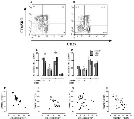
FIG 1Ex vivophenotypes of CD4⫹and CD8⫹T cells. The phenotypes of CD4⫹and CD8⫹T cells from non-TBi, LTBi, and ATB were evaluated according to the expression of CD45RO and CD27 as described in Materials and Methods. A representative example of the expression of CD45RO and CD27 on CD4 (A) and CD8 (B) T cells from one LTBi is shown. Frequencies of CD4⫹(C) and CD8⫹(D) T cells from non-TBi (n⫽12) (white bars), LTBi (n⫽11) (gray bars), and ATB (n⫽24) (black bars). Two-way ANOVA test with Bonferroni’s posttest: *,Pⱕ0.05; ***,Pⱕ0.001. Pearson’s correlation coefficient between the percentages of CD4⫹CD45RO⫹CD27⫹and CD4⫹CD45RO⫺CD27⫹T cells from ATB (r⫽ ⫺0.7317,P⫽0.0001) (E) and non-TBi (white circles) (Pearsonr⫽ ⫺0.7417;P⫽0.0058) and LTBi (black circles) (Pearsonr⫽ ⫺0.2190;P⫽0.5176) (F). Correlations between CD8⫹CD45RO⫹ CD27⫹and CD8⫹CD45RO⫺CD27⫹T cells in ATB (Pearsonr⫽ ⫺0.0844;P⫽0.6947) (G) and non-TBi (Pearsonr⫽ ⫺0.3545;P⫽0.2581) and LTBi (Pearsonr⫽ ⫺0.7951;P⫽0.0034) (H).
on August 17, 2020 by guest
http://cvi.asm.org/
CD27⫺cells among CD4⫹T cells compared to the CD8⫹T cells (Fig. 1CandD), irrespective of the group studied. Among CD4⫹T cells, the subsets expressing CD27 were more frequent than the subsets lacking CD27 expression, regardless of the expression of
CD45RO (Fig. 1C), while among CD8⫹T cells, there was a higher
proportion of cells lacking the CD45RO expression, regardless of
the expression of CD27 (Fig. 1D). Comparative analysis of these
subsets among non-TBi, LTBi, and ATB showed that ATB had a
lower frequency of CD4⫹CD45RO⫹CD27⫹T cells (Pⱕ0.001) than
did LTBi and also a higher frequency of CD4⫹CD45RO⫺CD27⫹T
cells than those for non-TBi (Pⱕ0.05) and LTBi (Pⱕ0.001), but no
differences were observed between non-TBi and LTBi (Fig. 1C).
Re-garding CD8⫹T cells, there were no differences in the percentages of
these subsets among the studied groups (Fig. 1D).
To evaluate whether the reduced frequency of CD4⫹
CD45RO⫹CD27⫹T cells observed in ATB is associated with
changes in the other T cell subsets, correlation analyses were done.
In ATB, the frequency of CD4⫹CD45RO⫹CD27⫹memory T
cells correlated negatively with the frequency of CD4⫹CD45RO⫺
CD27⫹T cells (Pearsonr⫽ ⫺0.7317;P⫽0.0001) (Fig. 1E). A
similar pattern was also found in non-TBi (Pearsonr⫽ ⫺0.7417;
P⫽0.0058) but not in LTBi (Pearsonr⫽ ⫺0.2190;P⫽0.5176)
(Fig. 1F). Regarding CD8⫹ T cells, the frequency of CD8⫹
CD45RO⫹CD27⫹T cells negatively correlated with the frequency
of CD8⫹CD45RO⫺CD27⫹T cells in LTBi (Pearsonr⫽ ⫺0.7951;
P⫽0.0034) but not in non-TBi (Fig. 1H) (Pearsonr⫽ ⫺0.3545;
P⫽0.2581) nor in ATB (Pearsonr⫽ ⫺0.0844;P⫽0.6947) (Fig.
1G). These results show that active TB causes a reduction in the
percentage of circulating TCMcells, while there is an increase of
circulating Tearly/naïvecells, which may occur as a compensatory
mechanism to balance the proportion of circulating T cell subsets.
Evaluation of IFN-␥- and IL-17-producing cells in 24-h and 144-h cultures from non-TBi, LTBi, and ATB individuals.Th1
and Th17 seem to have opposite roles during active TB (71); thus,
low Th1 and high Th17 responses have been previously reported in patients with active TB in long-term and short-term cultures,
respectively (57,74). Therefore, the frequency of IFN-␥- and
IL-17-producing CD4⫹and CD8⫹T cells in response to
mycobacte-rial antigens was determined in 24-h whole-blood and 144-h PBMC cultures stimulated or not with CFP-10 and PPD. There
was a negligible percentage of IFN-␥- and IL-17-producing CD4⫹
and CD8⫹T cells in both 24-h and 144-h cultures from non-TBi.
At 24 h, there were not significant differences in the frequencies of
IFN-␥- and IL-17-producing CD4⫹T cells among non-TBi, LTBi,
and ATB in response to CFP-10 or PPD (Fig. 2AandB). At 144 h,
LTBi had a higher frequency of CD4⫹IFN-␥⫹T cells in response to
CFP-10 and PPD than did non-TBi (Pⱕ0.01) (Fig. 2E). No
differ-ences were observed between ATB and non-TBi or LTBi regardless of
the antigen used (Fig. 2E). With respect to IL-17-producing cells, at
144 h ATB had a higher frequency of CD4⫹IL-17⫹T cells than did
LTBi in response to PPD (Pⱕ0.05) (Fig. 2F). There were no
differ-ences between non-TBi and LTBi groups in the percentages of
IL-17-producing cells at this time point. Regarding CD8⫹T cells, the
fre-quencies of IFN-␥- or IL-17-producing CD8⫹ T cells were not
different in response to CFP-10 and PPD among the studied groups (Fig. 2CandDand2GandH). These results suggest that evaluation of
IFN-␥and IL-17 responses in 24-h whole-blood cultures did not
discriminate infected from noninfected persons. However, in 144-h
PBMC cultures, the IFN-␥production elicited after CFP-10 and PPD
stimulation discriminated infected from noninfected individuals, while the high production of IL-17 was a characteristic of ATB pa-tients.
FIG 2Frequencies of IFN-␥- and IL-17-producing CD4⫹and CD8⫹T cells in non-TBi, LTBi, and ATB individuals. CD4⫹(A, B, E, and F) and CD8⫹(C, D, G, and H) T cells from 24-h whole blood (A to D) and 144-h PBMC (E to H) cultures stimulated with CFP-10 and PPD were intracellularly stained with anti-IFN-␥–PE–Cy7 (A, C, E, and G) and anti-IL-17–PE (B, D, F, and H). The frequencies of IFN-␥- and IL-17-producing CD4⫹and CD8⫹T cells were evaluated in non-TBi (white circles), LTBi (gray circles), and ATB (black circles). Kruskal-Wallis test with Dunn’s multiple comparison test: *,Pⱕ0.05; **,Pⱕ0.01.
Marín et al.
on August 17, 2020 by guest
http://cvi.asm.org/
Memory phenotypes of IFN-␥- and IL-17-producing cells in short- and long-term cultures from non-TBi, LTBi, and ATB individuals.Studies addressing the contributions of different
sub-sets of effector and memory CD4⫹and CD8⫹T cells in ATB and
LTBi to the recall response against mycobacterial antigens have been done, and differences in the memory phenotypes of
respond-ing cells have been reported (23,46,62). Therefore, considering
the phenotypic and functional features of the effectors and mem-ory T cells established above, we speculated that the phenotype of
IFN-␥- and IL-17-producing cells should be different in
short-and long-term cultures from LTBi short-and ATB. Thus, phenotypic
characterization of IFN-␥- and IL-17-producing CD4⫹ and
CD8⫹T cells was done by evaluating the expression of CD45RO
and CD27 in cells from 24-h and 144-h cultures stimulated with CFP-10 and PPD. One representative example of the gating
strat-egy for the phenotypic and functional evaluation of CD4⫹T cells
stimulated for 24 h (Fig. 3A) or 144 h (Fig. 3B) with CFP-10 is
shown. In short-term cultures (24 h) stimulated with CFP-10 (Fig.
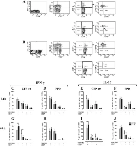
FIG 3Phenotypes of IFN-␥- and IL-17-producing CD4⫹T cells in non-TBi, LTBi, and ATB individuals. The phenotypes of IFN-␥- and IL-17-producing CD4⫹ T cells were evaluated based on the expression of CD45RO and CD27 as described in Materials and Methods. One representative example of the gating strategy used to evaluate the phenotypes of IFN-␥⫹and IL-17⫹CD4⫹T cells from whole-blood 24-h (A) and PBMC 144-h (B) cultures stimulated with CFP-10 from one ATB is shown. The phenotypes of IFN-␥(C, D, G, and H)- and IL-17 (E, F, I, and J)-producing CD4⫹T cells from 24-h whole-blood (C to F) and 144-h PBMC (G to J) cultures stimulated with CFP-10 (C, E, G, and I) and PPD (D, F, H, and J) were evaluated by the expression of CD27 and CD45RO. Gray bars, LTBi; black bars, ATB. Two-way ANOVA test with Bonferroni’s posttest: *,Pⱕ0.05; **,Pⱕ0.01.
on August 17, 2020 by guest
http://cvi.asm.org/
3C) or PPD (Fig. 3D), IFN-␥-producing CD4⫹T cells showed a
heterogeneous phenotype, although CD45RO⫹CD27⫹T cells
were the main producers, followed by CD45RO⫹CD27⫺and
CD45RO⫺ CD27⫹ T cells and a negligible percentage of
CD45RO⫺CD27⫺T cells. IL-17 was mainly produced by CD4⫹
CD27⫹T cells, irrespective of the CD45RO expression, with
ap-proximately 60% of the IL-17⫹cells exhibiting the CD45RO⫹
CD27⫹phenotype (Fig. 3EandF). At this time point, no
signifi-cant differences were observed between LTBi and ATB regarding
the frequency of each CD4⫹T cell subset producing IFN-␥or
IL-17 in response to CFP-10 and PPD (Fig. 3CtoF).
In long-term cultures (144 h), both CFP-10 and PPD induced
IFN-␥and IL-17 production mainly by CD4⫹CD45RO⫹CD27⫹
T cells, although CD4⫹CD45RO⫹CD27⫺also contributed to
IFN-␥production (Fig. 3GtoJ). In response to CFP-10, ATB had
a higher frequency of IFN-␥- and IL-17-producing CD4⫹
CD45RO⫹CD27⫹T cells compared to LTBi (Fig. 3GandI),
whereas ATB showed a lower frequency of IFN-␥-producing
CD4⫹CD45RO⫹CD27⫺T cells than did LTBi (Fig. 3G).
Regarding CD8⫹T cells (Fig. 4), in short-term cultures all the
cell subsets produced IFN-␥(Fig. 4AandB), while IL-17, similar
to CD4⫹T cells, was produced mainly by CD27⫹T cells, either
CD45RO⫹or CD45RO⫺(Fig. 4CandD). No differences were
observed in the phenotypes of IFN-␥- and IL-17-producing CD8⫹
T cells between LTBi and ATB in response to CFP-10 or PPD (Fig.
4AtoD). In long-term cultures, IFN-␥and IL-17 were mainly
produced by CD45RO⫹CD27⫹CD8⫹T cells, without statistically
significant differences between the studied groups after CFP-10 or
PPD stimulation (Fig. 4EtoH). These results show differences in
the phenotypes of IFN-␥and IL-17 after short-term stimulation
with mycobacterial antigens, since IFN-␥was basically produced
by CD45RO⫹T cells while IL-17 was mainly produced by CD27⫹
T cells. However, after long-termin vitrostimulation, both of
them were produced almost exclusively by central memory
CD45RO⫹CD27⫹T cells, with a higher frequency of this subset
producing IFN-␥and IL-17 in ATB after CFP-10 stimulation.
Recent evidence suggests that the profile and magnitude of the antimycobacterial immune response in ATB correlate with patho-gen load, and its reduction after completion of the anti-TB
ther-apy impacts the profile and magnitude of cytokine response (1,9,
45). However, the frequency ofex vivoCD4⫹and CD8⫹T cells
with different phenotypes did not show any relationship with AFB positivity in sputum smears in our TB patients (data not shown).
Nevertheless, there was a trend toward lower numbers of IFN-␥
-producing CD4⫹and CD8⫹T cells with increasing AFB smear
sputum positivity in 144-h cultures stimulated with CFP-10 (Fig.
5). Furthermore, theex vivophenotypes of CD4⫹and CD8⫹T
FIG 5Frequencies of IFN-␥-producing CD4⫹and CD8⫹T cells in ATB with different AFB smear values. PBMC from ATB were stimulated for 144 h with CFP-10 and then evaluated for the frequency of IFN-␥-producing cells as described in Materials and Methods. Frequencies of IFN-␥-producing CD4⫹ and CD8⫹T cells in patients with AFB smear values of 1⫹(white circles), 2⫹ (gray circles), and 3⫹(black circles). Kruskal-Wallis test with Dunn’s multiple comparison test.
FIG 4Phenotypes of IFN-␥- and IL-17-producing CD8⫹T cells in non-TBi, LTBi, and ATB individuals. The phenotypes of IFN-␥(A, B, E, and F)- and IL-17 (C, D, G, and H)-producing CD8⫹T cells from 24-h whole-blood (A to D) and 144-h PBMC (E to H) cultures after stimulation with CFP-10 (A, C, E, and G) and PPD (B, D, F, and H) were evaluated by the expression of CD27 and CD45RO. Gray bars, LTBi; black bars, ATB. Two-way ANOVA test with Bonferroni’s posttest.
Marín et al.
on August 17, 2020 by guest
http://cvi.asm.org/
cells and the frequencies of IFN-␥- and IL-17-producing CD4⫹
and CD8⫹T cells following stimulation with CFP-10 were
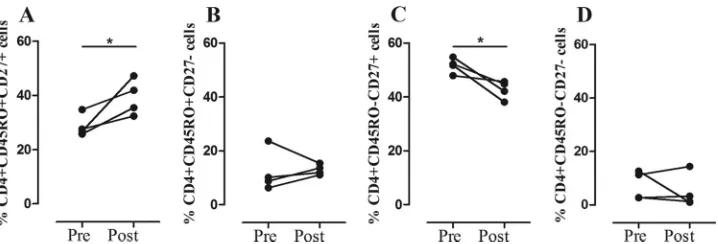
evalu-ated in four cured TB patients, and the results were compared with the pretreatment measurements. In all patients, there was an
in-crease in the frequency of circulating CD4⫹CD45RO⫹CD27⫹T
cells (Pⱕ0.05) (Fig. 6A) and a decrease in frequency of CD4⫹
CD45RO⫺CD27⫹T cells (Pⱕ0.05) (Fig. 6C) after the
comple-tion of their anti-TB therapy. No changes were observed in the
frequencies of CD4⫹ CD45RO⫹ CD27⫺ (Fig. 6B) and CD4⫹
CD45RO⫺CD27⫺cells (Fig. 6D) or in the frequencies of the four
CD8⫹T cell subsets (data not shown). Likewise, the frequencies of
IFN-␥- and IL-17-producing CD4⫹and CD8⫹T cells evaluated at
24 h and 144 h following stimulation with CFP-10 and PPD di-minished in ATB after anti-TB treatment, but the change did not reach statistical significance. These results show that the
func-tional response of CD4⫹or CD8⫹T cells seems to be affected by
the bacterial load and also that an effective anti-TB therapy re-stores the immunological components which were altered during the active disease, thus recovering the immune homeostasis.
DISCUSSION
This study shows alterations in the phenotypes of circulating
CD4⫹and CD8⫹T cells and the frequencies and phenotypes of
IFN-␥- and IL-17-producing CD4⫹and CD8⫹T cells in patients
with active tuberculosis. A special feature of our study is that short-term (24-h) whole-blood and long-term (144-h) PBMC cultures were compared. Because in short-term cultures there is not enough time for cellular division, their evaluation has been
considered anex vivomeasurement of the host’s immune status,
and cytokines produced in these cultures are considered to be
produced mainly by effector cells or effector memory T cells (25,
26,41). Conversely, long-term assays (5 to 7 days) allow the
eval-uation of antigen-specific memory T cells expanded after stimu-lation with the cognate antigen. In addition, long-term assays have been reported to be more sensitive and specific in detecting latent infection than short-term cultures, especially in areas where TB is
endemic (6,10,25).
Alterations in the frequencies of circulating memory T cells
have been reported in ATB (31,46,75). Most of these studies have
been carried outex vivoor after short-termin vitrostimulation
with mycobacterial antigens (31,62,75). Here, identification ofex
vivoand short- and long-term cultured CD4⫹and CD8⫹memory
phenotypes was done using the combination of CD45RO with
CD27. Four subsets of CD4⫹and CD8⫹T cells were defined:
CD45RO⫹CD27⫺, CD45RO⫹CD27⫹, CD45RO⫺CD27⫺, and
CD45RO⫺ CD27⫹, which, according to previous reports and
their expression pattern on the T cell subsets, would correspond to
TEM, TCM, TEF, and Tearly/naïvecells, respectively (19,22,38,69). It
is noteworthy that among CD8⫹T cells, a particular subset known
as TEMRA(effector memory CD45RA⫹) expressing CD45RA, but
not CD27 (64), overlaps with the CD45RO⫺CD27⫺subset
de-scribed in our study (N. D. Marín, Y. M. Ortiz, S. C. París, M. Rojas, and L. F. García, in preparation). Thus, the presence of
TEMRAcells among CD8⫹T cells may explain the increased
fre-quency of CD8⫹CD45RO⫺CD27⫺T cells compared to CD4⫹T
cells, although no differences in this specific subset were found among the studied groups.
The analysis of the phenotype of circulating CD4⫹ T cells
showed a decreased frequency of TCMCD45RO⫹CD27⫹T cells
and a higher frequency of CD45RO⫺CD27⫹T cells in ATB than
in LTBi. The frequencies of these subsets showed a negative cor-relation between them, not found with the other subsets,
suggest-ing that the generation of CD45RO⫺ CD27⫹ and CD45RO⫹
CD27⫹T cells is a dynamic process where the increase of the first
one could be a compensatory mechanism for the reduced
fre-quency of TCMCD4⫹CD45RO⫹CD27⫹T cells observed in ATB.
This finding and the irreversible loss of CD27 expression
associ-ated with terminal effector T cell differentiation (28) would
sup-port the idea that CD45RO⫺CD27⫹T cells might be direct
pre-cursors of CD45RO⫹CD27⫹T cells and that the latter would give
rise to CD45RO⫹CD27⫺TEMand terminally differentiated or
effector CD45RO⫺CD27⫺T cells. Decreased frequency of TCM
(CD45RO⫹CD27⫹) has been also reported in children (31) and
adults (75) with active TB. However, although there are many
studies addressing the phenotypic characterization of T cell sub-sets in TB, the mechanism(s) underlying the reduced frequency of memory T cells in TB is still poorly understood.
Prolonged exposure to antigen has been associated with
defec-tive generation of TCMby induction of effector and exhausted cells
(40,65). Since TB is a chronic infection where T cells are
con-stantly exposed to mycobacterial antigens, it is possible to hypoth-esize that this persistent, chronic stimulation may explain the
de-creased TCMcells observed in TB. In this regard, we found that the
frequency of CD4⫹CD45RO⫹CD27⫹T cells was restored in ATB
after the completion of their anti-TB treatment, while the
fre-quency of CD4⫹CD45RO⫺CD27⫹T cells was reduced. These
changes may be related to an effective control of the infection with low levels of antigens available to activate antigen-specific T cells,
thus favoring generation of TCMcells, as previously proposed (40).
FIG 6Phenotypic changes in T cells associated with anti-TB therapy.Ex vivofrequencies of CD4⫹CD45RO⫹CD27⫹(A), CD4⫹CD45RO⫹CD27⫺(B), CD4⫹ CD45RO⫺CD27⫹(C), and CD4⫹CD45RO⫺CD27⫺(D) T cells in four ATB patients before and after anti-TB treatment. Paired Student’sttest: *,Pⱕ0.05.
on August 17, 2020 by guest
http://cvi.asm.org/
The importance of memory T cells inM. tuberculosisinfection has been clearly demonstrated in mice, but in humans its role is less clear. In mice, the generation of memory T cells correlates with an increased recall response and enhanced protection after
M. tuberculosisinfection (33,63). However, in humans its role is controversial and the only anti-TB vaccine available, BCG, does not induce an effective memory T cell response, showing variable protection among human populations, since it seems to be
effec-tive only in protecting children from severe forms of TB (3). In
addition, the observation that cured TB patients may become
re-infected and suffer active TB further supports the idea that anti-M.
tuberculosismemory T cells are not protective in TB. Despite this, the generation of memory T cells is the basis for the development
of the new TB vaccines that would protect againstM. tuberculosis
and help to control its spread around the world, mainly in areas of high endemicity.
In addition to the phenotypic evaluation of T cells, we also evaluated the functional capacity of mycobacterial
antigen-spe-cific T cells from ATB, non-TBi, and LTBi to produce IFN-␥or
IL-17 after stimulation with mycobacterial antigens. IFN-␥has
been considered the most important cytokine in the defense
againstM. tuberculosisinfection, and its production in response to
mycobacterium-specific antigens has been used as a correlate of
protection (2,17,58). However, at the moment there is a
consen-sus that IFN-␥is necessary but not sufficient to protect againstM.
tuberculosisinfection in humans (42,66). In fact, our group and
others have shown that high levels of IFN-␥are associated with
susceptibility to developing ATB in recently exposed contacts (16,
29); therefore, it is important to study the role of other cytokines
or immune components that may help to elucidate the immune profiles associated with susceptibility to or protection from TB.
Here, we evaluated the frequency of IFN-␥- and IL-17-producing
cells in short- and long-term cultures. We, as well as others (60),
did not find differences in the frequencies of CD4⫹and CD8⫹T
cells producing these cytokines in response to CFP-10 and PPD in short-term cultures among non-TBi, LTBi, and ATB. However, it is important to mention that non-TBi had the lowest response in all cases.
Unlike short-term cultures, in long-term cultures the
propor-tion of IFN-␥⫹CD4⫹T cells was higher in LTBi in response to
both CFP-10 and PPD than it was in non-TBi. Although LTBi tend
to show a high proportion of CD4⫹IFN-␥⫹T cells compared to
that in ATB, our results and those of others did not show
signifi-cant differences in the frequency of IFN-␥-producing cells
be-tween them (60,68). Our group, however, using PBMC cultures
supplemented with autologous serum and afterin vitro
restimu-lation at 120 h of culture, has previously reported a lower
fre-quency of IFN-␥-producing cells in ATB than in LTBi after
stim-ulation with specific mycobacterial antigens or PPD (55,57). The
clinical heterogeneity of ATB as shown by the AFB sputum results
ranging from 1⫹to 3⫹may explain the high variability found in
the specific response to CFP-10 and PPD in this group.
Addition-ally, the time of exposure of TB contacts to the index case (16,68)
and theM. tuberculosisstrains infecting the patients may also
im-pact the IFN-␥response (54).
Th17 cells producing IL-17 seem to play opposite roles in early
and late stages ofM. tuberculosisinfection. During early stages of
M. tuberculosisinfection, IL-17-producing cells are suggested to be important in granuloma formation, favoring defense against
M. tuberculosisinfection when IL-12 is absent and also by
acceler-ating the recruitment of IFN-␥-producing CD4⫹T cells into the
lungs (35,36,49,73). However, a pathogenic role of Th17 cells in
later stages of theM. tuberculosisinfection has also been proposed.
IL-17 has been implicated in induction and recruitment of
neu-trophils into the lungs (47,73) through the induction of
granulo-cyte colony-stimulating factor (G-CSF) and IL-8 (37,44).
Neutro-phils in TB have been mainly associated with the development of pathology by increasing the local inflammation and granuloma
necrosis which favorsM. tuberculosisreplication and
dissemina-tion (18,70). Thus, given the dual role played by Th17 cells in early
and late stages ofM. tuberculosisinfection and its particular
inter-action with Th1 cells, the cross-regulation of these two T cell sub-sets is critical for the control of bacterial growth and tissue
dam-age. In this regard, and similarly to previous studies (68,74), we
found a higher frequency of IL-17-producing CD4⫹T cells in ATB
than in LTBi after long-termin vitrostimulation with PPD.
These results suggest that by increasing IL-17 production,M.
tuberculosismay cause a decreased production of IFN-␥during active disease; however, it may also occur that a reduction in
IFN-␥production may favor the expansion of IL-17-producing
cells. In this regard, IFN-␥regulates the development of Th17 cells
(14,47), and in TB, this may be a mechanism to control the
im-munopathology associated with Th17 during mycobacterial
infec-tions (14, 47, 67). Moreover, we and others have previously
reported that IL-17-producing cells are less sensitive to
Treg-me-diated suppression than are IFN-␥-producing cells following
stimulation with mycobacterial antigens (4,43). Thus, the change
of Th1 toward a nonprotective Th17 profile may exacerbate the inflammation in the lungs by favoring the recruitment of
neutro-phils. These events hamper adequate control ofM. tuberculosis,
worsen the prognosis of TB, and may also promote the reactiva-tion of TB in latently infected persons.
The inhibition of anM. tuberculosis-specific response has been
associated with the bacterial load and the severity of the disease
(11,32,53). In concordance with these studies, we found an
in-verse relationship between the frequency of IFN-␥-producing
cells and the severity of the disease as measured by the AFB smear, although it did not reach statistical significance. Furthermore, ef-fective anti-TB therapy has also been associated with a reduction
in the frequency of antigen-specific CD4⫹and CD8⫹T cells
pro-ducing IFN-␥(1,9,45). Analysis of the immune response before
and after anti-TB therapy showed a trend toward a reduction in
the frequency of IFN-␥- and IL-17-producing CD4⫹and CD8⫹T
cells elicited by stimulation with CFP-10 and PPD, in both short-and long-term cultures. Therefore, dynamic changes in the
fre-quencies of IFN-␥- and IL-17-producing cells could be used as an
immunological correlate of bacterial load and may also be used to evaluate the efficiency of anti-TB therapy.
Regarding the phenotype of IFN-␥- and IL-17-producing cells
in short-term cultures, both TCMand TEMCD4⫹and CD8⫹T cells
were the main producers of IFN-␥, followed by CD45RO⫺CD27⫹
T cells, whereas CD45RO⫺CD27⫺T cells had the lowest
contri-bution to the production of both IFN-␥and IL-17. It should be
noted that unlike IFN-␥-producing cells, IL-17-producing cells
were mainly CD27⫹T cells, suggesting a differential requirement
of this costimulatory molecule in the production of this cytokine
after stimulation with specific antigens. Pepper et al. (50) found
that IFN-␥- and IL-17-producing cells have differential
expres-sion of CD27 in a model of infection withListeria monocytogenes,
but unlike our results, they reported that IL-17-producing cells
Marín et al.
on August 17, 2020 by guest
http://cvi.asm.org/
were mainly short-lived CD27⫺T cells. In addition, Scriba et al.
(62), based on the expression of CD45RA, CCR7, and CD27,
found that after short-termin vitrostimulation, IL-17-producing
cells were mainly central memory cells, whereas IFN-␥-producing
cells were predominantly effector cells. However, our finding that
an important percentage of IL-17-producing cells are CD45RO⫺
CD27⫹may suggest that T cells in the early differentiation stages
are capable of producing this cytokine.
Long-term cultures are reported to detect mainly memory T
cell responses (10, 41), and in this regard, we found that both
CD4⫹and CD8⫹TCM(CD45RO⫹CD27⫹) cells were the major
subset producing both IFN-␥and IL-17. Despite the reduced
fre-quency of CD4⫹CD45RO⫹CD27⫹T cells foundex vivoin ATB,
the high proportion of TCMcells producing IFN-␥and IL-17 in
ATB after long-term stimulation suggests that although TCM
dif-ferentiation may be impaired in ATB, due to the persistent myco-bacterial stimulation, they are still able to proliferate and produce
cytokines after stimulation with mycobacterial antigensin vitro.
Of note, the percentages of CD4⫹and CD8⫹dead cells, as
deter-mined by FasL, annexin V, and 7-AAD staining, were always very low in long-term cultures, and there were no differences between LTBi and ATB (data not shown), suggesting that the differences in the cytokine responses observed in long-term cultures were not due to increased cell death.
Altogether, these results demonstrate thatM. tuberculosis
in-fection alters the generation of memory T cells and the functional profile of T cells by biasing the protective Th1 profile toward the pathological Th17 response during active disease, which could exacerbate the immune-related pathology. Despite the reduction
of TCMcells, this T cell subset was functionally able to produce
more IFN-␥and IL-17 than were memory cells from latently
in-fected individuals after long-term stimulation with specific myco-bacterial antigens. These findings provide a better understanding of immunological alterations occurring during active TB that could be used for the development of new diagnostic and thera-peutic strategies.
ACKNOWLEDGMENTS
We thank the TB patients and noninfected and latently infected individ-uals who consented to participate in this study and the Tuberculosis Con-trol Programs of the Servicio Seccional de Salud de Antioquia and the Secretaria de Salud de Medellín, who contributed in the recruitment of the patients and allowed us to have access to the clinical records of the pa-tients. We also thank Thomas J. Scriba and Andrés Baena for their critical reading of the manuscript.
This study was supported by Colciencias (Bogotá, Colombia) grant 1115-408-20488 and Estrategia de Sostenibilidad (Comité para el De-sarrollo de la Investigación, CODI; Universidad de Antioquia). N.D.M. is the recipient of a predoctoral scholarship from Colciencias, Bogotá, Colombia.
REFERENCES
1.Adetifa IM, et al.2010. Decay kinetics of an interferon gamma release assay with anti-tuberculosis therapy in newly diagnosed tuberculosis cases. PLoS One5:e12502. doi:10.1371/journal.pone.0012502.
2.Agger EM, Andersen P.2001. Tuberculosis subunit vaccine develop-ment: on the role of interferon-gamma. Vaccine19:2298 –2302. 3. Andersen P, Doherty TM.2005. The success and failure of BCG—
implications for a novel tuberculosis vaccine. Nat. Rev. Microbiol.3:656 – 662.
4.Annunziato F, et al.2007. Phenotypic and functional features of human Th17 cells. J. Exp. Med.204:1849 –1861.
5.Appay V, van Lier RA, Sallusto F, Roederer M.2008. Phenotype and
function of human T lymphocyte subsets: consensus and issues. Cyto-metry A73:975–983.
6.Beveridge NE, et al.2008. A comparison of IFNgamma detection meth-ods used in tuberculosis vaccine trials. Tuberculosis (Edinb.)88:631– 640. 7.Beverley PC.1992. Functional analysis of human T cell subsets defined by
CD45 isoform expression. Semin. Immunol.4:35– 41.
8.Caccamo N, et al.2009. Analysis of Mycobacterium tuberculosis-specific CD8 T-cells in patients with active tuberculosis and in individuals with latent infection. PLoS One4:e5528. doi:10.1371/journal.pone.0005528. 9.Casey R, et al.2010. Enumeration of functional T-cell subsets by
fluores-cence-immunospot defines signatures of pathogen burden in tuberculo-sis. PLoS One5:e15619. doi:10.1371/journal.pone.0015619.
10. Cehovin A, Cliff JM, Hill PC, Brookes RH, Dockrell HM.2007. Ex-tended culture enhances sensitivity of a gamma interferon assay for latent Mycobacterium tuberculosis infection. Clin. Vaccine Immunol.14:796 – 798.
11. Chen X, et al.2010. Reduced Th17 response in patients with tuberculosis correlates with IL-6R expression on CD4⫹T cells. Am. J. Respir. Crit. Care Med.181:734 –742.
12. Cooper AM, Khader SA.2008. The role of cytokines in the initiation, expansion, and control of cellular immunity to tuberculosis. Immunol. Rev.226:191–204.
13. Cooper AM, Solache A, Khader SA.2007. Interleukin-12 and tubercu-losis: an old story revisited. Curr. Opin. Immunol.19:441– 447. 14. Cruz A, et al.2006. Cutting edge: IFN-gamma regulates the induction and
expansion of IL-17-producing CD4 T cells during mycobacterial infec-tion. J. Immunol.177:1416 –1420.
15. De Jong R, et al.1992. The CD27- subset of peripheral blood memory CD4⫹lymphocytes contains functionally differentiated T lymphocytes that develop by persistent antigenic stimulation in vivo. Eur. J. Immunol.
22:993–999.
16. del Corral H, et al.2009. IFNgamma response to Mycobacterium tubercu-losis, risk of infection and disease in household contacts of tuberculosis pa-tients in Colombia. PLoS One4:e8257. doi:10.1371/journal.pone.0008257. 17. Ellner JJ, Hirsch CS, Whalen CC.2000. Correlates of protective
immu-nity to Mycobacterium tuberculosis in humans. Clin. Infect. Dis.30(Suppl 3):S279 –S282.
18. Eum SY, et al.2010. Neutrophils are the predominant infected phagocytic cells in the airways of patients with active pulmonary TB. Chest137:122– 128.
19. Fallen PR, et al.2003. Identification of non-naive CD4⫹CD45RA⫹T cell subsets in adult allogeneic haematopoietic cell transplant recipients. Bone Marrow Transplant.32:609 – 616.
20. Flynn JL, et al.1993. An essential role for interferon gamma in resistance to Mycobacterium tuberculosis infection. J. Exp. Med.178:2249 –2254. 21. Fritsch RD, et al.2005. Stepwise differentiation of CD4 memory T cells
defined by expression of CCR7 and CD27. J. Immunol.175:6489 – 6497. 22. Geldmacher C, et al.2010. Preferential infection and depletion of
Myco-bacterium tuberculosis-specific CD4 T cells after HIV-1 infection. J. Exp. Med.207:2869 –2881.
23. Goletti D, et al.2006. Region of difference 1 antigen-specific CD4⫹ memory T cells correlate with a favorable outcome of tuberculosis. J. In-fect. Dis.194:984 –992.
24. Hamann D, et al.1997. Phenotypic and functional separation of memory and effector human CD8⫹T cells. J. Exp. Med.186:1407–1418. 25. Hanekom WA, et al.2008. Immunological outcomes of new tuberculosis
vaccine trials: WHO panel recommendations. PLoS Med.5:e145. doi: 10.1371/journal.pmed.0050145.
26. Hanekom WA, et al.2004. Novel application of a whole blood intracel-lular cytokine detection assay to quantitate specific T-cell frequency in field studies. J. Immunol. Methods291:185–195.
27. Henao-Tamayo MI, et al.2010. Phenotypic definition of effector and memory T-lymphocyte subsets in mice chronically infected with Myco-bacterium tuberculosis. Clin. Vaccine Immunol.17:618 – 625.
28. Hendriks J, et al.2000. CD27 is required for generation and long-term maintenance of T cell immunity. Nat. Immunol.1:433– 440.
29. Higuchi K, Harada N, Fukazawa K, Mori T.2008. Relationship between whole-blood interferon-gamma responses and the risk of active tubercu-losis. Tuberculosis (Edinb.)88:244 –248.
30. Jacobs M, et al.2007. Tumor necrosis factor is critical to control tuber-culosis infection. Microbes Infect.9:623– 628.
31. Jacobsen M, et al.2007. Clonal expansion of CD8⫹effector T cells in childhood tuberculosis. J. Immunol.179:1331–1339.
on August 17, 2020 by guest
http://cvi.asm.org/
32. Jurado JO, et al.2012. IL-17 and IFN-gamma expression in lymphocytes from patients with active tuberculosis correlates with the severity of the disease. J. Leukoc. Biol.91:991–1002.
33. Kamath A, Woodworth JS, Behar SM.2006. Antigen-specific CD8⫹T cells and the development of central memory during Mycobacterium tu-berculosis infection. J. Immunol.177:6361– 6369.
34. Keane J, et al.2001. Tuberculosis associated with infliximab, a tumor necrosis factor alpha-neutralizing agent. N. Engl. J. Med.345:1098 –1104. 35. Khader SA, et al.2007. IL-23 and IL-17 in the establishment of protective pulmonary CD4⫹T cell responses after vaccination and during Mycobac-terium tuberculosis challenge. Nat. Immunol.8:369 –377.
36. Khader SA, et al.2005. IL-23 compensates for the absence of IL-12p70 and is essential for the IL-17 response during tuberculosis but is dispens-able for protection and antigen-specific IFN-gamma responses if IL-12p70 is available. J. Immunol.175:788 –795.
37. Kolls JK, Linden A.2004. Interleukin-17 family members and inflamma-tion. Immunity21:467– 476.
38. Kwa D, Vingerhoed J, Boeser-Nunnink B, Broersen S, Schuitemaker H.
2001. Cytopathic effects of non-inducing and syncytium-inducing human immunodeficiency virus type 1 variants on different CD4(⫹)-T-cell subsets are determined only by coreceptor expression. J. Virol.75:10455–10459.
39. Lalvani A, Millington KA.2008. T cells and tuberculosis: beyond inter-feron-gamma. J. Infect. Dis.197:941–943.
40. Lanzavecchia A, Sallusto F. 2005. Understanding the generation and function of memory T cell subsets. Curr. Opin. Immunol.17:326 –332. 41. Leyten EM, et al.2007. Discrepancy between Mycobacterium
tuberculo-sis-specific gamma interferon release assays using short and prolonged in vitro incubation. Clin. Vaccine Immunol.14:880 – 885.
42. Majlessi L, et al.2006. An increase in antimycobacterial Th1-cell re-sponses by prime-boost protocols of immunization does not enhance pro-tection against tuberculosis. Infect. Immun.74:2128 –2137.
43. Marin ND, et al.2010. Regulatory T cell frequency and modulation of IFN-gamma and IL-17 in active and latent tuberculosis. Tuberculosis (Ed-inb.)90:252–261.
44. McKenzie BS, Kastelein RA, Cua DJ.2006. Understanding the IL-23-IL-17 immune pathway. Trends Immunol.27:17–23.
45. Millington KA, et al.2007. Dynamic relationship between IFN-gamma and IL-2 profile of Mycobacterium tuberculosis-specific T cells and anti-gen load. J. Immunol.178:5217–5226.
46. Mueller H, et al. 2008. Mycobacterium tuberculosis-specific CD4⫹, IFNgamma⫹, and TNFalpha⫹multifunctional memory T cells coexpress GM-CSF. Cytokine43:143–148.
47. Nandi B, Behar SM. 2011. Regulation of neutrophils by interferon-gamma limits lung inflammation during tuberculosis infection. J. Exp. Med.208:2251–2262.
48. Newport MJ, et al.1996. A mutation in the interferon-gamma-receptor gene and susceptibility to mycobacterial infection. N. Engl. J. Med.335: 1941–1949.
49. Okamoto Yoshida Y, et al.2010. Essential role of IL-17A in the formation of a mycobacterial infection-induced granuloma in the lung. J. Immunol.
184:4414 – 4422.
50. Pepper M, et al.2010. Different routes of bacterial infection induce long-lived TH1 memory cells and short-lived TH17 cells. Nat. Immunol.
11:83– 89.
51. Picker LJ, et al.1995. Direct demonstration of cytokine synthesis heter-ogeneity among human memory/effector T cells by flow cytometry. Blood
86:1408 –1419.
52. Qiao D, et al.2011. ESAT-6- and CFP-10-specific Th1, Th22 and Th17 cells in tuberculous pleurisy may contribute to the local immune response against Mycobacterium tuberculosis infection. Scand. J. Immunol.73: 330 –337.
53. Qiu Z, et al.2012. Multifunctional CD4 T cell responses in patients with active tuberculosis. Sci. Rep.2:216. doi:10.1038/srep00216.
54. Rakotosamimanana N, et al.2010. Variation in gamma interferon re-sponses to different infecting strains of Mycobacterium tuberculosis in
acid-fast bacillus smear-positive patients and household contacts in An-tananarivo, Madagascar. Clin. Vaccine Immunol.17:1094 –1103. 55. Riano F, et al.2012. T cell responses to DosR and Rpf proteins in actively
and latently infected individuals from Colombia. Tuberculosis (Edinb.)
92:148 –159.
56. Romero P, et al.2007. Four functionally distinct populations of human effector-memory CD8⫹T lymphocytes. J. Immunol.178:4112– 4119. 57. Rueda CM, Marin ND, Garcia LF, Rojas M.2010. Characterization of
CD4 and CD8 T cells producing IFN-gamma in human latent and active tuberculosis. Tuberculosis (Edinb.)90:346 –353.
58. Sahiratmadja E, et al. 2007. Dynamic changes in pro- and anti-inflammatory cytokine profiles and gamma interferon receptor signaling integrity correlate with tuberculosis disease activity and response to cura-tive treatment. Infect. Immun.75:820 – 829.
59. Sallusto F, Geginat J, Lanzavecchia A.2004. Central memory and effec-tor memory T cell subsets: function, generation, and maintenance. Annu. Rev. Immunol.22:745–763.
60. Sargentini V, et al.2009. Cytometric detection of antigen-specific IFN-gamma/IL-2 secreting cells in the diagnosis of tuberculosis. BMC Infect. Dis.9:99. doi:10.1186/1471-2334-9-99.
61. Schiott A, Lindstedt M, Johansson-Lindbom B, Roggen E, Borrebaeck CA.2004. CD27- CD4⫹memory T cells define a differentiated memory population at both the functional and transcriptional levels. Immunology
113:363–370.
62. Scriba TJ, et al. 2008. Distinct, specific IL-17- and IL-22-producing CD4⫹T cell subsets contribute to the human anti-mycobacterial immune response. J. Immunol.180:1962–1970.
63. Serbina NV, Flynn JL.2001. CD8(⫹) T cells participate in the memory immune response to Mycobacterium tuberculosis. Infect. Immun.69: 4320 – 4328.
64. Shen T, et al.2010. PD-1 expression on peripheral CD8⫹TEM/TEMRA subsets closely correlated with HCV viral load in chronic hepatitis C pa-tients. Virol. J.7:310. doi:10.1186/1743-422X-7-310.
65. Shin H, Wherry EJ.2007. CD8 T cell dysfunction during chronic viral infection. Curr. Opin. Immunol.19:408 – 415.
66. Soares AP, et al.2008. Bacillus Calmette-Guerin vaccination of human newborns induces T cells with complex cytokine and phenotypic profiles. J. Immunol.180:3569 –3577.
67. Stumhofer JS, Silver J, Hunter CA.2007. Negative regulation of Th17 responses. Semin. Immunol.19:394 –399.
68. Sutherland JS, de Jong BC, Jeffries DJ, Adetifa IM, Ota MO. 2010. Production of TNF-alpha, IL-12(p40) and IL-17 can discriminate be-tween active TB disease and latent infection in a West African cohort. PLoS One5:e12365. doi:10.1371/journal.pone.0012365.
69. Tapaninen P, Korhonen A, Pusa L, Seppala I, Tuuminen T. 2010. Effector memory T-cells dominate immune responses in tuberculosis treatment: antigen or bacteria persistence? Int. J. Tuberc. Lung Dis.14: 347–355.
70. Torrado E, Cooper AM. 2010. IL-17 and Th17 cells in tuberculosis. Cytokine Growth Factor Rev.21:455– 462.
71. Torrado E, Robinson RT, Cooper AM.2011. Cellular response to my-cobacteria: balancing protection and pathology. Trends Immunol.32: 66 –72.
72. Tufariello JM, Chan J, Flynn JL.2003. Latent tuberculosis: mechanisms of host and bacillus that contribute to persistent infection. Lancet Infect. Dis.3:578 –590.
73. Umemura M, et al.2007. IL-17-mediated regulation of innate and ac-quired immune response against pulmonary Mycobacterium bovis bacille Calmette-Guerin infection. J. Immunol.178:3786 –3796.
74. Wang T, et al.2011. Increased frequencies of T helper type 17 cells in tuberculous pleural effusion. Tuberculosis (Edinb.)91:231–237. 75. Wang X, et al. 2010. Association of mycobacterial antigen-specific
CD4(⫹) memory T cell subsets with outcome of pulmonary tuberculosis. J. Infect.60:133–139.
76. World Health Organization.2011. Global tuberculosis control: WHO report 2011. World Health Organization, Geneva, Switzerland. Marín et al.