Targeted mutagenesis: analysis of phenotype
without germ line transmission.
A Nagy, J Rossant
J Clin Invest. 1996;
97(6)
:1360-1365.
https://doi.org/10.1172/JCI118555
.
The available techniques for directed gene manipulation in the mouse are unprecedented in
any multicellular organism and make the mouse an invaluable tool for unraveling all
aspects of mammalian biology. To realize fully the potential of these genetic tools requires
that phenotypic analysis be efficient, rapid, and complete. Genetic chimeras and mosaics, in
which mutant cells are mixed with wild-type cells, can be used to augment standard
analysis of intact mutant animals and alleviate the time required and the expense involved
in generating and maintaining multiple strains of mutant mice.
Perspective
Find the latest version:
J. Clin. Invest.
© The American Society for Clinical Investigation, Inc. 0021-9738/96/03/1360/06 $2.00
Volume 97, Number 6, March 1996, 1360–1365
“Molecular Medicine in Genetically Engineered Animals”
Series Editor, Kenneth R. Chien
January 1 Gene modification via “plug and socket” gene targeting ... Jada Lewis, Baoli Yang, Biological insights through genomics: mouse to man ... Pete Detloff, and Oliver Smithies January 15 Biological insights through genomics: mouse to man ... Edward M. Rubin and Gregory S. Barsh February 1 In vitro differentiation of murine embryonic stem cells:
new approaches to old problems ... Mitchell J. Weiss and Stuart H. Orkin February 15 Genes and physiology: molecular physiology in genetically
engineered animals... Kenneth R. Chien March 1 Animal models of human disease for gene therapy ... James M. Wilson March 15 Targeted mutagenesis: analysis of phenotype without germ line
transmission ... Andras Nagy and Janet Rossant April 1 Transgenesis in the rat and larger mammals... Linda Mullins and John Mullins April 15 The zebrafish: heritable diseases in transparent embryos ... Wolfgang Driever and Mark Fishman May 1 Recent advances in conditional gene mutation by
site-directed recombination ... Jamey Marth
Summary
The available techniques for directed gene manipulation in the mouse are unprecedented in any multicellular organism and make the mouse an invaluable tool for unraveling all aspects of mammalian biology. To realize fully the potential of these genetic tools requires that phenotypic analysis be efficient, rapid, and complete. Genetic chimeras and mosaics, in which mutant cells are mixed with wild-type cells, can be used to aug-ment standard analysis of intact mutant animals and alleviate the time required and the expense involved in generating and maintaining multiple strains of mutant mice.
Introduction
The mouse has long been the most widely used mammal for studying development and cellular physiology and has pro-vided many useful models for studying human disease. In the
last few years, the strength of the mouse as a model system has been dramatically augmented by the development of powerful techniques for manipulating the mouse genome. Most notable among these has been targeted mutagenesis (1), which allows directed alterations to be made and introduced into the mouse germ line. Initially, these mutations were fairly simple gene disruptions, but there are now more sophisticated techniques under development to generate subtle mutations, lineage-spe-cific and inducible mutations, and large-scale genome alter-ations (2).
With all these diverse capabilities available to generate tar-geted alterations in the mouse genome, one of the challenges ahead will be determining how to analyze the phenotypic ef-fects in depth in an efficient and cost-effective manner. Trans-mission through the germ line and examination of the pheno-type of the resulting homozygous mutant mice is slow and laborious. Further, the phenotypes may be confusing and the exact site of action of the mutation hard to discern. Generation of homozygous mutant embryonic stem (ES) cells provides a tool to overcome both of these problems. Mutant ES cells al-low study of effects on differentiation in vitro, can be used to generate mutant ES-derived embryos to determine phenotype without germ line transmission, and can be “rescued” in chi-meras of various sorts, revealing aspects of the phenotype not apparent in the mutants themselves.
Address correspondence to Dr. Andras Nagy and Dr. Janet Rossant, Samuel Lunenfeld Research Institute, 600 University Avenue, Tor-onto, Ontario, Canada, M5G 1X5. Phone: 586-8267; FAX: 416-586-8588; E-mail: [email protected] and [email protected]
Received for publication 15 December 1995.
Perspectives Series:
Molecular Medicine in Genetically Engineered Animals
Targeted Mutagenesis: Analysis of Phenotype without Germ Line Transmission
A. Nagy and J. Rossant
Generation of homozygous mutant ES cells
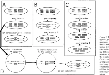
There are various methods for generating homozygous mutant ES cells once a heterozygous targeted mutant cell line has been generated (Fig. 1). For cell lines in which the targeted al-lele contains the neomycin resistance gene, the simplest method is to increase the concentration of the antibiotic, G418, thus selecting for cells that have higher neor activity (3) (Fig. 1
A). Most cells that survive high-dose selection have duplicate copies of the neor allele and lack the wild-type allele. The exact
mechanism of reduction to homozygosity in these cells is not clear but seems likely to involve complete or partial chromo-some loss and reduplication, rather than a localized gene con-version, in most cases. Some caution should be applied in using such cells as the sole determinant of mutant phenotype, since any chance mutations linked to the targeted allele may also be rendered homozygous and any parental imprinting effects on the chromosome may also become evident.
Direct targeting of the second allele is preferable but en-tails more work. A second targeting vector carrying a different drug selection marker, such as hygromycin or puromycin resis-tance, can be made and used to generate homozygous cells (Fig. 1 B). The necessity of making a second targeting vector can be avoided by ensuring that the neogene in the first vector is surrounded by loxP sites, the recognition sequence of the Cre site-specific recombinase (4). Heterozygous cells can then be transiently transfected with a Cre-expressing vector to ex-cise neo and retargeted with the same neo-containing vector (5) (Fig. 1 C). With the current excellent ES cells available, cells that have gone through these multiple manipulations re-main fully pluripotent.
Finally, new ES cell lines can be made directly from ho-mozygous embryos, after the original mutation has been
trans-mitted through the germ line (Fig. 1 D). This does not, of course, provide any assistance in the early analysis of the mu-tant phenotype, but it does provide cell lines that are unsullied by any defects that may have arisen during passage in culture, and it allows introduction of other marker genes or mutations onto the mutant background by intercrossing mouse lines.
Generation of completely ES-derived fetuses
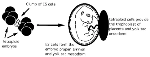
When ES cells are combined with tetraploid mouse embryos, fetuses and, in the best case scenario, viable offspring can be generated in which all tissues are ES derived (6, 7). This situa-tion is brought about by the impaired capacity of tetraploid cells to contribute to the embryonic lineages, such that survival is only possible if the ES cells take over the embryonic com-partment. ES cells have limited ability to make trophoblast and primitive endoderm, the extraembryonic lineages, but the tetraploid cells can survive and populate these lineages. The end result is a fetus with an ES-derived embryonic compart-ment and tetraploid-derived extraembryonic lineages (Fig. 2). Clearly, this approach has the potential to allow examination of mutant phenotypes derived from genetically altered ES cells very rapidly without the need for germ line transmission and further breeding.
[image:3.612.64.503.435.738.2]Can this potential be realized in practice? We have shown that the ability of ES cells to generate viable offspring after tet-raploid aggregation is very dependent on the quality of the cell line used; survival of ES-derived fetuses beyond term has only been achieved with early passage or selected subclones of cells (7). Thus, this approach is unlikely to be very effective for phe-notypes that are viable as homozygotes. However, survival of ES-derived fetuses is much better earlier in gestation, so that ES–tetraploid aggregations can be a valid way of analyzing
embryonic lethal phenotypes. Tetraploid cells are excluded from the embryonic compartment soon after implantation (8), so phenotypes that affect embryonic lineages (but not ex-traembryonic) between implantation and birth are accessible to analysis. We have recently been able to show that we can replicate the homozygous phenotypes observed in HNF3b(9, 10) and FGFR1 (11, 12) mutants after aggregation of homozy-gous mutant cells with wild-type tetraploid embryos, thus vali-dating the future use of this approach as a primary screen for mutant phenotype. The embryonic lethal phenotype resulting from complete loss of function of the endothelial growth fac-tor, VEGF, has recently been analyzed by ES–tetraploid ag-gregation (12a), allowing complete analysis of the mutant phe-notype within a much shorter time-frame than by standard analysis. In this particular case, the homozygous phenotype was inaccessible by standard breeding, because heterozygosity for the mutation was also embryonic lethal. The ES–tetraploid system is the only method to assess the effect of homozygosity in such cases.
Again, the availability of excellent ES cells worldwide and the high costs of maintaining mouse breeding stocks should lead to an increased use of this technique in the future, not only for mutant analysis but also for rapid analysis of all kinds of genetic manipulations performed in wild-type or mutant cells.
Lineage-specific mutant analysis
Lineage-specific effects of mutations can be studied by taking advantage of the ability of ES cells to populate lineages that are developmentally compromised in the host embryo. In tet-raploid aggregates, it is the whole epiblast lineage that is com-promised, making it possible to separate the effects of a partic-ular mutation on the embryonic versus extraembryonic lineage. For example, mutation of the Mash2 transcription factor gene, which is predominantly expressed in the extraembryonic tro-phoblast, leads to fetal death, apparently from placental insuf-ficiency in mid-gestation (13). Mash2 mutant embryos were rescued by aggregation with tetraploid embryos, simulta-neously proving that lethality was a direct consequence of tro-phoblast failure and showing that, in this case, the gene had no obvious role in any other lineage.
There are a number of mutations described in the literature that result in embryonic lethality as a result of likely extraem-bryonic lineage defects, precluding analysis of later emextraem-bryonic or adult roles for the genes. For example, mutation in the HNF4 transcription factor causes early embryonic lethality (14), apparently as a result of primitive endoderm defects, pre-cluding analysis of its proposed role in the liver. Similarly, mu-tation in the EGF receptor, a signaling molecule likely to be used in many different tissues, leads to problems at different
stages of gestation, dependent on genetic background (15–17). Again, the main defect causing lethality seems to be in the pro-liferation of the trophoblast lineage. In both these cases, and others, the ES–tetraploid rescue should allow rescue of the early lethality and elucidation of later defects.
Combination of mutant ES cells with embryos from strains with cell-autonomous defects in specific lineages within the embryo itself can provide more limited lineage-specific rescue, which can be very useful in delineating the roles of a given gene in a particular lineage. The most widely used system of this sort to date is the so-called RAG2 blastocyst complemen-tation assay (18). Mice lacking either RAG1 or RAG2, the re-combination-activating genes, contain no mature T or B lym-phocytes, because of the failure of VDJ recombination (19, 20). This defect can be rescued by introduction of wild-type ES cells into RAG2-deficient blastocysts: the entire mature lym-phocyte population of the resulting chimeras is ES derived. This assay has been used both to examine the effects on lym-phopoiesis of mutations that are embryonic lethal in the ho-mozygous state (e.g., Vav references 21–23; c-jun reference 24) and to analyze rapidly, without germ line transmission, the phenotypic effects of known immune function genes (e.g.,
CD40).
The general approach exemplified by the RAG2 comple-mentation assay could be applied to other lineages, if suitable deficient strains can be identified. Lineage deficiencies that are homozygous viable, like the RAG2 deficiency, would be most suitable, but even deficiencies that are homozygous lethal could be used, provided that easy genotype assays can be de-veloped to identify the one in four chimeras that would have the reconstituted lineage. A number of mutations have been reported to lead to complete loss of certain tissues or organs, e.g., loss of pancreas in insulin promoter factor-1 mutants (25), loss of endothelial cells inflk1mutants (26), and loss of cere-bellum inEn1 mutants (27). These kinds of mice might be suit-able hosts for lineage-specific rescue, if the defects are truly cell autonomous.
Diploid chimera analysis
Although the use of compromised embryos as chimeric part-ners can be a powerful way of analyzing lineage-specific de-fects, the generation of chimeras between mutant ES cells and normal diploid wild-type embryos has broader application. When ES cells are aggregated with eight-cell embryos or in-jected into blastocysts, they become finely intermixed with host cells, generating mosaic embryos and mice in which all cell and tissue types are likely to contain contributions from the two founding cell populations (28). As has been demon-strated over many years in mice and other organisms, such mo-saics allow analysis of several aspects of mutant cell behavior not necessarily apparent in the mutants themselves. In the past, two difficulties arose in applying such analysis to lethal mouse mutants. One was the fact that only one quarter of the offspring of a heterozygous cross would be the required ho-mozygous mutants, making identification of the mutant chime-ras difficult. Homozygous ES cells solve that problem. The sec-ond was the availability of suitable independent cell lineage markers to follow the distribution of normal and mutant cells. While this problem is not completely solved, there are now a number of good cell marker systems, the most common of which is the use of mice carrying theE. coli b-galactosidase
[image:4.612.55.297.57.149.2]gene inserted into a widely expressed locus (29).
What kind of information can be gleaned from chimera analysis? First, the cell autonomy of the defect can be ascer-tained. For example, embryos homozygous for the 413d retro-viral insertion die at gastrulation with severe problems in mesoderm generation (30). However, chimeras made with ho-mozygous ES cells survive and show contributions of mutant cells in many different lineages, suggesting that the defect in 413d is not cell autonomous (30). 413d is an insertion in the TGF-b–related nodal gene (31), a secreted factor, consistent with the non–cell autonomous nature of the mutation.
Second, more information on the lineage specificity of a de-fect can be obtained by detailed analysis of mutant cell distri-bution in chimeras. Exclusion of mutant cells from a given lineage implicates the gene in that lineage. For example, muta-tion of the twist transcription factor leads to failure of neural tube closure (32). Analysis of the distribution of mutant ES cells in chimeras showed that they were excluded from cranial mesenchyme but not neural crest or neural tube, pinpointing a specific defect in the head mesenchyme as the cause of the de-fects observed.
Third, defects in cell behavior can be seen much more clearly in chimeras than in mutants. Primary defects in cell ad-hesion or cell migration may become much more apparent when the mutant cells are forced to interact with wild-type cells. For example, the mouse Brachyury (T)mutation results in a truncation of the posterior axis and notochord defects (33). Chimeras with markedT/T ES cells show an accumula-tion of mutant cells at the primitive streak and a relative pau-city of cells in the nascent mesoderm populations (34). This
suggests that one of the main roles of the Brachyury gene is to regulate the changing cell adhesion properties necessary for normal gastrulation.
Fourth, rescue of mutant cells in chimeras can allow exami-nation of possible later roles for the gene in question. For ex-ample, mutation of GATA-2, a hematopoietic transcription factor, causes lethality in mid-gestation because of severe ane-mia, suggesting that GATA-2 is required for yolk sac hemato-poiesis (35). However, the embryos die too early to reveal any effects on definitive hematopoiesis. Chimeras made with ho-mozygous mutant cells allowed such analysis and showed that definitive hematopoiesis was severely affected, too, implicat-ing GATA-2 as a critical gene in the proliferation of hemato-poietic progenitors.
Mosaic analysis: lineage-specific gene knockout and gene repair
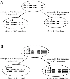
[image:5.612.58.356.401.736.2]Generation of chimeras with mutant ES cells can be a very powerful way of dissecting the multiple roles of genes in devel-opment and differentiation. However, the new techniques of genome alteration themselves offer the opportunity to gener-ate mosaic mice with lineage-specific genetic alterations, thus extending the power of this approach. This technology re-quires two steps. First, a specific alteration is made by homolo-gous recombination in a gene of interest that introduces two loxP sites, either around an essential part of the gene, leaving the gene functional (Fig. 3 A), or around an integrating exoge-nous sequence, interfering with the gene function (Fig. 3 B). Second, the Cre recombinase under the control of a lineage-specific promoter is introduced into embryos homozygous for
the above alteration. The recombinase then excises the essen-tial part (Fig. 3 A, lineage-specific gene knockout) or the inter-fering part (Fig. 3 B, lineage-specific gene repair) of the gene, creating a mosaic embryo or animal. Both of these approaches are theoretical extensions of the chimeric complementation as-says. They remove the limitation of the complementation chi-meras that presently exists because of the small number of available mutant hosts with cell-autonomous ablations. The approach of a lineage-specific knockout or repair is limited only by the need for reliable tissue/cell type–specific expres-sion of the Cre recombinase at a level providing high-fidelity excision in the targeted lineage and gene.
There have been relatively few published cases as yet of successful application of this approach (36, 37), but the cre-ation of an easily accessible panel of Cretransgenic lines would be extremely useful for the increasing number of researchers wishing to attempt lineage-specific excision or repair. To this end, one of us (A. Nagy) is organizing a data base listing the
Cre transgenic lines made, planned, and being produced in many laboratories.
Still more things to do with mutants
Not only do mutant ES cells allow sophisticated phenotypic analysis in vivo without germ line transmission, but they can, of course, be used to study effects on differentiation in vitro under various conditions, as already described in this series (38). From such differentiating ES cells, it may be possible to isolate other lines of more restricted stem cells that could be extremely useful tools in dissecting genetic hierarchies of lin-eage control. Mutant primary fibroblasts and other cells and tissues can also be obtained directly from mutant embryos, even those showing early lethality, and can be very informative in understanding the cell biology underlying a mutant defect. Such analysis need not mean maintaining large numbers of heterozygous mice, since essentially unlimited supplies of ho-mozygous embryos can be generated by ES–tetraploid aggre-gation. In the future, we can expect to see increased exploita-tion of techniques of experimental embryology and cell biology, in combination with the power of targeted mutagene-sis, to provide unprecedented insights into all aspects of mam-malian biology.
Acknowledgments
Apologies to all our colleagues whose experiments were not cited for lack of space. Those interested in the Cre transgenic database should contact A. Nagy by E-mail at [email protected]
J. Rossant is a Terry Fox Research Scientist of the National Can-cer Institute of Canada and a Howard Hughes Medical Institute In-ternational Scholar.
References
1. Capecchi, M.R. 1989. Altering the genome by homologous recombina-tion. Science (Wash. DC). 244:1288–1292.
2. Rossant, J., and A. Nagy. 1995. Genome engineering: the new mouse ge-netics. Nat. Med. 1:592–594.
3. Mortensen, R.M., D.A. Conner, S. Chao, A.A.T. Geisterfer-Lowrance, and J.G. Seidman. 1992. Production of homozygous mutant ES cells with a sin-gle targeting construct. Mol. Cell. Biol. 12:2391–2395.
4. Sauer, B., and N. Henderson. 1988. Site-specific recombination in mam-malian cells by the Cre recombinase of bacteriophage P1. Proc. Natl. Acad. Sci.
USA. 85:5166–5170.
5. Puri, M.C., J. Rossant, K. Alitalo, A. Bernstein, and J. Partanen. 1995. The receptor tyrosine kinase TIE is required for the integrity and survival of
vascular endothelial cells. EMBO (Eur. Mol. Biol. Organ.) J. 14:5886–5891. 6. Nagy, A., E. Gocza, E.M. Diaz, V.R. Prideaux, E. Ivanyi, M. Markkula, and J. Rossant. 1990. Embryonic stem cells alone are able to support fetal
de-velopment in the mouse. Development (Camb.). 110:815–822.
7. Nagy, A., J. Rossant, R. Nagy, W. Abramow-Newerley, and J.C. Roder. 1993. Derivation of completely cell culture-derived mice from early passage embryonic stem cells. Proc. Natl. Acad. Sci. USA. 90:8424–8428.
8. James, R.M., A.H. Klerkx, M. Keighren, J.H. Flockhart, and J.D. West. 1995. Restricted distribution of tetraploid cells in mouse tetraploid↔diploid chimaeras. Dev. Biol. 167:213–226.
9. Ang, S.-L., and J. Rossant. 1994. HNF-3b is essential for node and noto-chord formation in mouse development. Cell. 78:561–574.
10. Weinstein, D.C., A. Ruiz-in-Altaba, W.S. Chen, P. Hoodless, V.R. Prez-ioso, T.M. Jessell, and J.E. Darnell, Jr. 1993. The winged-helix transcription fac-tor HNF-3b is required for notochord development in the mouse embryo. Cell. 78:575–588.
11. Yamaguchi, T., K. Harpal, M. Henkemeyer, and J. Rossant. 1994. fgfr-1
is required for embryonic growth and mesodermal patterning during mouse gastrulation. Genes & Dev. 8:3032–3044.
12. Deng, C.-X., A. Synshaw-Boris, M.M. Shen, C. Daugherty, D.M. Ornitz, and P. Leder. 1994. Murine FGFR-1 is required for early postimplantation growth and axial organization. Genes & Dev. 8:3045–3057.
12a. Carmeliet, P., V. Ferreira, G. Breier, S. Pollefeyt, L. Kieckens, M. Gertsenstein, M. Fahrig, A. Vandenhoeck, K. Harpal, C. Eberhardt, et al. 1996. Abnormal blood vessel development and lethality in embryos lacking a single vascular endothelial growth factor allele. Nature (Lond.). In press.
13. Guillemot, F., A. Nagy, A. Auerbach, J. Rossant, and A.L. Joyner. 1994. Essential role of Mash-2 in extraembryonic development. Nature (Lond.). 371: 333–336.
14. Chen, W.S., K. Manova, D.C. Weinstein, S.A. Duncan, A.S. Plump, V.R. Prezioso, R.F. Bachvarova, and J.E. Darnell, Jr. 1994. Disruption of the HNF-4 gene, expressed in visceral endoderm, leads to cell death in embryonic ectoderm and impaired gastrulation of mouse embryos. Genes & Dev. 8:2466– 2477.
15. Threadgill, D.W., A.A. Dlugosz, L.A. Hansen, T. Tennenbaum, U. Li-chti, D. Yee, C. LaMantia, T. Mourton, K. Herrup, R.C. Harris, J.A. Barnard, S.H. Yuspa, R.J. Coffey, and T. Magnuson. 1995. Targeted disruption of mouse
EGF receptor: effect of genetic background on mutant phenotype. Science
(Wash. DC). 269:230–234.
16. Sibilia, M., and E.F. Wagner. 1995. Strain-dependent epithelial defects in mice lacking the EGF receptor. Science (Wash. DC).269:234–238.
17. Miettinen, P.J., J.E. Berger, J. Meneses, Y. Phung, R.A. Pedersen, Z. Werb, and R. Derynck. 1995. Epithelial immaturity and multiorgan failure in mice lacking epidermal growth factor receptor. Nature (Lond.). 376:337–341.
18. Chen, J., R. Lansford, V. Stewart, F. Young, and F.W. Alt. RAG-2-defi-cient blastocyst complementation: an assay of gene function in lymphocyte de-velopment. 1993. Proc. Natl. Acad. Sci. USA. 90:4528–4532.
19. Mombaerts, P., J. Iacomini, R.S. Johnson, K. Herrup, S. Tonegawa, and V.E. Papaioannou. 1992. RAG-1-deficient mice have no mature B and T lym-phocytes. Cell. 68:869–878.
20. Shinkai, Y., G. Rathbun, K.-P. Lam, E.M. Ottz, V. Stewart, M. Mendel-sohn, J. Charron, M. Datta, F. Young, A.M. Stall, and F.W. Alt. 1992. RAG-2-deficient mice lack mature lymphocytes owing to inability to initiate V(D)J re-arrangement. Cell. 68:855–868.
21. Tarakhovsky, A., M. Turner, S. Schaal, P.J. Mee, L.P. Duddy, K. Rajew-sky, and V.L. Tybulewicz. 1995. Defective antigen receptor-mediated prolifera-tion of B and T cells in the absence of Vav. Nature (Lond.). 374:467–470.
22. Zhang, R., F.W. Alt, L. Davidson, S.H. Orkin, and W. Swat. 1995. De-fective signalling through the T- and B-cell antigen receptors in lymphoid cells lacking the vav proto-oncogene. Nature (Lond.). 374:470–473.
23. Fischer, K.D., A. Zmuldzinas, S. Gardner, M. Barbacid, A. Bernstein, and C. Guidos. 1995. Defective T-cell receptor signalling and positive selection of Vav-deficient CD41 CD81 thymocytes. Nature (Lond.). 374:474–477.
24. Chen, J., V. Stewart, G. Spyrou, F. Hilberg, E.F. Wagner, and F.W. Alt.
1994. Generation of normal T and B lymphocytes by c-jun deficient embryonic
stem cells. Immunity. 1:65–72.
25. Jonsson, J., L. Carlsson, T. Edlund, and H. Edlund. 1994.
Insulin-pro-moter-factor 1 is required for pancreas development in mice. Nature (Lond.).
371:606–609.
26. Shalaby, F., J. Rossant, T.P. Yamaguchi, M. Gertsenstein, X.-F. Wu, M.L. Breitman, and A.C. Schuh. 1995. Failure of blood island formation and vasculogenesis in flk-1 deficient mice. Nature (Lond.). 376:62–66.
27. Wurst, W., A.B. Auerbach, and A.L. Joyner. 1994. Multiple develop-mental defects in Engrailed-1 mutant mice: an early mid-hindbrain deletion and
patterning defects in forelimbs and sternum. Development (Camb.). 120:2065–
2075.
28. Beddington, R.S.P., and E.J. Robertson. 1989. An assessment of the de-velopmental potential of embryonic stem cells in the midgestation mouse em-bryo. Development (Camb.). 105:733–737.
30. Conlon, F.L., K.S. Barth, and E.J. Robertson. 1991. A novel retrovirally induced embryonic lethal mutation in the mouse: assessment of the develop-mental fate of embryonic stem cells homozygous for the 413.d proviral integra-tion. Development (Camb.). 111:969–981.
31. Zhou, X., H. Sasaki, L. Lowe, B.L.M. Hogan, and M.R. Kuehn. 1993. Nodal is a novel TGF-b-like gene expressed in the mouse node during gastrula-tion. Nature (Lond.). 361:543–547.
32. Chen, Z.-F., and R.R. Behringer. 1995. twist is required in head mesen-chyme for cranial neural tube morphogenesis. Genes & Dev. 9:686–699.
33. Herrmann, B.G., and A. Kispert. 1994. The T genes in embryogenesis.
Trends Genet. 10:280–286.
34. Wilson, V., L. Manson, W.C. Skarnes, and R.S.P. Beddington. 1995. The
T gene is necessary for normal mesodermal morphogenetic cell movements
during gastrulation. Development (Camb.).121:877–886.
35. Tsai, F.-Y., G. Keller, F.C. Kuo, M. Weiss, J. Chen, M. Rosenblatt, F.W. Alt, and S.H. Orkin. 1994. An early haematopoietic defect in mice lacking the transcription factor GATA-2. Nature (Lond.). 371:221–226.
36. Gu, H., J.D. Marth, P.-C. Orban, H. Mossmann, and K. Rajewsky. 1994. Deletion of a DNA polymerase B gene segment in T cells using cell type-spe-cific gene targeting. Science (Wash. DC). 265:103–106.
37. Kuhn, R., F. Schwenk, M. Aguet, and K. Rajewsky. 1995. Inducible gene targeting in mice. Science (Wash. DC). 269:1427–1429.