ISSN Online: 1937-688X ISSN Print: 1937-6871
EIT Image Reconstruction by Modified Data
B. Gong, B. Schullcke, S. Krueger-Ziolek, K. Moeller
Institute of Technical Medicine, Furtwangen University, Villingen-Schwenningen, Germany
Abstract
Electrical impedance tomography (EIT) is a radiation-free imaging method. Canon-ically, in lung EIT, 16 electrodes are placed horizontally on the thorax skin. By in-jecting currents through electrodes attached to the skin, a set of induced voltage measurements can be collected. The conductivity distribution on the chest plane can be obtained from these electrical boundary conditions. It has been reported that the adjacent current injection pattern is sub-optimal for EIT reconstruction. However, this adjacent current injection pattern is commonly used in commercially available EIT devices. In this study, we modify the boundary conditions according to the su-perposition principle of the electrical field. As a result, boundary conditions of the adjacent current pattern will be transformed to those corresponding to “skip-3” cur-rent injection pattern. Simulation results indicated that reconstruction benefits from the modified boundary conditions.
Keywords
Electrical Impedance Tomography, Current Pattern, Superposition Principle
1. Introduction
Electrical Impedance Tomography (EIT) is a radiation-free imaging method. It at-tempts to reveal the conductivity distribution changes inside the human body of two time instants through electrical data obtained via the electrodes attached to the boun-dary. In lung EIT, commonly 16 electrodes are placed equidistantly on the boundary of a horizontal chest plane.
We denote the conductivity of the domain changes between two time steps by Δs and the measured voltage changes on the electrodes by a vector ΔV. Under the FEM
framework with M elements, the conductivity change Δs is represented by a M × 1
vec-tor. Approximately, there exists the following relation:
J∙Δs ≈ Δv (1)
How to cite this paper: Gong, B., Schullcke, B., Krueger-Ziolek, S. and Moeller, K. (2016) EIT Image Reconstruction by Modified Data. J. Biomedical Science and Engineering, 9, 99-106.
http://dx.doi.org/10.4236/jbise.2016.910B013
where J denotes the Jacobian matrix calculated at the constant conductivity 1:
1
i ij
j
V J
s ∆ =
∆ (2)
Jacobian matrix is commonly calculated by studying the first order perturbation of conductivity on each element [1]. Briefly, under FEM framework, the potential distri-bution can be solved by forward model. The perturbations on each element can be de-termined by the simulated potential information [1].
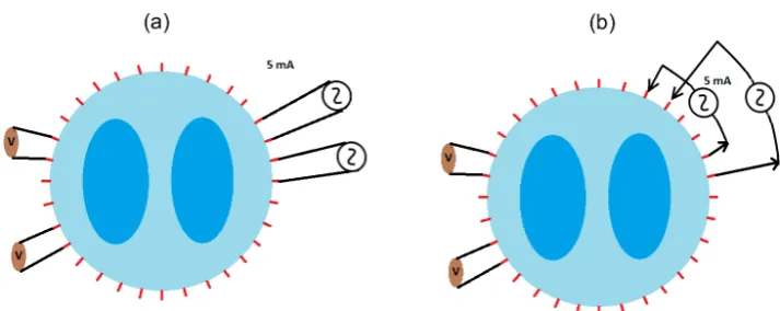
In commercially obtained EIT devices (PulmoVista 500, Dräger Medical, Lübeck, Germany), electrical currents are injected through the electrodes adjacently (Figure 1(a)). Other current injection patterns have been studied by A. Andy et al. in simula-tion [2]. The authors compared several current injecsimula-tion patterns based on a predefined distinguish ability parameter. They concluded that the adjacent current injection pat-terns are suboptimal in distinguish ability, especially in the inner-most part of the do-main. The authors further suggested injecting currents through electrodes by skipping several electrodes in between (Figure 1(b)). Incited by this result, we propose a frame-work that reforms the measurement data that are collected under adjacent current in-jection pattern by the superposition principle of the electrical field.
2. Method
2.1. Superposition Principle
Electric fields satisfy the superposition principle. This is because Maxwell’s equations are linear. In this study, we apply this principle to modify the boundary conditions
in-duced by the adjacent injection pattern (Figure 1(a)). For simplicity, we assume ,
(
e ei j)
and
(
e ej, k)
to be neighboring electrode pairs in a 16-electrode EIT system.Suppose an alternative electrical current of 5 mA is injected into two electrodes
(
)
,e ei j . Thereby, an electric field Eij will be generated. The induced voltage Vij on two
electrodes
(
e ei′ ′, j)
can be measured. Similarly, if the same amount of current isin-jected into electrodes
(
,e ej k)
, the corresponding electrical field Ejk and the voltage Vjkon electrodes
(
e ei′ ′, j)
can be determined. In addition, if the current is injected throughelectrodes
(
,e ej k)
, a new of electric field and set of voltage measurement on(
e ei′ ′, j)
,denoted by Eik and Vik, can be obtained. It follows from the superposition principle
that:
ik ij jk
E =E +E . (3)
Hence, the induced voltages between electrodes ei′ and e′j satisfy the following
re-lation:
ik ij jk
V =V +V . (4)
Figure 1. Different current patterns. (a) Demonstrates the adjacent current injection pattern. The so called “skip-3” current injection pattern is shown in (b). Note that for both current injection patterns, the measurement pattern is fixed to be adjacent in this study.
In the real world, all measurements are contaminated by noises. The measurement noise in EIT is commonly assumed to be independent between each electrode pair [2]. For simplicity, we assume an additive white noise on the voltage measurements. Let
ij ij ij
V =V +ε and Vjk=Vjk+εjk be two noised voltage measurements with
(
0,)
N
ε σ , the voltages relation Vik =Vij+Vjk implies that the voltage Vik contains
a white noise with standard deviation 2σ. By noticing this noise magnification fact, we
suggest applying superposition principle on a smaller number of boundary conditions. In this study, we modify the voltage measurement under adjacent injection pattern (Figure 1(a)). Using the superposition principle we generate a set of hypothetical mea-surement data corresponding to the skip-3 injection pattern shown in Figure 1(b).
In commercial EIT devices, the voltage measurements employing the current injec-tion electrodes are missed. Using the adjacent pattern, in each current injecinjec-tion, there are only 13 independent voltage measurements that can be obtained. The number of independent measurements is further reduced using the proposed modification
proce-dure. This is because, to form a hypothetic current injection from electrodes
(
,e ei k)
, itis necessary to employ all the current injections between these two electrodes. Within the hypothetical skip-3 pattern, for each current injection, there are only 10 indepen-dent voltage measurements that can be obtained.
2.2. Sensitivity Analysis
Figure 2. Sensitivity with respect to one of the voltage measurements. The left figure plots the sensitivity of a voltage measurement with respect to each FEM element under the adjacent cur-rent injection pattern. The right image showing the same plot regards to the skip-3 curcur-rent injec-tion pattern. Both plots are fixed with the same color scale.
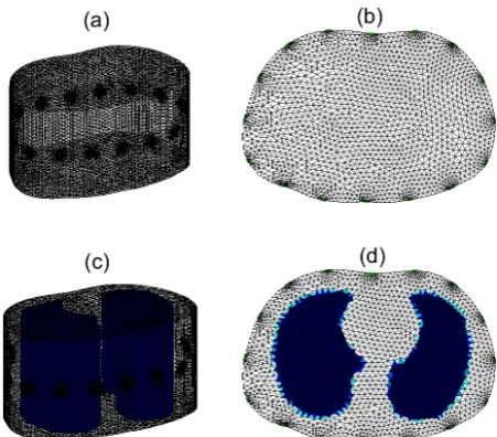
Figure 3. Ground truth for simulation. (a) Is the 3D homogeneous model. Each element has conductivity 1 Sm−1. (b) Is a 2D simplification of the 3D model (a). The reconstruction will be performed on this 2D model. The 3D ground truth is displayed in (c). Two contrasts with lung shape are embedded into the homogenous model. These contrasts have conductivity 0.95 Sm−1. The 2D simplification of the ground truth on the electrodes level is plotted in (d).
2.3. Reconstruction with Modified Data
In this study, we employ the canonical one step Gauss-Newton solver to reconstruct images. Tikhonov regularization was applied to circumvent the ill-posedness of the in-verse problem. It is assume that the original voltage measurements are collected with respect to the adjacent current injection pattern. At two time instants, two modified
voltages can be obtained according to the method proposed in Section 2.2. Let ∆V be
[image:4.595.261.487.253.451.2] 2 2 2 2
1 arg min
2
s
s ∆ V J s α s
∆ = ∆ − ⋅∆ + ⋅ ∆ (5)
where α is the Tikhonov regularization parameter. The solution of this problem can be explicitly written by:
(
)
1 T T
s J J α I − J V
∆ = ⋅ + ⋅ ⋅ ⋅ ∆ (6)
2.4. Simulation Data Acquisition
The performance of the proposed dada modification method was evaluated through simulation. A 3D simulated homogeneous thorax model was constructed with the help of MATLAB toolbox EIDORS [3]. This model has 50,586 tetrahedron elements. The
background conductivity of this homogeneous model was fixed to be 1 Sm−1. In this
ar-ticle, all simulated 3D models had a height equal to half of the width. Sixteen electrodes were attached equidistantly around the boundary on the middle level. The contact im-pedance was fixed to be 100 Ω.
Currents of 10 mA were injected into these electrodes with respect to adjacent as well as skip-3 injection patterns. Corresponding to the skip-3 injection pattern, a Jacobian matrix J can be calculated by the forward solver provided by EIDORS. For reconstruc-tion, the 2.5D model is employed. That is, the simulation data and Jacobian are calcu-lated on 3D model, but the reconstruction is applied on 2D FEM. This model requires a modification of Jacobian matrix by projecting the Jacobian matrix to 2D into the elec-trodes plane. For detail of the 2.5D framework please see [4].
Simulated voltage measurements are calculated using much finer independent 3D FEM models. The voltage measurements are calculated with respect to the adjacent
in-jection pattern. Using a homogeneous model, the boundary voltage Vh can be
calcu-lated using the forward solver.
Simulated lungs with conductivity 0.95 Sm−1 were embedded into an independent
homogeneous 3D phantom in order to build an inhomogeneous model (Figure 3(c)). This inhomogeneous model is considered as the 3D ground truth. After embedding the contrasts, another set of voltage measurements under the adjacent injection pattern can
be simulated. We denoted this voltage measurement by Vi. The voltage differences
i h
V V V
δ = − will be used for reconstruction. Let NL∈
( )
0,1 denote a noise level, awhite noise, defined by
( )
Noise=NL std⋅ δV ⋅χ (7)
was added to the voltage differences δV for Monte-Carlo simulations. Here std
( )
⋅denotes the standard deviation and χ
( )
0,1 is a random vector with Gaussiannormal distribution. This noised data will be further modified by the proposed super-position method to form a new set of voltage differences. The original voltage differ-ences and the modified one are both employed in simulation in Section 3 for comparison.
2.5. Evaluation Parameters
parameter is defined by:
2
2
: sol gr
gr s s RE s ∆ − ∆ =
∆ (8)
where ∆ssol and ∆sgr represent the 2D reconstructed images and the ground truth
respectively.
The system of figure of merit [5] was also employed. These parameters are based on a
new image ∆sq, which records the finite elements that have effective conductivity
changes. Given a reconstructed image with conductivity changes ∆s, the value of the
i-th finite element of ∆sq is defined by:
1 1max
(
( )
)
50
i q
i
if abs s abs s
s otherwise ∆ ≥ ∆ ∆ =
(9)
In addition, a binary mask on ∆sgr is defined by:
1 0 0 gr i C i s s otherwise ∆ ≠ ∆ =
(10)
Three figure of merits parameters are defined based on ∆sq:
Position error: PE= −rt rq 2 , where rq and rt are the reconstructed and the
original centers of gravity of the contrasts. Position errors of left and right lungs are calculated separately. The sum of these position errors is defined to be the total PE. For this study, we only present the total PE. The position error is expected to be small.
Shape deformation: SD= ∆ − ∆sq sC1 ∆sC 1. Shape deformation measures the
rel-ative area of misshaping of the reconstruction.
Ring effect: & 0
( )
( )
i
i C s i i C i
RNG=
∑
∉ ∆ < abs ∆s∑
∈ abs ∆s , where C is the trueregion of the inclusions. The figure of merits parameters are expected to have small va-riability.
3. Simulation Results
Performances of different methods are evaluated by simulation. The simulation model and data were constructed as described in Section 2.4. Four noises levels NL = 0, 0.05, 0.1, 0.2 were used to simulate the measurement noise. One step Gauss-Newton solver (Section 2.3) was employed to reconstruct images. The regularization parameter α used in Equation (5) was determined heuristically to get the best performance. This parame-ter was set regarding the noise levels. In Figure 4, the reconstructed images with the measurement data with a noise at level 0.05 was demonstrated. Furthermore, the re-construction error parameters were evaluated in Monte Carlo simulations with 50 in-dependent runs at each noise level. These results are shown in Figure 5.
4. Discussion and Conclusion
Figure 4. Reconstructed images using original data obtained by adjacent current injection pat-tern and the modified data proposed in Section 2.3. These reconstructed images are based on the measurement data with a white noise at level 0.05.
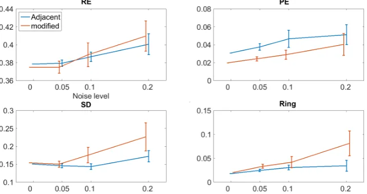
Figure 5. Error analysis based on Monte Carlo simulation. The errors were studied with different noise levels. The image error evaluation parameters were calculated after 50 independent runs of the simulation.
modified data can be observed. The conductivity changes in the central part of the do-main are less regarded when using the original data. As comparing the central parts of the two images of Figure 4, it can be observed that reconstructed conductivity changes in the left image are much smaller than the ground truth. As shown by the Monte Carlo simulation results (Figure 5), the position error can be largely corrected by using the modified data.
On the other hand, using original data, the edges of contrasts are better recon-structed around the boundary.
[image:7.595.195.553.262.451.2]voltage data. Consequently, a mixed voltage data set can be constructed. In addition, the Jacobian matrix is required to be modified in the same way to make it consistent with the mixed data.
In this article, EIT imaging using a data modification strategy was studied. The vol-tage measurements corresponding to adjacent current injection pattern are trans-formed to those from the skip-3 current injection pattern. Simulation results showed that using the modified data reconstruction is more robust in detecting the central area of the domain, while producing less position error. However, since some independent measurements are lost during data modification, the reconstruction using the modified dada is not stable. To circumvent this difficulty, we suggest completing the modified data with the original data in further studies.
Acknowledgements
This work is partially supported by the Federal Ministry of Education and Research (BMBF) under grant no. 03FH038I3 (MOSES).
References
[1] Holder, D.S. (2004) Electrical Impedance Tomography: Methods, History and Applications. CRC Press.http://dx.doi.org/10.1201/9781420034462
[2] Adler, A., Gaggero, P.O. and Maimaitijiang, Y. (2011) Adjacent Stimulation and Measure-ment Patterns Considered Harmful. Physiological Measurement, 32, 731.
http://dx.doi.org/10.1088/0967-3334/32/7/S01
[3] Adler, A. and Lionheart, W.R. (2006) Uses and Abuses of EIDORS: An Extensible Software Base for EIT. Physiological Measurement, 27, S25.
http://dx.doi.org/10.1088/0967-3334/27/5/s03
[4] Mamatjan, Y., Borsic, A., Gürsoy, D. and Adler, A. (2013) An Experimental Clinical Evalu-ation of EIT Imaging with ℓ1 Data and Image Norms. Physiological Measurement, 34, 1027. http://dx.doi.org/10.1088/0967-3334/34/9/1027
Submit or recommend next manuscript to SCIRP and we will provide best service for you:
Accepting pre-submission inquiries through Email, Facebook, LinkedIn, Twitter, etc. A wide selection of journals (inclusive of 9 subjects, more than 200 journals)
Providing 24-hour high-quality service User-friendly online submission system Fair and swift peer-review system
Efficient typesetting and proofreading procedure
Display of the result of downloads and visits, as well as the number of cited articles Maximum dissemination of your research work
Submit your manuscript at: http://papersubmission.scirp.org/