0095-1137/10/$12.00
doi:10.1128/JCM.00939-10
Copyright © 2010, American Society for Microbiology. All Rights Reserved.
Development and Application of Multiprobe Real-Time PCR Method
Targeting the
hsp65
Gene for Differentiation of
Mycobacterium
Species from Isolates and Sputum Specimens
䌤
Kijeong Kim,
2Hyungki Lee,
1Mi-Kyung Lee,
3Seoung-Ae Lee,
1Tae-Sun Shim,
4Seong Yong Lim,
5Won-Jung Koh,
6Jae-Joon Yim,
7Bazarragchaa Munkhtsetseg,
2Wonyong Kim,
2Sang-In Chung,
2Yoon-Hoh Kook,
1and Bum-Joon Kim
1*
Department of Microbiology and Immunology, Cancer Research Institute, Liver Research Institute, and SNUMRC, College of Medicine,
1and
Department of Internal Medicine and Lung Institute, Seoul National University College of Medicine,
7Seoul 110-799, Department of
Microbiology
2and Department of Laboratory Medicine,
3College of Medicine, Chung-Ang University, Seoul 156-756, Division of
Pulmonary and Critical Care Medicine, Department of Internal Medicine, University of Ulsan College of Medicine,
Asan Medical Center, Seoul 138-600,
4and Division of Pulmonary and Critical Care Medicine, Department of
Medicine, Kangbuk Samsung Hospital,
5and Department of Medicine, Samsung Medical Center,
6Sungkyunkwan University School of Medicine, Seoul, Republic of Korea
Received 11 May 2010/Returned for modification 1 July 2010/Accepted 8 July 2010
We developed a multiprobe real-time PCR assay targeting
hsp65
(HMPRT-PCR) to detect and identify
mycobacterial isolates and isolates directly from sputum specimens. Primers and probes for HMPRT-PCR
were designed on the basis of the
hsp65
gene sequence, enabling the recognition of seven pathogenic
myco-bacteria, including
Mycobacterium tuberculosis
,
M. avium
,
M. intracellulare
,
M. kansasii
,
M. abscessus
,
M.
mas-siliense
, and
M. fortuitum.
This technique was applied to 24 reference and 133 clinical isolates and differentiated
between all strains with 100% sensitivity and specificity. Furthermore, this method was applied to sputum
specimens from 117 consecutive smear-positive patients with smear results of from a trace to 3
ⴙ
. These results
were then compared to those obtained using the
rpoB
PCR-restriction analysis method with samples from
cultures of the same sputum specimens. The HMPRT-PCR method correctly identified the mycobacteria in 89
samples (76.0%, 89/117), and moreover, the sensitivity level was increased to 94.3% (50/53) for sputa with an
acid-fast bacillus score equal to or greater than 2
ⴙ
. Our data suggest that this novel HMPRT-PCR method
could be a promising approach for detecting pathogenic mycobacterial species from sputum samples and
culture isolates routinely in a clinical setting.
Of the known species in the genus
Mycobacterium
,
Mycobac-terium tuberculosis
is the most common and most important
pathogen, causing 2 million deaths and over 8 million cases of
tuberculosis worldwide annually (2, 3, 4, 7). In addition to
M.
tuberculosis
, infections with nontuberculosis mycobacteria
(NTM) can also cause clinical problems. Because of the
dif-ferent pathogenic potentials and susceptibilities of difdif-ferent
mycobacterial species, the treatments of mycobacterial
infec-tions are different (13, 30, 33, 34). Thus, it is very important to
differentiate between mycobacteria at the species level during
early-stage diagnostics.
Instead of a culture-based identification scheme, which may
take 4 to 6 weeks or longer to identify slowly growing
myco-bacteria, PCR-based protocols (sequencing or PCR-restriction
analysis [PRA]) targeting chronometer molecules, such as 16S
rRNA (5, 6, 28),
hsp65
(17, 19, 25), and
rpoB
(1, 16, 21), have
been widely used to identify mycobacteria. However, in spite of
the successful application of these conventional PCR-based
methods to culture isolates, there are some drawbacks in their
direct application to clinical specimens. This is especially true
for sputum samples, which also contain numbers of commensal
bacteria from the respiratory tract, producing confusing results
by the simultaneous amplification of both commensals and
mycobacterial strains. We have recently developed several
methods for mycobacterial species identification based on
am-plification of
hsp65
gene sequences directly from sputum
sam-ples (15, 27). Limitations due to the intrinsic features of
conventional PCR prevented feasible identification of
myco-bacterial species from sputum samples using this method.
The use of the real-time PCR assay in the diagnosis of many
infectious diseases has been increasing, as it represents an
appealing alternative to conventional PCR. It is an
improve-ment over conventional methods because of its increased
sen-sitivity and specificity, low contamination risk, and ease of
performance and speed (8). In particular, fluorescence
reso-nance energy transfer (FRET)-based real-time PCR permits
not only the simultaneous identification of multiple target
spe-cies but also the direct identification of target spespe-cies from
primary specimens such as sputum specimens through melting
curve analysis of the amplification product (8). These
charac-teristics of FRET-based real-time PCR provide a useful
ad-vantage for the identification of mycobacteria from sputum
samples. Recently, several real-time PCR-based methods for
mycobacterial detection and identification have been
devel-oped and evaluated (9, 22, 23, 26, 29). However, direct
appli-* Corresponding author. Mailing address: Department of
Microbi-ology and ImmunMicrobi-ology, Liver Research Institute, Cancer Research
Institute, and SNUMRC, College of Medicine, Seoul National
Uni-versity, 28 Yongon-dong, Chongno-gu, Seoul 110-799, Republic of
Korea. Phone: (82) 2-740-8316. Fax: (82) 2-743-0881. E-mail:
[email protected].
䌤
Published ahead of print on 14 July 2010.
3073
on May 16, 2020 by guest
http://jcm.asm.org/
cation of the real-time PCR-based method to primary
speci-mens was generally limited to
M. tuberculo
sis alone (11, 26). So
far, a method which can simultaneously identify several
patho-genic NTM as well as
M. tuberculosis
from primary sputum
samples in a single reaction has not been developed.
In the present study, we sought to develop a multiprobe
real-time PCR targeting the
hsp65
gene (HMPRT-PCR) based
on melting curve analysis (HybProbes). This enabled the
si-multaneous identification of several pathogenic mycobacteria,
including
M. tuberculosis
, in a single PCR performed on
cul-tures and sputum samples. The usefulness of these methods
was evaluated by blindly applying them to cultured and sputum
samples.
MATERIALS AND METHODS
Mycobacterial strains and sputa.Twenty-four mycobacterial reference strains (Table 1), 133 clinical isolates (Table 2), and 117 sputum specimens suspected of harboring mycobacteria were used in this study. Twenty-two of the 24 mycobac-terial reference strains were provided by the Korean Institute of Tuberculosis (KIT).M. intracellulare05-1390 andM. aviumATCC 4006 were provided by the Seoul National University College of Medicine (SNUMC) and the Samsung Medical Center (SMC), respectively. One hundred thirty-three clinical isolates which had already been identified by molecular biology-based assays, such as
rpoBPRA (21) andhsp65-based sequencing analysis (17, 19), were provided by SMC (51 strains), SNUMC (49 strains), the Kangbuk Samsung Medical Center (KBSMC) (24 strains), the Asan Medical Center (AMC) (5 strains), and the Chung-Ang University Medical Center (CAUMC) (4 strains). One hundred seventeen sputum specimens from different patients with positive acid-fast ba-cillus (AFB) smears detected between 1 April 2008 and 31 July 2008 at AMC, Seoul, Republic of Korea, were included in this study. The sputa were digested, decontaminated, and concentrated as recommended by the WHO (14). The
processed sediment was stained using the Ziehl-Neelsen method (14). The re-sults of the AFB smears were graded according to the recommendations of the American Thoracic Society and the Centers for Disease Control and Prevention (32). Sputa with trace AFB smear results (one to two bacilli in 300 fields) were also included in this study. The protocol for this study was approved by the Institutional Review Board of AMC.
DNA extraction.Chromosomal DNA was extracted from the clinical isolates and sputum samples by the bead beater-phenol extraction method, as described previously (19).
Primer and oligonucleotide probe design.To obtain appropriate primer se-quences for the amplification of mycobacterialhsp65DNA and identify multiple probe sequences for the species-specific detection of mycobacteria, thehsp65
[image:2.585.43.544.90.361.2]DNA sequences of a total of 140 mycobacterium-related strains were analyzed using SeqMan II software (DNAStar) (data not shown). AnM. tuberculosis
TABLE 1. Mycobacterial reference strains used in the present study and specificity of
hsp65
multiprobe real-time PCR for identification of
target
Mycobacterium
species
Species Strain Sourcea MeasuredTm(°C)
b
Real-time PCR identification
CH. 610 CH. 640 CH. 670 CH. 705
M. abscessus
19977
TATCC
—
—
—
68.2
ⴞ
0.51
M. abscessus
M. avium
25291
TATCC
60.9
⫾
0.00
—
74.0
ⴞ
0.06
60.3
⫾
0.03
M. avium
M. celatum
51131
TATCC
—
—
—
—
M. chelonae
35749
TATCC
—
—
64.9
⫾
0.01
62.6
⫾
0.57
M. flavescens
14474
TATCC
—
—
67.2
⫾
0.05
57.9
⫾
0.69
M. fortuitum
6841
TATCC
—
60.5
ⴞ
0.01
—
—
M. fortuitum
-
M. peregrinum
complex
M. gastri
15754
TATCC
61.2
⫾
0.03
57.7
⫾
0.08
—
57.9
⫾
0.18
M. genavense
51233
TATCC
—
—
69.1
⫾
0.23
—
M. gordonae
14470
TATCC
61.6
⫾
0.02
—
66.6
⫾
0.13
—
M. haemophilum
29548
TATCC
61.4
⫾
0.11
—
—
58.3
⫾
1.15
M. intracellulare
13950
TATCC
61.4
⫾
0.05
73.0
ⴞ
0.02
—
58.1
⫾
0.12
M. intracellulare
M. kansasii
12478
TATCC
—
67.0
ⴞ
0.01
—
57.4
⫾
0.23
M. kansasii
M. malmoense
29571
TATCC
61.6
⫾
0.04
—
—
61.9
⫾
0.20
M. massiliense
19086
KCTC
—
—
—
70.8
ⴞ
0.18
M. massiliense
M. marinum
29571
TATCC
61.6
⫾
0.04
—
63.1
⫾
0.14
58.1
⫾
0.02
M. peregrinum
27294
TATCC
—
60.0
ⴞ
0.07
—
—
M. fortuitum
-
M. peregrinum
complex
M. phlei
35784
ATCC
—
—
67.5
⫾
0.05
—
M. scrofulaceum
19981
TATCC
61.5
⫾
0.15
61.5
⫾
0.17
61.6
⫾
0.04
—
M. smegmatis
607
ATCC
61.6
⫾
0.09
—
—
61.5
⫾
0.33
M. szulgai
35799
TATCC
—
—
58.8
⫾
0.21
—
M. terrae
15755
TATCC
—
—
60.3
⫾
0.01
60.1
⫾
0.20
M. tuberculosis
27294
TATCC
66.9
ⴞ
0.02
—
—
57.8
⫾
0.13
M. tuberculosis
M. ulcerans
19423
TATCC
61.7
⫾
0.12
—
63.2
⫾
0.05
57.9
⫾
0.18
M. xenopi
19250
TATCC
—
—
—
—
aATCC, American Type Culture Collection; KCTC, Korean Collection for Type Cultures. bT
ms were obtained by duplicate real-time PCR and melting curve analyses, and the data represent the means⫾standard deviations. Boldface, species-specificTm;
—, no significantTm.
TABLE 2. Identification of mycobacterial clinical isolates
by HMPRT-PCR
Species
No. of isolates detected by:
rpoBPRA HMPRT-PCR
M. abscessus
33
16
aM. massiliense
17
aM. avium
18
18
M. fortuitum
7
7
M. intracellulare
26
26
M. kansasii
25
25
M. tuberculosis
24
24
Total
133
133
a
Differentiation betweenM. abscessusandM. massiliensewas confirmed by
hsp65-based sequencing analysis.
3074
KIM ET AL.
J. C
LIN. M
ICROBIOL.
on May 16, 2020 by guest
http://jcm.asm.org/
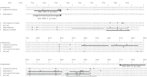
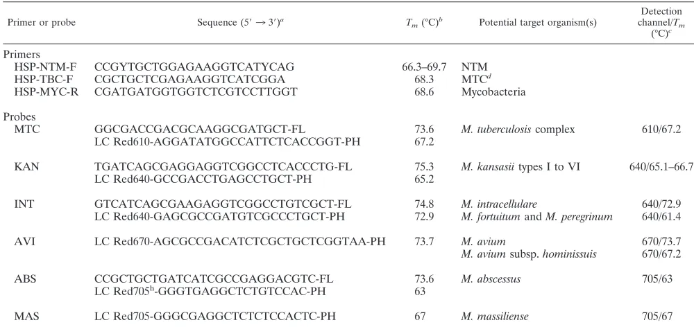
[image:2.585.301.542.588.707.2]complex (TBC)-specific forward primer and an NTM-specific forward primer which are different from each other at 3 bases at the 3⬘end and a reverse primer for all mycobacterial species were designed using the Oligo (version 6.5) program (Molecular Biology Insights). These primers produced 304-bphsp65amplicons (from the 698th to 991st nucleotides in theM. tuberculosis hsp65gene) (Fig. 1; Table 3). We designed a set of 10 hybridization probes (an anchor probe and a sensor probe; HybProbe) for the specific simultaneous detection in a single reaction of seven representative mycobacterial species or complexes that are the most clinically important and most frequently isolated from the patients using LC PDS (version 2.0) software (Fig. 1; Table 3). We used four channels (CH.) for probes specific to seven mycobacterial species. We used CH. 610 for the detec-tion ofM. tuberculosis; CH. 640 for the detection ofM. kansasii,M. intracellulare, and theM. fortiutum-M. peregrinumcomplex; CH. 670 for the detection ofM. avium; and CH. 705 for the detection ofM. abscessusandM. massiliense(Table 1). We determined species-specific regions of each target organism that ap-peared to be dissimilar from the sequences of the other organisms by more than 2 bases at the sensor binding position or 4 bases at the anchor probe-binding position in the sequences of the otherMycobacteriumspecies to ensure a highly specific signal. When designing the HybProbes, we ensured that the anchor and sensor probes were adjacently hybridized to the complementary target DNA for successful fluorescence emission by a mechanism of FRET. The potential presence of cross-complementarities among all the primers and probes was checked by using the LC PDS (version 2.0) software, and to obtain high sensitivity, we modified the primer or probe sequences while maintaining the primer and probe specificities. The diagnostic melting temperatures (Tms) of the
designed probes and their specificities were investigated by calculation of theTms
of the sensor and anchor probes hybridizing to the target and nontarget myco-bacterial DNAs by using the LC PDS software (Table 4). The probe specificity for each targetMycobacterium species was confirmed by comparing the se-quences from 240 mycobacterial or related strains via BLAST analysis (http: //blast.ncbi.nlm.nih.gov/Blast.cgi). The designed probes were purchased from Metabion.
Real-time PCR.A LightCycler (version 2.0) system was used for real-time PCR, and its four detection channels were calibrated for color compensation and activated for the experiment. The LightCycler Faststart DNA master HP kit (Roche Diagnostics) was used for the preparation of the master mixture,
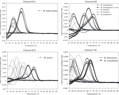
accord-ing to the protocol provided with the kit. A 10-l reaction mixture was prepared for each sample as follows: 1lTaqbuffer (which contains a deoxynucleoside triphosphate mixture and 10 mM MgCl2), additional 2 mM MgCl2, 0.4M NTM-specific forward primer (primer HSP-NTM-F), 0.3M TBC-specific for-ward primer (primer HSP-TBC-F), 1M mycobacterium-specific reverse primer (primer HSP-MYC-R), 0.2M HybProbes (which are presented in Table 3), 2 l of culture-extracted DNA, or 4l of sputum-extracted DNA with 0.1 mg/ml bovine serum albumin (New England Biolabs) as templates, and sterile distilled water. The cycling conditions were 10 min at 95°C and 45 cycles of 10 s at 95°C, 20 s at 65°C (single acquisition of fluorescence signals), and 20 s at 72°C. Melting curve analysis followed by use of cycling for 10 s at 95°C and 30 s at 57°C, and the temperature was then increased from 57°C to 90°C at a temperature transition rate of 0.1°C/s, during which the fluorescence signal was continuously acquired. To determine the precise melting temperatures of the probes designed for the target species by real-time PCR, triplicate experiments were performed; and the averageTms for theMycobacteriumspecies were determined (Table 4; Fig. 2).
The probeTmspecificity was verified from duplicate measurements of theTms by
real-time PCR with a panel of reference mycobacterial DNAs (Table 1). DNA from a total of 133 cultures (Table 2) and 117 sputum specimens (Table 5) was subsequently tested for species identification.
RESULTS
[image:3.585.46.539.63.321.2]Determining the melting temperatures specific for target
mycobacterial species by HMPRT-PCR.
To enable the
species-specific identification of mycobacteria, we analyzed the melting
temperatures specific for the 24 reference strains by melting
curve analysis by HMPRT-PCR. All PCR products from the
seven species (
M. tuberculosis
,
M. avium
,
M. intracellulare
,
M.
kansasii
,
M. abscessus
,
M. massiliense
, and
M. fortuitum
) were
clearly separated, showing species-specific melting
tempera-tures similar to theoretical calculations (Table 1; Fig. 2).
M.
tuberculosis
could be clearly separated in CH. 610, showing a
FIG. 1. Primer and probe positions developed for the identification of
Mycobacterium
species on the basis of the
hsp65
DNA sequence
alignment. Arrows indicate the primer positions. The primer sequence that is different from the one indicated by one of the arrows is shown under
the arrow. Underlines indicate the probe positions and sequences designed for the specific identification of the respective species. The numbers
indicate the nucleotide positions on the
hsp65
DNA sequence of
M. tuberculosis
. Boldface bases denote the bases different from the ones in the
consensus sequence. The strains used were as follows:
M. tuberculosis
, H37Rv;
M. kansasii
, ATCC 12478
T;
M. intracellulare
, ATCC 13950
T;
M.
avium
, ATCC 25291
T;
M. abscessus
, ATCC 19977
T;
M. massiliense
, Icheon.
on May 16, 2020 by guest
http://jcm.asm.org/
melting temperature 5°C higher than the melting temperatures
for the other reference strains. Differentiation between
M.
kansasii
and
M. gastri
, which is not possible using the 16S
rRNA gene-targeting method, was easily done, with an almost
10°C difference in the melting temperatures between two
spe-cies being shown in CH. 640.
M. avium
could be clearly
distin-guished, as it had a distinct melting temperature 5°C higher
than the melting temperatures for the other strains in CH. 670.
Separation of two members of the
M. abscessus
complex (
M.
abscessus
and
M. massiliense
) was also possible, with a more
than 2°C difference in melting temperatures occurring in CH.
705. Even for
M. avium
(ATCC 25291
Tand ATCC 4006) and
M. intracellulare
(ATCC 13950
Tand SNUMC 05-1390),
differ-ent melting temperatures between strains were observed
(Ta-ble 1). Only separation of two
M. fortuitum
complex strains (
M.
fortuitum
and
M. peregrinum
) was not possible, since both had
similar melting temperatures in CH. 640.
Differentiation of clinical mycobacterial strains by
HMPRT-PCR.
To evaluate the usefulness of the method developed for
the identification of clinical isolates, we blind tested clinical
isolates that had been identified by
rpoB
PRA and compared
the results obtained by the two methods. In this experiment, a
total of 133 clinical isolates were analyzed, including
M.
tuber-culosis
,
M. avium
,
M. intracellulare
,
M. kansasii
,
M. abscessus
,
[image:4.585.41.541.80.316.2]M. massiliense
, and
M. fortuitum
isolates. All 133 isolates could
be clearly differentiated by HMPRT-PCR, showing melting
temperatures almost identical to those of the reference strains.
Differentiation between
M. abscessus
and
M. massiliense
, which
TABLE 3. Primers and HybProbes developed for identification of
Mycobacterium
species on the basis of the
hsp65
sequences in this study
Primer or probe Sequence (5⬘33⬘)a T
m(°C)b Potential target organism(s)
Detection channel/Tm
(°C)c
Primers
HSP-NTM-F
CCGYTGCTGGAGAAGGTCATYCAG
66.3–69.7
NTM
HSP-TBC-F
CGCTGCTCGAGAAGGTCATCGGA
68.3
MTC
dHSP-MYC-R
CGATGATGGTGGTCTCGTCCTTGGT
68.6
Mycobacteria
Probes
MTC
GGCGACCGACGCAAGGCGATGCT-FL
73.6
M. tuberculosis
complex
610/67.2
LC Red610-AGGATATGGCCATTCTCACCGGT-PH
67.2
KAN
TGATCAGCGAGGAGGTCGGCCTCACCCTG-FL
75.3
M. kansasii
types I to VI
640/65.1–66.7
LC Red640-GCCGACCTGAGCCTGCT-PH
65.2
INT
GTCATCAGCGAAGAGGTCGGCCTGTCGCT-FL
74.8
M. intracellulare
640/72.9
LC Red640-GAGCGCCGATGTCGCCCTGCT-PH
72.9
M. fortuitum
and
M. peregrinum
640/61.4
AVI
LC Red670-AGCGCCGACATCTCGCTGCTCGGTAA-PH
73.7
M. avium
670/73.7
M. avium
subsp.
hominissuis
670/67.2
ABS
CCGCTGCTGATCATCGCCGAGGACGTC-FL
73.6
M. abscessus
705/63
LC Red705
h-GGGTGAGGCTCTGTCCAC-PH
63
MAS
LC Red705-GGGCGAGGCTCTCTCCACTC-PH
67
M. massiliense
705/67
aFL, fluorescein; LC Red610, LightCycler dye Red610; PH, phosphate; LC Red640, LightCycler dye Red640; LC Red670, LightCycler dye Red670; LC Red705,
LightCycler dye Red705.
bPrimer or probeT
mcalculated by using LC PDS software (version 2.0). cT
mfor species detection at the specified channel calculated by using LC PDS software (version 2.0). dMTC,M. tuberculosiscomplex.
TABLE 4. Measurement of melting temperatures of target
Mycobacterium
species by
hsp65
multiprobe real-time PCR
Species Strain Sourcea T
m(°C)b
MeasuredTm(°C)c
CH. 610 CH. 640 CH. 670 CH. 705
M. tuberculosis
27294
TATCC
67.2
66.9
ⴞ
0.02
—
—
57.8
⫾
0.10
M. kansasii
12478
TATCC
65.1–66.7
—
67.0
ⴞ
0.01
—
57.4
⫾
0.18
M. intracellulare
13950
TATCC
72.9
61.5
⫾
0.02
73.0
ⴞ
0.02
—
58.2
⫾
0.31
M. intracellulare
05–1390
SNUMC
70.7
61.6
⫾
0.02
70.0
ⴞ
0.06
—
58.4
⫾
0.18
M. fortuitum
6841
TATCC
61.4
—
60.6
ⴞ
0.06
—
—
M. avium
25291
TATCC
73.7
60.9
⫾
0.00
—
74.0
ⴞ
0.05
60.4
⫾
0.03
M. avium
4006
SMC
67.2
60.7
⫾
0.05
—
70.5
ⴞ
0.03
60.0
⫾
0.07
M. abscessus
19977
TATCC
63.0
—
—
—
68.3
ⴞ
0.37
M. massiliense
19086
KCTC
67.1
—
—
—
71.0
ⴞ
0.07
aATCC, American Type Culture Collection; SNU, Seoul National University College of Medicine; KCTC, Korean Collection for Type Cultures.
b
Calculated by using LC PDS software (version 2.0).
c
Tms were obtained by triplicate real-time PCR and melting curve analyses, and the data represent the means⫾standard deviations. Boldface, species-specificTm;
—, no significantTm.
3076
KIM ET AL.
J. C
LIN. M
ICROBIOL.
on May 16, 2020 by guest
http://jcm.asm.org/
[image:4.585.42.542.575.691.2]is not possible by
rpoB
PRA, could be achieved by
HMPRT-PCR. A total of 33 strains identified as
M. abscessus
by
rpoB
PRA were further separated into 16
M. abscessus
and 17
M.
massiliense
strains, and this result was completely concordant
with the results obtained by
hsp65
sequence analysis (Table 2).
Enrolled patients and specimens.
Sputa from 117 different
patients were stained and found to be positive by AFB smear
during the study period. Among the 117 patients, 58 patients
(49.6%) were male and their median age was 59 years. A trace
AFB smear was the most common (36 sputum specimens,
30.8%), followed by results of 1
⫹
(28 sputum specimens,
23.9%), 3
⫹
(25 sputum specimens, 21.4%), 2
⫹
(16 sputum
specimens, 13.7%), and 4
⫹
(12 sputum specimens sputa,
10.3%) (Table 5).
Application of HMPRT-PCR to sputum samples.
HMPRT-PCR was directly applied to 117 sputum samples with diverse
AFB staining scores. We blind tested the samples, and the
results were compared with those obtained by the
rpoB
PRA
method performed with the same cultures (18). Of the 117
samples, 81 (77.8%) were successfully analyzed by
HMPRT-PCR. For 79 (97.5%) samples, complete agreement (71/81,
87.7%) or partial agreement (8/81, 9.9%) between the results
of the two different methods were observed. In general, a
higher sensitivity of this method was achieved with sputa
show-ing higher AFB scores. While the sensitivity was only 60.9%
(39/64) for sputa with a trace or 1
⫹
score, the sensitivity was
94.3% (50/53) for sputa with AFB scores equal to or greater
than 2
⫹
(Table 5).
Comparing the sensitivity levels of HMPRT-PCR for
detec-tion of the four most common mycobacterial species (
M.
tu-berculosis
,
M. avium
,
M. intracellulare
, and
M. abscessus
), the
highest levels of sensitivity were observed for the detection of
[image:5.585.44.539.67.463.2]M. tuberculosis
(100%, 21/21) and
M. intracellulare
(84.3%,
27/32). The sensitivities for the
M. abscessus-M. massiliense
FIG. 2.
hsp
real-time PCR melting curve analysis to identify the major target
Mycobacterium
species,
M. tuberculosis
,
M. intracellulare
,
M. avium
,
M. kansasii
,
M. abscessus
, and
M. massiliense.
All the target species were differentially identified by measurement of their specific melting
temperatures. The strains tested are the same ones listed in Table 1 and were tested in duplicate or triplicate. Only the data for target strains are
shown in triplicate to avoid complex plots. The
y
axis indicates the negative differential of fluorescence over temperature at the detection channel
and normalized by the background fluorescence at channel 530.
on May 16, 2020 by guest
http://jcm.asm.org/
complex (66.7%, 20/30) and
M. avium
(63.3%, 19/30) followed
(Table 5).
The two sputum specimens showing different results
be-tween two methods belonged to the group having trace AFB
scores. These were identified as
M. abscessus
by culture-based
rpoB
PRA but as
M. tuberculosis
by HMPRT-PCR. Repeated
assays showed the same results (data not shown). In the case
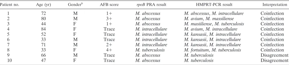
of the eight sputum specimens whose results showed partial
agreement between the two methods, all the sputum
speci-mens were identified being infected with a single species by
rpoB
PRA but were proven to be coinfected with two
My-cobacterium
spp. by HMPRT-PCR. The most frequently
en-countered case of coinfection is simultaneous infection with
M. intracellulare
and
M. kansasii
(three cases). The species
involved most frequently in coinfection was
M. intracellulare
(five cases) (Table 6).
DISCUSSION
The most important advantage of our HMPRT-PCR
method is that it enables the simultaneous identification of
seven mycobacterial species, the most commonly encountered
mycobacteria in clinical settings, in a single reaction.
Particu-larly in the Republic of Korea,
M. tuberculosis
infections
ac-count for more than 93% of mycobacterial infections; and
almost 98% of clinically significant NTM infections are known
to be caused by
M. avium
,
M. intracellulare
, the
M. abscessus
complex, the
M. fortuitum
complex, and
M. kansasii
(20). So, at
least in the Republic of Korea, this assay can detect nearly all
infections caused by mycobacteria. To apply our method to
species differentiation, species- or strain-specific characteristics
should be evaluated. If there were different profiles between
strains of a given species, our method would be too
compli-cated for mycobacterial species identification. A substantial
number of clinical isolates (133 isolates) have now been
iden-tified using our new HMPRT-PCR method in parallel with the
[image:6.585.42.541.90.233.2]rpoB
PRA method. All the isolates were clearly identified at
the species level using our method, as they showed species- or
strain-specific melting temperature profiles. One characteristic
feature of the HMPRT-PCR method is that it can differentiate
between two members of
M. abscessus
complex strains,
M.
abscessus
and
M. massiliense
. Recently, their separation has
been gaining importance since it was reported that there are
differences in the antibiotic susceptibility patterns,
epidemio-logic features, and clinical outcomes between the two strains
(18, 31). However, separation of these two strains is difficult
due to the high degree of sequence conservation between
them. Therefore, sequencing of
hsp65
(18) or
rpoB
(1) has
TABLE 5. Comparison of identification results between sputum HMPRT-PCR and same culture-based
rpoB
PRA for 117 sputum samples,
according to mycobacterial species
Species Strain score by HMPRT-PCR for the following scores obtained by culture-basedrpoBPRA
a
: Sensitivity
(%)b
Trace 1⫹ 2⫹ 3⫹ 4⫹ Total
M. intracellulare
12 (8/0/4)
11 (10/0/1)
4 (4/0/0)
4 (4/0/0)
1 (1/0/0)
32 (27/0/5)
84.3
M. avium
13 (6/0/7)
3 (0/0/3)
4 (4/0/0)
7 (6/0/1)
3 (3/0/0)
30 (19/0/11)
63.3
M. abscessus
8 (4/2/2)
12 (7/0/5)
4 (4/0/0)
6 (5/0/1)
0
30 (20/2/8)
c66.7
M. tuberculosis
3 (3/0/0)
1 (1/0/0)
3 (3/0/0)
7 (7/0/0)
7 (7/0/0)
21 (21/0/0)
100
M. fortuitum
0
0
0
0
1 (1/0/0)
1 (1/0/0)
100
M. kansasii
0
0
1 (1/0/0)
0
0
1 (1/0/0)
100
M. szulgai
0
1 (0/0/1)
0
0
0
1 (0/0/1)
0
M. celatum
0
0
0
1 (0/0/1)
0
1 (0/0/1)
0
Total
36 (21/2/13)
28 (18/0/10)
16 (16/0/0)
25 (22/0/3)
12 (12/0/0)
117 (89/2/26)
Sensitivity (%)
58.3
64.3
100
88
100
76.1
aData in parentheses represent the number of isolates for which the results of the two methods are in agreement/number of isolates for which the results are in
disagreement/number of isolates not identified by HMPRT-PCR.
bCalculated from the concordant results between the two assays,rpoBPRA and HMPRT-PCR.
cDifferentiation betweenM. abscessus(12 samples) andM. massiliense(8 samples) was possible by HMPRT-PCR.
TABLE 6. Sputum samples from the same culture showing discrepancies between results by HNPRT-PCR and
rpoB
PRA
aPatient no. Age (yr) Genderb AFB score rpoBPRA result HMPRT-PCR result Interpretation
1
72
M
1
⫹
M. abscessus
M. abscessus
,
M. intracellulare
Coinfection
2
80
M
3
⫹
M. abscessus
M. avium
,
M. massiliense
Coinfection
3
44
F
1
⫹
M. abscessus
M. masiiliense
,
M. tuberculosis
Coinfection
4
84
F
Trace
M. intracellulare
M. avium
,
M. intracellulare
Coinfection
5
52
F
Trace
M. intracellulare
M. kansasii
,
M. intracellulare
Coinfection
6
33
M
Trace
M. intracellulare
M. kansasii
,
M. intracellulare
Coinfection
7
71
M
2
⫹
M. intracellulare
M. kansasii
,
M. intracellulare
Coinfection
8
33
F
4
⫹
M. tuberculosis
M. fortuitum
,
M. tuberculosis
Coinfection
9
66
M
Trace
M. abscessus
M. tuberculosis
Disagreement
10
47
F
Trace
M. abscessus
M. tuberculosis
Disagreement
a
Most discrepancies may be caused by the overgrowth of a rapid grower.
b
M, male; F, female.
3078
KIM ET AL.
J. C
LIN. M
ICROBIOL.
on May 16, 2020 by guest
http://jcm.asm.org/
[image:6.585.44.541.595.707.2]generally been performed to separate these strains. To our
knowledge, the HMPRT-PCR method described here is the
only real-time PCR-based identification method that can
dif-ferentiate between the two strains. These results suggest that
our HMPRT-PCR method can effectively be used for the
iden-tification of mycobacterial culture isolates in the clinical
labo-ratory.
Another advantage of our HMPRT-PCR method is that it
can directly identify mycobacterial infections from sputum
samples. In particular, this assay showed a sensitivity level of
94.3% (50/53 sputum samples) for the identification of sputum
samples with an AFB score equal to or greater than 2
⫹
, which
seems to be acceptable for the identification of mycobacterial
organisms from sputum in a clinical laboratory. However, a
relatively low level of sensitivity in identification was observed
for sputum samples with 1
⫹
and trace AFB scores. To improve
the sensitivity of this assay, use of a nested PCR strategy should
be considered for samples with AFB scores lower than 1
⫹
.
Moreover, our HMPRT-PCR method can identify
mycobac-terial coinfections directly from sputa. Recently, reports
re-garding mycobacterial coinfections have increased along with
the increase in the incidence of AIDS and
immune-compro-mised patients (10, 12, 24). Thus, the detection of coinfection
in clinical specimens has gained much attention in the
myco-bacterial diagnostic field. Culture-based methods can
underes-timate the incidence of genuine coinfections in specimens,
probably due to physiological differences, particularly
differ-ences in the growth rates between
Mycobacterium
spp. or host
immune status. Our method could detect coinfections in eight
sputum samples identified as having a single infection by a
culture-based protocol (Table 6). Identical results were found
in repeated experiments (data not shown).
Interestingly, the two misidentified cases observed in sputum
samples with trace AFB scores were similar. In both cases, the
organisms were identified as
M. abscessus
by culture-based
rpoB
PRA but were identified as
M. tuberculosis
by the
HMPRT-PCR method (Table 6). Although we had no clinical
evidence proving
M. tuberculosis
infections in these cases, the
possibility that the difference in the results obtained by the two
methods may be due to differences in the sensitivity of the
HMPRT-PCR method in the detection of these two strains
cannot be excluded. Our data showed that the sensitivity level
of this method was lower for the detection of
M. abscessus
(66.7%, 20/30) than for the detection of
M. tuberculosis
(100%,
21/21) (Table 5). The other possible explanation is that the
difference in the results obtained by the two methods may be
due to the difference in the growth rates between the slowly
growing species,
M. tuberculosis
, and the rapidly growing
spe-cies,
M. abscessus
. Because the two mismatched cases were
found exclusively for sputum samples with trace AFB scores, it
is possible that increasing the sensitivity of the HMPRT-PCR
method using a nested PCR strategy may solve this problem.
The mycobacteria in 2 of 26 sputum specimens whose
iden-tification could not be achieved by HMPRT-PCR were
con-firmed to be
M. celatum
and
M. szulgai
by culture-based
hsp65
sequencing analysis (Table 5). Identification of the two strains
is not possible through our HMPRT-PCR method. Although
both strains are known to be rarely encountered in the
Repub-lic of Korea, the addition of probes to detect these two strains
deserves to be considered for the next version of this assay.
In conclusion, our data suggest that a novel HMPRT-PCR
method to detect pathogenic mycobacterial species from
spu-tum samples, as well as culture isolates, could be effective as a
routine method in the clinical setting.
ACKNOWLEDGMENT
This study was supported by grant A101205 from the Korean
Healthcare Technology R&D project, Ministry for Health, Welfare &
Family Affairs, Republic of Korea, and in part supported by grant
04-2008-0860 from the SNUH Research Fund.
REFERENCES
1.Ade´kambi, T., P. Colson, and M. Drancourt.2003.rpoB-based identification of nonpigmented and late-pigmenting rapidly growing mycobacteria. J. Clin. Microbiol.41:5699–5708.
2.Barnes, P. F., A. B. Bloch, P. T. Davison, and D. E. Sneider, Jr.1991. Tuberculosis in patients with human immunodeficiency virus infection. N. Engl. J. Med.324:1644–1649.
3.Bloom, B. R.1992. Back to a frightening future. Nature358:538–539. 4.Bloom, B. R., and C. J. L. Murray.1992. Tuberculosis: commentary on a
reemergent killer. Science257:1055–1064.
5.Cloud, J. L., H. Neal, R. Rosenberry, C. Y. Turenne, M. JAMA, D. R. Hillyard, and K. C. Carroll.2002. Identification ofMycobacteriumspp. by using a commercial 16S ribosomal DNA sequencing kit and additional se-quencing libraries. J. Clin. Microbiol.40:400–406.
6.Coll, P., M. Garrigo, C. Moreno, and N. Marti.2003. Routine use of Gen-Probe AmplifiedMycobacterium TuberculosisDirect (MTD) test for detec-tion ofMycobacterium tuberculosiswith smear-positive and smear-negative specimens. Int. J. Tuber. Lung Dis.7:886–891.
7.Dye, C., S. Scheele, P. Dolin, V. Pathania, and M. C. Raviglione.1999. Global burden of tuberculosis. Estimated incidence, prevalence, and mor-tality by country. JAMA282:677–686.
8.Espy, M. J., J. R. Uhl, L. M. Sloan, S. P. Buckwalter, M. F. Jones, E. A. Vetter, J. D. Yao, N. L. Wengenack, J. E. Rosenblatt, F. R. Cockerill, III, and T. F. Smith.2006. Real-time PCR in clinical microbiology: applications for routine laboratory testing. Clin. Microbiol. Rev.19:165–256.
9.Foongladda, S., S. Pholwat, B. Eampokalap, P. Kiratisin, and R. Sutthent.
2009. Multi-probe real-time PCR identification of commonMycobacterium
species in blood culture broth. J. Mol. Diagn.11:42–48.
10.Gopinath, K., and S. Singh.2009. Multiplex PCR assay for simultaneous detection and differentiation ofMycobacterium tuberculosis,Mycobacterium aviumcomplexes and other mycobacterial species directly from clinical spec-imens. J. Appl. Microbiol.107:425–435.
11.Halse, T. A., J. Edwards, P. L. Cunningham, W. J. Wolfgang, N. B. Dumas, V. E. Escuyer, and K. A. Musser.2010. Combined real-time PCR andrpoB
gene pyrosequencing for rapid identification ofMycobacterium tuberculosis
and determination of rifampin resistance directly in clinical specimens. J. Clin. Microbiol.48:1182–1188.
12.Huminer, D., S. Dux, Z. Samra, L. Kaufman, A. Lavy, C. S. Block, and S. D. Pitlik.1993.Mycobacterium simiaeinfection in Israeli patients with AIDS. Clin. Infect. Dis.17:508–509.
13.Joh, J. S., C. H. Lee, J. E. Lee, Y. K. Park, G. H. Bai, E. C. Kim, S. K. Han, Y. S. Shim, and J. J. Yim.2007. The interval between initiation of anti-tuberculosis treatment in patients with culture-positive pulmonary tubercu-losis and receipt of drug-susceptibility test results. J. Korean Med. Sci.
22:26–29.
14.Kent, P., and G. Kubica.1985. Public health mycobacteriology—a guide for the level III laboratory. Centers for Disease Control and Prevention, At-lanta, GA.
15.Kim, B. J., J. H. Park, S. A. Lee, H. Kim, C. Y. Cha, Y. H. Kook, E. C. Kim, S. I. Joo, J. S. Lee, and J. J. Yim.2008. Differentiation of mycobacteria in sputa by duplex polymerase chain reaction for mycobacterialhsp65gene. Diagn. Microbiol. Infect. Dis.62:193–198.
16.Kim, B. J., S. H. Lee, M. A. Lyu, S. J. Kim, G. H. Bai, G. T. Chae, E. C. Kim, C. Y. Cha, and Y. H. Kook.1999. Identification of mycobacterial species by comparative sequence analysis of the RNA polymerase gene (rpoB). J. Clin. Microbiol.37:1714–1720.
17.Kim, H., S. H. Kim, T. S. Shim, M. N. Kim, G. H. Bai, Y. G. Park, S. H. Lee, G. T. Chae, C. Y. Cha, Y. H. Kook, and B. J. Kim.2005. Differentiation of
Mycobacteriumspecies by analysis of the heat-shock protein 65 gene (hsp65). Int. J. Syst. Evol. Microbiol.55:1649–1656.
18.Kim, H. Y., Y. Kook, Y. J. Yun, C. G. Park, N. Y. Lee, T. S. Shim, B. J. Kim, and Y. H. Kook.2008. Proportions ofMycobacterium massilienseand Myco-bacterium bolletiistrains among KoreanMycobacterium chelonae-Mycobac-terium abscessusgroup isolates. J. Clin. Microbiol.46:3384–3890. 19.Kim, H. J., H. S. Mun, H. Kim, E. J. Oh, Y. Ha, G. H. Bai, Y. G. Park, C. Y.
Cha, Y. H. Kook, and B. J. Kim.2006. Differentiation of mycobacterial species by hsp65 duplex PCR followed by duplex-PCR-based restriction analysis and direct sequencing. J. Clin. Microbiol.44:3855–3862.
on May 16, 2020 by guest
http://jcm.asm.org/
20.Koh, W. J., O. J. Kwon, and K. S. Lee.2005. Diagnosis and treatment of nontuberculous mycobacterial pulmonary diseases: a Korean perspective. J. Korean Med. Sci.20:913–925.
21.Lee, H., H. J. Park, S. N. Cho, G. H. Bai, and S. J. Kim.2000. Species identification of mycobacteria by PCR-restriction fragment length polymor-phism of therpoBgene. J. Clin. Microbiol.38:2966–2971.
22.Leung, K. L., C. W. Yip, W. F. Cheung, A. C. Lo, W. M. Ko, and K. M. Kam.
2009. Development of a simple and low-cost real-time PCR method for the identification of commonly encountered mycobacteria in a high throughput laboratory. J. Appl. Microbiol.107:1433–1439.
23.Lim, S. Y., B. J. Kim, M. K. Lee, and K. Kim.2008. Development of a real-time PCR-based method for rapid differential identification of Myco-bacteriumspecies. Lett. Appl. Microbiol.46:101–106.
24.Massenkeil, G., M. Opravil, M. Salfinger, A. von Graevenitz, and R. Lu¨thy.
1992. Disseminated coinfection withMycobacterium aviumcomplex and My-cobacterium kansasiiin a patient with AIDS and liver abscess. Clin. Infect. Dis.14:618–619.
25.McNabb, A., K. Adie, M. Rodrigues, W. A. Black, and J. Isaac-Renton.2006. Direct identification of mycobacteria in primary liquid detection media by partial sequencing of the 65-kilodalton heat shock protein gene. J. Clin. Microbiol.44:60–66.
26.Miller, N., T. Cleary, G. Kraus, A. K. Young, G. Spruill, and H. J. Hnatyszyn.
2002. Rapid and specific detection ofMycobacterium tuberculosisfrom acid-fast bacillus smear-positive respiratory specimens and BacT/ALERT MP culture bottles by using fluorogenic probes and real-time PCR. J. Clin. Microbiol.40:4143–4147.
27.Mun, H. S., H. J. Kim, E. J. Oh, H. Kim, Y. G. Park, G. H. Bai, J. Do, C. Y.
Cha, Y. H. Kook, and B. J. Kim.2007. Direct application ofAvaII PCR restriction fragment length polymorphism analysis (AvaII PRA) targeting 644 bp heat shock protein 65 (hsp65) gene to sputum samples. Microbiol. Immunol.51:105–110.
28.Nakanaga, K., N. Ishii, K. Suzuki, K. Tanigawa, M. Goto, T. Okabe, H. Imada, A. Kodama, T. Iwamoto, H. Takahashi, and H. Saito.2007. “ Myco-bacterium ulcerans subsp.shinshuense” isolated from a skin ulcer lesion: identification based on 16S rRNA gene sequencing. J. Clin. Microbiol.45:
3840–3843.
29.Pinsky, B. A., and N. Banaei.2008. Multiplex real-time PCR assay for rapid identification ofMycobacterium tuberculosiscomplex members to the species level. J. Clin. Microbiol.46:2241–2246.
30.Rosenzweig, D. Y.1996. Nontuberculous mycobacterial disease in the immu-nocompetent adult. Semin. Respir. Infect.11:252–261.
31.Simmon, K. E., J. I. Pounder, J. N. Greene, F. Walsh, C. M. Anderson, S. Cohen, and C. A. Petti.2007. Identification of an emerging pathogen, My-cobacterium massiliense, byrpoBsequencing of clinical isolates collected in the United States. J. Clin. Microbiol.45:1978–1980.
32.Taylor, Z., C. M. Nolan, and H. M. Blumberg.2005. Controlling tuberculosis in the United States. Recommendations from the American Thoracic Soci-ety, CDC, and the Infectious Diseases Society of America. MMWR Recom-mend. Rep.54(RR-12):1–81.
33.Wagner, D., and L. S. Young.2004. Nontuberculous mycobacterial infec-tions: a clinical review. Infection32:257–270.
34.Wolinsky, E.1992. Mycobacterial diseases other than tuberculosis Clin. Infect. Dis.15:1–10.